Tag Archives: University of Rome Tor Vergata

Shooting drugs to an infection site with a slingshot

It seems as if I’ve been writing up nanomedicine research a lot lately, so I would have avoided this piece. However, since I do try to cover Canadian nanotechnology regardless of the topic and this work features researchers from l’Université de Montréal (Québec, Canada), here’s one of the latest innovations in the field of nanomedicine. (I have some additional comments about the nano scene in Canada and one major issue concerning nanomedicine at the end of this posting.) From a May 8, 2017 news item on ScienceDaily,

An international team of researchers from the University of Rome Tor Vergata and the University of Montreal has reported, in a paper published this week in Nature Communications, the design and synthesis of a nanoscale molecular slingshot made of DNA that is 20,000 times smaller than a human hair. This molecular slingshot could “shoot” and deliver drugs at precise locations in the human body once triggered by specific disease markers.

A May 8, 2017 University of Montreal news release (also on EurekAlert), which originated the news item, delves further into the research (Note: A link has been removed),

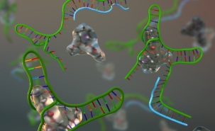
The molecular slingshot is only a few nanometres long and is composed of a synthetic DNA strand that can load a drug and then effectively act as the rubber band of the slingshot. The two ends of this DNA “rubber band” contain two anchoring moieties that can specifically stick to a target antibody, a Y-shaped protein expressed by the body in response to different pathogens such as bacteria and viruses. When the anchoring moieties of the slingshot recognize and bind to the arms of the target antibody the DNA “rubber band” is stretched and the loaded drug is released.

“One impressive feature about this molecular slingshot,” says Francesco Ricci, Associate Professor of Chemistry at the University of Rome Tor Vergata, “is that it can only be triggered by the specific antibody recognizing the anchoring tags of the DNA ‘rubber band’. By simply changing these tags, one can thus program the slingshot to release a drug in response to a variety of specific antibodies. Since different antibodies are markers of different diseases, this could become a very specific weapon in the clinician’s hands.”

“Another great property of our slingshot,” adds Alexis Vallée-Bélisle, Assistant Professor in the Department of Chemistry at the University of Montreal, “is its high versatility. For example, until now we have demonstrated the working principle of the slingshot using three different trigger antibodies, including an HIV antibody, and employing nucleic acids as model drugs. But thanks to the high programmability of DNA chemistry, one can now design the DNA slingshot to ‘shoot’ a wide range of threrapeutic molecules.”

“Designing this molecular slingshot was a great challenge,” says Simona Ranallo, a postdoctoral researcher in Ricci’s team and principal author of the new study. “It required a long series of experiments to find the optimal design, which keeps the drug loaded in ‘rubber band’ in the absence of the antibody, without affecting too much its shooting efficiency once the antibody triggers the slingshot.”

The group of researchers is now eager to adapt the slingshot for the delivery of clinically relevant drugs, and to demonstrate its clinical efficiency. [emphasis mine] “We envision that similar molecular slingshots may be used in the near future to deliver drugs to specific locations in the body. This would drastically improve the efficiency of drugs as well as decrease their toxic secondary effects,” concludes Ricci.

Here’s a link to and a citation for the paper,

Antibody-powered nucleic acid release using a DNA-based nanomachine by Simona Ranallo, Carl Prévost-Tremblay, Andrea Idili, Alexis Vallée-Bélisle, & Francesco Ricci. Nature Communications 8, Article number: 15150 (2017) doi:10.1038/ncomms15150 Published online: 08 May 2017

This is an open access paper.

A couple of comments

The Canadian nanotechnology scene is pretty much centered in Alberta and Québec. The two provinces have invested a fair amount of money in their efforts. Despite the fact that the province of Alberta also hosts the federal government’s National Institute of Nanotechnology, it seems that the province of Québec is the one making the most progress in its various ‘nano’ fields of endeavour. Another province that should be mentioned with regard to its ‘nano’ efforts is Ontario. As far as I can tell, nanotechnology there doesn’t enjoy the same level of provincial funding support as the other two but there is some important work coming out of Ontario.

My other comment has to do with nanomedicine. While it is an exciting field, there is a tendency toward a certain hyperbole. For anyone who got excited about the ‘slingshot’, don’t forget this hasn’t been tested on any conditions close to the conditions found in a human body nor have they even used, “... clinically relevant drugs,  … .”  It’s also useful to know that less than 1% of the drugs used in nanoparticle-delivery systems make their way to the affected site (from an April 27, 2016 posting about research investigating the effectiveness of nanoparticle-based drug delivery systems). By the way, it was a researcher at the University of Toronto (Ontario, Canada) who first noted this phenomenon after a meta-analysis of the research,

More generally, the authors argue that, in order to increase nanoparticle delivery efficiency, a systematic and coordinated long-term strategy is necessary. To build a strong foundation for the field of cancer nanomedicine, researchers will need to understand a lot more about the interactions between nanoparticles and the body’s various organs than they do today. …

It’s not clear from the news release, the paper, or the May 8, 2017 article by Sherry Noik for the Canadian Broadcasting Corporation’s News Online website, how this proposed solution would be administered but presumably the same factors which affect other nano-based drug deliveries could affect this new one,

Scientists have for many years been working on improving therapies like chemo and radiation on that score, but most efforts have focused on modifying the chemistry rather than altering the delivery of the drug.

“It’s all about tuning the concentration of the drug optimally in the body: high concentration where you want it to be active, and low concentration where you don’t want to affect other healthy parts,” says Prof. Alexis Vallée-Bélisle of the University of Montreal, co-author of the report published this week in Nature Communications.

“If you can increase the concentration of that drug at the specific location, that drug will be more efficient,” he told CBC News in an interview.

‘Like a weapon’

Restricting the movement of the drug also reduces potentially harmful secondary effects on other parts of the body — for instance, the hair loss that can result from toxic cancer treatments, or the loss of so-called good bacteria due to antibiotic use.

The idea of the slingshot is to home in on the target cells at a molecular level.

The two ends of the strand anchor themselves to the antibody, stretching the strand taut and catapulting the drug to its target.

“Imagine our slingshot like a weapon, and this weapon is being used by our own antibody,” said Vallée-Bélisle, who heads the Laboratory of Biosensors & Nanomachines at U of M. “We design a specific weapon targeting, for example, HIV. We provide the weapon in the body with the bullet — the drug. If the right solider is there, the soldier can use the weapon and shoot the problem.”

Equally important: if the wrong soldier is present, the weapon won’t be deployed.

So rather than delay treatment for an unidentified infection that could be either viral or bacterial, a patient could receive the medication for both and their body would only use the one it needed.

Getting back to my commentary, how does the drug get to its target? Through the bloodstream?  Does it get passed through various organs? How do we increase the amount of medication (in nano-based drug delivery systems) reaching affected areas from less than 1%?

The researchers deserve to be congratulated for this work and given much encouragement and thanks as they grapple with the questions I’ve posed and with all of the questions I don’t know how to ask.

Perovskite, nanorods, and solar energy

As the authors, Azhar Fakharuddin, Rajan Jose, and Thomas Brown, note in an Aug. 7, 2015 Nanowerk Spotlight article , securing energy sources is a global pursuit and pervoskite (a new wonder material for solar cells) has presented a challenge (Note: A link has been removed),

Energy security has been a top global concern motivating researchers to seek it from renewable and cost-effective resources. Solar cells, that convert sun light into electricity, hold the promise as a cheap energy alternative. The silicon and thin film photovoltaic industry have taken many strides to lower energy prices; however, continued research is required in order to extensively compete with fossil fuels.

The development of perovskite solar cells, first reported in 2009 (and with a record power conversion efficiency of 20.1 percent so far), is a possible route towards high efficiency photovoltaics that are also cost-effectiveness, owing to to their easy-processing from solution.

Question marks have however remained on their stability.

The authors (members of a research team) have recently published a paper about a method that could make perovskite solar cells more stable,

Now, a research team from University Malaysia Pahang, focussing on renewable energy, working in in collaboration with scientists from University of Rome ‘Tor Vergata’, Italy, has developed the world’s first nanorod-based perovskite solar module.

Among the three types of electron transport layers investigated, the nanorod-based devices retained the original efficiency values even after 2500 hours of shelf-life investigation, a protocol used to gauge initial stability and indoor lifetime performance.
The device employing a conventional TiO2 nanoparticle material showed nearly 60% of original performance, whereas planar devices employing a compact TiO2 layer showed below 5% of original performance, measured at similar experimental conditions.
A chemical analysis of the devices hinted that the peculiar conformation of nanorods facilitates a stable perovskite phase due to their inherent stability and macroporous nature.

If you want more detail, the research team’s Nanowerk Spotlight article is the place to look (it’s almost like a Reddit session except there’s no ‘ask me anything’ option). There’s also the team’s paper,

Vertical TiO2 Nanorods as a Medium for Stable and High-Efficiency Perovskite Solar Modules by Azhar Fakharuddin, Francesco Di Giacomo, Alessandro L. Palma, Fabio Matteocci, Irfan Ahmed, Stefano Razza, Alessandra D’Epifanio, Silvia Licoccia, Jamil Ismail, Aldo Di Carlo, Thomas M. Brown, and Rajan Jose. ACS Nano, Article ASAP DOI: 10.1021/acsnano.5b03265 Publication Date (Web): July 24, 2015

Copyright © 2015 American Chemical Society

This paper is behind a paywall.

One final note, I’ve been meaning to publish a post about perovskite-based solar cells for a while now as the material seems to be sweeping the solar energy community and, now, it’s done.

Italians and Polish collaborate on nanoscale study of vanishing Da Vinci self-portrait

In addition to a new nondamaging technique to examine paintings (my June 2, 2014 post: Damage-free art authentication and spatially offset Raman spectroscopy [SORS]), there’s a new report in a June 3, 2014 news item on ScienceDaily about a nondamaging technique to examine paper such as the paper on which holds a Da Vinci self-portrait,

One of Leonardo da Vinci’s masterpieces, drawn in red chalk on paper during the early 1500s and widely believed to be a self-portrait, is in extremely poor condition. Centuries of exposure to humid storage conditions or a closed environment has led to widespread and localized yellowing and browning of the paper, which is reducing the contrast between the colors of chalk and paper and substantially diminishing the visibility of the drawing.

A group of researchers from Italy and Poland with expertise in paper degradation mechanisms was tasked with determining whether the degradation process has now slowed with appropriate conservation conditions — or if the aging process is continuing at an unacceptable rate.

Caption: This is Leonardo da Vinci's self-portrait as acquired during diagnostic studies carried out at the Central Institute for the Restoration of Archival and Library Heritage in Rome, Italy. Credit: M. C. Misiti/Central Institute for the Restoration of Archival and Library Heritage, Rome

Caption: This is Leonardo da Vinci’s self-portrait as acquired during diagnostic studies carried out at the Central Institute for the Restoration of Archival and Library Heritage in Rome, Italy.
Credit: M. C. Misiti/Central Institute for the Restoration of Archival and Library Heritage, Rome

The June 3, 2014 American Institute of Physics news release on EurekAlert provides more detail about the work,

… the team developed an approach to nondestructively identify and quantify the concentration of light-absorbing molecules known as chromophores in ancient paper, the culprit behind the “yellowing” of the cellulose within ancient documents and works of art.

“During the centuries, the combined actions of light, heat, moisture, metallic and acidic impurities, and pollutant gases modify the white color of ancient paper’s main component: cellulose,” explained Joanna Łojewska, a professor in the Department of Chemistry at Jagiellonian University in Krakow, Poland. “This phenomenon is known as ‘yellowing,’ which causes severe damage and negatively affects the aesthetic enjoyment of ancient art works on paper.”

Chromophores are the key to understanding the visual degradation process because they are among the chemical products developed by oxidation during aging and are, ultimately, behind the “yellowing” within cellulose. Yellowing occurs when “chromophores within cellulose absorb the violet and blue range of visible light and largely scatter the yellow and red portions — resulting in the characteristic yellow-brown hue,” said Olivia Pulci, a professor in the Physics Department at the University of Rome Tor Vergata.

To determine the degradation rate of Leonardo’s self-portrait, the team created a nondestructive approach that centers on identifying and quantifying the concentration of chromophores within paper. It involves using a reflectance spectroscopy setup to obtain optical reflectance spectra of paper samples in the near-infrared, visible, and near-ultraviolet wavelength ranges.

Once reflectance data is gathered, the optical absorption spectrum of cellulose fibers that form the sheet of paper can be calculated using special spectroscopic data analysis.

Then, computational simulations based on quantum mechanics — in particular, Time-Dependent Density Functional Theory, which plays a key role in studying optical properties in theoretical condensed matter physics — are tapped to calculate the optical absorption spectrum of chromophores in cellulose.

“Using our approach, we were able to evaluate the state of degradation of Leonardo da Vinci’s self-portrait and other paper specimens from ancient books dating from the 15th century,” said Adriano Mosca Conte, a researcher at the University of Rome Tor Vergata. “By comparing the results of ancient papers with those of artificially aged samples, we gained significant insights into the environmental conditions in which Leonardo da Vinci’s self-portrait was stored during its lifetime.”

Their work revealed that the type of chromophores present in Leonardo’s self portrait are “similar to those found in ancient and modern paper samples aged in extremely humid conditions or within a closed environment, which agrees with its documented history,” said Mauro Missori, a researcher at the Institute for Complex Systems, CNR, in Rome, Italy.

One of the most significant implications of their work is that the state of degradation of ancient paper can be measured and quantified by evaluation of the concentrations of chromophores in cellulose fibers. “The periodic repetition of our approach is fundamental to establishing the formation rate of chromophores within the self-portrait. Now our approach can serve as a precious tool to preserve and save not only this invaluable work of art, but others as well,” Conte noted.

Absolutely fascinating stuff to those of use who care about yellowing paper. (Having worked in an archives, I care deeply.) Here’s a link to and a citation for the study,

Visual degradation in Leonardo da Vinci’s iconic self-portrait: A nanoscale study by A. Mosca Conte, O. Pulci, M. C. Misiti, J. Lojewska, L. Teodonio1, C. Violante, and M. Missori. Appl. Phys. Lett. 104, 224101 (2014); http://dx.doi.org/10.1063/1.4879838

This is an open access study.

Biosensing cocaine

Amusingly, the Feb. 13, 2013 news item on Nanowerk highlights the biosensing aspect of the work in its title,

New biosensing nanotechnology adopts natural mechanisms to detect molecules

(Nanowerk News) Since the beginning of time, living organisms have developed ingenious mechanisms to monitor their environment.

The Feb. 13, 2013 news release from the University of Montreal (Université de Montréal) takes a somewhat different tack by focusing on cocaine,

Detecting cocaine “naturally”

Since the beginning of time, living organisms have developed ingenious mechanisms to monitor their environment. As part of an international study, a team of researchers has adapted some of these natural mechanisms to detect specific molecules such as cocaine more accurately and quickly. Their work may greatly facilitate the rapid screening—less than five minutes—of many drugs, infectious diseases, and cancers.

Professor Alexis Vallée-Bélisle of the University of Montreal Department of Chemistry has worked with Professor Francesco Ricci of the University of Rome Tor Vergata and Professor Kevin W. Plaxco of the University of California at Santa Barbara to improve a new biosensing nanotechnology. The results of the study were recently published in the Journal of American Chemical Society (JACS).

The scientists have provided an interesting image to illustrate their work,

Artist's rendering: the research team used an existing cocaine biosensor (in green) and revised its design to react to a series of inhibitor molecules (in blue). They were able to adapt the biosensor to respond optimally even within a large concentration window. Courtesy: University of Montreal

Artist’s rendering: the research team used an existing cocaine biosensor (in green) and revised its design to react to a series of inhibitor molecules (in blue). They were able to adapt the biosensor to respond optimally even within a large concentration window. Courtesy: University of Montreal

The news release provides some insight into the current state of biosensing and what the research team was attempting to accomplish,

“Nature is a continuing source of inspiration for developing new technologies,” says Professor Francesco Ricci, senior author of the study. “Many scientists are currently working to develop biosensor technology to detect—directly in the bloodstream and in seconds—drug, disease, and cancer molecules.”

“The most recent rapid and easy-to-use biosensors developed by scientists to determine the levels of various molecules such as drugs and disease markers in the blood only do so when the molecule is present in a certain concentration, called the concentration window,” adds Professor Vallée-Bélisle. “Below or above this window, current biosensors lose much of their accuracy.”

To overcome this limitation, the international team looked at nature: “In cells, living organisms often use inhibitor or activator molecules to automatically program the sensitivity of their receptors (sensors), which are able to identify the precise amount of thousand of molecules in seconds,” explains Professor Vallée-Bélisle. “We therefore decided to adapt these inhibition, activation, and sequestration mechanisms to improve the efficiency of artificial biosensors.”

The researchers put their idea to the test by using an existing cocaine biosensor and revising its design so that it would respond to a series of inhibitor molecules. They were able to adapt the biosensor to respond optimally even with a large concentration window. “What is fascinating,” says Alessandro Porchetta, a doctoral student at the University of Rome, “is that we were successful in controlling the interactions of this system by mimicking mechanisms that occur naturally.”

“Besides the obvious applications in biosensor design, I think this work will pave the way for important applications related to the administration of cancer-targeting drugs, an area of increasing importance,” says Professor Kevin Plaxco. “The ability to accurately regulate biosensor or nanomachine’s activities will greatly increase their efficiency.”

The funders for this project are (from the news release),

… the Italian Ministry of Universities and Research (MIUR), the Bill & Melinda Gates Foundation Grand Challenges Explorations program, the European Commission Marie Curie Actions program, the U.S. National Institutes of Health, and the Fonds de recherche du Québec Nature et Technologies.

Here’s a citation and a link to the research paper,

Using Distal-Site Mutations and Allosteric Inhibition To Tune, Extend, and Narrow the Useful Dynamic Range of Aptamer-Based Sensors by Alessandro Porchetta, Alexis Vallée-Bélisle, Kevin W. Plaxco, and Francesco Ricci. J. Am. Chem. Soc., 2012, 134 (51), pp 20601–20604 DOI: 10.1021/ja310585e Publication Date (Web): December 6, 2012

Copyright © 2012 American Chemical Society

This article is behind a paywall.

One final note, Alexis Vallée-Bélisle has been mentioned here before in the context of a ‘Grand Challenges Canada programme’ (not the Bill and Melinda Gates ‘Grand Challenges’) announcement of several fundees  in my Nov. 22, 2012 posting. That funding appears to be for a difference project.