Tag Archives: University of São Paulo (USP)

Adding as little as 0.1% of rosmarinic acid reduced amount of sunscreen needed to protect skin and more

Since metallic nanoparticles are now pretty much accepted as being relatively safe ingredients, I don’t write about sunscreens very often anymore. Of course metallic nanoparticles had to be rebranded as ‘minerals’ after some civil society groups raised a great fuss. (See my February 9, 2012 posting “Unintended consequences: Australians not using sunscreens to avoid nanoparticles?” for a rundown of the situation.)

This April 5, 2023 news item about a different kind of sunscreen ingredient on phys.org caught my eye,

An article published in the journal Cosmetics reports an investigation of the effects of including rosmarinic acid, an active antioxidant, in a sunscreen along with two conventional ultraviolet light filters, ethylhexyl methoxycinnamate (against UVB) and avobenzone (against UVA).

They don’t seem to have tested this new ingredient in any ‘mineral’ sunscreens but it seems an intriguing possibility. Here’s more about rosmarinic acid and why it may be a good addition to sunscreens from an April 5, 2023 Fundação de Amparo à Pesquisa do Estado de São Paulo (São Paulo Research Foundation; FAPESP) press release (also on EurekAlert), which originated the news item, Note: Links have been removed)

The research group increased the sunscreen’s photoprotective efficacy by adding rosmarinic acid at 0.1%, a very small proportion compared with those of conventional UV filters. They believe their findings suggest that incorporating natural molecules with antioxidant activities into sunscreens could decrease the proportion of conventional UV filters in the final product, with the advantage of providing other functional properties.

The product’s performance improved without the need to increase active principle levels, reducing both the amount of sunscreen needed to protect the same skin area and the volume of synthetic chemicals discharged into the environment.

In vitro and clinical trials obtained a 41% increase in sun protection factor (SPF). The higher the SPF, the more sunburn protection increases.

Another advantage of including rosmarinic acid was the addition of antioxidant activity to photoprotection so that the product could be used in antiaging cosmetics, for example.

“Our research on photoprotective systems aims primarily to evaluate potential sunscreen enhancement strategies. We’re interested above all in discovering ways to increase sunburn protection while also improving the stability of the product so that it remains safe and effective for longer,” said pharmaceutical scientist and biochemist André Rolim Baby, last author of the article and a professor at the University of São Paulo’s School of Pharmaceutical Sciences (FCF-USP) in Brazil.

“We’re also looking for products or systems with less environmental impact and ways of reducing the concentration of conventional filters by including natural ingredients that enhance the formulation. And we’re very interested in mapping other cosmetic properties of photoprotective molecules, such as anti-free radical action and protection of biomarkers in the outermost skin layers.”

Multifunctional compound

The investigation was part of a project supported by FAPESP to map chemopreventive properties of various UV filters.

In addition to being an antioxidant, rosmarinic acid, a natural polyphenol antioxidant found in rosemary, as well as sage, peppermint and many other herbal plants, has antiviral, anti-inflammatory, immunomodulatory, antibiotic and anticancer properties.

In a review article published in 2022 in the journal Nutrients, the research group highlighted the beneficial effects of rosmarinic acid as a food supplement, such as improvement in skin firmness and wrinkle reduction.

“In another investigation, we found potential benefits of rosmarinic acid for skin surface hydration, reinforcing the need for more research on the substance in the field of cosmetology,” Baby said.

Here’s a link to and a citation for the research team’s latest paper,

Photoprotective Efficacy of the Association of Rosmarinic Acid 0.1% with Ethylhexyl Methoxycinnamate and Avobenzone by Maíra de Oliveira Bispo, Ana Lucía Morocho-Jácome, Cassiano Carlos Escudeiro, Renata Miliani Martinez, Claudinéia Aparecida Sales de Oliveira Pinto, Catarina Rosado, Maria Valéria Robles Velasco and André Rolim Baby. Cosmetics 2023, 10(1), 11; https://doi.org/10.3390/cosmetics10010011 Published: 5 January 2023 (This article belongs to the Special Issue Feature Papers in Cosmetics in 2022)

This paper is open access.
Mystery of North American insect bioluminescent systems unraveled by Brazilian scientists

I’ve always been fond of ‘l’ words and so it is that I’m compelled to post a story about a “luciferin-luciferase system” or, in this case, a story about insect bioluminescence.

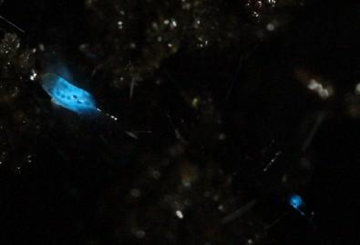
Caption: Researchers isolated molecules present in the larvae of the fungus gnat Orfelia fultoni Credit: Vadim Viviani, UFSCar

A September 9, 2020 Fundação de Amparo à Pesquisa do Estado de São Paulo (FAPESP) press release (also on EurekAlert but published Sept. 11, 2020) announces research into ‘blue’ bioluminescence,

Molecules belonging to an almost unknown bioluminescent system found in larvae of the fungus gnat Orfelia fultoni (subfamily Keroplatinae) have been isolated for the first time by researchers at the Federal University of São Carlos (UFSCar) in the state of São Paulo, Brazil. The small fly is one of the few terrestrial organisms that produce blue light. It inhabits riverbanks in the Appalachian Mountains in the eastern United States. A key part of its bioluminescent system is a molecule also present in two recently discovered Brazilian flies.

The study, supported by Paulo Research Foundation – FAPESP, is published in Scientific Reports. Five authors are affiliated with UFSCar and two with universities in the United States.

The bioluminescent systems of glow-worms, fireflies and other insects are normally made up of luciferin (a low molecular weight molecule) and luciferase, an enzyme that catalyzes the oxidation of luciferin by oxygen, producing light. While some bioluminescent systems are well known and even used in biotechnological applications, others are poorly understood, including blue light-emitting systems, such as that of O. fultoni.

“In the published paper, we describe the properties of the insect’s luciferase and luciferin and their anatomical location in its larvae. We also specify several possible proteins that are possible candidates for the luciferase. We don’t yet know what type of protein it is, but it’s likely to be a hexamerin. In insects, hexamerins are storage proteins that provide amino acids, besides having other functions, such as binding low molecular weight compounds, like luciferin,” said Vadim Viviani, a professor in UFSCar’s Sustainability Science and Technology Center (CCTS) in Sorocaba, São Paulo, and principal investigator for the study.

The study was part of the FAPESP-funded project “Arthropod bioluminescence“. The partnership with United States-based researchers dates from a previous project, supported by FAPESP and the United States National Science Foundation (NSF), in partnership with Vanderbilt University (VU), located in Nashville, Tennessee.

In addition to luciferin and luciferase, researchers began characterizing a complex found in insects of the family Keroplatidae, which, in addition to O. fultoni, also includes a Brazilian species in the genus Neoditomyia that produces only luciferin and hence does not emit light.

Because they do not use it to emit light, the luciferin in O. fultoni and the Brazilian Neoditomyia has been named keroplatin. In larvae of this subfamily, keroplatin is associated with “black bodies” – large cells containing dark granules, proteins and probably mitochondria (energy-producing organelles). Researchers are still investigating the biological significance of this association between keroplatin and mitochondria.

“It’s a mystery,” Viviani said. “This luciferin may play a role in the mitochondrial energy metabolism. At night, probably in the presence of a natural chemical reducer, the luciferin is released by these black bodies and reacts with the surrounding luciferase to produce blue light. These are possibilities we plan to study.”

Brazilian cousins

An important factor in the elucidation of the United States insect’s bioluminescent system was the discovery of a larva that lives in Intervales State Park in São Paulo in 2018. It does not emit light but produces luciferin, similar to O. fultoni (read more at: agencia.fapesp.br/29066).

In their latest study, the group injected purified luciferase from the United States species into larvae of the Brazilian species, which then produced blue light. The nonluminescent Brazilian species is more abundant in nature than the United States species, so a larger amount of the material could be obtained for study purposes, especially to characterize the luciferin (keroplatin) present in both species.

In 2019, the group discovered and described Neoceroplatus betaryensis, a new species of fungus gnat, in collaboration with Cassius Stevani, a professor at the University of São Paulo’s Institute of Chemistry (IQ-USP). It was the first blue light-emitting insect found in South America and was detected in a privately held forest reserve near the Upper Ribeira State Tourist Park (PETAR) in the southern portion of the state of São Paulo. A close relative of O. fultoni, N. betaryensis inhabits fallen tree trunks in humid places (read more at: agencia.fapesp.br/31797).

“We show that the bioluminescent system of this Brazilian species is identical to that of O. fultoni. However, the insect is very rare, and so it’s hard to obtain sufficient material for research purposes,” Viviani said.

The researchers are now cloning the insect’s luciferase and characterizing it in molecular terms. They are also analyzing the chemical structure of its luciferin and the morphology of its lanterns.

“Once all this has been determined, we’ll be able to synthesize the luciferin and luciferase in the lab and use these systems in a range of biotech applications, such as studying cells. This will help us understand more about human diseases, among other things,” Viviani said.

Here’s a link to and a citation for the paper,

A new brilliantly blue-emitting luciferin-luciferase system from Orfelia fultoni and Keroplatinae (Diptera) by Vadim R. Viviani, Jaqueline R. Silva, Danilo T. Amaral, Vanessa R. Bevilaqua, Fabio C. Abdalla, Bruce R. Branchini & Carl H. Johnson. Scientific Reports volume 10, Article number: 9608 (2020) DOI: https://doi.org/10.1038/s41598-020-66286-1 Published 15 June 2020

This paper is open access.

Evolution-in-materio and unconventional computing

Training materials such as carbon nanotubes to imitate electronic circuits? Welcome to the world of evolution-in-materio and unconventional computing. From an April 7, 2015 news item on ScienceDaily,

As we approach the miniaturization limits of conventional electronics, alternatives to silicon-based transistors — the building blocks of the multitude of electronic devices we’ve come to rely on — are being hotly pursued.

Inspired by the way living organisms have evolved in nature to perform complex tasks with remarkable ease, a group of researchers from Durham University in the U.K. and the University of São Paulo-USP in Brazil is exploring similar “evolutionary” methods to create information processing devices.

An April 7, 2015 American Institute of Physics (AIP) news release on EurekAlert, which originated the news item, delves into the research itself and the emerging field to which it belongs,

In the Journal of Applied Physics, from AIP Publishing, the group describes using single-walled carbon nanotube composites (SWCNTs) as a material in “unconventional” computing. By studying the mechanical and electrical properties of the materials, they discovered a correlation between SWCNT concentration/viscosity/conductivity and the computational capability of the composite.

“Instead of creating circuits from arrays of discrete components (transistors in digital electronics), our work takes a random disordered material and then ‘trains’ the material to produce a desired output,” said Mark K. Massey, research associate, School of Engineering and Computing Sciences at Durham University.

This emerging field of research is known as “evolution-in-materio,” a term coined by Julian Miller at the University of York in the U.K. What exactly is it? An interdisciplinary field blends together materials science, engineering and computer science. Although still in its early stages, the concept has already shown that by using an approach similar to natural evolution, materials can be trained to mimic electronic circuits–without needing to design the material structure in a specific way.

“The material we use in our work is a mixture of carbon nanotubes and polymer, which creates a complex electrical structure,” explained Massey. “When voltages (stimuli) are applied at points of the material, its electrical properties change. When the correct signals are applied to the material, it can be trained or ‘evolved’ to perform a useful function.”

While the group doesn’t expect to see their method compete with high-speed silicon computers, it could turn out to be a complementary technology. “With more research, it could lead to new techniques for making electronics devices,” he noted. The approach may find applications within the realm of “analog signal processing or low-power, low-cost devices in the future.”

Beyond pursuing the current methodology of evolution-in-materio, the next stage of the group’s research will be to investigate evolving devices as part of the material fabrication “hardware-in-the-loop” evolution. “This exciting approach could lead to further enhancements in the field of evolvable electronics,” said Massey.

Here’s a link to and a citation for the paper,

Computing with carbon nanotubes: Optimization of threshold logic gates using disordered nanotube/polymer composites by using disordered nanotube/polymer composites by M. K. Massey, A. Kotsialos, F. Qaiser, D. A. Zeze, C. Pearson, D. Volpati, L. Bowen, and M. C. Petty. J. Appl. Phys. 117, 134903 (2015); http://dx.doi.org/10.1063/1.4915343

This paper appears to be open access.

Also, the researchers have produced a video,

Credit: Mark Massey/Durham University

Final comment, I am gobsmacked and fascinated.