Tag Archives: University of Sydney

Physical neural network based on nanowires can learn and remember ‘on the fly’

A November 1, 2023 news item on Nanowerk announced new work on neuromorphic engineering from Australia,

For the first time, a physical neural network has successfully been shown to learn and remember ‘on the fly’, in a way inspired by and similar to how the brain’s neurons work.

The result opens a pathway for developing efficient and low-energy machine intelligence for more complex, real-world learning and memory tasks.

Key Takeaways
*The nanowire-based system can learn and remember ‘on the fly,’ processing dynamic, streaming data for complex learning and memory tasks.

*This advancement overcomes the challenge of heavy memory and energy usage commonly associated with conventional machine learning models.

*The technology achieved a 93.4% accuracy rate in image recognition tasks, using real-time data from the MNIST database of handwritten digits.

*The findings promise a new direction for creating efficient, low-energy machine intelligence applications, such as real-time sensor data processing.

Nanowire neural network
Caption: Electron microscope image of the nanowire neural network that arranges itself like ‘Pick Up Sticks’. The junctions where the nanowires overlap act in a way similar to how our brain’s synapses operate, responding to electric current. Credit: The University of Sydney

A November 1, 2023 University of Sydney news release (also on EurekAlert), which originated the news item, elaborates on the research,

Published today [November 1, 2023] in Nature Communications, the research is a collaboration between scientists at the University of Sydney and University of California at Los Angeles.

Lead author Ruomin Zhu, a PhD student from the University of Sydney Nano Institute and School of Physics, said: “The findings demonstrate how brain-inspired learning and memory functions using nanowire networks can be harnessed to process dynamic, streaming data.”

Nanowire networks are made up of tiny wires that are just billionths of a metre in diameter. The wires arrange themselves into patterns reminiscent of the children’s game ‘Pick Up Sticks’, mimicking neural networks, like those in our brains. These networks can be used to perform specific information processing tasks.

Memory and learning tasks are achieved using simple algorithms that respond to changes in electronic resistance at junctions where the nanowires overlap. Known as ‘resistive memory switching’, this function is created when electrical inputs encounter changes in conductivity, similar to what happens with synapses in our brain.

In this study, researchers used the network to recognise and remember sequences of electrical pulses corresponding to images, inspired by the way the human brain processes information.

Supervising researcher Professor Zdenka Kuncic said the memory task was similar to remembering a phone number. The network was also used to perform a benchmark image recognition task, accessing images in the MNIST database of handwritten digits, a collection of 70,000 small greyscale images used in machine learning.

“Our previous research established the ability of nanowire networks to remember simple tasks. This work has extended these findings by showing tasks can be performed using dynamic data accessed online,” she said.

“This is a significant step forward as achieving an online learning capability is challenging when dealing with large amounts of data that can be continuously changing. A standard approach would be to store data in memory and then train a machine learning model using that stored information. But this would chew up too much energy for widespread application.

“Our novel approach allows the nanowire neural network to learn and remember ‘on the fly’, sample by sample, extracting data online, thus avoiding heavy memory and energy usage.”

Mr Zhu said there were other advantages when processing information online.

“If the data is being streamed continuously, such as it would be from a sensor for instance, machine learning that relied on artificial neural networks would need to have the ability to adapt in real-time, which they are currently not optimised for,” he said.

In this study, the nanowire neural network displayed a benchmark machine learning capability, scoring 93.4 percent in correctly identifying test images. The memory task involved recalling sequences of up to eight digits. For both tasks, data was streamed into the network to demonstrate its capacity for online learning and to show how memory enhances that learning.

Here’s a link to and a citation for the paper,

Online dynamical learning and sequence memory with neuromorphic nanowire networks by Ruomin Zhu, Sam Lilak, Alon Loeffler, Joseph Lizier, Adam Stieg, James Gimzewski & Zdenka Kuncic. Nature Communications volume 14, Article number: 6697 (2023) DOI: https://doi.org/10.1038/s41467-023-42470-5 Published: 01 November 2023

This paper is open access.

You’ll notice a number of this team’s members are also listed in the citation in my June 21, 2023 posting “Learning and remembering like a human brain: nanowire networks” and you’ll see some familiar names in the citation in my June 17, 2020 posting “A tangle of silver nanowires for brain-like action.”

Synthetic human embryos—what now? (1 of 2)

Usually, there’s a rough chronological order to how I introduce the research, but this time I’m looking at the term used to describe it, following up with the various news releases and commentaries about the research, and finishing with a Canadian perspective.

After writing this post (but before it was published), the Weizmann Institute of Science (Israel) made their September 6, 2023 announcement and things changed a bit. That’s in Part two.

Say what you really mean (a terminology issue)

First, it might be useful to investigate the term, ‘synthetic human embryos’ as Julian Hitchcock does in his June 29, 2023 article on Bristows website (h/t Mondaq’s July 5, 2023 news item), Note: Links have been removed,

Synthetic Embryos” are neither Synthetic nor Embryos. So why are editors giving that name to stem cell-based models of human development?

One of the less convincing aspects of the last fortnight’s flurry of announcements about advances in simulating early human development (see here) concerned their name. Headlines galore (in newspapers and scientific journals) referred to “synthetic embryos“.

But embryo models, however impressive, are not embryos. To claim that the fundamental stages of embryo development that we learnt at school – fertilisation, cleavage and compaction – could now be bypassed to achieve the same result would be wrong. Nor are these objects “synthesised”: indeed, their interest to us lies in the ways in which they organise themselves. The researchers merely place the stem cells in a matrix in appropriate conditions, then stand back and watch them do it. Scientists were therefore unhappy about this use of the term in news media, and relieved when the International Society for Stem Cell Research (ISSCR) stepped in with a press release:

“Unlike some recent media reports describing this research, the ISSCR advises against using the term “synthetic embryo” to describe embryo models, because it is inaccurate and can create confusion. Integrated embryo models are neither synthetic nor embryos. While these models can replicate aspects of the early-stage development of human embryos, they cannot and will not develop to the equivalent of postnatal stage humans. Further, the ISSCR Guidelines prohibit the transfer of any embryo model to the uterus of a human or an animal.”

Although this was the ISSCR’s first attempt to put that position to the public, it had already made that recommendation to the research community two years previously. Its 2021 Guidelines for Stem Cell Research and Clinical Translation had recommended researchers to “promote accurate, current, balanced, and responsive public representations of stem cell research”. In particular:

“While organoids, chimeras, embryo models, and other stem cell-based models are useful research tools offering possibilities for further scientific progress, limitations on the current state of scientific knowledge and regulatory constraints must be clearly explained in any communications with the public or media. Suggestions that any of the current in vitro models can recapitulate an intact embryo, human sentience or integrated brain function are unfounded overstatements that should be avoided and contradicted with more precise characterizations of current understanding.”

Here’s a little bit about Hitchcock from his Bristows profile page,

  • Diploma Medical School, University of Birmingham (1975-78)
  • LLB, University of Wolverhampton
  • Diploma in Intellectual Property Law & Practice, University of Bristol
  • Qualified 1998

Following an education in medicine at the University of Birmingham and a career as a BBC science producer, Julian has focused on the law and regulation of life science technologies since 1997, practising in England and Australia. He joined Bristows with Alex Denoon in 2018.

Hitchcock’s June 29, 2023 article comments on why this term is being used,

I have a lot of sympathy with the position of the science writers and editors incurring the scientists’ ire. First, why should journalists have known of the ISSCR’s recommendations on the use of the term “synthetic embryo”? A journalist who found Recommendation 4.1 of the ISSCR Guidelines would probably not have found them specific enough to address the point, and the academic introduction containing the missing detail is hard to find. …

My second reason for being sympathetic to the use of the terrible term is that no suitable alternative has been provided, other than in the Stem Cell Reports paper, which recommends the umbrella terms “embryo models” or “stem cell based embryo models”. …

When asked why she had used the term “synthetic embryo”, the journalist I contacted remarked that, “We’re still working out the right language and it’s something we’re discussing and will no doubt evolve along with the science”.

It is absolutely in the public’s interest (and in the interest of science), that scientific research is explained in terms that the public understands. There is, therefore, a need, I think, for the scientific community to supply a name to the media or endure the penalties of misinformation …

In such an intensely competitive field of research, disagreement among researchers, even as to names, is inevitable. In consequence, however, journalists and their audiences are confronted by a slew of terms which may or may not be synonymous or overlapping, with no agreed term [emphasis mine] for the overall class of stem cell based embryo models. We cannot blame them if they make up snappy titles of their own [emphasis mine]. …

The announcement

The earliest date for the announcement at the International Society for Stem Cell Researh meeting that I can find is Hannah Devlin’s June 14, 2023 article in The Guardian newspaper, Note: A link has been removed,

Scientists have created synthetic human embryos using stem cells, in a groundbreaking advance that sidesteps the need for eggs or sperm.

Scientists say these model embryos, which resemble those in the earliest stages of human development, could provide a crucial window on the impact of genetic disorders and the biological causes of recurrent miscarriage.

However, the work also raises serious ethical and legal issues as the lab-grown entities fall outside current legislation in the UK and most other countries.

The structures do not have a beating heart or the beginnings of a brain, but include cells that would typically go on to form the placenta, yolk sac and the embryo itself.

Prof Magdalena Żernicka-Goetz, of the University of Cambridge and the California Institute of Technology, described the work in a plenary address on Wednesday [June 14, 2023] at the International Society for Stem Cell Research’s annual meeting in Boston.

The (UK) Science Media Centre made expert comments available in a June 14, 2023 posting “expert reaction to Guardian reporting news of creation of synthetic embryos using stem cells.”

Two days later, this June 16, 2023 essay by Kathryn MacKay, Senior Lecturer in Bioethics, University of Sydney (Australia), appeared on The Conversation (h/t June 16, 2023 news item on phys.org), Note: Links have been removed,

Researchers have created synthetic human embryos using stem cells, according to media reports. Remarkably, these embryos have reportedly been created from embryonic stem cells, meaning they do not require sperm and ova.

This development, widely described as a breakthrough that could help scientists learn more about human development and genetic disorders, was revealed this week in Boston at the annual meeting of the International Society for Stem Cell Research.

The research, announced by Professor Magdalena Żernicka-Goetz of the University of Cambridge and the California Institute of Technology, has not yet been published in a peer-reviewed journal. But Żernicka-Goetz told the meeting these human-like embryos had been made by reprogramming human embryonic stem cells.

So what does all this mean for science, and what ethical issues does it present?

MacKay goes on to answer her own questions, from the June 16, 2023 essay, Note: A link has been removed,

One of these quandaries arises around whether their creation really gets us away from the use of human embryos.

Robin Lovell-Badge, the head of stem cell biology and developmental genetics at the Francis Crick Institute in London UK, reportedly said that if these human-like embryos can really model human development in the early stages of pregnancy, then we will not have to use human embryos for research.

At the moment, it is unclear if this is the case for two reasons.

First, the embryos were created from human embryonic stem cells, so it seems they do still need human embryos for their creation. Perhaps more light will be shed on this when Żernicka-Goetz’s research is published.

Second, there are questions about the extent to which these human-like embryos really can model human development.

Professor Magdalena Żernicka-Goetz’s research is published

Almost two weeks later the research from the Cambridge team (there are other teams and countries also racing; see Part two for the news from Sept. 6, 2023) was published, from a June 27, 2023 news item on ScienceDaily,

Cambridge scientists have created a stem cell-derived model of the human embryo in the lab by reprogramming human stem cells. The breakthrough could help research into genetic disorders and in understanding why and how pregnancies fail.

Published today [Tuesday, June 27, 2023] in the journal Nature, this embryo model is an organised three-dimensional structure derived from pluripotent stem cells that replicate some developmental processes that occur in early human embryos.

Use of such models allows experimental modelling of embryonic development during the second week of pregnancy. They can help researchers gain basic knowledge of the developmental origins of organs and specialised cells such as sperm and eggs, and facilitate understanding of early pregnancy loss.

A June 27, 2023 University of Cambridge press release (also on EurekAlert), which originated the news item, provides more detail about the work,

“Our human embryo-like model, created entirely from human stem cells, gives us access to the developing structure at a stage that is normally hidden from us due to the implantation of the tiny embryo into the mother’s womb,” said Professor Magdalena Zernicka-Goetz in the University of Cambridge’s Department of Physiology, Development and Neuroscience, who led the work.

She added: “This exciting development allows us to manipulate genes to understand their developmental roles in a model system. This will let us test the function of specific factors, which is difficult to do in the natural embryo.”

In natural human development, the second week of development is an important time when the embryo implants into the uterus. This is the time when many pregnancies are lost.

The new advance enables scientists to peer into the mysterious ‘black box’ period of human development – usually following implantation of the embryo in the uterus – to observe processes never directly observed before.

Understanding these early developmental processes holds the potential to reveal some of the causes of human birth defects and diseases, and to develop tests for these in pregnant women.

Until now, the processes could only be observed in animal models, using cells from zebrafish and mice, for example.

Legal restrictions in the UK currently prevent the culture of natural human embryos in the lab beyond day 14 of development: this time limit was set to correspond to the stage where the embryo can no longer form a twin. [emphasis mine]

Until now, scientists have only been able to study this period of human development using donated human embryos. This advance could reduce the need for donated human embryos in research.

Zernicka-Goetz says the while these models can mimic aspects of the development of human embryos, they cannot and will not develop to the equivalent of postnatal stage humans.

Over the past decade, Zernicka-Goetz’s group in Cambridge has been studying the earliest stages of pregnancy, in order to understand why some pregnancies fail and some succeed.

In 2021 and then in 2022 her team announced in Developmental Cell, Nature and Cell Stem Cell journals that they had finally created model embryos from mouse stem cells that can develop to form a brain-like structure, a beating heart, and the foundations of all other organs of the body.

The new models derived from human stem cells do not have a brain or beating heart, but they include cells that would typically go on to form the embryo, placenta and yolk sac, and develop to form the precursors of germ cells (that will form sperm and eggs).

Many pregnancies fail at the point when these three types of cells orchestrate implantation into the uterus begin to send mechanical and chemical signals to each other, which tell the embryo how to develop properly.

There are clear regulations governing stem cell-based models of human embryos and all researchers doing embryo modelling work must first be approved by ethics committees. Journals require proof of this ethics review before they accept scientific papers for publication. Zernicka-Goetz’s laboratory holds these approvals.

“It is against the law and FDA regulations to transfer any embryo-like models into a woman for reproductive aims. These are highly manipulated human cells and their attempted reproductive use would be extremely dangerous,” said Dr Insoo Hyun, Director of the Center for Life Sciences and Public Learning at Boston’s Museum of Science and a member of Harvard Medical School’s Center for Bioethics.

Zernicka-Goetz also holds position at the California Institute of Technology and is NOMIS Distinguished Scientist and Scholar Awardee.

The research was funded by the Wellcome Trust and Open Philanthropy.

(There’s more about legal concerns further down in this post.)

Here’s a link to and a citation for the paper,

Pluripotent stem cell-derived model of the post-implantation human embryo by Bailey A. T. Weatherbee, Carlos W. Gantner, Lisa K. Iwamoto-Stohl, Riza M. Daza, Nobuhiko Hamazaki, Jay Shendure & Magdalena Zernicka-Goetz. Nature (2023) DOI: https://doi.org/10.1038/s41586-023-06368-y Published: 27 June 2023

This paper is open access.

Published the same day (June 27, 2023) is a paper (citation and link follow) also focused on studying human embryonic development using stem cells. First, there’s this from the Abstract,

Investigating human development is a substantial scientific challenge due to the technical and ethical limitations of working with embryonic samples. In the face of these difficulties, stem cells have provided an alternative to experimentally model inaccessible stages of human development in vitro …

This time the work is from a US/German team,

Self-patterning of human stem cells into post-implantation lineages by Monique Pedroza, Seher Ipek Gassaloglu, Nicolas Dias, Liangwen Zhong, Tien-Chi Jason Hou, Helene Kretzmer, Zachary D. Smith & Berna Sozen. Nature (2023) DOI: https://doi.org/10.1038/s41586-023-06354-4 Published: 27 June 2023

The paper is open access.

Legal concerns and a Canadian focus

A July 25, 2023 essay by Françoise Baylis and Jocelyn Downie of Dalhousie University (Nova Scotia, Canada) for The Conversation (h/t July 25, 2023 article on phys.org) covers the advantages of doing this work before launching into a discussion of legislation and limits in the UK and, more extensively, in Canada, Note: Links have been removed,

This research could increase our understanding of human development and genetic disorders, help us learn how to prevent early miscarriages, lead to improvements in fertility treatment, and — perhaps — eventually allow for reproduction without using sperm and eggs.

Synthetic human embryos — also called embryoid bodies, embryo-like structures or embryo models — mimic the development of “natural human embryos,” those created by fertilization. Synthetic human embryos include the “cells that would typically go on to form the embryo, placenta and yolk sac, and develop to form the precursors of germ cells (that will form sperm and eggs).”

Though research involving natural human embryos is legal in many jurisdictions, it remains controversial. For some people, research involving synthetic human embryos is less controversial because these embryos cannot “develop to the equivalent of postnatal stage humans.” In other words, these embryos are non-viable and cannot result in live births.

Now, for a closer look at the legalities in the UK and in Canada, from the July 25, 2023 essay, Note: Links have been removed,

The research presented by Żernicka-Goetz at the ISSCR meeting took place in the United Kingdom. It was conducted in accordance with the Human Fertilization and Embryology Act, 1990, with the approval of the U.K. Stem Cell Bank Steering Committee.

U.K. law limits the research use of human embryos to 14 days of development. An embryo is defined as “a live human embryo where fertilisation is complete, and references to an embryo include an egg in the process of fertilisation.”

Synthetic embryos are not created by fertilization and therefore, by definition, the 14-day limit on human embryo research does not apply to them. This means that synthetic human embryo research beyond 14 days can proceed in the U.K.

The door to the touted potential benefits — and ethical controversies — seems wide open in the U.K.

While the law in the U.K. does not apply to synthetic human embryos, the law in Canada clearly does. This is because the legal definition of an embryo in Canada is not limited to embryos created by fertilization [emphasis mine].

The Assisted Human Reproduction Act (the AHR Act) defines an embryo as “a human organism during the first 56 days of its development following fertilization or creation, excluding any time during which its development has been suspended.”

Based on this definition, the AHR Act applies to embryos created by reprogramming human embryonic stem cells — in other words, synthetic human embryos — provided such embryos qualify as human organisms.

A synthetic human embryo is a human organism. It is of the species Homo sapiens, and is thus human. It also qualifies as an organism — a life form — alongside other organisms created by means of fertilization, asexual reproduction, parthenogenesis or cloning.

Given that the AHR Act applies to synthetic human embryos, there are legal limits on their creation and use in Canada.

First, human embryos — including synthetic human embryos – can only be created for the purposes of “creating a human being, improving or providing instruction in assisted reproduction procedures.”

Given the state of the science, it follows that synthetic human embryos could legally be created for the purpose of improving assisted reproduction procedures.

Second, “spare” or “excess” human embryos — including synthetic human embryos — originally created for one of the permitted purposes, but no longer wanted for this purpose, can be used for research. This research must be done in accordance with the consent regulations which specify that consent must be for a “specific research project.”

Finally, all research involving human embryos — including synthetic human embryos — is subject to the 14-day rule. The law stipulates that: “No person shall knowingly… maintain an embryo outside the body of a female person after the fourteenth day of its development following fertilization or creation, excluding any time during which its development has been suspended.”

Putting this all together, the creation of synthetic embryos for improving assisted human reproduction procedures is permitted, as is research using “spare” or “excess” synthetic embryos originally created for this purpose — provided there is specific consent and the research does not exceed 14 days.

This means that while synthetic human embryos may be useful for limited research on pre-implantation embryo development, they are not available in Canada for research on post-implantation embryo development beyond 14 days.

The authors close with this comment about the prospects for expanding Canada’s14-day limit, from the July 25, 2023 essay,

… any argument will have to overcome the political reality that the federal government is unlikely to open up the Pandora’s box of amending the AHR Act.

It therefore seems likely that synthetic human embryo research will remain limited in Canada for the foreseeable future.

As mentioned, in September 2023 there was a new development. See: Part two.
Learning and remembering like a human brain: nanowire networks

It’s all about memory in this April 21, 2023 news item on Nanowerk, Note: A link has been removed,

An international team led by scientists at the University of Sydney has demonstrated nanowire networks can exhibit both short- and long-term memory like the human brain.

The research has been published today in the journal Science Advances (“Neuromorphic learning, working memory, and metaplasticity in nanowire networks”), led by Dr Alon Loeffler, who received his PhD in the School of Physics, with collaborators in Japan.

An April 24, 2023 University of Sydney (Australia) press release (also on EurekAlert but published April 21, 2023), which originated news item, offers more detail about the research,

“In this research we found higher-order cognitive function, which we normally associate with the human brain, can be emulated in non-biological hardware,” Dr Loeffler said.

“This work builds on our previous research in which we showed how nanotechnology could be used to build a brain-inspired electrical device with neural network-like circuitry and synapse-like signalling.

“Our current work paves the way towards replicating brain-like learning and memory in non-biological hardware systems and suggests that the underlying nature of brain-like intelligence may be physical.”

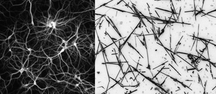
Nanowire networks are a type of nanotechnology typically made from tiny, highly conductive silver wires that are invisible to the naked eye, covered in a plastic material, which are scattered across each other like a mesh. The wires mimic aspects of the networked physical structure of a human brain.

Advances in nanowire networks could herald many real-world applications, such as improving robotics or sensor devices that need to make quick decisions in unpredictable environments.

“This nanowire network is like a synthetic neural network because the nanowires act like neurons, and the places where they connect with each other are analogous to synapses,” senior author Professor Zdenka Kuncic, from the School of Physics, said.

“Instead of implementing some kind of machine learning task, in this study Dr Loeffler has actually taken it one step further and tried to demonstrate that nanowire networks exhibit some kind of cognitive function.”

To test the capabilities of the nanowire network, the researchers gave it a test similar to a common memory task used in human psychology experiments, called the N-Back task.

For a person, the N-Back task might involve remembering a specific picture of a cat from a series of feline images presented in a sequence. An N-Back score of 7, the average for people, indicates the person can recognise the same image that appeared seven steps back.

When applied to the nanowire network, the researchers found it could ‘remember’ a desired endpoint in an electric circuit seven steps back, meaning a score of 7 in an N-Back test.

“What we did here is manipulate the voltages of the end electrodes to force the pathways to change, rather than letting the network just do its own thing. We forced the pathways to go where we wanted them to go,” Dr Loeffler said.

“When we implement that, its memory had much higher accuracy and didn’t really decrease over time, suggesting that we’ve found a way to strengthen the pathways to push them towards where we want them, and then the network remembers it.

“Neuroscientists think this is how the brain works, certain synaptic connections strengthen while others weaken, and that’s thought to be how we preferentially remember some things, how we learn and so on.”

The researchers said when the nanowire network is constantly reinforced, it reaches a point where that reinforcement is no longer needed because the information is consolidated into memory.

“It’s kind of like the difference between long-term memory and short-term memory in our brains,” Professor Kuncic said.

“If we want to remember something for a long period of time, we really need to keep training our brains to consolidate that, otherwise it just kind of fades away over time.

“One task showed that the nanowire network can store up to seven items in memory at substantially higher than chance levels without reinforcement training and near-perfect accuracy with reinforcement training.”

COI [Conflict of Interest] Statement

Professor Zdenka Kuncic is with Emergentia [can be found here], Inc. The authors declare that they have no other competing interests.

Caption: Neural network (left) nanowire network (right) Credit: Loeffler et al.

I have a link to and citation for the paper in Science Advances (another link and citation follows),

Neuromorphic learning, working memory, and metaplasticity in nanowire networks by Alon Loeffler, Adrian Diaz-Alvarez, Ruomin Zhu, Natesh Ganesh, James M. Shine, Tomonobu Nakayama, and Zdenka Kuncic. Science Advances 21 Apr 2023 Vol 9, Issue 16 DOI: 10.1126/sciadv.adg3289

This paper is open access.

Never having seen this organization’s (Zenodo.org) setup before I’m a little confused by it,

Neuromorphic Learning, Working Memory and Metaplasticity in Nanowire Networks by Loeffler, Alon; Diaz-Alvarez, Adrian; Zhu, Ruomin; Ganesh, Natesh; Shine, James. M; Nakayama, Tomonobu; Kuncic, Zdenka, https://zenodo.org/record/7633958#.ZEv_2EnMKpo Published: February 12, 2023

I’m not sure if they’re including an early version of the article (I don’t think so) but they do have other files, which are open access and they reference the Science Advances study published in April 2023.

It seems their focus is data, from the About Zenodo webpage,

Every last detail

To fully understand and reproduce research performed by others, it is necessary to have all the details. In the digital age, that means all the digital artefacts, which are all welcomed in Zenodo.

To be an effective catch­-all, that eliminates barriers to adopting data sharing practices, Zenodo does not impose any requirements on format, size, access restrictions or licence. Quite literally we wish there to be no reason for researchers not to share!

Data, software and other artefacts in support of publications may be the core, but equally welcome are the materials associated with the conferences, projects or the institutions themselves, all of which are necessary to understand the scholarly process.

Don’t wait until the publication date!

Publication may happen months or years after completion of the research, so collecting together all the research artefacts at that stage to publish openly is often challenging. Zenodo therefore offers the possibility to house closed and restricted content, so that artefacts can be captured and stored safely whilst the research is ongoing, such that nothing is missing when they are openly shared later in the research workflow.

Additionally, to help publishing, research materials for the review process can be safely uploaded to Zenodo in restricted records and then protected links can be shared with the reviewers. Content can also be embargoed and automatically opened when the associated paper is published.

To support all these use cases, the simple web interface is supplemented by a rich API which allows third ­party tools and services to use Zenodo as a backend in their workflow.

Help scientists identify why dead frogs are unexpectedly turning up across eastern Australia

Australian scientists are calling on citizen scientists to help them understand why frogs in eastern Australia are dying in what seems to be record numbers.

Here’s more from a July 28, 2021 essay by Jodi Rowley (curator, Amphibian & Reptile Conservation Biology, Australian Museum at the University of New South Wales [UNSW]), and Karrie Rose (Australian Registry of Wildlife Health – Taronga Conservation Society, University of Sydney) for The Conversation (can also be found as a July 28, 2021 news item on phys.org), Note: Links have been removed,

Over the past few weeks, we’ve received a flurry of emails from concerned people who’ve seen sick and dead frogs across eastern Victoria, New South Wales and Queensland.

One person wrote:

“About a month ago, I noticed the Green Tree Frogs living around our home showing signs of lethargy & ill health. I was devastated to find about 7 of them dead.”

In most circumstances, it’s rare to see a dead frog. Most frogs are secretive in nature and, when they die, they decompose rapidly. So the growing reports of dead and dying frogs from across eastern Australia over the last few months are surprising, to say the least.

While the first cold snap of each year can be accompanied by a few localised frog deaths, this outbreak has affected more animals over a greater range than previously encountered.

This is truly an unusual amphibian mass mortality event.

In this outbreak, frogs appear to be either darker or lighter than normal, slow, out in the daytime (they’re usually nocturnal), and are thin. Some frogs have red bellies, red feet, and excessive sloughed skin.

The iconic green tree frog (Litoria caeulea) seems hardest hit in this event, with the often apple-green and plump frogs turning brown and shrivelled.

This frog is widespread and generally rather common. [emphasis mine] In fact, it’s the ninth most commonly recorded frog in the national citizen science project, FrogID. But it has disappeared from parts of its former range. [emphasis mine]

We simply don’t know the true impacts of this event on Australia’s frog species, particularly those that are rare, cryptic or living in remote places. Well over 100 species of frog live within the geographic range of this outbreak. Dozens of these are considered threatened, including the booroolong Frog (Litoria booroolongensis) and the giant barred frog (Mixophyes iteratus).

Here’s more about the Australian agencies investigating the mass mortality event and some information about how you can help, from the July 28, 2021 essay by Rowley and Rose,

… the Australian Registry of Wildlife Health is working with the Australian Museum, government biosecurity and environment agencies as part of the investigation.

While we suspect a combination of the amphibian chytrid fungus and the chilly temperatures, we simply don’t know what factors may be contributing to the outbreak.

We also aren’t sure how widespread it is, what impact it will have on our frog populations, or how long it will last.

While the temperatures stay low, we suspect our frogs will continue to succumb. If we don’t investigate quickly, we will lose the opportunity to achieve a diagnosis and understand what has transpired.

We need your help to solve this mystery.

Please send any reports of sick or dead frogs (and if possible, photos) to us, via the national citizen science project FrogID, or email calls@frogid.net.au.

You can find FrogID here. At this writing (Monday, Aug. 2, 2021), there doesn’t seem to be a specific link to the current investigation on the FrogID homepage, which is devoted to reporting frog sounds. However, at the bottom of the homepage there is a ‘Contact us’ section with a ‘Research Enquiries’ option.

For any Canadians who are reading this and are unable to participate but would still like to contribute to frog welfare, there’s a Canadian effort, frogwatch. For anyone in the UK, there’s Froglife. Both of which, like FrogID, are citizen science projects.

A tangle of silver nanowires for brain-like action

I’ve been meaning to get to this news item from late 2019 as it features work from a team that I’ve been following for a number of years now. First mentioned here in an October 17, 2011 posting, James Gimzewski has been working with researchers at the University of California at Los Angeles (UCLA) and researchers at Japan’s National Institute for Materials Science (NIMS) on neuromorphic computing.

This particular research had a protracted rollout with the paper being published in October 2019 and the last news item about it being published in mid-December 2019.

A December 17, 2029 news item on Nanowerk was the first to alert me to this new work (Note: A link has been removed),

UCLA scientists James Gimzewski and Adam Stieg are part of an international research team that has taken a significant stride toward the goal of creating thinking machines.

Led by researchers at Japan’s National Institute for Materials Science, the team created an experimental device that exhibited characteristics analogous to certain behaviors of the brain — learning, memorization, forgetting, wakefulness and sleep. The paper, published in Scientific Reports (“Emergent dynamics of neuromorphic nanowire networks”), describes a network in a state of continuous flux.

A December 16, 2019 UCLA news release, which originated the news item, offers more detail (Note: A link has been removed),

“This is a system between order and chaos, on the edge of chaos,” said Gimzewski, a UCLA distinguished professor of chemistry and biochemistry, a member of the California NanoSystems Institute at UCLA and a co-author of the study. “The way that the device constantly evolves and shifts mimics the human brain. It can come up with different types of behavior patterns that don’t repeat themselves.”

The research is one early step along a path that could eventually lead to computers that physically and functionally resemble the brain — machines that may be capable of solving problems that contemporary computers struggle with, and that may require much less power than today’s computers do.

The device the researchers studied is made of a tangle of silver nanowires — with an average diameter of just 360 nanometers. (A nanometer is one-billionth of a meter.) The nanowires were coated in an insulating polymer about 1 nanometer thick. Overall, the device itself measured about 10 square millimeters — so small that it would take 25 of them to cover a dime.

Allowed to randomly self-assemble on a silicon wafer, the nanowires formed highly interconnected structures that are remarkably similar to those that form the neocortex, the part of the brain involved with higher functions such as language, perception and cognition.

One trait that differentiates the nanowire network from conventional electronic circuits is that electrons flowing through them cause the physical configuration of the network to change. In the study, electrical current caused silver atoms to migrate from within the polymer coating and form connections where two nanowires overlap. The system had about 10 million of these junctions, which are analogous to the synapses where brain cells connect and communicate.

The researchers attached two electrodes to the brain-like mesh to profile how the network performed. They observed “emergent behavior,” meaning that the network displayed characteristics as a whole that could not be attributed to the individual parts that make it up. This is another trait that makes the network resemble the brain and sets it apart from conventional computers.

After current flowed through the network, the connections between nanowires persisted for as much as one minute in some cases, which resembled the process of learning and memorization in the brain. Other times, the connections shut down abruptly after the charge ended, mimicking the brain’s process of forgetting.

In other experiments, the research team found that with less power flowing in, the device exhibited behavior that corresponds to what neuroscientists see when they use functional MRI scanning to take images of the brain of a sleeping person. With more power, the nanowire network’s behavior corresponded to that of the wakeful brain.

The paper is the latest in a series of publications examining nanowire networks as a brain-inspired system, an area of research that Gimzewski helped pioneer along with Stieg, a UCLA research scientist and an associate director of CNSI.

“Our approach may be useful for generating new types of hardware that are both energy-efficient and capable of processing complex datasets that challenge the limits of modern computers,” said Stieg, a co-author of the study.

The borderline-chaotic activity of the nanowire network resembles not only signaling within the brain but also other natural systems such as weather patterns. That could mean that, with further development, future versions of the device could help model such complex systems.

In other experiments, Gimzewski and Stieg already have coaxed a silver nanowire device to successfully predict statistical trends in Los Angeles traffic patterns based on previous years’ traffic data.

Because of their similarities to the inner workings of the brain, future devices based on nanowire technology could also demonstrate energy efficiency like the brain’s own processing. The human brain operates on power roughly equivalent to what’s used by a 20-watt incandescent bulb. By contrast, computer servers where work-intensive tasks take place — from training for machine learning to executing internet searches — can use the equivalent of many households’ worth of energy, with the attendant carbon footprint.

“In our studies, we have a broader mission than just reprogramming existing computers,” Gimzewski said. “Our vision is a system that will eventually be able to handle tasks that are closer to the way the human being operates.”

The study’s first author, Adrian Diaz-Alvarez, is from the International Center for Material Nanoarchitectonics at Japan’s National Institute for Materials Science. Co-authors include Tomonobu Nakayama and Rintaro Higuchi, also of NIMS; and Zdenka Kuncic at the University of Sydney in Australia.

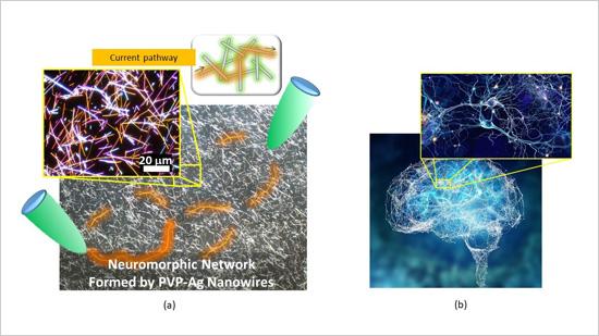
Caption: (a) Micrograph of the neuromorphic network fabricated by this research team. The network contains of numerous junctions between nanowires, which operate as synaptic elements. When voltage is applied to the network (between the green probes), current pathways (orange) are formed in the network. (b) A Human brain and one of its neuronal networks. The brain is known to have a complex network structure and to operate by means of electrical signal propagation across the network. Credit: NIMS

A November 11, 2019 National Institute for Materials Science (Japan) press release (also on EurekAlert but dated December 25, 2019) first announced the news,

An international joint research team led by NIMS succeeded in fabricating a neuromorphic network composed of numerous metallic nanowires. Using this network, the team was able to generate electrical characteristics similar to those associated with higher order brain functions unique to humans, such as memorization, learning, forgetting, becoming alert and returning to calm. The team then clarified the mechanisms that induced these electrical characteristics.

The development of artificial intelligence (AI) techniques has been rapidly advancing in recent years and has begun impacting our lives in various ways. Although AI processes information in a manner similar to the human brain, the mechanisms by which human brains operate are still largely unknown. Fundamental brain components, such as neurons and the junctions between them (synapses), have been studied in detail. However, many questions concerning the brain as a collective whole need to be answered. For example, we still do not fully understand how the brain performs such functions as memorization, learning and forgetting, and how the brain becomes alert and returns to calm. In addition, live brains are difficult to manipulate in experimental research. For these reasons, the brain remains a “mysterious organ.” A different approach to brain research?in which materials and systems capable of performing brain-like functions are created and their mechanisms are investigated?may be effective in identifying new applications of brain-like information processing and advancing brain science.

The joint research team recently built a complex brain-like network by integrating numerous silver (Ag) nanowires coated with a polymer (PVP) insulating layer approximately 1 nanometer in thickness. A junction between two nanowires forms a variable resistive element (i.e., a synaptic element) that behaves like a neuronal synapse. This nanowire network, which contains a large number of intricately interacting synaptic elements, forms a “neuromorphic network”. When a voltage was applied to the neuromorphic network, it appeared to “struggle” to find optimal current pathways (i.e., the most electrically efficient pathways). The research team measured the processes of current pathway formation, retention and deactivation while electric current was flowing through the network and found that these processes always fluctuate as they progress, similar to the human brain’s memorization, learning, and forgetting processes. The observed temporal fluctuations also resemble the processes by which the brain becomes alert or returns to calm. Brain-like functions simulated by the neuromorphic network were found to occur as the huge number of synaptic elements in the network collectively work to optimize current transport, in the other words, as a result of self-organized and emerging dynamic processes..

The research team is currently developing a brain-like memory device using the neuromorphic network material. The team intends to design the memory device to operate using fundamentally different principles than those used in current computers. For example, while computers are currently designed to spend as much time and electricity as necessary in pursuit of absolutely optimum solutions, the new memory device is intended to make a quick decision within particular limits even though the solution generated may not be absolutely optimum. The team also hopes that this research will facilitate understanding of the brain’s information processing mechanisms.

This project was carried out by an international joint research team led by Tomonobu Nakayama (Deputy Director, International Center for Materials Nanoarchitectonics (WPI-MANA), NIMS), Adrian Diaz Alvarez (Postdoctoral Researcher, WPI-MANA, NIMS), Zdenka Kuncic (Professor, School of Physics, University of Sydney, Australia) and James K. Gimzewski (Professor, California NanoSystems Institute, University of California Los Angeles, USA).

Here at last is a link to and a citation for the paper,

Emergent dynamics of neuromorphic nanowire networks by Adrian Diaz-Alvarez, Rintaro Higuchi, Paula Sanz-Leon, Ido Marcus, Yoshitaka Shingaya, Adam Z. Stieg, James K. Gimzewski, Zdenka Kuncic & Tomonobu Nakayama. Scientific Reports volume 9, Article number: 14920 (2019) DOI: https://doi.org/10.1038/s41598-019-51330-6 Published: 17 October 2019

This paper is open access.

CRISPR (clustered regularly interspaced short palindromic repeats) has a metaphor issue?

Elinor Hortie at the University of Sydney (Australia) has written a very interesting essay about CRISPR ‘scissors’, a metaphor she find misleading. From Hortie’s July 4, 2019 essay on The Conversation,

Last week I read an article about CRISPR, the latest tool scientists are using to edit DNA. It was a great piece – well researched, beautifully written, factually accurate. It covered some of the amazing projects scientist are working on using CRISPR, like bringing animals back from extinction and curing diseases. It also gave me the heebies, but not for the reason you might expect.

Take CRISPR. It’s most often described as a pair of molecular scissors that can be used to modify DNA, the blueprint for life. And when we read that, I think most of us start imagining something like a child with her Lego bricks strewn in front of her, instruction booklet in one hand, scissors in the other. One set of pictograms, one model; one gene, one disease; one snip, one cure. We’re there in a blink. CRISPR seems like it can work miracles.

I want to stress that the molecular scissors metaphor is pretty damn accurate as far as it goes. But in focusing on the relatively simple relationship between CRISPR and DNA, we miss the far more complicated relationship between DNA and the rest of the body. This metaphor ignores an entire ecosystem of moving parts that are crucial for understanding the awe-inspiring, absolutely insane thing scientists are trying to do when they attempt gene editing.

Hortie proposes a different metaphor,

In my research I use CRISPR from time to time. To design experiments and interpret results effectively, I need a solid way to conceptualise what it can (and can’t) do. I do not think of CRISPR as molecular scissors.

Instead I imagine a city. The greater metropolis represents the body, the suburbs are organs, the buildings are cells, the people are proteins, and the internet is DNA.

In this metaphor CRISPR is malware. More precisely, CRISPR is malware that can search for any chosen 20-character line of code and corrupt it. This is not a perfect metaphor by any stretch, but it gets me closer to understanding than almost anything else.

Hortie offers an example from her own work demonstrating how a CRISPR ‘malware’ metaphor/analogy more accurately represents the experience of using the gene-editing system,

As an example, let’s look at Alzheimer’s, one of the diseases CRISPR is being touted to cure. The headlines are usually some variation of “CRISPR to correct Alzheimer’s gene!”, and the molecular scissors analogy is never far behind.

It seems reasonable to me that someone could read those words and assume that chopping away the disease-gene with the DNA-shears should be relatively simple. When the cure doesn’t appear within five years, I can understand why that same person would come to ask me why Big Pharma is holding out (this has happened to me more than once).

Now let’s see how it looks using the malware metaphor. The consensus is that Alzheimer’s manifests when a specific protein goes rogue, causing damage to cells and thereby stopping things from working properly inside the brain. It might have a genetic cause, but it’s complicated. In our allegorical city, what would that look like?

I think riots would come close. Rampaging humans (proteins) destroying houses and property (cells), thereby seriously derailing the normal functioning of a specific suburb (the brain).

And you want to fix that with malware?

It’s hard to predict the domino effect

Can you imagine for a second trying to stop soccer hooligans smashing things on the streets of Buenos Aires by corrupting roughly three words in the FIFA by-laws with what’s essentially a jazzed-up command-F function?

I’m not saying it’s not possible – it absolutely is.

But think of all the prior knowledge you need, and all the pieces that have to fall in place for that to work. You’d have to know that the riots are caused by football fans. You’d have to understand which rule was bothering them (heaven help you if it’s more than one), and if that rule causes drama at every game. You’d have to find a 20-character phrase that, when corrupted, would change how the rule was read, rather than just making a trivial typo.

You’d have to know that the relevant footballers have access to the updated rule book, and you’d have to know there were no other regulations making your chosen rule redundant. You’d have to know there aren’t any similar 20-character phrases anywhere on the internet that might get corrupted at the same time (like in the rules for presidential succession say, or in the nuclear warhead codes). Even then you’d still be rolling the dice.

Even if you stop the riots successfully, which of us really know the long-term consequences of changing the World Game forever?

That’s stretching the metaphor as Hortie notes herself later in the essay. And, she’s not the only one concerned about metaphors and CRISPR. There’s a December 8, 2017* article by Rebecca Robbins for STAT news which covers ten analogies/metaphors ranked from worst to best,

… Some of these analogies are better than others. To compile the definitive ranking, I sat down with STAT’s senior science writer Sharon Begley, a wordsmith who has herself compared CRISPR to “1,000 monkeys editing a Word document” and the kind of dog “you can train to retrieve everything from Frisbees to slippers to a cold beer.”

Sharon and I evaluated each of the metaphors we found by considering these three questions: Is it creative? Is it clear? And is it accurate? Below, our rankings of CRISPR analogies, ordered from worst to best:

0. A knockout punch


9. The hand of God


8. A bomb removal squad

It’s a very interesting list with a description of why each does and doesn’t work as an analogy. By the way, ‘scissors’ was not the top analogy. The number one spot went to ‘A Swiss army knife’.

There are many more essays than I would have believed concerning CRISPR and metaphors/analogies. I’m glad to see them as the language we use to describe our work and our world helps us understand it and can constrain us in unexpected ways. Critiques such as Hortie’s and the others can help us to refine the language and to recognize its limitations.

h/t July 4, 2019 news item on phys.org

*”December 8, 0217′ corrected to ‘December 8, 2017’ on Jan.20.21

Trounce biofouling with nanowrinkles

This is an example of what the researchers mean by ‘nanowrinkles’,

Caption: The Nepenthes pitcher plant (left) and its nanowrinkled ‘mouth’ (centre) inspired the engineered nanomaterial (right). Credit: Sydney Nano Courtesy: University of Sydney

As for ‘biofouling’, here’s my rough and ready description for anyone who might find it helpful, if you’ve been to the beach and slipped on some rocks, that slip was probably due to a biofilm and biofilm contributes to ‘biofouling’.

Australian scientists have announced research on a new technique for the eradication of biofouling according to a January 17, 2018 University of Sydney press release (also on EurekAlert but dated Jan. 16, 2018),

Sydney scientists have developed nanowrinkled coatings that could avoid the build-up of damaging biological material and save some of the $320 million annually spent by the Australian shipping industry because of biofouling.

A team of chemistry researchers from the University of Sydney Nano Institute has developed nanostructured surface coatings that have anti-fouling properties without using any toxic components.

Biofouling – the build-up of damaging biological material – is a huge economic issue, costing the aquaculture and shipping industries billions of dollars a year in maintenance and extra fuel usage. It is estimated that the increased drag on ship hulls due to biofouling costs the shipping industry in Australia $320 million a year a b.

Since the banning of the toxic anti-fouling agent tributyltin, the need for new non-toxic methods to stop marine biofouling has been pressing.

Leader of the research team, Associate Professor Chiara Neto, said: “We are keen to understand how these surfaces work and also push the boundaries of their application, especially for energy efficiency. Slippery coatings are expected to be drag-reducing, which means that objects, such as ships, could move through water with much less energy required.”

The new materials were tested tied to shark netting in Sydney’s Watson Bay, showing that the nanomaterials were efficient at resisting biofouling in a marine environment.

The research has been published in ACS Applied Materials & Interfaces.

The new coating uses ‘nanowrinkles’ inspired by the carnivorous Nepenthes pitcher plant. The plant traps a layer of water on the tiny structures around the rim of its opening. This creates a slippery layer causing insects to aquaplane on the surface, before they slip into the pitcher where they are digested.

Nanostructures utilise materials engineered at the scale of billionths of a metre – 100,000 times smaller than the width of a human hair. Associate Professor Neto’s group at Sydney Nano is developing nanoscale materials for future development in industry.

Biofouling can occur on any surface that is wet for a long period of time, for example aquaculture nets, marine sensors and cameras, and ship hulls. The slippery surface developed by the Neto group stops the initial adhesion of bacteria, inhibiting the formation of a biofilm from which larger marine fouling organisms can grow.

The interdisciplinary University of Sydney team included biofouling expert Professor Truis Smith-Palmer of St Francis Xavier University in Nova Scotia, Canada, who was on sabbatical visit to the Neto group for a year, partially funded by the Faculty of Science scheme for visiting women.

In the lab, the slippery surfaces resisted almost all fouling from a common species of marine bacteria, while control Teflon samples without the lubricating layer were completely fouled. Not satisfied with testing the surfaces under highly controlled lab conditions with only one type of bacteria the team also tested the surfaces in the ocean, with the help of marine biologist Professor Ross Coleman.

Test surfaces were attached to swimming nets at Watsons Bay baths in Sydney Harbour for a period of seven weeks. In the much harsher marine environment, the slippery surfaces were still very efficient at resisting fouling.

The antifouling coatings are mouldable and transparent, making their application ideal for underwater cameras and sensors.

Sources for economic data:
a) M. P. Schultz, J. A. Bendick, E. R. Holm, W. M. Hertel, Biofouling 2011, 27, 87-98;
b) Western Australia Departmet of Fisheries, in Fisheries Occasional Publication No. 115, 2012

Even though there’s a link to the paper in the excerpt, here’s a citation and another link to the paper,

Marine Antifouling Behavior of Lubricant-Infused Nanowrinkled Polymeric Surfaces by
Cameron S. Ware, Truis Smith-Palmer, Sam Peppou-Chapman, Liam R. J. Scarratt, Erin M. Humphries, Daniel Balzer, and Chiara Neto. ACS Appl. Mater. Interfaces, Article ASAP DOI: 10.1021/acsami.7b14736 Publication Date (Web): December 18, 2017

Copyright © 2017 American Chemical Society

This paper is behind a paywall.

Making graphene cheaply by using soybeans

One of the issues with new materials is being able to produce them in a commercially viable fashion and it seems that researchers in Australia may have helped  to do that with graphene. From a Feb. 15, 2017 news item on phys.org,

A breakthrough by CSIRO-led [Australia’s Commonwealth Scientific and Industrial Research Organisation] scientists has made the world’s strongest material more commercially viable, thanks to the humble soybean.

From a Feb. 15, (?) 2017 CSIRO press release (also on EurekAlert), which originated the news item, expands on the theme (Note: A link has been removed),

Graphene is a carbon material that is one atom thick.

Its thin composition and high conductivity means it is used in applications ranging from miniaturised electronics to biomedical devices.

These properties also enable thinner wire connections; providing extensive benefits for computers, solar panels, batteries, sensors and other devices.

Until now, the high cost of graphene production has been the major roadblock in its commercialisation.

Previously, graphene was grown in a highly-controlled environment with explosive compressed gases, requiring long hours of operation at high temperatures and extensive vacuum processing.

CSIRO scientists have developed a novel “GraphAir” technology which eliminates the need for such a highly-controlled environment.

The technology grows graphene film in ambient air with a natural precursor, making its production faster and simpler.

“This ambient-air process for graphene fabrication is fast, simple, safe, potentially scalable, and integration-friendly,” CSIRO scientist Dr Zhao Jun Han, co-author of the paper published today in Nature Communications said.

“Our unique technology is expected to reduce the cost of graphene production and improve the uptake in new applications.”

GraphAir transforms soybean oil – a renewable, natural material – into graphene films in a single step.

“Our GraphAir technology results in good and transformable graphene properties, comparable to graphene made by conventional methods,” CSIRO scientist and co-author of the study Dr Dong Han Seo said.

With heat, soybean oil breaks down into a range of carbon building units that are essential for the synthesis of graphene.

The team also transformed other types of renewable and even waste oil, such as those leftover from barbecues or cooking, into graphene films.

“We can now recycle waste oils that would have otherwise been discarded and transform them into something useful,” Dr Seo said.

The potential applications of graphene include water filtration and purification, renewable energy, sensors, personalised healthcare and medicine, to name a few.

Graphene has excellent electronic, mechanical, thermal and optical properties as well.

Its uses range from improving battery performance in energy devices, to cheaper solar panels.

CSIRO are looking to partner with industry to find new uses for graphene.

Researchers from The University of Sydney, University of Technology Sydney and The Queensland University of Technology also contributed to this work.

Here’s a link to and a citation for the paper,

Single-step ambient-air synthesis of graphene from renewable precursors as electrochemical genosensor by Dong Han Seo, Shafique Pineda, Jinghua Fang, Yesim Gozukara, Samuel Yick, Avi Bendavid, Simon Kwai Hung Lam, Adrian T. Murdock, Anthony B. Murphy, Zhao Jun Han, & Kostya (Ken) Ostrikov. Nature Communications 8, Article number: 14217 (2017) doi:10.1038/ncomms14217 Published online: 30 January 2017

This is an open access paper.

Seeing the future with quantum computing

Researchers at the University of Sydney (Australia) have demonstrated the ability to see the ‘quantum future’ according to a Jan. 16, 2017 news item on ScienceDaily,

Scientists at the University of Sydney have demonstrated the ability to “see” the future of quantum systems, and used that knowledge to preempt their demise, in a major achievement that could help bring the strange and powerful world of quantum technology closer to reality.

The applications of quantum-enabled technologies are compelling and already demonstrating significant impacts — especially in the realm of sensing and metrology. And the potential to build exceptionally powerful quantum computers using quantum bits, or qubits, is driving investment from the world’s largest companies.

However a significant obstacle to building reliable quantum technologies has been the randomisation of quantum systems by their environments, or decoherence, which effectively destroys the useful quantum character.

The physicists have taken a technical quantum leap in addressing this, using techniques from big data to predict how quantum systems will change and then preventing the system’s breakdown from occurring.

A Jan. 14, 2017 University of Sydney press release (also on EurekAlert), which originated the news item, expands on the theme,

“Much the way the individual components in mobile phones will eventually fail, so too do quantum systems,” said the paper’s senior author Professor Michael J.  Biercuk.

“But in quantum technology the lifetime is generally measured in fractions of a second, rather than years.”

Professor Biercuk, from the University of Sydney’s School of Physics and a chief investigator at the Australian Research Council’s Centre of Excellence for Engineered Quantum Systems, said his group had demonstrated it was possible to suppress decoherence in a preventive manner. The key was to develop a technique to predict how the system would disintegrate.

Professor Biercuk highlighted the challenges of making predictions in a quantum world: “Humans routinely employ predictive techniques in our daily experience; for instance, when we play tennis we predict where the ball will end up based on observations of the airborne ball,” he said.

“This works because the rules that govern how the ball will move, like gravity, are regular and known.  But what if the rules changed randomly while the ball was on its way to you?  In that case it’s next to impossible to predict the future behavior of that ball.

“And yet this situation is exactly what we had to deal with because the disintegration of quantum systems is random. Moreover, in the quantum realm observation erases quantumness, so our team needed to be able to guess how and when the system would randomly break.

“We effectively needed to swing at the randomly moving tennis ball while blindfolded.”

The team turned to machine learning for help in keeping their quantum systems – qubits realised in trapped atoms – from breaking.

What might look like random behavior actually contained enough information for a computer program to guess how the system would change in the future. It could then predict the future without direct observation, which would otherwise erase the system’s useful characteristics.

The predictions were remarkably accurate, allowing the team to use their guesses preemptively to compensate for the anticipated changes.

Doing this in real time allowed the team to prevent the disintegration of the quantum character, extending the useful lifetime of the qubits.

“We know that building real quantum technologies will require major advances in our ability to control and stabilise qubits – to make them useful in applications,” Professor Biercuk said.

Our techniques apply to any qubit, built in any technology, including the special superconducting circuits being used by major corporations.

“We’re excited to be developing new capabilities that turn quantum systems from novelties into useful technologies. The quantum future is looking better all the time,” Professor Biercuk said.

Here’s a link to and a  citation for the paper,

Prediction and real-time compensation of qubit decoherence via machine learning by Sandeep Mavadia, Virginia Frey, Jarrah Sastrawan, Stephen Dona, & Michael J. Biercuk. Nature Communications 8, Article number: 14106 (2017) doi:10.1038/ncomms14106 Published online: 16 January 2017

This paper is open access.

‘Smart’ fabric that’s bony

Researchers at Australia’s University of New South of Wales (UNSW) have devised a means of ‘weaving’ a material that mimics *bone tissue, periosteum according to a Jan. 11, 2017 news item on ScienceDaily,

For the first time, UNSW [University of New South Wales] biomedical engineers have woven a ‘smart’ fabric that mimics the sophisticated and complex properties of one nature’s ingenious materials, the bone tissue periosteum.

Having achieved proof of concept, the researchers are now ready to produce fabric prototypes for a range of advanced functional materials that could transform the medical, safety and transport sectors. Patents for the innovation are pending in Australia, the United States and Europe.

Potential future applications range from protective suits that stiffen under high impact for skiers, racing-car drivers and astronauts, through to ‘intelligent’ compression bandages for deep-vein thrombosis that respond to the wearer’s movement and safer steel-belt radial tyres.

A Jan. 11, 2017 UNSW press release on EurekAlert, which originated the news item, expands on the theme,

Many animal and plant tissues exhibit ‘smart’ and adaptive properties. One such material is the periosteum, a soft tissue sleeve that envelops most bony surfaces in the body. The complex arrangement of collagen, elastin and other structural proteins gives periosteum amazing resilience and provides bones with added strength under high impact loads.

Until now, a lack of scalable ‘bottom-up’ approaches by researchers has stymied their ability to use smart tissues to create advanced functional materials.

UNSW’s Paul Trainor Chair of Biomedical Engineering, Professor Melissa Knothe Tate, said her team had for the first time mapped the complex tissue architectures of the periosteum, visualised them in 3D on a computer, scaled up the key components and produced prototypes using weaving loom technology.

“The result is a series of textile swatch prototypes that mimic periosteum’s smart stress-strain properties. We have also demonstrated the feasibility of using this technique to test other fibres to produce a whole range of new textiles,” Professor Knothe Tate said.

In order to understand the functional capacity of the periosteum, the team used an incredibly high fidelity imaging system to investigate and map its architecture.

“We then tested the feasibility of rendering periosteum’s natural tissue weaves using computer-aided design software,” Professor Knothe Tate said.

The computer modelling allowed the researchers to scale up nature’s architectural patterns to weave periosteum-inspired, multidimensional fabrics using a state-of-the-art computer-controlled jacquard loom. The loom is known as the original rudimentary computer, first unveiled in 1801.

“The challenge with using collagen and elastin is their fibres, that are too small to fit into the loom. So we used elastic material that mimics elastin and silk that mimics collagen,” Professor Knothe Tate said.

In a first test of the scaled-up tissue weaving concept, a series of textile swatch prototypes were woven, using specific combinations of collagen and elastin in a twill pattern designed to mirror periosteum’s weave. Mechanical testing of the swatches showed they exhibited similar properties found in periosteum’s natural collagen and elastin weave.

First author and biomedical engineering PhD candidate, Joanna Ng, said the technique had significant implications for the development of next-generation advanced materials and mechanically functional textiles.

While the materials produced by the jacquard loom have potential manufacturing applications – one tyremaker believes a titanium weave could spawn a new generation of thinner, stronger and safer steel-belt radials – the UNSW team is ultimately focused on the machine’s human potential.

“Our longer term goal is to weave biological tissues – essentially human body parts – in the lab to replace and repair our failing joints that reflect the biology, architecture and mechanical properties of the periosteum,” Ms Ng said.

An NHMRC development grant received in November [2016] will allow the team to take its research to the next phase. The researchers will work with the Cleveland Clinic and the University of Sydney’s Professor Tony Weiss to develop and commercialise prototype bone implants for pre-clinical research, using the ‘smart’ technology, within three years.

In searching for more information about this work, I found a Winter 2015 article (PDF; pp. 8-11) by Amy Coopes and Steve Offner for UNSW Magazine about Knothe Tate and her work (Note: In Australia, winter would be what we in the Northern Hemisphere consider summer),

Tucked away in a small room in UNSW’s Graduate School of Biomedical Engineering sits a 19th century–era weaver’s wooden loom. Operated by punch cards and hooks, the machine was the first rudimentary computer when it was unveiled in 1801. While on the surface it looks like a standard Jacquard loom, it has been enhanced with motherboards integrated into each of the loom’s five hook modules and connected to a computer. This state-of-the-art technology means complex algorithms control each of the 5,000 feed-in fibres with incredible precision.

That capacity means the loom can weave with an extraordinary variety of substances, from glass and titanium to rayon and silk, a development that has attracted industry attention around the world.

The interest lies in the natural advantage woven materials have over other manufactured substances. Instead of manipulating material to create new shades or hues as in traditional weaving, the fabrics’ mechanical properties can be modulated, to be stiff at one end, for example, and more flexible at the other.

“Instead of a pattern of colours we get a pattern of mechanical properties,” says Melissa Knothe Tate, UNSW’s Paul Trainor Chair of Biomedical Engineering. “Think of a rope; it’s uniquely good in tension and in bending. Weaving is naturally strong in that way.”


The interface of mechanics and physiology is the focus of Knothe Tate’s work. In March [2015], she travelled to the United States to present another aspect of her work at a meeting of the international Orthopedic Research Society in Las Vegas. That project – which has been dubbed “Google Maps for the body” – explores the interaction between cells and their environment in osteoporosis and other degenerative musculoskeletal conditions such as osteoarthritis.

Using previously top-secret semiconductor technology developed by optics giant Zeiss, and the same approach used by Google Maps to locate users with pinpoint accuracy, Knothe Tate and her team have created “zoomable” anatomical maps from the scale of a human joint down to a single cell.

She has also spearheaded a groundbreaking partnership that includes the Cleveland Clinic, and Brown and Stanford universities to help crunch terabytes of data gathered from human hip studies – all processed with the Google technology. Analysis that once took 25 years can now be done in a matter of weeks, bringing researchers ever closer to a set of laws that govern biological behaviour. [p. 9]

I gather she was recruited from the US to work at the University of New South Wales and this article was to highlight why they recruited her and to promote the university’s biomedical engineering department, which she chairs.

Getting back to 2017, here’s a link to and citation for the paper,

Scale-up of nature’s tissue weaving algorithms to engineer advanced functional materials by Joanna L. Ng, Lillian E. Knothe, Renee M. Whan, Ulf Knothe & Melissa L. Knothe Tate. Scientific Reports 7, Article number: 40396 (2017) doi:10.1038/srep40396 Published online: 11 January 2017

This paper is open access.

One final comment, that’s a lot of people (three out of five) with the last name Knothe in the author’s list for the paper.

*’the bone tissue’ changed to ‘bone tissue’ on July 17,2017.