Tag Archives: University of Texas at San Antonio

The inside scoop on beetle exoskeletons

In the past I’ve covered work on the Namib beetle and its bumps which allow it to access condensation from the air in one of the hottest places on earth and work on jewel beetles and how their structural colo(u)r is derived. Now, there’s research into a beetle’s body armor from the University of Nebraska-Lincoln according to a Feb. 22, 2017 news item on ScienceDaily,

Beetles wear a body armor that should weigh them down — think medieval knights and turtles. In fact, those hard shells protecting delicate wings are surprisingly light, allowing even flight.

Better understanding the structure and properties of beetle exoskeletons could help scientists engineer lighter, stronger materials. Such materials could, for example, reduce gas-guzzling drag in vehicles and airplanes and reduce the weight of armor, lightening the load for the 21st-century knight.

But revealing exoskeleton architecture at the nanoscale has proven difficult. Nebraska’s Ruiguo Yang, assistant professor of mechanical and materials engineering, and his colleagues found a way to analyze the fibrous nanostructure. …

A Feb. 22, 2017 University of Nebraska-Lincoln news release by Gillian Klucas (also on EurekAlert), which originated the news item, describes skeletons and the work in more detail,

The lightweight exoskeleton is composed of chitin fibers just around 20 nanometers in diameter (a human hair measures approximately 75,000 nanometers in diameter) and packed and piled into layers that twist in a spiral, like a spiral staircase. The small diameter and helical twisting, known as Bouligand, make the structure difficult to analyze.

Yang and his team developed a method of slicing down the spiral to reveal a surface of cross-sections of fibers at different orientations. From that viewpoint, the researchers were able to analyze the fibers’ mechanical properties with the aid of an atomic force microscope. This type of microscope applies a tiny force to a test sample, deforms the sample and monitors the sample’s response. Combining the experimental procedure and theoretical analysis, the researchers were able to reveal the nanoscale architecture of the exoskeleton and the material properties of the nanofibers.

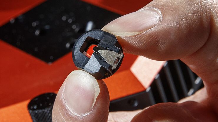
Yang holds a piece of the atomic force microscope used to measure the beetle's surface. A small wire can barely be seen in the middle of the piece. Unseen is a two-nano-size probe attached to the wire, which does the actual measuring.

Craig Chandler | University Communication

Yang holds a piece of the atomic force microscope used to measure the beetle’s surface. A small wire can barely be seen in the middle of the piece. Unseen is a two-nano-size probe attached to the wire, which does the actual measuring.

They made their discoveries in the common figeater beetle, Cotinis mutabilis, a metallic green native of the western United States. But the technique can be used on other beetles and hard-shelled creatures and might also extend to artificial materials with fibrous structures, Yang said.

Comparing beetles with differing demands on their exoskeletons, such as defending against predators or environmental damage, could lead to evolutionary insights as well as a better understanding of the relationship between structural features and their properties.

Yang’s co-authors are Alireza Zaheri and Horacio Espinosa of Northwestern University; Wei Gao of the University of Texas at San Antonio; and Cheryl Hayashi of the University of California, Riverside.

Here’s a link to and a citation for the paper,

Exoskeletons: AFM Identification of Beetle Exocuticle: Bouligand Structure and Nanofiber Anisotropic Elastic Properties by Ruiguo Yang, Alireza Zaheri,Wei Gao, Charely Hayashi, Horacio D. Espinosa. Adv. Funct. Mater. vol. 27 (6) 2017 DOI: 10.1002/adfm.201770031 First published: 8 February 2017

This paper is behind a paywall.
Nanorods as multistate switches

This research goes beyond the binary (0 or 1) and to an analog state that resembles quantum states. Fascinating, yes? An Oct. 10, 2016 news item on phys.org tells more,

Rice University scientists have discovered how to subtly change the interior structure of semi-hollow nanorods in a way that alters how they interact with light, and because the changes are reversible, the method could form the basis of a nanoscale switch with enormous potential.

“It’s not 0-1, it’s 1-2-3-4-5-6-7-8-9-10,” said Rice materials scientist Emilie Ringe, lead scientist on the project, which is detailed in the American Chemical Society journal Nano Letters. “You can differentiate between multiple plasmonic states in a single particle. That gives you a kind of analog version of quantum states, but on a larger, more accessible scale.”

Ringe and colleagues used an electron beam to move silver from one location to another inside gold-and-silver nanoparticles, something like a nanoscale Etch A Sketch. The result is a reconfigurable optical switch that may form the basis for a new type of multiple-state computer memory, sensor or catalyst.

An Oct. 10, 2016 Rice University news release, which originated the news item, describes the work in additional detail,

At about 200 nanometers long, 500 of the metal rods placed end-to-end would span the width of a human hair. However, they are large in comparison with modern integrated circuits. Their multistate capabilities make them more like reprogrammable bar codes than simple memory bits, she said.

“No one has been able to reversibly change the shape of a single particle with the level of control we have, so we’re really excited about this,” Ringe said.

Altering a nanoparticle’s internal structure also alters its external plasmonic response. Plasmons are the electrical ripples that propagate across the surface of metallic materials when excited by light, and their oscillations can be easily read with a spectrometer — or even the human eye — as they interact with visible light.

The Rice researchers found they could reconfigure nanoparticle cores with pinpoint precision. That means memories made of nanorods need not be merely on-off, Ringe said, because a particle can be programmed to emit many distinct plasmonic patterns.

The discovery came about when Ringe and her team, which manages Rice’s advanced electron microscopy lab, were asked by her colleague and co-author Denis Boudreau, a professor at Laval University in Quebec, to characterize hollow nanorods made primarily of gold but containing silver.

“Most nanoshells are leaky,” Ringe said. “They have pinholes. But we realized these nanorods were defect-free and contained pockets of water that were trapped inside when the particles were synthesized. We thought: We have something here.”

Ringe and the study’s lead author, Rice research scientist Sadegh Yazdi, quickly realized how they might manipulate the water. “Obviously, it’s difficult to do chemistry there, because you can’t put molecules into a sealed nanoshell. But we could put electrons in,” she said.

Focusing a subnanometer electron beam on the interior cavity split the water and inserted solvated electrons – free electrons that can exist in a solution. “The electrons reacted directly with silver ions in the water, drawing them to the beam to form silver,” Ringe said. The now-silver-poor liquid moved away from the beam, and its silver ions were replenished by a reaction of water-splitting byproducts with the solid silver in other parts of the rod.

“We actually were moving silver in the solution, reconfiguring it,” she said. “Because it’s a closed system, we weren’t losing anything and we weren’t gaining anything. We were just moving it around, and could do so as many times as we wished.”

The researchers were then able to map the plasmon-induced near-field properties without disturbing the internal structure — and that’s when they realized the implications of their discovery.

“We made different shapes inside the nanorods, and because we specialize in plasmonics, we mapped the plasmons and it turned out to have a very nice effect,” Ringe said. “We basically saw different electric-field distributions at different energies for different shapes.” Numerical results provided by collaborators Nicolas Large of the University of Texas at San Antonio and George Schatz of Northwestern University helped explain the origin of the modes and how the presence of a water-filled pocket created a multitude of plasmons, she said.

The next challenge is to test nanoshells of other shapes and sizes, and to see if there are other ways to activate their switching potentials. Ringe suspects electron beams may remain the best and perhaps only way to catalyze reactions inside particles, and she is hopeful.

“Using an electron beam is actually not as technologically irrelevant as you might think,” she said. “Electron beams are very easy to generate. And yes, things need to be in vacuum, but other than that, people have generated electron beams for nearly 100 years. I’m sure 40 years ago people were saying, ‘You’re going to put a laser in a disk reader? That’s crazy!’ But they managed to do it.

“I don’t think it’s unfeasible to miniaturize electron-beam technology. Humans are good at moving electrons and electricity around. We figured that out a long time ago,” Ringe said.

The research should trigger the imaginations of scientists working to create nanoscale machines and processes, she said.

“This is a reconfigurable unit that you can access with light,” she said. “Reading something with light is much faster than reading with electrons, so I think this is going to get attention from people who think about dynamic systems and people who think about how to go beyond current nanotechnology. This really opens up a new field.”

Here’s a link to and a citation for the paper,

Reversible Shape and Plasmon Tuning in Hollow AgAu Nanorods by Sadegh Yazdi, Josée R. Daniel, Nicolas Large, George C. Schatz, Denis Boudreau, and Emilie Ringe. Nano Lett., Article ASAP DOI: 10.1021/acs.nanolett.6b02946 Publication Date (Web): October 5, 2016

Copyright © 2016 American Chemical Society

This paper is behind a paywall.

The researchers have made this video available for the public,

Gold-144 is a polymorph

Au-144 (also known as Gold-144) is an iconic gold nanocluster according to a June 14, 2016 news item announcing its polymorphic nature on ScienceDaily,

Chemically the same, graphite and diamonds are as physically distinct as two minerals can be, one opaque and soft, the other translucent and hard. What makes them unique is their differing arrangement of carbon atoms.

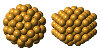
Polymorphs, or materials with the same composition but different structures, are common in bulk materials, and now a new study in Nature Communications confirms they exist in nanomaterials, too. Researchers describe two unique structures for the iconic gold nanocluster Au144(SR)60, better known as Gold-144, including a version never seen before. Their discovery gives engineers a new material to explore, along with the possibility of finding other polymorphic nanoparticles.

A June 14, 2016 Columbia University news release (also on EurekAlert), which originated the news item, provides more insight into the work,

“This took four years to unravel,” said Simon Billinge, a physics professor at Columbia Engineering and a member of the Data Science Institute. “We weren’t expecting the clusters to take on more than one atomic arrangement. But this discovery gives us more handles to turn when trying to design clusters with new and useful properties.”

Gold has been used in coins and jewelry for thousands of years for its durability, but shrink it to a size 10,000 times smaller than a human hair [at one time one billionth of a meter or a nanometer was said to be 1/50,000, 1/60,000 or 1/100,000 of the diameter of a human hair], and it becomes wildly unstable and unpredictable. At the nanoscale, gold likes to split apart other particles and molecules, making it a useful material for purifying water, imaging and killing tumors, and making solar panels more efficient, among other applications.

Though a variety of nanogold particles and molecules have been made in the lab, very few have had their secret atomic arrangement revealed. But recently, new technologies are bringing these miniscule structures into focus.

Under one approach, high-energy x-ray beams are fired at a sample of nanoparticles. Advanced data analytics are used to interpret the x-ray scattering data and infer the sample’s structure, which is key to understanding how strong, reactive or durable the particles might be.

Billinge and his lab have pioneered a method, the atomic Pair Distribution Function (PDF) analysis, for interpreting this scattering data. To test the PDF method, Billinge asked chemists at the Colorado State University to make tiny samples of Gold-144, a molecule-sized nanogold cluster first isolated in 1995. Its structure had been theoretically predicted in 2009, and though never confirmed, Gold-144 has found numerous applications, including in tissue-imaging.

Hoping the test would confirm Gold-144’s structure, they analyzed the clusters at the European Synchrotron Radiation Source in Grenoble, and used the PDF method to infer their structure. To their surprise, they found an angular core, and not the sphere-like icosahedral core predicted. When they made a new sample and tried the experiment again, this time using synchrotrons at Brookhaven and Argonne national laboratories, the structure came back spherical.

“We didn’t understand what was going on, but digging deeper, we realized we had a polymorph,” said study coauthor Kirsten Jensen, formerly a postdoctoral researcher at Columbia, now a chemistry professor at the University of Copenhagen.

Further experiments confirmed the cluster had two versions, sometimes found together, each with a unique structure indicating they behave differently. The researchers are still unsure if Gold-144 can switch from one version to the other or, what exactly, differentiates the two forms.

To make their discovery, the researchers solved what physicists call the nanostructure inverse problem. How can the structure of a tiny nanoparticle in a sample be inferred from an x-ray signal that has been averaged over millions of particles, each with different orientations?

“The signal is noisy and highly degraded,” said Billinge. “It’s the equivalent of trying to recognize if the bird in the tree is a robin or a cardinal, but the image in your binoculars is too blurry and distorted to tell.”

“Our results demonstrate the power of PDF analysis to reveal the structure of very tiny particles,” added study coauthor Christopher Ackerson, a chemistry professor at Colorado State. “I’ve been trying, off and on, for more than 10 years to get the single-crystal x-ray structure of Gold-144. The presence of polymorphs helps to explain why this molecule has been so resistant to traditional methods.”

The PDF approach is one of several rival methods being developed to bring nanoparticle structure into focus. Now that it has proven itself, it could help speed up the work of describing other nanostructures.

The eventual goal is to design nanoparticles by their desired properties, rather than through trial and error, by understanding how form and function relate. Databases of known and predicted structures could make it possible to design new materials with a few clicks of a mouse.

The study is a first step.

“We’ve had a structure model for this iconic gold molecule for years and then this study comes along and says the structure is basically right but it’s got a doppelgänger,” said Robert Whetten, a professor of chemical physics at the University of Texas, San Antonio, who led the team that first isolated Gold-144. “It seemed preposterous, to have two distinct structures that underlie its ubiquity, but this is a beautiful paper that will persuade a lot of people.”

Here’s an image illustrating the two shapes,

Setting out to confirm the predicted structure of Gold-144, researchers discovered an entirely unexpected atomic arrangement (right). The two structures, described in detail for the first time, each have 144 gold atoms, but are uniquely shaped, suggesting they also behave differently. (Courtesy of Kirsten Ørnsbjerg Jensen)

Setting out to confirm the predicted structure of Gold-144, researchers discovered an entirely unexpected atomic arrangement (right). The two structures, described in detail for the first time, each have 144 gold atoms, but are uniquely shaped, suggesting they also behave differently. (Courtesy of Kirsten Ørnsbjerg Jensen)

Here’s a link to and a citation for the paper,

Polymorphism in magic-sized Au144(SR)60 clusters by Kirsten M.Ø. Jensen, Pavol Juhas, Marcus A. Tofanelli, Christine L. Heinecke, Gavin Vaughan, Christopher J. Ackerson, & Simon J. L. Billinge.  Nature Communications 7, Article number: 11859  doi:10.1038/ncomms11859 Published 14 June 2016

This is an open access paper.