According to the two items I have here, the answer is: yes, we have neurons that are specific to math and to the sound of singing.
Math neurons
A February 14, 2022 news item on ScienceDaily explains how specific the math neurons are,
The brain has neurons that fire specifically during certain mathematical operations. This is shown by a recent study conducted by the Universities of Tübingen and Bonn [both in Germany]. The findings indicate that some of the neurons detected are active exclusively during additions, while others are active during subtractions. They do not care whether the calculation instruction is written down as a word or a symbol. The results have now been published in the journal Current Biology.
…
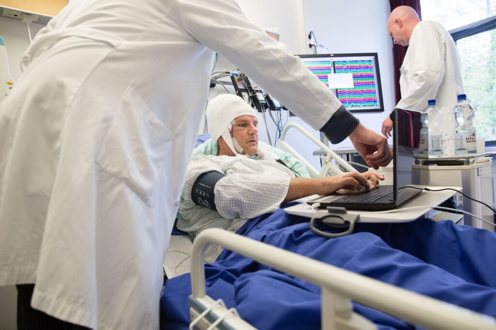
A February 14, 2022 University of Bonn press release (also on EurekAlert), which originated the news item, delves further,
Most elementary school children probably already know that three apples plus two apples add up to five apples. However, what happens in the brain during such calculations is still largely unknown. The current study by the Universities of Bonn and Tübingen now sheds light on this issue.
The researchers benefited from a special feature of the Department of Epileptology at the University Hospital Bonn. It specializes in surgical procedures on the brains of people with epilepsy. In some patients, seizures always originate from the same area of the brain. In order to precisely localize this defective area, the doctors implant several electrodes into the patients. The probes can be used to precisely determine the origin of the spasm. In addition, the activity of individual neurons can be measured via the wiring.
Some neurons fire only when summing up
Five women and four men participated in the current study. They had electrodes implanted in the so-called temporal lobe of the brain to record the activity of nerve cells. Meanwhile, the participants had to perform simple arithmetic tasks. “We found that different neurons fired during additions than during subtractions,” explains Prof. Florian Mormann from the Department of Epileptology at the University Hospital Bonn.
It was not the case that some neurons responded only to a “+” sign and others only to a “-” sign: “Even when we replaced the mathematical symbols with words, the effect remained the same,” explains Esther Kutter, who is doing her doctorate in Prof. Mormann’s research group. “For example, when subjects were asked to calculate ‘5 and 3’, their addition neurons sprang back into action; whereas for ‘7 less 4,’ their subtraction neurons did.”
This shows that the cells discovered actually encode a mathematical instruction for action. The brain activity thus showed with great accuracy what kind of tasks the test subjects were currently calculating: The researchers fed the cells’ activity patterns into a self-learning computer program. At the same time, they told the software whether the subjects were currently calculating a sum or a difference. When the algorithm was confronted with new activity data after this training phase, it was able to accurately identify during which computational operation it had been recorded.
Prof. Andreas Nieder from the University of Tübingen supervised the study together with Prof. Mormann. “We know from experiments with monkeys that neurons specific to certain computational rules also exist in their brains,” he says. “In humans, however, there is hardly any data in this regard.” During their analysis, the two working groups came across an interesting phenomenon: One of the brain regions studied was the so-called parahippocampal cortex. There, too, the researchers found nerve cells that fired specifically during addition or subtraction. However, when summing up, different addition neurons became alternately active during one and the same arithmetic task. Figuratively speaking, it is as if the plus key on the calculator were constantly changing its location. It was the same with subtraction. Researchers also refer to this as “dynamic coding.”
“This study marks an important step towards a better understanding of one of our most important symbolic abilities, namely calculating with numbers,” stresses Mormann. The two teams from Bonn and Tübingen now want to investigate exactly what role the nerve cells found play in this.
Funding:
The study was funded by the German Research Foundation (DFG) and the Volkswagen Foundation.
Here’s a link to and a citation for the paper,
Neuronal codes for arithmetic rule processing in the human brain by Esther F. Kutter, Jan Boström, Christian E. Elger, Andreas Nieder, Florian Mormann. Current Biology, 2022; DOI: 10.1016/j.cub.2022.01.054 Published February 14, 2022
This paper appears to be open access.
Neurons for the sounds of singing
This work from the Massachusetts Institute of Technology (MIT) according to a February 22, 2022 news item on ScienceDaily,
For the first time, MIT neuroscientists have identified a population of neurons in the human brain that lights up when we hear singing, but not other types of music.
…
Pretty nifty, eh? As is the news release headline with its nod to a classic Hollywood musical and song, from a February 22, 2022 MIT news release (also on EurekAlert),
Singing in the brain
…
These neurons, found in the auditory cortex, appear to respond to the specific combination of voice and music, but not to either regular speech or instrumental music. Exactly what they are doing is unknown and will require more work to uncover, the researchers say.
“The work provides evidence for relatively fine-grained segregation of function within the auditory cortex, in a way that aligns with an intuitive distinction within music,” says Sam Norman-Haignere, a former MIT postdoc who is now an assistant professor of neuroscience at the University of Rochester Medical Center.
The work builds on a 2015 study in which the same research team used functional magnetic resonance imaging (fMRI) to identify a population of neurons in the brain’s auditory cortex that responds specifically to music. In the new work, the researchers used recordings of electrical activity taken at the surface of the brain, which gave them much more precise information than fMRI.
“There’s one population of neurons that responds to singing, and then very nearby is another population of neurons that responds broadly to lots of music. At the scale of fMRI, they’re so close that you can’t disentangle them, but with intracranial recordings, we get additional resolution, and that’s what we believe allowed us to pick them apart,” says Norman-Haignere.
Norman-Haignere is the lead author of the study, which appears today in the journal Current Biology. Josh McDermott, an associate professor of brain and cognitive sciences, and Nancy Kanwisher, the Walter A. Rosenblith Professor of Cognitive Neuroscience, both members of MIT’s McGovern Institute for Brain Research and Center for Brains, Minds and Machines (CBMM), are the senior authors of the study.
Neural recordings
In their 2015 study, the researchers used fMRI to scan the brains of participants as they listened to a collection of 165 sounds, including different types of speech and music, as well as everyday sounds such as finger tapping or a dog barking. For that study, the researchers devised a novel method of analyzing the fMRI data, which allowed them to identify six neural populations with different response patterns, including the music-selective population and another population that responds selectively to speech.
In the new study, the researchers hoped to obtain higher-resolution data using a technique known as electrocorticography (ECoG), which allows electrical activity to be recorded by electrodes placed inside the skull. This offers a much more precise picture of electrical activity in the brain compared to fMRI, which measures blood flow in the brain as a proxy of neuron activity.
“With most of the methods in human cognitive neuroscience, you can’t see the neural representations,” Kanwisher says. “Most of the kind of data we can collect can tell us that here’s a piece of brain that does something, but that’s pretty limited. We want to know what’s represented in there.”
Electrocorticography cannot be typically be performed in humans because it is an invasive procedure, but it is often used to monitor patients with epilepsy who are about to undergo surgery to treat their seizures. Patients are monitored over several days so that doctors can determine where their seizures are originating before operating. During that time, if patients agree, they can participate in studies that involve measuring their brain activity while performing certain tasks. For this study, the MIT team was able to gather data from 15 participants over several years.
For those participants, the researchers played the same set of 165 sounds that they used in the earlier fMRI study. The location of each patient’s electrodes was determined by their surgeons, so some did not pick up any responses to auditory input, but many did. Using a novel statistical analysis that they developed, the researchers were able to infer the types of neural populations that produced the data that were recorded by each electrode.
“When we applied this method to this data set, this neural response pattern popped out that only responded to singing,” Norman-Haignere says. “This was a finding we really didn’t expect, so it very much justifies the whole point of the approach, which is to reveal potentially novel things you might not think to look for.”
That song-specific population of neurons had very weak responses to either speech or instrumental music, and therefore is distinct from the music- and speech-selective populations identified in their 2015 study.
Music in the brain
In the second part of their study, the researchers devised a mathematical method to combine the data from the intracranial recordings with the fMRI data from their 2015 study. Because fMRI can cover a much larger portion of the brain, this allowed them to determine more precisely the locations of the neural populations that respond to singing.
“This way of combining ECoG and fMRI is a significant methodological advance,” McDermott says. “A lot of people have been doing ECoG over the past 10 or 15 years, but it’s always been limited by this issue of the sparsity of the recordings. Sam is really the first person who figured out how to combine the improved resolution of the electrode recordings with fMRI data to get better localization of the overall responses.”
The song-specific hotspot that they found is located at the top of the temporal lobe, near regions that are selective for language and music. That location suggests that the song-specific population may be responding to features such as the perceived pitch, or the interaction between words and perceived pitch, before sending information to other parts of the brain for further processing, the researchers say.
The researchers now hope to learn more about what aspects of singing drive the responses of these neurons. They are also working with MIT Professor Rebecca Saxe’s lab to study whether infants have music-selective areas, in hopes of learning more about when and how these brain regions develop.
Here’s a link to and a citation for the paper,
A neural population selective for song in human auditory cortex by Sam V. Norman-Haignere, Jenelle Feather, Dana Boebinger, Peter Brunner, Anthony Ritaccio, Josh H. Mcdermott, Gerwin Schalk, Nancy Kanwisher. Current Biology, 2022 DOI: 10.1016/j.cub.2022.01.069 Published February 22, 2022.
This paper appears to be open access.
I couldn’t resist,