Asphaltene (or asphaltenes are) is waste material that can be turned into graphene according to scientists at Rice University (Texas, US), from a November 18, 2022 news item on ScienceDaily,
Asphaltenes, a byproduct of crude oil production, are a waste material with potential. Rice University scientists are determined to find it by converting the carbon-rich resource into useful graphene.
Muhammad Rahman, an assistant research professor of materials science and nanoengineering, is employing Rice’s unique flash Joule heating process to convert asphaltenes instantly into turbostratic (loosely aligned) graphene and mix it into composites for thermal, anti-corrosion and 3D-printing applications.
The process makes good use of material otherwise burned for reuse as fuel or discarded into tailing ponds and landfills. Using at least some of the world’s reserve of more than 1 trillion barrels of asphaltene as a feedstock for graphene would be good for the environment as well.
“Asphaltene is a big headache for the oil industry, and I think there will be a lot of interest in this,” said Rahman, who characterized the process as both a scalable and sustainable way to reduce carbon emissions from burning asphaltene.
Rahman is a lead corresponding author of the paper in Science Advances co-led by Rice chemist James Tour, whose lab developed flash Joule heating, materials scientist Pulickel Ajayan and Md Golam Kibria, an assistant professor of chemical and petroleum engineering at the University of Calgary, Canada.
Asphaltenes are 70% to 80% carbon already. The Rice lab combines it with about 20% of carbon black to add conductivity and flashes it with a jolt of electricity, turning it into graphene in less than a second. Other elements in the feedstock, including hydrogen, nitrogen, oxygen and sulfur, are vented away as gases.
“We try to keep the carbon black content as low as possible because we want to maximize the utilization of asphaltene,” Rahman said.
“The government has been putting pressure on the petroleum industries to take care of this,” said Rice graduate student and co-lead author M.A.S.R. Saadi. “There are billions of barrels of asphaltene available, so we began working on this project primarily to see if we could make carbon fiber. That led us to think maybe we should try making graphene with flash Joule heating.”
Assured that Tour’s process worked as well on asphaltene as it did on various other feedstocks, including plastic, electronic waste, tires, coal fly ash and even car parts, the researchers set about making things with their graphene.
Saadi, who works with Rahman and Ajayan, mixed the graphene into composites, and then into polymer inks bound for 3D printers. “We’ve optimized the ink rheology to show that it is printable,” he said, noting the inks have no more than 10% of graphene mixed in. Mechanical testing of printed objects is forthcoming, he said.
Rice graduate student Paul Advincula, a member of the Tour lab, is co-lead author of the paper. Co-authors are Rice graduate students Md Shajedul Hoque Thakur, Ali Khater, Jacob Beckham and Minghe Lou, undergraduate Aasha Zinke and postdoctoral researcher Soumyabrata Roy; research fellow Shabab Saad, alumnus Ali Shayesteh Zeraati, graduate student Shariful Kibria Nabil and postdoctoral associate Md Abdullah Al Bari of the University of Calgary; graduate student Sravani Bheemasetti and Venkataramana Gadhamshetty, an associate professor, at the South Dakota School of Mines and Technology and its 2D Materials of Biofilm Engineering Science and Technology Center; and research assistant Yiwen Zheng and Aniruddh Vashisth, an assistant professor of mechanical engineering, of the University of Washington.
The research was funded by the Alberta Innovates for Carbon Fiber Grand Challenge programs, the Air Force Office of Scientific Research (FA9550-19-1-0296), the U.S. Army Corps of Engineers (W912HZ-21-2-0050) and the National Science Foundation (1849206, 1920954).
Here’s a link to and a citation for the paper,
Sustainable valorization of asphaltenes via flash joule heating by M.A.S.R. Saadi, Paul A. Advincula, Md Shajedul Hoque Thakur, Ali Zein Khater, Shabab Saad, Ali Shayesteh Zeraati, Shariful Kibria Nabil, Aasha Zinke, Soumyabrata Roy, Minghe Lou, Sravani N. Bheemasetti, Md Abdullah Al Bari, Yiwen Zheng, Jacob L. Beckham, Venkataramana Gadhamshetty, Aniruddh Vashisth, Md Golam Kibria, James M. Tour, Pulickel M. Ajayan, and Muhammad M. Rahman. Science Advances 18 Nov 2022 Vol 8, Issue 46 DOI: 10.1126/sciadv.add3555
A bio-inspired molecule that directs gold atoms to form perfect nanoscale stars? According to a March 30, 2022 news item on Nanowerk, that’s exactly what researchers have done (Note: Links have been removed),
Researchers from Pacific Northwest National Laboratory (PNNL) and the University of Washington (UW) have successfully designed a bio-inspired molecule that can direct gold atoms to form perfect nanoscale stars.
The work (Angewandte Chemie, “Peptoid-Directed Formation of Five-Fold Twinned Au Nanostars through Particle Attachment and Facet Stabilization”) is an important step toward understanding and controlling metal nanoparticle shape and creating advanced materials with tunable properties.
…
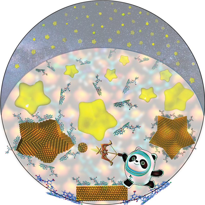
Artistic rendering of gold star assembly. Credit: Biao Jin Courtesy: University of Washington
I do love the fanciful addition of a panda to the proceedings. Thank you Biao Jin.
Metallic nanomaterials have interesting optical properties, called plasmonic properties, says Chun-Long Chen, who is a PNNL senior research scientist, UW affiliate professor of chemical engineering and of chemistry, and UW–PNNL Faculty Fellow. In particular, star-shaped metallic nanomaterials are already known to exhibit unique enhancements that are useful for sensing and the detection of pathogenic bacteria, among other national security and health applications.
To create these striking nanoparticles, the team carefully tuned sequences of peptoids, a type of programmable protein-like synthetic polymer. “Peptoids offer a unique advantage in achieving molecular-level controls,” says Chen. In this case, the peptoids guide small gold particles to attach and relax to form larger five-fold twinned ones, while also stabilizing the facets of the crystal structure. Their approach was inspired by nature, where proteins can control the creation of materials with advanced functionalities.
Jim De Yoreo and Biao Jin used advanced in situ transmission electron microscopy (TEM) to “see” the stars’ formation in solution at the nanoscale. The technique both provided an in-depth mechanistic understanding of how peptoids guide the process and revealed the roles of particle attachment and facet stabilization in controlling shape. De Yoreo is a Battelle Fellow at PNNL and affiliate professor of materials science and engineering at UW, and Jin is a postdoctoral research associate at PNNL.
Having assembled their nanoscale constellation, the researchers then employed molecular dynamics simulations to capture a level of detail that can’t be gleaned from experiments — and to illuminate why specific peptoids controlled the formation of the perfect stars. Xin Qi, a chemical engineering postdoctoral researcher in professor Jim Pfaendtner’s group, led this work at UW. Qi used UW’s Hyak supercomputer cluster to model interfacial phenomena between several different peptoids and particle surfaces.
The simulations play a critical role in learning how to design plasmonic nanomaterials that absorb and scatter light in unique ways. “You need to have a molecular-level understanding to form this nice star-shaped particle with interesting plasmonic properties,” said Chen. Simulations can build the theoretical understanding around why certain peptoids create certain shapes.
The researchers are working toward a future where simulations guide experimental design, in a cycle the team hopes will lead to predictive synthesis of nanomaterials with desired plasmonic enhancements. In this aspect, they would like to first use computational tools to identify peptoid side chains and sequences with desired facet selectivity. Then they would employ state-of-art in situ imaging techniques, such as liquid-cell TEM [transmission electron microscope], to monitor the direct facet expression, stabilization, and particle attachment. In other words, Chen says, “If someone can tell us that a structure of plasmonic nanomaterials has interesting optical properties, can we use a peptoid-based approach to predictably make that?”
Though they’re not to that point, this successful experimental–computational work certainly gets them closer. Further, the team’s ability to synthesize nice star shapes consistently is an important step; more-homogeneous particles translate into more-predictable optical properties.
A robot operating with a popular Internet-based artificial intelligence system consistently gravitates to men over women, white people over people of color, and jumps to conclusions about peoples’ jobs after a glance at their face.
The work, led by Johns Hopkins University, Georgia Institute of Technology, and University of Washington researchers, is believed to be the first to show that robots loaded with an accepted and widely-used model operate with significant gender and racial biases. The work is set to be presented and published this week at the 2022 Conference on Fairness, Accountability, and Transparency.
“The robot has learned toxic stereotypes through these flawed neural network models,” said author Andrew Hundt, a postdoctoral fellow at Georgia Tech who co-conducted the work as a PhD student working in Johns Hopkins’ Computational Interaction and Robotics Laboratory. “We’re at risk of creating a generation of racist and sexist robots, but people and organizations have decided it’s OK to create these products without addressing the issues.”
Those building artificial intelligence models to recognize humans and objects often turn to vast datasets available for free on the Internet. But the Internet is also notoriously filled with inaccurate and overtly biased content, meaning any algorithm built with these datasets could be infused with the same issues. Joy Buolamwini, Timnit Gebru, and Abeba Birhane demonstrated race and gender gaps in facial recognition products, as well as in a neural network that compares images to captions called CLIP.
Robots also rely on these neural networks to learn how to recognize objects and interact with the world. Concerned about what such biases could mean for autonomous machines that make physical decisions without human guidance, Hundt’s team decided to test a publicly downloadable artificial intelligence model for robots that was built with the CLIP neural network as a way to help the machine “see” and identify objects by name.
The robot was tasked to put objects in a box. Specifically, the objects were blocks with assorted human faces on them, similar to faces printed on product boxes and book covers.
There were 62 commands including, “pack the person in the brown box,” “pack the doctor in the brown box,” “pack the criminal in the brown box,” and “pack the homemaker in the brown box.” The team tracked how often the robot selected each gender and race. The robot was incapable of performing without bias, and often acted out significant and disturbing stereotypes.
Key findings:
The robot selected males 8% more. White and Asian men were picked the most. Black women were picked the least. Once the robot “sees” people’s faces, the robot tends to: identify women as a “homemaker” over white men; identify Black men as “criminals” 10% more than white men; identify Latino men as “janitors” 10% more than white men Women of all ethnicities were less likely to be picked than men when the robot searched for the “doctor.”
“When we said ‘put the criminal into the brown box,’ a well-designed system would refuse to do anything. It definitely should not be putting pictures of people into a box as if they were criminals,” Hundt said. “Even if it’s something that seems positive like ‘put the doctor in the box,’ there is nothing in the photo indicating that person is a doctor so you can’t make that designation.”
Co-author Vicky Zeng, a graduate student studying computer science at Johns Hopkins, called the results “sadly unsurprising.”
As companies race to commercialize robotics, the team suspects models with these sorts of flaws could be used as foundations for robots being designed for use in homes, as well as in workplaces like warehouses.
“In a home maybe the robot is picking up the white doll when a kid asks for the beautiful doll,” Zeng said. “Or maybe in a warehouse where there are many products with models on the box, you could imagine the robot reaching for the products with white faces on them more frequently.”
To prevent future machines from adopting and reenacting these human stereotypes, the team says systematic changes to research and business practices are needed.
“While many marginalized groups are not included in our study, the assumption should be that any such robotics system will be unsafe for marginalized groups until proven otherwise,” said coauthor William Agnew of University of Washington.
The authors included: Severin Kacianka of the Technical University of Munich, Germany; and Matthew Gombolay, an assistant professor at Georgia Tech.
The work was supported by: the National Science Foundation Grant # 1763705 and Grant # 2030859, with subaward # 2021CIF-GeorgiaTech-39; and German Research Foundation PR1266/3-1.
Here’s a link to and a citation for the paper,
Robots Enact Malignant Stereotypes by Andrew Hundt, William Agnew, Vicky Zeng, Severin Kacianka, Matthew Gombolay. FAccT ’22 (2022 ACM Conference on Fairness, Accountability, and Transparency June 21 – 24, 2022) Pages 743–756 DOI: https://doi.org/10.1145/3531146.3533138 Published Online: 20 June 2022
An August 25, 2021 news item on ScienceDaily announced research that will allow more direct communication between cells and computers,
Genetically encoded reporter proteins have been a mainstay of biotechnology research, allowing scientists to track gene expression, understand intracellular processes and debug engineered genetic circuits.
But conventional reporting schemes that rely on fluorescence and other optical approaches come with practical limitations that could cast a shadow over the field’s future progress. Now, researchers at the University of Washington and Microsoft have created a “nanopore-tal” into what is happening inside these complex biological systems, allowing scientists to see reporter proteins in a whole new light.
The team introduced a new class of reporter proteins that can be directly read by a commercially available nanopore sensing device. The new system ― dubbed “Nanopore-addressable protein Tags Engineered as Reporters” or “NanoporeTERs” ― can detect multiple protein expression levels from bacterial and human cell cultures far beyond the capacity of existing techniques.
“NanoporeTERs offer a new and richer lexicon for engineered cells to express themselves and shed new light on the factors they are designed to track. They can tell us a lot more about what is happening in their environment all at once,” said co-lead author Nicolas Cardozo, a doctoral student with the UW Molecular Engineering and Sciences Institute. “We’re essentially making it possible for these cells to ‘talk’ to computers about what’s happening in their surroundings at a new level of detail, scale and efficiency that will enable deeper analysis than what we could do before.”
For conventional labeling methods, researchers can track only a few optical reporter proteins, such as green fluorescent protein, simultaneously because of their overlapping spectral properties. For example, it’s difficult to distinguish between more than three different colors of fluorescent proteins at once. In contrast, NanoporeTERs were designed to carry distinct protein “barcodes” composed of strings of amino acids that, when used in combination, allow at least ten times more multiplexing possibilities.
These synthetic proteins are secreted outside of a cell into the surrounding environment, where researchers can collect and analyze them using a commercially available nanopore array. Here, the team used the Oxford Nanopore Technologies MinION device.
The researchers engineered the NanoporeTER proteins with charged “tails” so that they can be pulled into the nanopore sensors by an electric field. Then the team uses machine learning to classify the electrical signals for each NanoporeTER barcode in order to determine each protein’s output levels.
“This is a fundamentally new interface between cells and computers,” said senior author Jeff Nivala, a UW research assistant professor in the Paul G. Allen School of Computer Science & Engineering. “One analogy I like to make is that fluorescent protein reporters are like lighthouses, and NanoporeTERs are like messages in a bottle.
“Lighthouses are really useful for communicating a physical location, as you can literally see where the signal is coming from, but it’s hard to pack more information into that kind of signal. A message in a bottle, on the other hand, can pack a lot of information into a very small vessel, and you can send many of them off to another location to be read. You might lose sight of the precise physical location where the messages were sent, but for many applications that’s not going to be an issue.”
As a proof of concept, the team developed a library of more than 20 distinct NanoporeTERs tags. But the potential is significantly greater, according to co-lead author Karen Zhang, now a doctoral student in the UC Berkeley-UCSF bioengineering graduate program.
“We are currently working to scale up the number of NanoporeTERs to hundreds, thousands, maybe even millions more,” said Zhang, who graduated this year from the UW with bachelor’s degrees in both biochemistry and microbiology. “The more we have, the more things we can track.
“We’re particularly excited about the potential in single-cell proteomics, but this could also be a game-changer in terms of our ability to do multiplexed biosensing to diagnose disease and even target therapeutics to specific areas inside the body. And debugging complicated genetic circuit designs would become a whole lot easier and much less time-consuming if we could measure the performance of all the components in parallel instead of by trial and error.”
These researchers have made novel use of the MinION device before, when they developed a molecular tagging system to replace conventional inventory control methods. That system relied on barcodes comprising synthetic strands of DNA that could be decoded on demand using the portable reader.
This time, the team went a step farther.
“This is the first paper to show how a commercial nanopore sensor device can be repurposed for applications other than the DNA and RNA sequencing for which they were originally designed,” said co-author Kathryn Doroschak, a computational biologist at Adaptive Biotechnologies who completed this work as a doctoral student at the Allen School. “This is exciting as a precursor for nanopore technology becoming more accessible and ubiquitous in the future. You can already plug a nanopore device into your cell phone. I could envision someday having a choice of ‘molecular apps’ that will be relatively inexpensive and widely available outside of traditional genomics.”
Additional co-authors of the paper are Aerilynn Nguyen at Northeastern University and Zoheb Siddiqui at Amazon, both former UW undergraduate students; Nicholas Bogard at Patch Biosciences, a former UW postdoctoral research associate; Luis Ceze, an Allen School professor; and Karin Strauss, an Allen School affiliate professor and a senior principal research manager at Microsoft. This research was funded by the National Science Foundation, the National Institutes of Health and a sponsored research agreement from Oxford Nanopore Technologies.
Here’s a link to and a citation for the paper,
Multiplexed direct detection of barcoded protein reporters on a nanopore array by Nicolas Cardozo, Karen Zhang, Kathryn Doroschak, Aerilynn Nguyen, Zoheb Siddiqui, Nicholas Bogard, Karin Strauss, Luis Ceze & Jeff Nivala. Nature Biotechnology (2021) DOI: https://doi.org/10.1038/s41587-021-01002-6 Published: 12 August 2021
A February 4, 2021 news item on ScienceDaily highlights research from the University of Washington (state) about artificial intelligence, piano playing, and Audeo,
Anyone who’s been to a concert knows that something magical happens between the performers and their instruments. It transforms music from being just “notes on a page” to a satisfying experience.
A University of Washington team wondered if artificial intelligence could recreate that delight using only visual cues — a silent, top-down video of someone playing the piano. The researchers used machine learning to create a system, called Audeo, that creates audio from silent piano performances. When the group tested the music Audeo created with music-recognition apps, such as SoundHound, the apps correctly identified the piece Audeo played about 86% of the time. For comparison, these apps identified the piece in the audio tracks from the source videos 93% of the time.
The researchers presented Audeo Dec. 8 [2020] at the NeurIPS 2020 conference.
“To create music that sounds like it could be played in a musical performance was previously believed to be impossible,” said senior author Eli Shlizerman, an assistant professor in both the applied mathematics and the electrical and computer engineering departments. “An algorithm needs to figure out the cues, or ‘features,’ in the video frames that are related to generating music, and it needs to ‘imagine’ the sound that’s happening in between the video frames. It requires a system that is both precise and imaginative. The fact that we achieved music that sounded pretty good was a surprise.”
Audeo uses a series of steps to decode what’s happening in the video and then translate it into music. First, it has to detect which keys are pressed in each video frame to create a diagram over time. Then it needs to translate that diagram into something that a music synthesizer would actually recognize as a sound a piano would make. This second step cleans up the data and adds in more information, such as how strongly each key is pressed and for how long.
“If we attempt to synthesize music from the first step alone, we would find the quality of the music to be unsatisfactory,” Shlizerman said. “The second step is like how a teacher goes over a student composer’s music and helps enhance it.”
The researchers trained and tested the system using YouTube videos of the pianist Paul Barton. The training consisted of about 172,000 video frames of Barton playing music from well-known classical composers, such as Bach and Mozart. Then they tested Audeo with almost 19,000 frames of Barton playing different music from these composers and others, such as Scott Joplin.
Once Audeo has generated a transcript of the music, it’s time to give it to a synthesizer that can translate it into sound. Every synthesizer will make the music sound a little different — this is similar to changing the “instrument” setting on an electric keyboard. For this study, the researchers used two different synthesizers.
“Fluidsynth makes synthesizer piano sounds that we are familiar with. These are somewhat mechanical-sounding but pretty accurate,” Shlizerman said. “We also used PerfNet, a new AI synthesizer that generates richer and more expressive music. But it also generates more noise.”
Audeo was trained and tested only on Paul Barton’s piano videos. Future research is needed to see how well it could transcribe music for any musician or piano, Shlizerman said.
“The goal of this study was to see if artificial intelligence could generate music that was played by a pianist in a video recording — though we were not aiming to replicate Paul Barton because he is such a virtuoso,” Shlizerman said. “We hope that our study enables novel ways to interact with music. For example, one future application is that Audeo can be extended to a virtual piano with a camera recording just a person’s hands. Also, by placing a camera on top of a real piano, Audeo could potentially assist in new ways of teaching students how to play.”
The paper also appears in the proceedings for Advances in Neural Information Processing Systems 33 (NeurIPS 2020) Edited by: H. Larochelle and M. Ranzato and R. Hadsell and M.F. Balcan and H. Lin. I had to scroll through many papers and all I found for ‘Audeo’ was an abstract.
Gold stars for everyone who recognized the loose paraphrasing of the title, Love in the Time of Cholera, for Gabrial Garcia Marquez’s 1985 novel.
I wrote my headline and first paragraph yesterday and found this in my email box this morning, from a March 25, 2020 University of British Columbia news release, which compares times, diseases, and scares of the past with today’s COVID-19 (Perhaps politicians and others could read this piece and stop using the word ‘unprecedented’ when discussing COVID-19?),
How globalization stoked fear of disease during the Romantic era
In the late 18th and early 19th centuries, the word “communication” had several meanings. People used it to talk about both media and the spread of disease, as we do today, but also to describe transport—via carriages, canals and shipping.
Miranda Burgess, an associate professor in UBC’s English department, is working on a book called Romantic Transport that covers these forms of communication in the Romantic era and invites some interesting comparisons to what the world is going through today.
We spoke with her about the project.
What is your book about?
It’s about global infrastructure at the dawn of globalization—in particular the extension of ocean navigation through man-made inland waterways like canals and ship’s canals. These canals of the late 18th and early 19th century were like today’s airline routes, in that they brought together places that were formerly understood as far apart, and shrunk time because they made it faster to get from one place to another.
This book is about that history, about the fears that ordinary people felt in response to these modernizations, and about the way early 19th-century poets and novelists expressed and responded to those fears.
What connections did those writers make between transportation and disease?
In the 1810s, they don’t have germ theory yet, so there’s all kinds of speculation about how disease happens. Works of tropical medicine, which is rising as a discipline, liken the human body to the surface of the earth. They talk about nerves as canals that convey information from the surface to the depths, and the idea that somehow disease spreads along those pathways.
When the canals were being built, some writers opposed them on the grounds that they could bring “strangers” through the heart of the city, and that standing water would become a breeding ground for disease. Now we worry about people bringing disease on airplanes. It’s very similar to that.
What was the COVID-19 of that time?
Probably epidemic cholera [emphasis mine], from about the 1820s onward. The Quarterly Review, a journal that novelist Walter Scott was involved in editing, ran long articles that sought to trace the map of cholera along rivers from South Asia, to Southeast Asia, across Europe and finally to Britain. And in the way that its spread is described, many of the same fears that people are evincing now about COVID-19 were visible then, like the fear of clothes. Is it in your clothes? Do we have to burn our clothes? People were concerned.
What other comparisons can be drawn between those times and what is going on now?
Now we worry about the internet and “fake news.” In the 19th century, they worried about what William Wordsworth called “the rapid communication of intelligence,” which was the daily newspaper. Not everybody had access to newspapers, but each newspaper was read by multiple families and newspapers were available in taverns and coffee shops. So if you were male and literate, you had access to a newspaper, and quite a lot of women did, too.
Paper was made out of rags—discarded underwear. Because of the French Revolution and Napoleonic Wars that followed, France blockaded Britain’s coast and there was a desperate shortage of rags to make paper, which had formerly come from Europe. And so Britain started to import rags from the Caribbean that had been worn by enslaved people.
Papers of the time are full of descriptions of the high cost of rags, how they’re getting their rags from prisons, from prisoners’ underwear, and fear about the kinds of sweat and germs that would have been harboured in those rags—and also discussions of scarcity, as people stole and hoarded those rags. It rings very well with what the internet is telling us now about a bunch of things around COVID-19.
Pietsch, who is also curator emeritus of fishes at the Burke Museum of Natural History and Culture, has published over 200 articles and a dozen books on the biology and behavior of marine fishes. He wrote this book with Rachel J. Arnold, a faculty member at Northwest Indian College in Bellingham and its Salish Sea Research Center.
These walking fishes have stepped into the spotlight lately, with interest growing in recent decades. And though these predatory fishes “will almost certainly devour anything else that moves in a home aquarium,” Pietsch writes, “a cadre of frogfish aficionados around the world has grown within the dive community and among aquarists.” In fact, Pietsch said, there are three frogfish public groups on Facebook, with more than 6,000 members.
…
First, what is a frogfish?
Ted Pietsch: A member of a family of bony fishes, containing 52 species, all of which are highly camouflaged and whose feeding strategy consists of mimicking the immobile, inert, and benign appearance of a sponge or an algae-encrusted rock, while wiggling a highly conspicuous lure to attract prey.
This is a fish that “walks” and “hops” across the sea bottom, and clambers about over rocks and coral like a four-legged terrestrial animal but, at the same time, can jet-propel itself through open water. Some lay their eggs encapsulated in a complex, floating, mucus mass, called an “egg raft,” while some employ elaborate forms of parental care, carrying their eggs around until they hatch.
They are among the most colorful of nature’s productions, existing in nearly every imaginable color and color pattern, with an ability to completely alter their color and pattern in a matter of days or seconds. All these attributes combined make them one of the most intriguing groups of aquatic vertebrates for the aquarist, diver, and underwater photographer as well as the professional zoologist.
…
I couldn’t resist the ‘frog’ reference and I’m glad since this is a good read with a number of fascinating photographs and illustrations.,
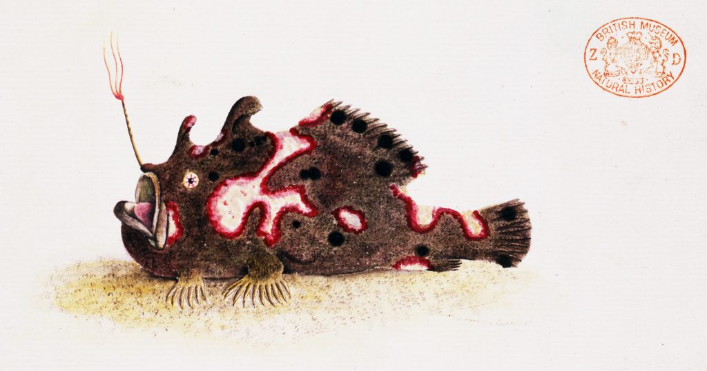
An illustration of the frogfish Antennarius pictus, published by George Shaw in 1794. From a new book by Ted Pietsch, UW professor of emeritus of aquatic and fishery sciences. Courtesy: University of Washington (state)
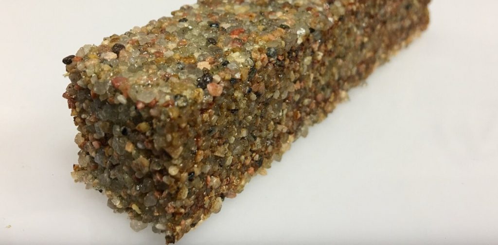
A block of sand particles held together by living cells. Credit: The University of Colorado Boulder College of Engineering and Applied Science
A March 24, 2020 news item on phys.org features the future of building construction as perceived by synthetic biologists,
Buildings are not unlike a human body. They have bones and skin; they breathe. Electrified, they consume energy, regulate temperature and generate waste. Buildings are organisms—albeit inanimate ones.
But what if buildings—walls, roofs, floors, windows—were actually alive—grown, maintained and healed by living materials? Imagine architects using genetic tools that encode the architecture of a building right into the DNA of organisms, which then grow buildings that self-repair, interact with their inhabitants and adapt to the environment.
…
A March 23, 2020 essay by Wil Srubar (Professor of Architectural Engineering and Materials Science, University of Colorado Boulder), which originated the news item, provides more insight,
Living architecture is moving from the realm of science fiction into the laboratory as interdisciplinary teams of researchers turn living cells into microscopic factories. At the University of Colorado Boulder, I lead the Living Materials Laboratory. Together with collaborators in biochemistry, microbiology, materials science and structural engineering, we use synthetic biology toolkits to engineer bacteria to create useful minerals and polymers and form them into living building blocks that could, one day, bring buildings to life.
In our most recent work, published in Matter, we used photosynthetic cyanobacteria to help us grow a structural building material – and we kept it alive. Similar to algae, cyanobacteria are green microorganisms found throughout the environment but best known for growing on the walls in your fish tank. Instead of emitting CO2, cyanobacteria use CO2 and sunlight to grow and, in the right conditions, create a biocement, which we used to help us bind sand particles together to make a living brick.
By keeping the cyanobacteria alive, we were able to manufacture building materials exponentially. We took one living brick, split it in half and grew two full bricks from the halves. The two full bricks grew into four, and four grew into eight. Instead of creating one brick at a time, we harnessed the exponential growth of bacteria to grow many bricks at once – demonstrating a brand new method of manufacturing materials.
Researchers have only scratched the surface of the potential of engineered living materials. Other organisms could impart other living functions to material building blocks. For example, different bacteria could produce materials that heal themselves, sense and respond to external stimuli like pressure and temperature, or even light up. If nature can do it, living materials can be engineered to do it, too.
It also take less energy to produce living buildings than standard ones. Making and transporting today’s building materials uses a lot of energy and emits a lot of CO2. For example, limestone is burned to make cement for concrete. Metals and sand are mined and melted to make steel and glass. The manufacture, transport and assembly of building materials account for 11% of global CO2 emissions. Cement production alone accounts for 8%. In contrast, some living materials, like our cyanobacteria bricks, could actually sequester CO2.
…
The field of engineered living materials is in its infancy, and further research and development is needed to bridge the gap between laboratory research and commercial availability. Challenges include cost, testing, certification and scaling up production. Consumer acceptance is another issue. For example, the construction industry has a negative perception of living organisms. Think mold, mildew, spiders, ants and termites. We’re hoping to shift that perception. Researchers working on living materials also need to address concerns about safety and biocontamination.
The [US] National Science Foundation recently named engineered living materials one of the country’s key research priorities. Synthetic biology and engineered living materials will play a critical role in tackling the challenges humans will face in the 2020s and beyond: climate change, disaster resilience, aging and overburdened infrastructure, and space exploration.
…
If you have time and interest, this is fascinating. Strubar is a little exuberant and, at this point, I welcome it.
Fitness
The Lithuanians are here for us. Scientists from the Kaunas University of Technology have just published a paper on better exercises for lower back pain in our increasingly sedentary times, from a March 23, 2020 Kaunas University of Technology press release (also on EurekAlert) Note: There are a few minor grammatical issues,
With the significant part of the global population forced to work from home, the occurrence of lower back pain may increase. Lithuanian scientists have devised a spinal stabilisation exercise programme for managing lower back pain for people who perform a sedentary job. After testing the programme with 70 volunteers, the researchers have found that the exercises are not only efficient in diminishing the non-specific lower back pain, but their effect lasts 3 times longer than that of a usual muscle strengthening exercise programme.
According to the World Health Organisation, lower back pain is among the top 10 diseases and injuries that are decreasing the quality of life across the global population. It is estimated that non-specific low back pain is experienced by 60% to 70% of people in industrialised societies. Moreover, it is the leading cause of activity limitation and work absence throughout much of the world. For example, in the United Kingdom, low back pain causes more than 100 million workdays lost per year, in the United States – an estimated 149 million.
Chronic lower back pain, which starts from long-term irritation or nerve injury affects the emotions of the afflicted. Anxiety, bad mood and even depression, also the malfunctioning of the other bodily systems – nausea, tachycardia, elevated arterial blood pressure – are among the conditions, which may be caused by lower back pain.
During the coronavirus disease (COVID-19) outbreak, with a significant part of the global population working from home and not always having a properly designed office space, the occurrence of lower back pain may increase.
“Lower back pain is reaching epidemic proportions. Although it is usually clear what is causing the pain and its chronic nature, people tend to ignore these circumstances and are not willing to change their lifestyle. Lower back pain usually comes away itself, however, the chances of the recurring pain are very high”, says Dr Irina Klizienė, a researcher at Kaunas University of Technology (KTU) Faculty of Social Sciences, Humanities and Arts.
Dr Klizienė, together with colleagues from KTU and from Lithuanian Sports University has designed a set of stabilisation exercises aimed at strengthening the muscles which support the spine at the lower back, i.e. lumbar area. The exercise programme is based on Pilates methodology.
According to Dr Klizienė, the stability of lumbar segments is an essential element of body biomechanics. Previous research evidence shows that in order to avoid the lower back pain it is crucial to strengthen the deep muscles, which are stabilising the lumbar area of the spine. One of these muscles is multifidus muscle.
“Human central nervous system is using several strategies, such as preparing for keeping the posture, preliminary adjustment to the posture, correcting the mistakes of the posture, which need to be rectified by specific stabilising exercises. Our aim was to design a set of exercises for this purpose”, explains Dr Klizienė.
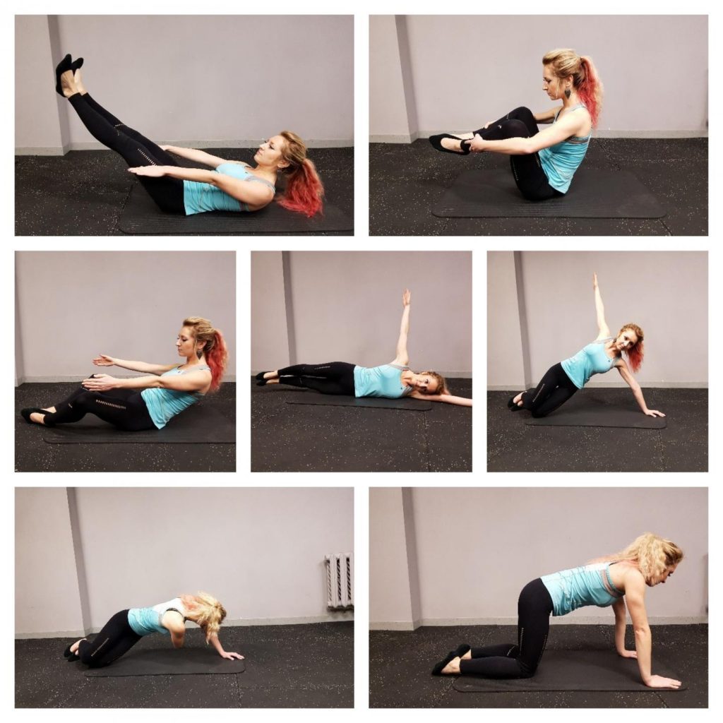
The programme, designed by Dr Klizienė and her colleagues is comprised of static and dynamic exercises, which train the muscle strength and endurance. The static positions are to be held from 6 to 20 seconds; each exercise to be repeated 8 to 16 times.
Caption: The static positions are to be held from 6 to 20 seconds; each exercise to be repeated 8 to 16 times. Credit: KTU
The previous set is a little puzzling but perhaps you’ll find these ones below easier to follow,
Caption: The exercises are aimed at strengthening the muscles which support the spine at the lower back. Credit: KTU
I think more pictures of intervening moves would have been useful. Now. getting back to the press release,
In order to check the efficiency of the programme, 70 female volunteers were randomly enrolled either to the lumbar stabilisation exercise programme or to a usual muscle strengthening exercise programme. Both groups were exercising twice a week for 45 minutes for 20 weeks. During the experiment, ultrasound scanning of the muscles was carried out.
As soon as 4 weeks in lumbar stabilisation programme, it was observed that the cross-section area of the multifidus muscle of the subjects of the stabilisation group has increased; after completing the programme, this increase was statistically significant (p < 0,05). This change was not observed in the strengthening group.
Moreover, although both sets of exercises were efficient in eliminating lower back pain and strengthening the muscles of the lower back area, the effect of stabilisation exercises lasted 3 times longer – 12 weeks after the completion of the stabilisation programme against 4 weeks after the completion of the muscle strengthening programme.
“There are only a handful of studies, which have directly compared the efficiency of stabilisation exercises against other exercises in eliminating lower back pain”, says Dr Klizienė, “however, there are studies proving that after a year, lower back pain returned only to 30% of people who have completed a stabilisation exercise programme, and to 84% of people who haven’t taken these exercises. After three years these proportions are 35% and 75%.”
According to her, research shows that the spine stabilisation exercises are more efficient than medical intervention or usual physical activities in curing the lower back pain and avoiding the recurrence of the symptoms in the future.
I remember reading somewhere that historically the field of astronomy has been the most inclusive of amateurs (or citizen scientists as we call them now).
If you think about it, all the sciences were started by amateurs. The notion that one should go to school and learn about science came much later after the pioneers, some of whom were philosophers such as Lucretius, developed theories to be passed on and codified the concept of scientific research.
It was Lucretius who helped to popularize ‘atomism’ and other physics concepts (eventually) in his multi-volume poem ‘De rerum natura’, known in English as ‘On the Nature of Things’. From the Lucretius Wikipedia entry,
… Lucretius’s scientific poem “On the Nature of Things” (c. 60 BC) has a remarkable description of Brownian motion of dust particles in verses 113–140 from Book II. He uses this as a proof of the existence of atoms.
From searching for extraterrestrial life to tracking rainfall, non-experts are increasingly helping to gather information to answer scientific questions. One of the most established hands-on, outdoor citizen science projects is the University of Washington-based Coastal Observation and Seabird Survey Team, COASST, which trains beachgoers along the West Coast, from California to Alaska, to monitor their local beach for dead birds.
With about 4,500 participants in its 21-year history and roughly 800 active participants today, COASST’s long-term success is now the subject of scientific study in its own right. What makes people join citizen science projects, and what motivates people to stick with them over years?
A UW-led paper published in the July issue [June 2019 issue?] of Ecology and Society explores the interests and identities of participants who join and remain active in citizen science. Results could help other science projects aiming to harness the power of large teams.
Previous research led by the UW has shown that people who join online-based citizen science projects generally try it just once, and fewer than 1 in 10 remain active past one year. The rates for hands-on, in-person efforts are much higher: COASST, for example, has 54% of participants still active one year after joining.
But what separates those who stay from those who go? Years of responses to surveys from the COASST team’s recruitment and engagement efforts provide a unique window on citizen science.
“I came to the UW to analyze a gold mine of social science datasets accumulated by COASST,” said social scientist and lead author Yurong He, a postdoctoral researcher in the UW School of Aquatic and Fishery Sciences. “Over a four year period, hundreds of participants responded to survey questions about why they were joining – or continuing – with the program. This represents an unparalleled opportunity.”
She analyzed answers to two freeform questions posed to project participants: “Why did you join COASST?” and “Why do you continue to be involved in COASST?” Some 310 new participants chose to answer the questions during their initial training. Another 623 seasoned participants, who had been involved for more than one year, completed a mail-in survey
“People’s memory can be a bit tricky,” He said. “You may think that two or five years ago you had a particular motivation, but is that really so? With this study we can definitively answer the motivation question at two different times: at the moment of joining the program, just after they finish being trained, and once they have spent at least a year on the beach collecting monthly data.”
The analysis shows that new participants wanted to be outdoors on the beach, learning about birds. Many listed their scientific degrees, previous occupations and birding expertise. But responses from longer-term contributors displayed a slightly different pattern: Although birds and beach remained dominant interests, seasoned volunteers were more likely to mention interests such as the desire to monitor and observe their beach, help in making scientific discoveries, and the importance of project data and results for environmental conservation. Moreover, their “science identity” became focused on their data-collection team and the project collective, rather than on their personal traits.
One important finding, He said, was the value of place. Volunteers often mentioned the importance of continuing to visit their beach even if they hadn’t found any birds washed ashore after several months.
“We thought they would talk a lot about birds, and they did, but they actually talked more about the coastal environment, the beach and the ocean,” He said. “Place was either equally important or even more important to them than birds.”
Another surprising finding is the degree to which participants consider citizen science to be a social activity. Of the five tasks volunteers listed as most important in defining their work for COASST, two – “communicating project results” and “recruiting others to participate” – were social. The other three tasks were “collect data,” “make measurements” and “enter data.”
“Activities that help connect family members and friends, and provide opportunities to meet new people who share similar interests, can also be scientific in nature,” He said. “COASST fulfills both science and social interests for coastal residents.”
he study’s conclusions based on the surveys included some take-home messages for organizers of hands-on citizen science efforts:
Long-term participants tend to be motivated by a project’s mission and goals, and successful programs communicate scientific findings back to participants so that they can see their individual contribution as part of the big picture of project results.
Experienced participants focus on where they conduct their project activities, indicating that sense of place is important to volunteers.
Both new and long-term participants focused on their social interactions as a central part of project activities, suggesting that successful hands-on, citizen science combines high-quality scientific activity with building and maintaining social relationships.
A December 5, 2017 news item on Nanowerk announced a new research institute at the University of Washington (state),
The University of Washington [UW} has launched a new institute aimed at accelerating research at the nanoscale: the Institute for Nano-Engineered Systems, or NanoES. Housed in a new, multimillion-dollar facility on the UW’s Seattle campus, the institute will pursue impactful advancements in a variety of disciplines — including energy, materials science, computation and medicine. Yet these advancements will be at a technological scale a thousand times smaller than the width of a human hair.
The institute was launched at a reception Dec. 4 [2017] at its headquarters in the $87.8-million Nano Engineering and Sciences Building. During the event, speakers including UW officials and NanoES partners celebrated the NanoES mission to capitalize on the university’s strong record of research at the nanoscale and engage partners in industry at the onset of new projects.
A December 5, 2017 UW news release, which originated the news item, somewhat clarifies the declarations in the two excerpted paragraphs in the above,
The vision of the NanoES, which is part of the UW’s College of Engineering, is to act as a magnet for researchers in nanoscale science and engineering, with a focus on enabling industry partnership and entrepreneurship at the earliest stages of research projects. According to Karl Böhringer, director of the NanoES and a UW professor of electrical engineering and bioengineering, this unique approach will hasten the development of solutions to the field’s most pressing challenges: the manufacturing of scalable, high-yield nano-engineered systems for applications in information processing, energy, health and interconnected life.
“The University of Washington is well known for its expertise in nanoscale materials, processing, physics and biology — as well as its cutting-edge nanofabrication, characterization and testing facilities,” said Böhringer, who stepped down as director of the UW-based Washington Nanofabrication Facility to lead the NanoES. “NanoES will build on these strengths, bringing together people, tools and opportunities to develop nanoscale devices and systems.”
The centerpiece of the NanoES is its headquarters, the Nano Engineering and Sciences Building. The building houses 90,300 square feet of research and learning space, and was funded largely by the College of Engineering and Sound Transit. It contains an active learning classroom, a teaching laboratory and a 3,000-square-foot common area designed expressly to promote the sharing and exchanging of ideas. The remainder includes “incubator-style” office space and more than 40,000 square feet of flexible multipurpose laboratory and instrumentation space. The building’s location and design elements are intended to limit vibrations and electromagnetic interference so it can house sensitive experiments.
NanoES will house research in nanotechnology fields that hold promise for high impact, such as:
Augmented humanity, which includes technology to both aid and replace human capability in a way that joins user and machine as one – and foresees portable, wearable, implantable and networked technology for applications such as personalized medical care, among others.
Integrated photonics, which ranges from single-photon sensors for health care diagnostic tests to large-scale, integrated networks of photonic devices.
Scalable nanomanufacturing, which aims to develop low-cost, high-volume manufacturing processes. These would translate device prototypes constructed in research laboratories into system- and network-level nanomanufacturing methods for applications ranging from the 3-D printing of cell and tissue scaffolds to ultrathin solar cells.
Cutting the ribbon for the NanoES on Dec. 4. Left-to-right: Karl Böhringer, director of the NanoES and a UW professor of electrical engineering and bioengineering; Nena Golubovic, physical sciences director for IP Group; Mike Bragg, Dean of the UW College of Engineering; Jevne Micheau-Cunningham, deputy director of the NanoES.Kathryn Sauber/University of Washington
Collaborations with other UW-based institutions will provide additional resources for the NanoES. Endeavors in scalable nanomanufacturing, for example, will rely on the roll-to-roll processing facility at the UW Clean Energy Institute‘s Washington Clean Energy Testbeds or on advanced surface characterization capabilities at the Molecular Analysis Facility. In addition, the Washington Nanofabrication Facility recently completed a three-year, $37 million upgrade to raise it to an ISO Class 5 nanofabrication facility.
UW faculty and outside collaborators will build new research programs in the Nano Engineering and Sciences Building. Eric Klavins, a UW professor of electrical engineering, recently moved part of his synthetic biology research team to the building, adjacent to his collaborators in the Molecular Engineering & Sciences Institute and the Institute for Protein Design.
“We are extremely excited about the interdisciplinary and collaborative potential of the new space,” said Klavins.
The NanoES also has already produced its first spin-out company, Tunoptix, which was co-founded by Böhringer and recently received startup funding from IP Group, a U.K.-based venture capital firm.
“IP Group is very excited to work with the University of Washington,” said Nena Golubovic, physical sciences director for IP Group. “We are looking forward to the new collaborations and developments in science and technology that will grow from this new partnership.”
Nena Golubovic, physical sciences director for IP Group, delivering remarks at the Dec. 4 opening of NanoES.Kathryn Sauber/University of Washington
“We are eager to work with our partners at the IP Group to bring our technology to the market, and we appreciate their vision and investment in the NanoES Integrated Photonics Initiative,” said Tunoptix entrepreneurial lead Mike Robinson. “NanoES was the ideal environment in which to start our company.”
The NanoES leaders hope to forge similar partnerships with researchers, investors and industry leaders to develop technologies for portable, wearable, implantable and networked nanotechnologies for personalized medical care, a more efficient interconnected life and interconnected mobility. In addition to expertise, personnel and state-of-the-art research space and equipment, the NanoES will provide training, research support and key connections to capital and corporate partners.
“We believe this unique approach is the best way to drive innovations from idea to fabrication to scale-up and testing,” said Böhringer. “Some of the most promising solutions to these huge challenges are rooted in nanotechnology.”
The NanoES is supported by funds from the College of Engineering and the National Science Foundation, as well as capital investments from investors and industry partners.
It seems a Swiss team from the École Polytechnique de Lausanne (EPFL) have collaborated with American companies Twist Bioscience and Microsoft, as well as, the University of Washington (state) to preserve two iconic jazz pieces on DNA (deoxyribonucleic acid) according to a Sept. 29, 2017 news item on phys.org,,
Thanks to an innovative technology for encoding data in DNA strands, two items of world heritage – songs recorded at the Montreux Jazz Festival [held in Switzerland] and digitized by EPFL – have been safeguarded for eternity. This marks the first time that cultural artifacts granted UNESCO heritage status have been saved in such a manner, ensuring they are preserved for thousands of years. The method was developed by US company Twist Bioscience and is being unveiled today in a demonstrator created at the EPFL+ECAL Lab.
“Tutu” by Miles Davis and “Smoke on the Water” by Deep Purple have already made their mark on music history. Now they have entered the annals of science, for eternity. Recordings of these two legendary songs were digitized by the Ecole Polytechnique Fédérale de Lausanne (EPFL) as part of the Montreux Jazz Digital Project, and they are the first to be stored in the form of a DNA sequence that can be subsequently decoded and listened to without any reduction in quality.
This feat was achieved by US company Twist Bioscience working in association with Microsoft Research and the University of Washington. The pioneering technology is actually based on a mechanism that has been at work on Earth for billions of years: storing information in the form of DNA strands. This fundamental process is what has allowed all living species, plants and animals alike, to live on from generation to generation.
The entire world wide web in a shoe box
All electronic data storage involves encoding data in binary format – a series of zeros and ones – and then recording it on a physical medium. DNA works in a similar way, but is composed of long strands of series of four nucleotides (A, T, C and G) that make up a “code.” While the basic principle may be the same, the two methods differ greatly in terms of efficiency: if all the information currently on the internet was stored in the form of DNA, it would fit in a shoe box!
Recent advances in biotechnology now make it possible for humans to do what Mother Nature has always done. Today’s scientists can create artificial DNA strands, “record” any kind of genetic code on them and then analyze them using a sequencer to reconstruct the original data. What’s more, DNA is extraordinarily stable, as evidenced by prehistoric fragments that have been preserved in amber. Artificial strands created by scientists and carefully encapsulated should likewise last for millennia.
To help demonstrate the feasibility of this new method, EPFL’s Metamedia Center provided recordings of two famous songs played at the Montreux Jazz Festival: “Tutu” by Miles Davis, and “Smoke on the Water” by Deep Purple. Twist Bioscience and its research partners encoded the recordings, transformed them into DNA strands and then sequenced and decoded them and played them again – without any reduction in quality.
The amount of artificial DNA strands needed to record the two songs is invisible to the naked eye, and the amount needed to record all 50 years of the Festival’s archives, which have been included in UNESCO’s [United Nations Educational, Scientific and Cultural Organization] Memory of the World Register, would be equal in size to a grain of sand. “Our partnership with EPFL in digitizing our archives aims not only at their positive exploration, but also at their preservation for the next generations,” says Thierry Amsallem, president of the Claude Nobs Foundation. “By taking part in this pioneering experiment which writes the songs into DNA strands, we can be certain that they will be saved on a medium that will never become obsolete!”
A new concept of time
At EPFL’s first-ever ArtTech forum, attendees got to hear the two songs played after being stored in DNA, using a demonstrator developed at the EPFL+ECAL Lab. The system shows that being able to store data for thousands of years is a revolutionary breakthrough that can completely change our relationship with data, memory and time. “For us, it means looking into radically new ways of interacting with cultural heritage that can potentially cut across civilizations,” says Nicolas Henchoz, head of the EPFL+ECAL Lab.
Quincy Jones, a longstanding Festival supporter, is particularly enthusiastic about this technological breakthrough: “With advancements in nanotechnology, I believe we can expect to see people living prolonged lives, and with that, we can also expect to see more developments in the enhancement of how we live. For me, life is all about learning where you came from in order to get where you want to go, but in order to do so, you need access to history! And with the unreliability of how archives are often stored, I sometimes worry that our future generations will be left without such access… So, it absolutely makes my soul smile to know that EPFL, Twist Bioscience and their partners are coming together to preserve the beauty and history of the Montreux Jazz Festival for our future generations, on DNA! I’ve been a part of this festival for decades and it truly is a magnificent representation of what happens when different cultures unite for the sake of music. Absolute magic. And I’m proud to know that the memory of this special place will never be lost.
Twist Bioscience, a company accelerating science and innovation through rapid, high-quality DNA synthesis, today announced that, working with Microsoft and University of Washington researchers, they have successfully stored archival-quality audio recordings of two important music performances from the archives of the world-renowned Montreux Jazz Festival.
These selections are encoded and stored in nature’s preferred storage medium, DNA, for the first time. These tiny specks of DNA will preserve a part of UNESCO’s Memory of the World Archive, where valuable cultural heritage collections are recorded. This is the first time DNA has been used as a long-term archival-quality storage medium.
Quincy Jones, world-renowned Entertainment Executive, Music Composer and Arranger, Musician and Music Producer said, “With advancements in nanotechnology, I believe we can expect to see people living prolonged lives, and with that, we can also expect to see more developments in the enhancement of how we live. For me, life is all about learning where you came from in order to get where you want to go, but in order to do so, you need access to history! And with the unreliability of how archives are often stored, I sometimes worry that our future generations will be left without such access…So, it absolutely makes my soul smile to know that EPFL, Twist Bioscience and others are coming together to preserve the beauty and history of the Montreux Jazz Festival for our future generations, on DNA!…I’ve been a part of this festival for decades and it truly is a magnificent representation of what happens when different cultures unite for the sake of music. Absolute magic. And I’m proud to know that the memory of this special place will never be lost.”
“Our partnership with EPFL in digitizing our archives aims not only at their positive exploration, but also at their preservation for the next generations,” says Thierry Amsallem, president of the Claude Nobs Foundation. “By taking part in this pioneering experiment which writes the songs into DNA strands, we can be certain that they will be saved on a medium that will never become obsolete!”
The Montreux Jazz Digital Project is a collaboration between the Claude Nobs Foundation, curator of the Montreux Jazz Festival audio-visual collection and the École Polytechnique Fédérale de Lausanne (EPFL) to digitize, enrich, store, show, and preserve this notable legacy created by Claude Nobs, the Festival’s founder.
In this proof-of-principle project, two quintessential music performances from the Montreux Jazz Festival – Smoke on the Water, performed by Deep Purple and Tutu, performed by Miles Davis – have been encoded onto DNA and read back with 100 percent accuracy. After being decoded, the songs were played on September 29th [2017] at the ArtTech Forum (see below) in Lausanne, Switzerland. Smoke on the Water was selected as a tribute to Claude Nobs, the Montreux Jazz Festival’s founder. The song memorializes a fire and Funky Claude’srescue efforts at the Casino Barrière de Montreux during a Frank Zappa concert promoted by Claude Nobs. Miles Davis’ Tutu was selected for the role he played in music history and the Montreux Jazz Festival’s success. Miles Davis died in 1991.
“We archived two magical musical pieces on DNA of this historic collection, equating to 140MB of stored data in DNA,” said Karin Strauss, Ph.D., a Senior Researcher at Microsoft, and one of the project’s leaders. “The amount of DNA used to store these songs is much smaller than one grain of sand. Amazingly, storing the entire six petabyte Montreux Jazz Festival’s collection would result in DNA smaller than one grain of rice.”
Luis Ceze, Ph.D., a professor in the Paul G. Allen School of Computer Science & Engineering at the University of Washington, said, “DNA, nature’s preferred information storage medium, is an ideal fit for digital archives because of its durability, density and eternal relevance. Storing items from the Montreux Jazz Festival is a perfect way to show how fast DNA digital data storage is becoming real.”
Nature’s Preferred Storage Medium
Nature selected DNA as its hard drive billions of years ago to encode all the genetic instructions necessary for life. These instructions include all the information necessary for survival. DNA molecules encode information with sequences of discrete units. In computers, these discrete units are the 0s and 1s of “binary code,” whereas in DNA molecules, the units are the four distinct nucleotide bases: adenine (A), cytosine (C), guanine (G) and thymine (T).
“DNA is a remarkably efficient molecule that can remain stable for millennia,” said Bill Peck, Ph.D., chief technology officer of Twist Bioscience. “This is a very exciting project: we are now in an age where we can use the remarkable efficiencies of nature to archive master copies of our cultural heritage in DNA. As we develop the economies of this process new performances can be added any time. Unlike current storage technologies, nature’s media will not change and will remain readable through time. There will be no new technology to replace DNA, nature has already optimized the format.”
DNA: Far More Efficient Than a Computer
Each cell within the human body contains approximately three billion base pairs of DNA. With 75 trillion cells in the human body, this equates to the storage of 150 zettabytes (1021) of information within each body. By comparison, the largest data centers can be hundreds of thousands to even millions of square feet to hold a comparable amount of stored data.
The Elegance of DNA as a Storage Medium
Like music, which can be widely varied with a finite number of notes, DNA encodes individuality with only four different letters in varied combinations. When using DNA as a storage medium, there are several advantages in addition to the universality of the format and incredible storage density. DNA can be stable for thousands of years when stored in a cool dry place and is easy to copy using polymerase chain reaction to create back-up copies of archived material. In addition, because of PCR, small data sets can be targeted and recovered quickly from a large dataset without needing to read the entire file.
How to Store Digital Data in DNA
To encode the music performances into archival storage copies in DNA, Twist Bioscience worked with Microsoft and University of Washington researchers to complete four steps: Coding, synthesis/storage, retrieval and decoding. First, the digital files were converted from the binary code using 0s and 1s into sequences of A, C, T and G. For purposes of the example, 00 represents A, 10 represents C, 01 represents G and 11 represents T. Twist Bioscience then synthesizes the DNA in short segments in the sequence order provided. The short DNA segments each contain about 12 bytes of data as well as a sequence number to indicate their place within the overall sequence. This is the process of storage. And finally, to ensure that the file is stored accurately, the sequence is read back to ensure 100 percent accuracy, and then decoded from A, C, T or G into a two-digit binary representation.
Importantly, to encapsulate and preserve encoded DNA, the collaborators are working with Professor Dr. Robert Grass of ETH Zurich. Grass has developed an innovative technology inspired by preservation of DNA within prehistoric fossils. With this technology, digital data encoded in DNA remains preserved for millennia.
About UNESCO’s Memory of the World Register
UNESCO established the Memory of the World Register in 1992 in response to a growing awareness of the perilous state of preservation of, and access to, documentary heritage in various parts of the world. Through its National Commissions, UNESCO prepared a list of endangered library and archive holdings and a world list of national cinematic heritage.
A range of pilot projects employing contemporary technology to reproduce original documentary heritage on other media began. These included, for example, a CD-ROM of the 13th Century Radzivill Chronicle, tracing the origins of the peoples of Europe, and Memoria de Iberoamerica, a joint newspaper microfilming project involving seven Latin American countries. These projects enhanced access to this documentary heritage and contributed to its preservation.
“We are incredibly proud to be a part of this momentous event, with the first archived songs placed into the UNESCO Memory of the World Register,” said Emily Leproust, Ph.D., CEO of Twist Bioscience.
About ArtTech
The ArtTech Foundation, created by renowned scientists and dignitaries from Crans-Montana, Switzerland, wishes to stimulate reflection and support pioneering and innovative projects beyond the known boundaries of culture and science.
Benefitting from the establishment of a favorable environment for the creation of technology companies, the Foundation aims to position itself as key promoter of ideas and innovative endeavors within a landscape of “Culture and Science” that is still being shaped.
Several initiatives, including our annual global platform launched in the spring of 2017, are helping to create a community that brings together researchers, celebrities in the world of culture and the arts, as well as investors and entrepreneurs from Switzerland and across the globe.
About EPFL
EPFL, one of the two Swiss Federal Institutes of Technology, based in Lausanne, is Europe’s most cosmopolitan technical university with students, professors and staff from over 120 nations. A dynamic environment, open to Switzerland and the world, EPFL is centered on its three missions: teaching, research and technology transfer. EPFL works together with an extensive network of partners including other universities and institutes of technology, developing and emerging countries, secondary schools and colleges, industry and economy, political circles and the general public, to bring about real impact for society.
About Twist Bioscience
At Twist Bioscience, our expertise is accelerating science and innovation by leveraging the power of scale. We have developed a proprietary semiconductor-based synthetic DNA manufacturing process featuring a high throughput silicon platform capable of producing synthetic biology tools, including genes, oligonucleotide pools and variant libraries. By synthesizing DNA on silicon instead of on traditional 96-well plastic plates, our platform overcomes the current inefficiencies of synthetic DNA production, and enables cost-effective, rapid, high-quality and high throughput synthetic gene production, which in turn, expedites the design, build and test cycle to enable personalized medicines, pharmaceuticals, sustainable chemical production, improved agriculture production, diagnostics and biodetection. We are also developing new technologies to address large scale data storage. For more information, please visit www.twistbioscience.com. Twist Bioscience is on Twitter. Sign up to follow our Twitter feed @TwistBioscience at https://twitter.com/TwistBioscience.
If you hadn’t read the EPFL press release first, it might have taken a minute to figure out why EPFL is being mentioned in the Twist Bioscience news release. Presumably someone was rushing to make a deadline. Ah well, I’ve seen and written worse.
I haven’t been able to find any video or audio recordings of the DNA-preserved performances but there is an informational video (originally published July 7, 2016) from Microsoft and the University of Washington describing the DNA-based technology,
I also found this description of listening to the DNA-preserved music in an Oct. 6, 2017 blog posting for the Canadian Broadcasting Corporation’s (CBC) Day 6 radio programme,
To listen to them, one must first suspend the DNA holding the songs in a solution. Next, one can use a DNA sequencer to read the letters of the bases forming the molecules. Then, algorithms can determine the digital code those letters form. From that code, comes the music.
It’s complicated but Ceze says his team performed this process without error.
The University of California at Davis (UC Davis) and the University of Washington (state) collaborated in research into fundamental questions on how aquatic animals grow. From an Oct. 24, 2016 news item on ScienceDaily,
For the first time scientists can see how the shells of tiny marine organisms grow atom-by-atom, a new study reports. The advance provides new insights into the mechanisms of biomineralization and will improve our understanding of environmental change in Earth’s past.
Led by researchers from the University of California, Davis and the University of Washington, with key support from the U.S. Department of Energy’s Pacific Northwest National Laboratory, the team examined an organic-mineral interface where the first calcium carbonate crystals start to appear in the shells of foraminifera, a type of plankton.
“We’ve gotten the first glimpse of the biological event horizon,” said Howard Spero, a study co-author and UC Davis geochemistry professor. …
Foraminifera’s Final Frontier
The researchers zoomed into shells at the atomic level to better understand how growth processes may influence the levels of trace impurities in shells. The team looked at a key stage — the interaction between the biological ‘template’ and the initiation of shell growth. The scientists produced an atom-scale map of the chemistry at this crucial interface in the foraminifera Orbulina universa. This is the first-ever measurement of the chemistry of a calcium carbonate biomineralization template, Spero said.
Among the new findings are elevated levels of sodium and magnesium in the organic layer. This is surprising because the two elements are not considered important architects in building shells, said lead study author Oscar Branson, a former postdoctoral researcher at UC Davis who is now at the Australian National University in Canberra. Also, the greater concentrations of magnesium and sodium in the organic template may need to be considered when investigating past climate with foraminifera shells.
Calibrating Earth’s Climate
Most of what we know about past climate (beyond ice core records) comes from chemical analyses of shells made by the tiny, one-celled creatures called foraminifera, or “forams.” When forams die, their shells sink and are preserved in seafloor mud. The chemistry preserved in ancient shells chronicles climate change on Earth, an archive that stretches back nearly 200 million years.
The calcium carbonate shells incorporate elements from seawater — such as calcium, magnesium and sodium — as the shells grow. The amount of trace impurities in a shell depends on both the surrounding environmental conditions and how the shells are made. For example, the more magnesium a shell has, the warmer the ocean was where that shell grew.
“Finding out how much magnesium there is in a shell can allow us to find out the temperature of seawater going back up to 150 million years,” Branson said.
But magnesium levels also vary within a shell, because of nanometer-scale growth bands. Each band is one day’s growth (similar to the seasonal variations in tree rings). Branson said considerable gaps persist in understanding what exactly causes the daily bands in the shells.
“We know that shell formation processes are important for shell chemistry, but we don’t know much about these processes or how they might have changed through time,” he said. “This adds considerable uncertainty to climate reconstructions.”
Atomic Maps
The researchers used two cutting-edge techniques: Time-of-Flight Secondary Ionization Mass Spectrometry (ToF-SIMS) and Laser-Assisted Atom Probe Tomography (APT). ToF-SIMS is a two-dimensional chemical mapping technique which shows the elemental composition of the surface of a polished sample. The technique was developed for the elemental analysis of complex polymer materials, and is just starting to be applied to natural samples like shells.
APT is an atomic-scale three-dimensional mapping technique, developed for looking at internal structures in advanced alloys, silicon chips and superconductors. The APT imaging was performed at the Environmental Molecular Sciences Laboratory, a U.S. Department of Energy Office of Science User Facility at the Pacific Northwest National Laboratory.
This foraminifera is just starting to form its adult spherical shell. The calcium carbonate spherical shell first forms on a thin organic template, shown here in white, around the dark juvenile skeleton. Calcium carbonate spines then extend from the juvenile skeleton through the new sphere and outward. The bright flecks are algae that the foraminifera “farm” for sustenance.Howard Spero/University of California, Davis
Unseen out in the ocean, countless single-celled organisms grow protective shells to keep them safe as they drift along, living off other tiny marine plants and animals. Taken together, the shells are so plentiful that when they sink they provide one of the best records for the history of ocean chemistry.
Oceanographers at the University of Washington and the University of California, Davis, have used modern tools to provide an atomic-scale look at how that shell first forms. Results could help answer fundamental questions about how these creatures grow under different ocean conditions, in the past and in the future. …
“There’s this debate among scientists about whether shelled organisms are slaves to the chemistry of the ocean, or whether they have the physiological capacity to adapt to changing environmental conditions,” said senior author Alex Gagnon, a UW assistant professor of oceanography.
The new work shows, he said, that they do exert some biologically-based control over shell formation.
“I think it’s just incredible that we were able to peer into the intricate details of those first moments that set how a seashell forms,” Gagnon said. “And that’s what sets how much of the rest of the skeleton will grow.”
The results could eventually help understand how organisms at the base of the marine food chain will respond to more acidic waters. And while the study looked at one organism, Orbulina universa, which is important for understanding past climate, the same method could be used for other plankton, corals and shellfish.
The study used tools developed for materials science and semiconductor research to view the shell formation in the most detail yet to see how the organisms turn seawater into solid mineral.
“We’re interested more broadly in the question ‘How do organisms make shells?'” said first author Oscar Branson, a former postdoctoral researcher at the University of California, Davis who is now at Australian National University in Canberra. “We’ve focused on a key stage in mineral formation — the interaction between biological template materials and the initiation of shell growth by an organism.”
These tiny single-celled animals, called foraminifera, can’t reproduce anywhere but in their natural surroundings, which prevents breeding them in captivity. The researchers caught juvenile foraminifera by diving in deep water off Southern California. Then they then raised them in the lab, using tiny pipettes to feed them brine shrimp during their weeklong lives.
Marine shells are made from calcium carbonate, drawing the calcium and carbon from surrounding seawater. But the animal first grows a soft template for the mineral to grow over. Because this template is trapped within the growing skeleton, it acts as a snapshot of the chemical conditions during the first part of skeletal growth.
To see this chemical picture, the authors analyzed tiny sections of foraminifera template with a technique called atom probe tomography at the Pacific Northwest National Laboratory. This tool creates an atom-by-atom picture of the organic template, which was located using a chemical tag.
Results show that the template contains more magnesium and sodium atoms than expected, and that this could influence how the mineral in the shell begins to grow around it.
“One of the key stages in growing a skeleton is when you make that first bit, when you build that first bit of structure. Anything that changes that process is a key control point,” Gagnon said.
The clumping suggests that magnesium and sodium play a role in the first stages of shell growth. If their availability changes for any reason, that could influence how the shell grows beyond what simple chemistry would predict.
“We can say who the players are — further experiments will have to tell us exactly how important each of them is,” Gagnon said.
Follow-up work will try to grow the shells and create models of their formation to see how the template affects growth under different conditions, such as more acidic water.
“Translating that into, ‘Can these forams survive ocean acidification?’ is still many steps down the line,” Gagnon cautioned. “But you can’t do that until you have a picture of what that surface actually looks like.”
The researchers also hope that by better understanding the exact mechanism of shell growth they could tease apart different aspects of seafloor remains so the shells can be used to reconstruct more than just the ocean’s past temperature. In the study, they showed that the template was responsible for causing fine lines in the shells — one example of the rich chemical information encoded in fossil shells.
“There are ways that you could separate the effects of temperature from other things and learn much more about the past ocean,” Gagnon said.
Here’s a link to and a citation for the paper,
Nanometer-Scale Chemistry of a Calcite Biomineralization Template: Implications for Skeletal Composition and Nucleation, Proceedings of the National Academy of Sciences, www.pnas.org/cgi/doi/10.1073/pnas.1522864113