This really is a ‘Frankenstein’ story complete with turtle cadavers. From a June 14, 2016 news item on ScienceDaily,
It was a dark and stormy night in the laboratory, and jagged bolts of lightning lit the sky as Dr. Kaplan and his assistant Bianca stitched the pieces of the lifeless creature back together.
Actually, it was a sunny day on the shores of Chesapeake Bay, but recent sea turtle research by Assistant Professor David Kaplan of the Virginia Institute of Marine Science and graduate student Bianca Santos easily brings to mind the classic tale of Dr. Frankenstein and his makeshift monster.
Santos, a master’s student in William & Mary’s School of Marine Science at VIMS, is working with Kaplan to reduce sea turtle mortality by trying to pinpoint where the hundreds of dead loggerhead sea turtles that wash up on Chesapeake Bay beaches each summer may have succumbed. With that knowledge, researchers could hone in on likely causes of sea-turtle death, while wildlife authorities could map out safe zones for these imperiled marine reptiles. One of Kaplan’s research specialties is the spatial management of marine ecosystems.
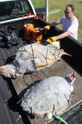
David Kaplan examines the Frankenturtles before their deployment. Also visible are the bucket drifters that more closely follow Bay currents. Courtesy: VIMS
A June 14, 2016 Virginia Institute of Marine Science news release by
The pair’s approach to the problem is ingenious if somewhat morbid: obtain dead sea turtles (from the Virginia Aquarium’s Stranding Response Program), replace the turtles’ inner organs with buoyant Styrofoam, “sew” their shells back together with zip ties, and then attach GPS units to track the path of the “Frankenturtles” as winds and currents disperse them from a mid-Bay release site.
“It might seem sort of gross, but it’s a good way to reuse a dead turtle that would otherwise be buried,” says Kaplan. “And hopefully, the deployment of our two Frankenturtles will ultimately help lower the number of turtle deaths in the future.”
Santos explains that the team is actually releasing three different types of drifters: the two Frankenturtles, two wooden-Styrofoam turtle models, and a pair of bucket drifters. By observing how the wind differentially affects the highly buoyant, sail-like wooden models; the partly emergent Frankenturtles; and the mostly submerged buckets, the researchers hope to better understand how a wind-driven carcass might deviate from the more predictable current patterns traced by the Bay’s surface waters. Sea turtles initially sink after dying, but quickly float back to the surface buoyed by gases from decomposing tissues.
“Our plan is to deploy the drifters on several different occasions—under a variety of wind and wave conditions—and in locations where mortality events could occur during the spring peak in strandings,” says Santos. “We’ll then use the separation rate between our bucket drifters, which closely track water movement, and our turtle carcasses to determine the amount of wind forcing to apply to simulated carcasses in our computer model.”
They initiated their field trials on June 13th [2016], deploying the drifters in open Bay waters about halfway between the mouth of the York River and Cape Charles on Virginia’s bayside Eastern Shore. One Frankenturtle comprises the remains of a 15-20 year old loggerhead killed by a boat strike. The other is a younger turtle whose mode of death remains a mystery despite a necropsy. Deploying these creatures wasn’t an easy job: in addition to the unforgettable and growing aroma of thawing turtle, the creatures are both heavy and unwieldy. The larger Frankenturtle weighs in at 150 pounds, the smaller at 70 pounds.
Modeling turtle movement
Once data from the Frankenturtle trials have allowed the researchers to properly configure their “turtle carcass drift model,” they’ll feed the model with historical records of stranding locations provided by the Virginia Aquarium’s Stranding Response Team. The team is the Commonwealth’s official entity for responding to reports of dead and injured sea turtles and other marine life in Bay and nearby coastal waters.
“If our model can accurately simulate how winds and currents act on a dead sea turtle, we should be able to backtrack from a stranding site to the place where the turtle likely died,” says Santos. “By knowing the ‘where,’” she adds, “we can better look at the ‘why.’”
The researchers plan to track the Frankenturtles and other drifters released on June 13th for 3-4 days before retrieving the GPS units for future use. Earlier experiments by Santos show that’s about how long dead turtles remain intact before they are dismembered and consumed by waves, birds, crabs, and fish. The public can view the motion of the drifters in real-time via the VIMS website at www.vims.edu/frankenturtle.
Sea turtle mortality
Mortality of loggerhead turtles in Chesapeake Bay is of continuing concern. “Strandings peaked in the early 2000s at around 200-400 per year,” says Kaplan. “Modifications to the pound-net fishery likely reduced the number to the current 100-300 per year, and it is these we’re trying to understand.” He adds that scientists don’t really don’t have a good idea what percentage of dead turtles these strandings represent. “The actual number could be much higher,” Kaplan says.
Evidence that strandings may represent only a small percentage of actual deaths comes from Santos’ decay experiments as well as the low odds of finding every dead turtle. “Bianca’s decay study shows that turtles remain intact for only 3-5 days after death, decreasing the likelihood that they might last long enough to wash up on a beach,” says Kaplan. “And of those that do wash ashore, many probably strand in remote or marshy areas where they are unlikely to be observed and reported by a beachgoer.”
Potential sources of mortality in the Bay include accidental capture in fishing gear, strikes by boat propellers, entanglement in plastic trash, and sudden drops in temperature.
Although loggerheads are the most common sea turtles in the Chesapeake, with 5,000-10,000 entering Bay waters each summer to feed, they are listed as “threatened” in U.S. waters under the Endangered Species Act due to the perils they face across their range, including loss of nesting habitat, disorientation of hatchlings by beachfront lighting, nest predation, and incidental capture in dredges and coastal fisheries. Measures to protect against these threats are enforced by NOAA Fisheries and the U.S. Fish and Wildlife Service.