Tag Archives: Vanderbilt University (VU)

Robot that can maneuver through living lung tissue

Caption: Overview of the semiautonomous medical robot’s three stages in the lungs. Credit: Kuntz et al.

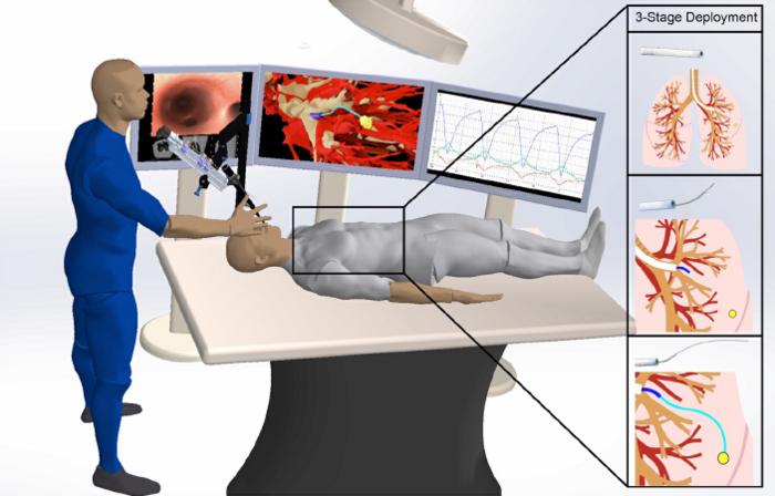
This looks like one robot operating on another robot; I guess the researchers want to emphasize the fact that this autonomous surgical procedure isn’t currently being tested on human beings.

There’s more in a September 21, 2023 news item on ScienceDaily,

Scientists have shown that their steerable lung robot can autonomously maneuver the intricacies of the lung, while avoiding important lung structures.

Lung cancer is the leading cause of cancer-related deaths in the United States. Some tumors are extremely small and hide deep within lung tissue, making it difficult for surgeons to reach them. To address this challenge, UNC -Chapel Hill and Vanderbilt University researchers have been working on an extremely bendy but sturdy robot capable of traversing lung tissue.

Their research has reached a new milestone. In a new paper, published in Science Robotics, Ron Alterovitz, PhD, in the UNC Department of Computer Science, and Jason Akulian, MD MPH, in the UNC Department of Medicine, have proven that their robot can autonomously go from “Point A” to “Point B” while avoiding important structures, such as tiny airways and blood vessels, in a living laboratory model.

Thankfully there’s a September 21, 2023 University of North Carolina (UNC) news release (also on EurekAlert), which originated the news item, to provide more information, Note: Links have been removed,

“This technology allows us to reach targets we can’t otherwise reach with a standard or even robotic bronchoscope,” said Dr. Akulian, co-author on the paper and Section Chief of Interventional Pulmonology and Pulmonary Oncology in the UNC Division of Pulmonary Disease and Critical Care Medicine. “It gives you that extra few centimeters or few millimeters even, which would help immensely with pursuing small targets in the lungs.”

The development of the autonomous steerable needle robot leveraged UNC’s highly collaborative culture by blending medicine, computer science, and engineering expertise. In addition to Alterovitz and Akulian, the development effort included Yueh Z. Lee, MD, PhD, at the UNC Department of Radiology, as well as Robert J. Webster III at Vanderbilt University and Alan Kuntz at the University of Utah.

The robot is made of several separate components. A mechanical control provides controlled thrust of the needle to go forward and backward and the needle design allows for steering along curved paths. The needle is made from a nickel-titanium alloy and has been laser etched to increase its flexibility, allowing it to move effortlessly through tissue.

As it moves forward, the etching on the needle allows it to steer around obstacles with ease. Other attachments, such as catheters, could be used together with the needle to perform procedures such as lung biopsies.

To drive through tissue, the needle needs to know where it is going. The research team used CT scans of the subject’s thoracic cavity and artificial intelligence to create three-dimensional models of the lung, including the airways, blood vessels, and the chosen target. Using this 3-D model and once the needle has been positioned for launch, their AI-driven software instructs it to automatically travel from “Point A” to “Point B” while avoiding important structures.

“The autonomous steerable needle we’ve developed is highly compact, but the system is packed with a suite of technologies that allow the needle to navigate autonomously in real-time,” said Alterovitz, the principal investigator on the project and senior author on the paper. “It’s akin to a self-driving car, but it navigates through lung tissue, avoiding obstacles like significant blood vessels as it travels to its destination.”

The needle can also account for respiratory motion. Unlike other organs, the lungs are constantly expanding and contracting in the chest cavity. This can make targeting especially difficult in a living, breathing subject. According to Akulian, it’s like shooting at a moving target.

The researchers tested their robot while the laboratory model performed intermittent breath holding. Every time the subject’s breath is held, the robot is programmed to move forward.

“There remain some nuances in terms of the robot’s ability to acquire targets and then actually get to them effectively,” said Akulian, who is also a member of the UNC Lineberger Comprehensive Cancer Center, “and while there’s still a lot of work to be done, I’m very excited about continuing to push the boundaries of what we can do for patients with the world-class experts that are here.”

“We plan to continue creating new autonomous medical robots that combine the strengths of robotics and AI to improve medical outcomes for patients facing a variety of health challenges while providing guarantees on patient safety,” added Alterovitz.

Here’s a link to and a citation for the paper,

Autonomous medical needle steering in vivo by Alan Kuntz, Maxwell Emerson, Tayfun Efe Ertop, Inbar Fried, Mengyu Fu, Janine Hoelscher, Margaret Rox, Jason Akulian, Erin A. Gillaspie, Yueh Z. Lee, Fabien Maldonado, Robert J. Webster III, and Ron Alterovitz. Science Robotics 20 Sep 2023 Vol 8, Issue 82 DOI: 10.1126/scirobotics.adf7614

This paper is behind a paywall.

Mystery of North American insect bioluminescent systems unraveled by Brazilian scientists

I’ve always been fond of ‘l’ words and so it is that I’m compelled to post a story about a “luciferin-luciferase system” or, in this case, a story about insect bioluminescence.

Caption: Researchers isolated molecules present in the larvae of the fungus gnat Orfelia fultoni Credit: Vadim Viviani, UFSCar

A September 9, 2020 Fundação de Amparo à Pesquisa do Estado de São Paulo (FAPESP) press release (also on EurekAlert but published Sept. 11, 2020) announces research into ‘blue’ bioluminescence,

Molecules belonging to an almost unknown bioluminescent system found in larvae of the fungus gnat Orfelia fultoni (subfamily Keroplatinae) have been isolated for the first time by researchers at the Federal University of São Carlos (UFSCar) in the state of São Paulo, Brazil. The small fly is one of the few terrestrial organisms that produce blue light. It inhabits riverbanks in the Appalachian Mountains in the eastern United States. A key part of its bioluminescent system is a molecule also present in two recently discovered Brazilian flies.

The study, supported by Paulo Research Foundation – FAPESP, is published in Scientific Reports. Five authors are affiliated with UFSCar and two with universities in the United States.

The bioluminescent systems of glow-worms, fireflies and other insects are normally made up of luciferin (a low molecular weight molecule) and luciferase, an enzyme that catalyzes the oxidation of luciferin by oxygen, producing light. While some bioluminescent systems are well known and even used in biotechnological applications, others are poorly understood, including blue light-emitting systems, such as that of O. fultoni.

“In the published paper, we describe the properties of the insect’s luciferase and luciferin and their anatomical location in its larvae. We also specify several possible proteins that are possible candidates for the luciferase. We don’t yet know what type of protein it is, but it’s likely to be a hexamerin. In insects, hexamerins are storage proteins that provide amino acids, besides having other functions, such as binding low molecular weight compounds, like luciferin,” said Vadim Viviani, a professor in UFSCar’s Sustainability Science and Technology Center (CCTS) in Sorocaba, São Paulo, and principal investigator for the study.

The study was part of the FAPESP-funded project “Arthropod bioluminescence“. The partnership with United States-based researchers dates from a previous project, supported by FAPESP and the United States National Science Foundation (NSF), in partnership with Vanderbilt University (VU), located in Nashville, Tennessee.

In addition to luciferin and luciferase, researchers began characterizing a complex found in insects of the family Keroplatidae, which, in addition to O. fultoni, also includes a Brazilian species in the genus Neoditomyia that produces only luciferin and hence does not emit light.

Because they do not use it to emit light, the luciferin in O. fultoni and the Brazilian Neoditomyia has been named keroplatin. In larvae of this subfamily, keroplatin is associated with “black bodies” – large cells containing dark granules, proteins and probably mitochondria (energy-producing organelles). Researchers are still investigating the biological significance of this association between keroplatin and mitochondria.

“It’s a mystery,” Viviani said. “This luciferin may play a role in the mitochondrial energy metabolism. At night, probably in the presence of a natural chemical reducer, the luciferin is released by these black bodies and reacts with the surrounding luciferase to produce blue light. These are possibilities we plan to study.”

Brazilian cousins

An important factor in the elucidation of the United States insect’s bioluminescent system was the discovery of a larva that lives in Intervales State Park in São Paulo in 2018. It does not emit light but produces luciferin, similar to O. fultoni (read more at: agencia.fapesp.br/29066).

In their latest study, the group injected purified luciferase from the United States species into larvae of the Brazilian species, which then produced blue light. The nonluminescent Brazilian species is more abundant in nature than the United States species, so a larger amount of the material could be obtained for study purposes, especially to characterize the luciferin (keroplatin) present in both species.

In 2019, the group discovered and described Neoceroplatus betaryensis, a new species of fungus gnat, in collaboration with Cassius Stevani, a professor at the University of São Paulo’s Institute of Chemistry (IQ-USP). It was the first blue light-emitting insect found in South America and was detected in a privately held forest reserve near the Upper Ribeira State Tourist Park (PETAR) in the southern portion of the state of São Paulo. A close relative of O. fultoni, N. betaryensis inhabits fallen tree trunks in humid places (read more at: agencia.fapesp.br/31797).

“We show that the bioluminescent system of this Brazilian species is identical to that of O. fultoni. However, the insect is very rare, and so it’s hard to obtain sufficient material for research purposes,” Viviani said.

The researchers are now cloning the insect’s luciferase and characterizing it in molecular terms. They are also analyzing the chemical structure of its luciferin and the morphology of its lanterns.

“Once all this has been determined, we’ll be able to synthesize the luciferin and luciferase in the lab and use these systems in a range of biotech applications, such as studying cells. This will help us understand more about human diseases, among other things,” Viviani said.

Here’s a link to and a citation for the paper,

A new brilliantly blue-emitting luciferin-luciferase system from Orfelia fultoni and Keroplatinae (Diptera) by Vadim R. Viviani, Jaqueline R. Silva, Danilo T. Amaral, Vanessa R. Bevilaqua, Fabio C. Abdalla, Bruce R. Branchini & Carl H. Johnson. Scientific Reports volume 10, Article number: 9608 (2020) DOI: https://doi.org/10.1038/s41598-020-66286-1 Published 15 June 2020

This paper is open access.