Tag Archives: VCU

New nanomapping technology: CRISPR-CAS9 as a programmable nanoparticle

A November 21, 2017 news item on Nanowerk describes a rather extraordinary (to me, anyway) approach to using CRRISP ( Clustered Regularly Interspaced Short Palindromic Repeats)-CAS9 (Note: A link has been removed),

A team of scientists led by Virginia Commonwealth University physicist Jason Reed, Ph.D., have developed new nanomapping technology that could transform the way disease-causing genetic mutations are diagnosed and discovered. Described in a study published today [November 21, 2017] in the journal Nature Communications (“DNA nanomapping using CRISPR-Cas9 as a programmable nanoparticle”), this novel approach uses high-speed atomic force microscopy (AFM) combined with a CRISPR-based chemical barcoding technique to map DNA nearly as accurately as DNA sequencing while processing large sections of the genome at a much faster rate. What’s more–the technology can be powered by parts found in your run-of-the-mill DVD player.

A November 21, 2017 Virginia Commonwealth University news release by John Wallace, which originated the news item, provides more detail,

The human genome is made up of billions of DNA base pairs. Unraveled, it stretches to a length of nearly six feet long. When cells divide, they must make a copy of their DNA for the new cell. However, sometimes various sections of the DNA are copied incorrectly or pasted together at the wrong location, leading to genetic mutations that cause diseases such as cancer. DNA sequencing is so precise that it can analyze individual base pairs of DNA. But in order to analyze large sections of the genome to find genetic mutations, technicians must determine millions of tiny sequences and then piece them together with computer software. In contrast, biomedical imaging techniques such as fluorescence in situ hybridization, known as FISH, can only analyze DNA at a resolution of several hundred thousand base pairs.

Reed’s new high-speed AFM method can map DNA to a resolution of tens of base pairs while creating images up to a million base pairs in size. And it does it using a fraction of the amount of specimen required for DNA sequencing.

“DNA sequencing is a powerful tool, but it is still quite expensive and has several technological and functional limitations that make it difficult to map large areas of the genome efficiently and accurately,” said Reed, principal investigator on the study. Reed is a member of the Cancer Molecular Genetics research program at VCU Massey Cancer Center and an associate professor in the Department of Physics in the College of Humanities and Sciences.

“Our approach bridges the gap between DNA sequencing and other physical mapping techniques that lack resolution,” he said. “It can be used as a stand-alone method or it can complement DNA sequencing by reducing complexity and error when piecing together the small bits of genome analyzed during the sequencing process.”

IBM scientists made headlines in 1989 when they developed AFM technology and used a related technique to rearrange molecules at the atomic level to spell out “IBM.” AFM achieves this level of detail by using a microscopic stylus — similar to a needle on a record player — that barely makes contact with the surface of the material being studied. The interaction between the stylus and the molecules creates the image. However, traditional AFM is too slow for medical applications and so it is primarily used by engineers in materials science.

“Our device works in the same fashion as AFM but we move the sample past the stylus at a much greater velocity and use optical instruments to detect the interaction between the stylus and the molecules. We can achieve the same level of detail as traditional AFM but can process material more than a thousand times faster,” said Reed, whose team proved the technology can be mainstreamed by using optical equipment found in DVD players. “High-speed AFM is ideally suited for some medical applications as it can process materials quickly and provide hundreds of times more resolution than comparable imaging methods.”

Increasing the speed of AFM was just one hurdle Reed and his colleagues had to overcome. In order to actually identify genetic mutations in DNA, they had to develop a way to place markers or labels on the surface of the DNA molecules so they could recognize patterns and irregularities. An ingenious chemical barcoding solution was developed using a form of CRISPR technology.

CRISPR has made a lot of headlines recently in regard to gene editing. CRISPR is an enzyme that scientists have been able to “program” using targeting RNA in order to cut DNA at precise locations that the cell then repairs on its own. Reed’s team altered the chemical reaction conditions of the CRISPR enzyme so that it only sticks to the DNA and does not actually cut it.

“Because the CRISPR enzyme is a protein that’s physically bigger than the DNA molecule, it’s perfect for this barcoding application,” Reed said. “We were amazed to discover this method is nearly 90 percent efficient at bonding to the DNA molecules. And because it’s easy to see the CRISPR proteins, you can spot genetic mutations among the patterns in DNA.”

To demonstrate the technique’s effectiveness, the researchers mapped genetic translocations present in lymph node biopsies of lymphoma patients. Translocations occur when one section of the DNA gets copied and pasted to the wrong place in the genome. They are especially prevalent in blood cancers such as lymphoma but occur in other cancers as well.

While there are many potential uses for this technology, Reed and his team are focusing on medical applications. They are currently developing software based on existing algorithms that can analyze patterns in sections of DNA up to and over a million base pairs in size. Once completed, it would not be hard to imagine this shoebox-sized instrument in pathology labs assisting in the diagnosis and treatment of diseases linked to genetic mutations.

Here’s a link to and a citation for the paper,

DNA nanomapping using CRISPR-Cas9 as a programmable nanoparticle by Andrey Mikheikin, Anita Olsen, Kevin Leslie, Freddie Russell-Pavier, Andrew Yacoot, Loren Picco, Oliver Payton, Amir Toor, Alden Chesney, James K. Gimzewski, Bud Mishra, & Jason Reed. Nature Communications 8, Article number: 1665 (2017) doi:10.1038/s41467-017-01891-9 Published online: 21 November 2017

This paper is open access.
Alternative to rare earth magnets synthesized at Virginia Commonwealth University (US)

There’s a lot of interest in finding alternatives to rare earths given that China has been restricting exports (this Nov. 25, 2010 post describes the situation which hasn’t changed much, as far as I know). Should the research at the Virginia Commonwealth University highlighted in a June 1, 2015 news item on Nanotechnology Now present a viable alternative to rare earths the geopolitical situation should undergo some interesting changes,

A team of scientists at Virginia Commonwealth University has synthesized a powerful new magnetic material that could reduce the dependence of the United States and other nations on rare earth elements produced by China.

“The discovery opens the pathway to systematically improving the new material to outperform the current permanent magnets,” said Shiv Khanna, Ph.D., a commonwealth professor in the Department of Physics in the College of Humanities and Sciences.

A June 1, 2015 Virginia Commonwealth University news release by Brian McNeill (also on EurekAlert), which originated the news item, describes the achievement in more detail,

The new material consists of nanoparticles containing iron, cobalt and carbon atoms with a magnetic domain size of roughly 5 nanometers. It can store information up to 790 kelvins with thermal and time-stable, long-range magnetic order, which could have a potential impact for data storage application.

When collected in powders, the material exhibits magnetic properties that rival those of permanent magnets that generally contain rare earth elements. The need to generate powerful magnets without rare earth elements is a strategic national problem as nearly 70 to 80 percent of the current rare earth materials are produced in China.

Permanent magnets, specifically those containing rare earth metals, are an important component used by the electronics, communications and automobile industries, as well as in radars and other applications.

Additionally, the emergence of green technology markets – such as hybrid and electric vehicles, direct drive wind turbine power systems and energy storage systems – have created an increased demand for permanent magnets.

However, China is the main supplier of world rare earth demands and has tried to impose restrictions on their export, creating an international problem.

The current paper is a joint experimental theoretical effort in which the new material was synthesized, characterized and showed improved characteristics following the theoretical prediction.

“This is good science along with addressing a problem with national importance,” said Ahmed El-Gendy, a former postdoctoral associate in the Department of Chemistry in the College of Humanities and Sciences and a co-author of the paper.

Here’s a link to and a citation for the paper,

Experimental evidence for the formation of CoFe2C phase with colossal magnetocrystalline-anisotropy by Ahmed A. El-Gendy, Massimo Bertino, Dustin Clifford, Meichun Qian, Shiv N. Khanna, and Everett E. Carpenter. Appl. Phys. Lett. 106, 213109 (2015); http://dx.doi.org/10.1063/1.4921789

This is an open access paper.

Graphene with a pentagonal pattern

Graphene has been viewed, until now, as having an hexgonal (six-sided) pattern. However, researchers have discovered a new graphene pattern according to a Feb. 3, 2015 news item on Nanowerk,

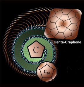
Researchers at Virginia Commonwealth University and universities in China and Japan have discovered a new structural variant of carbon called “penta-graphene” – a very thin sheet of pure carbon that has a unique structure inspired by a pentagonal pattern of tiles found paving the streets of Cairo.

The newly discovered material, called penta-graphene, is a single layer of carbon pentagons that resembles the Cairo tiling, and that appears to be dynamically, thermally and mechanically stable.

A Feb. 3, 2015 Virginia Commonwealth University (VCU) news release by Brian McNeill (also on EurekAlert), which originated the news item, provides more information about the research,

“The three last important forms of carbon that have been discovered were fullerene, the nanotube and graphene. Each one of them has unique structure. Penta-graphene will belong in that category,” said the paper’s senior author, Puru Jena, Ph.D., distinguished professor in the Department of Physics in VCU’s College of Humanities and Sciences.

Qian Wang, Ph.D., a professor at Peking University and an adjunct professor at VCU, was dining in a restaurant in Beijing with her husband when she noticed artwork on the wall depicting pentagon tiles from the streets of Cairo.

“I told my husband, “Come, see! This is a pattern composed only of pentagons,'” she said. “I took a picture and sent it to one of my students, and said, ‘I think we can make this. It might be stable. But you must check it carefully.’ He did, and it turned out that this structure is so beautiful yet also very simple.”

Most forms of carbon are made of hexagonal building blocks, sometimes interspersed with pentagons. Penta-graphene would be a unique two-dimensional carbon allotrope composed exclusively of pentagons.

Along with Jena and Wang, the paper’s authors include Shunhong Zhang, Ph.D candidate, from Peking University; Jian Zhou, Ph.D., a postdoctoral researcher at VCU; Xiaoshuang Chen, Ph.D., from the Chinese Academy of Science in Shanghai; and Yoshiyuki Kawazoe, Ph.D., from Tohoku University in Sendai, Japan.

The researchers simulated the synthesis of penta-graphene using computer modelling. The results suggest that the material might outperform graphene in certain applications, as it would be mechanically stable, possess very high strength, and be capable of withstanding temperatures of up to 1,000 degrees Kelvin.

“You know the saying, diamonds are forever? That’s because it takes a lot of energy to convert diamond back into graphite,” Jena said. “This will be similar.”

Penta-graphene has several interesting and unusual properties, Jena said. For example, penta-graphene is a semiconductor, whereas graphene is a conductor of electricity.

“When you take graphene and roll it up, you make what is called a carbon nanotube which can be metallic or semiconducting,” Jena said. “Penta-graphene, when you roll it up, will also make a nanotube, but it is always semiconducting.”

The way the material stretches is also highly unusual, the researchers said.

“If you stretch graphene, it will expand along the direction it is stretched, but contract along the perpendicular direction.” Wang said. “However, if you stretch penta-graphene, it will expand in both directions.”

The material’s mechanical strength, derived from a rare property known as Negative Poisson’s Ratio, may hold especially interesting applications for technology, the researchers said.

Penta-graphene’s properties suggest that it may have applications in electronics, biomedicine, nanotechnology and more.

The next step, Jena said, is for scientists to synthesize penta-graphene.

“Once you make it, it [will be] very stable. So the question becomes, how do you make it? In this paper, we have some ideas. Right now, the project is theoretical. It’s based on computer modelling, but we believe in this prediction quite strongly. And once you make it, it will open up an entirely new branch of carbon science. Two-dimensional carbon made completely of pentagons has never been known.”

Here’s a graphic representation of the new graphene material,

Caption: The newly discovered material, called penta-graphene, is a single layer of carbon pentagons that resembles the Cairo tiling, and that appears to be dynamically, thermally and mechanically stable. Credit: Virginia Commonwealth University

Caption: The newly discovered material, called penta-graphene, is a single layer of carbon pentagons that resembles the Cairo tiling, and that appears to be dynamically, thermally and mechanically stable.
Credit: Virginia Commonwealth University

Here’s a link to and a citation for the paper,

Penta-graphene: A new carbon allotrope by Shunhong Zhanga, Jian Zhou, Qian Wanga, Xiaoshuang Chen, Yoshiyuki Kawazoe, and Puru Jena. PNAS February 2, 2015 doi: 10.1073/pnas.1416591112 Published online before print February 2, 2015

This paper is behind a paywall.

Silky smooth tissue engineering

Virginia Commonwealth University (VCU) researchers have announced a new technique for tissue engineering that utilizes silk proteins. From a May 13, 2014 news item on Nanowerk,

When most people think of silk, the idea of a shimmering, silk scarf, or luxurious gown comes to mind.

But few realize, in its raw form, this seemingly delicate fiber is actually one of the strongest natural materials around – often compared to steel.

Silk, made up of the proteins fibroin and sericin, comes from the silkworm, and has been used in textiles and medical applications for thousands of years. The [US] Food and Drug Administration has classified silk as an approved biomaterial because it is nontoxic, biodegradable and biocompatible.

Those very properties make it an attractive candidate for use in widespread applications in tissue engineering. One day, silk could be an exciting route to create environmentally sound devices called “green devices,” instead of using plastics. However, forming complex architectures at the microscale or smaller, using silk proteins and other biomaterials has been a challenge for materials experts.

Now, a team of researchers from the Virginia Commonwealth University School of Engineering has found a way to fabricate precise, biocompatible architectures of silk proteins at the microscale.

A May 12, 2014 VCU news release by Sathya Achia Abraham, which originated the news item, describes the research underlying two recently published papers by the research team

    Kurland [Nicholas Kurland, Ph.D.] and Yadavalli [Vamsi Yadavalli, Ph.D., associate professor of chemical and life science engineering] successfully combined silk proteins with the technique of photolithography in a process they term “silk protein lithography” (SPL). Photolithography, or “writing using light,” is the method used to form circuits used in computers and smartphones, Yadavalli said.

According to Yadavalli, SPL begins by extracting the two main proteins from silk cocoons. These proteins are chemically modified to render them photoactive, and coated on glass or silicon surfaces as a thin film. As ultraviolet light passes through a stencil-like patterned mask, it crosslinks light-exposed proteins, turning them from liquid to solid.

The protein in unexposed areas is washed away, leaving behind patterns controllable to 1 micrometer. In comparison, a single human hair is 80-100 micrometers in diameter.

“These protein structures are high strength and excellent at guiding cell adhesion, providing precise spatial control of cells,” Yadavalli said.

“One day, we can envision implantable bioelectronic devices or tissue scaffolds that can safely disappear once they perform their intended function,” he said.

The team’s current research focuses on combining the photoreactive material with techniques such as rapid prototyping, and developing flexible bioelectronic scaffolds.

Study collaborators included S.C. Kundu, Ph.D., professor of biotechnology at the Indian Institute of Technology Kharagpur in India, and Tuli Dey, Ph.D., postdoctoral associate, at the Indian Institute of Technology Kharagpur in India, who provided the silk cocoons used in the study and assisted with cell culture experiments. VCU has recently filed a patent on this work.

Here’s a link to and a citation for both papers,

Silk Protein Lithography as a Route to Fabricate Sericin Microarchitectures by Nicholas E. Kurland, Tuli Dey, Congzhou Wang, Subhas C. Kundu and Vamsi K. Yadavalli. Article first published online: 16 APR 2014 DOI: 10.1002/adma.201400777

© 2014 WILEY-VCH Verlag GmbH & Co. KGaA, Weinheim

Precise Patterning of Silk Microstructures Using Photolithography by Nicholas E. Kurland, Tuli Dey, Subhas C. Kundu, and Vamsi K. Yadavalli. Advanced Materials Volume 25, Issue 43, pages 6207–6212, November 20, 2013 Article first published online: 20 AUG 2013 DOI: 10.1002/adma.201302823

© 2013 WILEY-VCH Verlag GmbH & Co. KGaA, Weinheim

Both papers are behind a paywall.

I have written about silk proteins in a Nov. 28, 2012 post (Producing stronger silk musically) that briefly mentioned tissue engineering with regard to a new technique for biosynthesising  materials.