Tag Archives: worms

Wormlike communication at the nanoscale

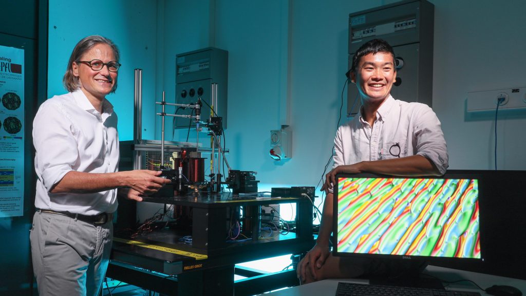
These days I need a little joy and these two researchers seem happy to share,

Prof. Dirk Grundler and doctoral assistant Sho Watanabe with a broadband spin-wave spectroscopy set up. Credit: EPFL / Alain Herzog

A July 15, 2020 news item on phys.org announces the development that so delights these researchers,

Researchers at EPFL [École polytechnique fédérale de Lausanne; Switzerland] have shown that electromagnetic waves coupled to precisely engineered structures known as artificial ferromagnetic quasicrystals allow for more efficient information transmission and processing at the nanoscale. Their research also represents the first practical demonstration of Conway worms, a theoretical concept for the description of quasicrystals.

A July 15, 2020 EPFL press release, which originated the news item, explains further,

High-frequency electromagnetic waves are used to transmit and process information in microelectronic devices such as smartphones. It’s already appreciated that these waves can be compressed using magnetic oscillations known as spin waves or magnons. This compression could pave the way for the design of nanoscale, multifunctional microwave devices with a considerably reduced footprint. But first, scientists need to gain a better understanding of spin waves – or precisely how magnons behave and propagate in different structures.

Learning more about aperiodic structures

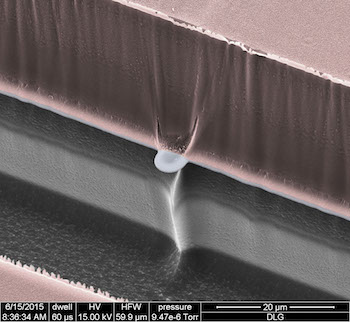
In a study conducted by the doctoral assistant Sho Watanabe, postdoctoral researcher Dr. Vinayak Bhat, and further team members, the scientists from EPFL’s Laboratory of Nanoscale Magnetic Materials and Magnonics (LMGN) examined how electromagnetic waves propagate, and how they could be manipulated, in precisely engineered nanostructures known as artificial ferromagnetic quasicrystals. The quasicrystals have a unique property: their structure is aperiodic, meaning that their constituent atoms or tailor-made elements do not follow a regular, repeating pattern but are still arranged deterministically. Although this characteristic makes materials especially useful for the design of everyday and high-tech devices, it remains poorly understood.

Faster, easier transmission of information

The LMGN team found that, under controlled conditions, a single electromagnetic wave coupled to an artificial quasicrystal splits into several spin waves, which then propagate within the structure. Each of these spin waves represents a different phase of the original electromagnetic wave, carrying different information. “It’s a very interesting discovery, because existing information-transmission methods follow the same principle,” says Dirk Grundler, an associate professor at EPFL’s School of Engineering (STI). “Except you need an extra device, a multiplexer, to split the input signal because – unlike in our study – it doesn’t divide on its own.”

Grundler also explains that, in conventional systems, the information contained in each wave can only be read at different frequencies – another inconvenience that the EPFL team overcame in their study. “In our two-dimensional quasicrystals, all the waves can be read at the same frequency,” he adds. The findings have been published in the journal Advanced Functional Materials.

Waves that spread like worms

The researchers also observed that, rather than propagating randomly, the waves often moved like so-called Conway worms, named after a well-known mathematician John Horton Conway who also developed a model to describe the behavior and feeding patterns of prehistoric worms. Conway discovered that, within two-dimensional quasicrystals, constituent elements arrange like meandering worms following a Fibonacci sequence. Thereby they form selected one-dimensional quasicrystals. “Our study represents the first practical demonstration of this theoretical concept, proving that the sequences induce interesting functional properties of waves in a quasicrystal,” says Grundler.

Take a look at that last paragraph. A mathematician develops a model for how prehistoric worms may have moved and applies it, theoretically, to 2D quasicrystals which these researchers believe they’ve observed in the laboratory and they believe this may have an impact on our future electronic devices. Sometimes I sit at home in wonder.

Here’s a link to and a citation for the paper,

Direct Observation of Worm‐Like Nanochannels and Emergent Magnon Motifs in Artificial Ferromagnetic Quasicrystals by Sho Watanabe, Vinayak S. Bhat, Korbinian Baumgaert, Dirk Grundler. Advanced Functional Materials DOI: https://doi.org/10.1002/adfm.202001388 First published: 15 July 2020

This is an open access paper.

The mention of quasicrystals reminded me of Daniel Schechtman who received the Nobel Prize for Chemistry in 2011 and who was mentioned in a December 24, 2013 posting here,

“I suggested earlier that this achievement has a fabulous quality and the Daniel Schechtman backstory is the reason. The winner of the 2011 Nobel Prize for Chemistry, Schechtman was reviled for years [emphasis mine] within his scientific community as Ian Sample notes in his Oct. 5, 2011 article on the announcement of Schechtman’s Nobel win written for the Guardian newspaper (Note: A link has been removed),

A scientist whose work was so controversial he was ridiculed and asked to leave his research group has won the Nobel Prize in Chemistry.

Daniel Shechtman, 70, a researcher at Technion-Israel Institute of Technology in Haifa, received the award for discovering seemingly impossible crystal structures in frozen gobbets of metal that resembled the beautiful patterns seen in Islamic mosaics.

Images of the metals showed their atoms were arranged in a way that broke well-establised rules of how crystals formed, a finding that fundamentally altered how chemists view solid matter.

You may want to click on the Guardian link to the full story about Schechtman and his quasicrystals. As for my December 24, 2013 posting, that features news of the creation of the first single-element quasicrystal in a laboratory along with an excerpt of the Schechtman story (scroll down about 50% of the way).

Gently measuring electrical signals in small animals with nano-SPEARs

This work comes from Rice University (Texas, US) according to an April 17, 2017 news item on Nanowerk,

Microscopic probes developed at Rice University have simplified the process of measuring electrical activity in individual cells of small living animals. The technique allows a single animal like a worm to be tested again and again and could revolutionize data-gathering for disease characterization and drug interactions.

The Rice lab of electrical and computer engineer Jacob Robinson has invented “nanoscale suspended electrode arrays” — aka nano-SPEARs — to give researchers access to electrophysiological signals from the cells of small animals without injuring them. Nano-SPEARs replace glass pipette electrodes that must be aligned by hand each time they are used.”

An April 17, 2017 Rice University news release (also on EurekAltert), which originated the news item, details the work,

“One of the experimental bottlenecks in studying synaptic behavior and degenerative diseases that affect the synapse is performing electrical measurements at those synapses,” Robinson said. “We set out to study large groups of animals under lots of different conditions to screen drugs or test different genetic factors that relate to errors in signaling at those synapses.”

Robinson’s early work at Rice focused on high-quality, high-throughput electrical characterization of individual cells. The new platform adapts the concept to probe the surface cells of nematodes, worms that make up 80 percent of all animals on Earth.

Most of what is known about muscle activity and synaptic transmission in the worms comes from the few studies that successfully used manually aligned glass pipettes to measure electrical activity from individual cells, Robinson said. However, this patch clamp technique requires time-consuming and invasive surgery that could negatively affect the data that is gathered from small research animals.

The platform developed by Robinson’s team works something like a toll booth for traveling worms. As each animal passes through a narrow channel, it is temporarily immobilized and pressed against one or several nano-SPEARS that penetrate its body-wall muscle and record electrical activity from nearby cells. That animal is then released, the next is captured and measured, and so on. Robinson said the device proved much faster to use than traditional electrophysiological cell measurement techniques.

The nano-SPEARs are created using standard thin-film deposition procedures and electron-beam or photolithography and can be made from less than 200 nanometers to more than 5 microns thick, depending on the size of animal to be tested. Because the nano-SPEARs can be fabricated on either silicon or glass, the technique easily combines with fluorescence microscopy, Robinson said.

The animals suitable for probing with a nano-SPEAR can be as large as several millimeters, like hydra, cousins of the jellyfish and the subject of an upcoming study. But nematodes known as Caenorhabditis elegans were practical for several reasons: First, Robinson said, they’re small enough to be compatible with microfluidic devices and nanowire electrodes. Second, there were a lot of them down the hall at the lab of Rice colleague Weiwei Zhong, who studies nematodes as transparent, easily manipulated models for signaling pathways that are common to all animals.

“I used to shy away from measuring electrophysiology because the conventional method of patch clamping is so technically challenging,” said Zhong, an assistant professor of biochemistry and cell biology and co-author of the paper. “Only a few graduate students or postdocs can do it. With Jacob’s device, even an undergraduate student can measure electrophysiology.”

“This meshes nicely with the high-throughput phenotyping she does,” Robinson said. “She can now correlate locomotive phenotypes with activity at the muscle cells. We believe that will be useful to study degenerative diseases centered around neuromuscular junctions.”

In fact, the labs have begun doing so. “We are now using this setup to profile worms with neurodegenerative disease models such as Parkinson’s and screen for drugs that reduce the symptoms,” Zhong said. “This would not be possible using the conventional method.”

Initial tests on C. elegans models for amyotrophic lateral sclerosis and Parkinson’s disease revealed for the first time clear differences in electrophysiological responses between the two, the researchers reported.

Testing the efficacy of drugs will be helped by the new ability to study small animals for long periods. “What we can do, for the first time, is look at electrical activity over a long period of time and discover interesting patterns of behavior,” Robinson said.

Some worms were studied for up to an hour, and others were tested on multiple days, said lead author Daniel Gonzales, a Rice graduate student in Robinson’s lab who took charge of herding nematodes through the microfluidic devices.

“It was in some way easier than working with isolated cells because the worms are larger and fairly sturdy,” Gonzales said. “With cells, if there’s too much pressure, they die. If they hit a wall, they die. But worms are really sturdy, so it was just a matter of getting them up against the electrodes and keeping them there.”

The team constructed microfluidic arrays with multiple channels that allowed testing of many nematodes at once. In comparison with patch-clamping techniques that limit labs to studying about one animal per hour, Robinson said his team measured as many as 16 nematodes per hour.

“Because this is a silicon-based technology, making arrays and producing recording chambers in high numbers becomes a real possibility,” he said.

A scanning electron micrograph shows a nano-SPEAR suspended midway between layers of silicon (grey) and photoresist material (pink) that form a recording chamber for immobilized nematodes. The high-throughput technology developed at Rice University can be adapted for other small animals and could enhance data-gathering for disease characterization and drug interactions. Courtesy of the Robinson Lab

Here’s a link to and a citation for the paper,

Scalable electrophysiology in intact small animals with nanoscale suspended electrode arrays by Daniel L. Gonzales, Krishna N. Badhiwala, Daniel G. Vercosa, Benjamin W. Avants, Zheng Liu, Weiwei Zhong, & Jacob T. Robinson. Nature Nanotechnology (2017) doi:10.1038/nnano.2017.55 Published online 17 April 2017

This paper is behind a paywall.

Creeping gel does ‘The Loco-Motion’

Now it’s the creeping gel’s turn, from an Oct. 24, 2016 news item on phys.org,

Directed motion seems simple to us, but the coordinated interplay of complex processes is needed, even for seemingly simple crawling motions of worms or snails. By using a gel that periodically swells and shrinks, researchers developed a model for the waves of muscular contraction and relaxation involved in crawling. As reported in the journal Angewandte Chemie, they were able to produce two types of crawling motion by using inhomogeneous irradiation.


Courtesy: Angewandte Chemie

Courtesy: Angewandte Chemie

An Oct. 24, 2016 Angewandte Chemie (Wiley) press release (also on EurekAlert), which originated the news item, explains further,

Crawling comes from waves that travel through muscle. These waves can travel in the same direction as the animal is crawling (direct waves), from the tail end toward the head, or in the opposite direction (retrograde waves), from the head toward the tail. While land snails use the former type of wave, earthworms and limpets use the latter. Chitons (polyplacophora) can switch between both types of movement.

With the aid of a chemical model in the form of a self-oscillating gel, researchers working with Qingyu Gao at the China University of Mining and Technology (Jiangsu, China) and Irving R. Epstein at Brandeis University (Waltham, Massachusetts, USA) have been able to answer some of the many questions about these crawling processes.

A gel is a molecular network with liquid bound in the gaps. In this case, the liquid contains all of the ingredients needed for an oscillating chemical reaction (“chemical clock”). The researchers incorporated one component of their reaction system into the network: a ruthenium complex. During the reaction, the ruthenium periodically switches between two oxidation states, Ru2+ and Ru3+. This switch changes the gel so that in one state it can hold more liquid than the other, so the gel swells and shrinks periodically. Like the chemical clock, these regions propagate in waves, similar to the waves of muscle contractions in crawling.

The complex used in this gel also changes oxidation state when irradiated with light. When the right half of the gel is irradiated more strongly than the left, the waves move from right to left, i.e., from a high- to a low-frequency region of gel oscillations. Once the difference in intensity of irradiation reaches a certain threshold, it causes a wormlike motion of the gel from left to right, retrograde wave locomotion. If the difference is increased further, the gel comes to a stop. A further increase in the difference causes the gel to move again, but in the opposite direction, i.e., direct wave locomotion. The nonuniform illumination plays a role analogous to that of anchoring segments and appendages (such as limbs and wings) during cell migration and animal locomotion, which control the direction of locomotion by strengthening direct movement and/or inhibiting the opposite movement.

By using computational models, the researchers were able to describe these processes. Within the gel, there are regions where pulling forces predominate; pushing forces predominate in other areas. Variations in the intensity of the irradiation lead to different changes in the friction forces and the tensions in the gel. When these effects are added up, it is possible to predict in which direction a particular grid element of the gel will move.

One important finding from this model: special changes in the viscoelastic properties of the slime excreted by the snails and worms as they crawl are not required for locomotion, whether retrograde or direct.

Here’s a link to and a citation for the paper,

Retrograde and Direct Wave Locomotion in a Photosensitive Self-Oscillating Gel by Lin Ren, Weibing She, Prof. Dr. Qingyu Gao, Dr. Changwei Pan, Dr. Chen Ji, and Prof. Dr. Irving R. Epstein. Angewandte Chemie International Edition DOI: 10.1002/anie.201608367 Version of Record online: 13 OCT 2016

© 2016 Wiley-VCH Verlag GmbH & Co. KGaA, Weinheim

This paper is behind a paywall.

For anyone curious about the song, there’s this from its Wikipedia entry (Note: Links have been removed),

“The Loco-Motion” is a 1962 pop song written by American songwriters Gerry Goffin and Carole King. “The Loco-Motion” was originally written for Dee Dee Sharp but Sharp turned the song down.[1] The song is notable for appearing in the American Top 5 three times – each time in a different decade, performed by artists from three different cultures: originally African American pop singer Little Eva in 1962 (U.S. No. 1);[2] then American band Grand Funk Railroad in 1974 (U.S. No. 1);[3] and finally Australian singer Kylie Minogue in 1988 (U.S. No. 3).[4]

The song is a popular and enduring example of the dance-song genre: much of the lyrics are devoted to a description of the dance itself, usually done as a type of line dance. However, the song came before the dance.

“The Loco-Motion” was also the second song to reach No. 1 by two different musical acts. The earlier song to do this was “Go Away Little Girl”, also written by Goffin and King. It is one of only nine songs to achieve this

I had not realized this song had such a storied past; there’s a lot more about it in the Wikipedia entry.

University of Missouri and the US Geological survey study carbon nanotubes in aquatic environments

The University of Missouri’s Aug. 22, 2012 news release (by Timothy Wall) announces the result of a carbon nanotube study in aquatic environments,

A joint study by the University of Missouri and United States Geological Survey found that they [carbon nanotubes or CNTs] can be toxic to aquatic animals. The researchers urge that care be taken to prevent the release of CNTs into the environment as the materials enter mass production.

“The great promise of carbon nanotubes must be balanced with caution and preparation,” said Baolin Deng, professor and chair of chemical engineering at the University of Missouri. “We don’t know enough about their effects on the environment and human health. The EPA and other regulatory groups need more studies like ours to provide information on the safety of CNTs.”

CNTs are microscopically thin cylinders of carbon atoms that can be hundreds of millions of times longer than they are wide, but they are not pure carbon. Nickel, chromium and other metals used in the manufacturing process can remain as impurities. Deng and his colleagues found that these metals and the CNTs themselves can reduce the growth rates or even kill some species of aquatic organisms. The four species used in the experiment were mussels (Villosa iris), small flies’ larvae (Chironomus dilutus), worms (Lumbriculus variegatus) and crustaceans (Hyalella azteca).

“One of the greatest possibilities of contamination of the environment by CNTs comes during the manufacture of composite materials,” said Hao Li, associate professor of mechanical and aerospace engineering at MU. “Good waste management and handling procedures can minimize this risk. Also, to control long-term risks, we need to understand what happens when these composite materials break down.”

I found the abstract for the team’s paper gave a good overview of how the research was conducted,

Carbon nanotubes (CNTs) are hydrophobic in nature and thus tend to accumulate in sediments if released into aquatic environments. As part of our overall effort to examine the toxicity of carbon-based nanomaterials to sediment-dwelling invertebrates, we have evaluated the toxicity of different types of CNTs in 14-d water-only exposures to an amphipod (Hyalella azteca), a midge (Chironomus dilutus), an oligochaete (Lumbriculus variegatus), and a mussel (Villosa iris) in advance of conducting whole-sediment toxicity tests with CNTs. The results of these toxicity tests conducted with CNTs added to water showed that 1.00 g/L (dry wt) of commercial sources of CNTs significantly reduced the survival or growth of the invertebrates. Toxicity was influenced by the type and source of the CNTs, by whether the materials were precleaned by acid, by whether sonication was used to disperse the materials, and by species of the test organisms. Light and electron microscope imaging of the surviving test organisms showed the presence of CNTs in the gut as well as on the outer surface of the test organisms, although no evidence was observed to show penetration of CNTs through cell membranes. The present study demonstrated that both the metals solubilized from CNTs such as nickel and the “metal-free” CNTs contributed to the toxicity.

Here’s the full citation and a link to the paper,

Toxicity of carbon nanotubes to freshwater aquatic invertebrates by Joseph N. Mwangi, Ning Wang, Christopher G. Ingersoll, Doug K. Hardesty, Eric L. Brunson, Hao Li, and Baolin Deng in Environmental Toxicology and Chemistry, Volume 31, Issue 8, pages 1823–1830, August 2012

For anyone who’s curious about what carbon nanotubes look like, here’s an image provided by the University of MIssouri,

Carbon Nanotubes Credit: Shaddack, Wikimedia Commons
Multi-walled carbon nanotubes. 3-15 walls, mean inner diameter 4nm, mean outer diameter 13-16 nm, length 1-10+ micrometers. Black clumpy powder, grains shown, partially smeared on paper. Scale in centimeters.

I could have included a larger version of the image but, given that we’re talking about the nanoscale, the smaller image seems more appropriate.