Tag Archives: wound healing

Nanomedicine: two stories about wound healing

Different strokes for different folks or, in this case, somewhat different approaches to healing different wounds.

Infected wounds

A July 17, 2024 news item on Nanowerk highlights work from China’s Research Center for Neutrophil Engineering Technology (affiliated with Suzhou Hospital of Nanjing Medical University), Note: A link has been removed,

Infectious wounds represent a critical challenge in healthcare, especially for diabetic patients grappling with ineffective antibiotics and escalating drug resistance. Conventional therapies often inadequately address deep tissue infections, highlighting the need for more innovative solutions. Engineered nanovesicles (NVs) from activated neutrophils provide a precise mechanism to combat pathogens deeply embedded in tissues, potentially revolutionizing the management of complex infectious wounds and boosting overall treatment efficacy.

Researchers at the Research Center for Neutrophil Engineering Technology have achieved a significant advancement in medical nanotechnology. Their findings, published in the journal Burns & Trauma (“Engineered nanovesicles from activated neutrophils with enriched bactericidal proteins have molecular debridement ability and promote infectious wound healing”), detail the creation of novel neutrophil-engineered NVs.

A July 17, 2024 Maximum Academic Press ‘press release’ on EurekAlert, which originated the news item, goes on to describe what the researchers discovered,

This study reveals that engineered NVs derived from activated neutrophils not only mimic the physical properties of exosomes but surpass them due to their rich content of bactericidal proteins. Extensively tested both in vitro and in vivo, these NVs effectively combat key pathogens like Staphylococcus aureus and Escherichia coli, which contribute to deep tissue infections. The NVs promote rapid debridement, significantly reduce bacterial populations, and boost collagen deposition, thus hastening the healing process. This research positions NVs as a formidable alternative to traditional antibiotics, introducing a novel method for treating resistant infections and advancing the field of wound care.

Dr. Bingwei Sun, the lead researcher, emphasized, “These engineered NVs mark a major advancement in the management of infectious diseases. By targeting the infection site with high levels of bactericidal proteins, we achieve swift and effective healing, thereby opening new paths for the treatment of chronic and resistant infections.”

The advent of activated neutrophil-derived NVs signifies a major leap in medical technology, potentially reducing healthcare costs and enhancing patient outcomes. This innovation not only promises to improve wound healing in diabetic and other chronic infection patients but also sets the stage for further development of biologically inspired therapeutic strategies.

Here’s a link to and a citation for the paper,

Engineered nanovesicles from activated neutrophils with enriched bactericidal proteins have molecular debridement ability and promote infectious wound healing by Hangfei Jin, Xiao Wen, Ran Sun, Yanzhen Yu, Zaiwen Guo, Yunxi Yang, Linbin Li, and Bingwei Sun. Burns & Trauma, Volume 12, 2024, tkae018, DOI: https://doi.org/10.1093/burnst/tkae018 Published: 20 June 2024

This paper is open access.

Diabetic wounds

A July 17, 2024 news item on phys.org announces work from another team developing its own approach to healing wounds, albeit, a different category of wounds,

Diabetic wounds are notoriously challenging to treat, due to prolonged inflammation and a high risk of infection. Traditional treatments generally offer only passive protection and fail to dynamically interact with the wound environment.

In a new article published in Burns & Trauma on June 5, 2024, a research team from Mudanjiang Medical University and allied institutions assesses the effectiveness of PLLA nanofibrous membranes.

Infused with curcumin and silver nanoparticles, these membranes are designed to substantially enhance the healing processes in diabetic wounds by targeting fundamental issues like excessive inflammation and infection.

This research centered on developing PLLA/C/Ag nanofibrous membranes through air-jet spinning, achieving a consistent fiber distribution essential for effective therapeutic delivery. The membranes boast dual benefits: antioxidant properties that reduce harmful reactive oxygen species in wound environments and potent antibacterial activity that decreases infection risks.

A July 17, 2024 Maximum Academic Press ‘press release‘ on EurekAlert provides more information about the research, Note 1: This press release appears to have originated the news item, which was then edited and rewritten; Note 2: Links have been removed,

In a pioneering study, researchers have developed a poly (L-lactic acid) (PLLA) nanofibrous membrane enhanced with curcumin and silver nanoparticles (AgNPs), aimed at improving the healing of diabetic wounds. This advanced dressing targets critical barriers such as inflammation, oxidative stress, and bacterial infections, which hinder the recovery process in diabetic patients. The study’s results reveal a promising therapeutic strategy that could revolutionize care for diabetes-related wounds.

Diabetic wounds are notoriously challenging to heal, with prolonged inflammation and a high risk of infection. Traditional treatments generally offer only passive protection and fail to dynamically interact with the wound environment. The creation of bioactive dressings like the poly (L-lactic acid) (PLLA) nanofibrous membranes incorporated with AgNPs and curcumin (PLLA/C/Ag) membranes signifies a crucial shift towards therapies that actively correct imbalances in the wound healing process, offering a more effective solution for managing diabetic wounds.

Published (DOI: 10.1093/burnst/tkae009) in Burns & Trauma on June 5, 2024, this trailblazing research by a team from Mudanjiang Medical University and allied institutions assesses the effectiveness of PLLA nanofibrous membranes. Infused with curcumin and silver nanoparticles, these membranes are designed to substantially enhance the healing processes in diabetic wounds by targeting fundamental issues like excessive inflammation and infection.

This research centered on developing PLLA/C/Ag nanofibrous membranes through air-jet spinning, achieving a consistent fiber distribution essential for effective therapeutic delivery. The membranes boast dual benefits: antioxidant properties that reduce harmful reactive oxygen species in wound environments and potent antibacterial activity that decreases infection risks. In vivo tests on diabetic mice demonstrated the membranes’ capability to promote crucial healing processes such as angiogenesis and collagen deposition. These findings illustrate that PLLA/C/Ag membranes not only protect wounds but also actively support and expedite the healing process, marking them as a significant therapeutic innovation for diabetic wound management with potential for broader chronic wound care applications.

Dr. Yanhui Chu, a principal investigator of the study, highlighted the importance of these developments: “The PLLA/C/Ag membranes are a significant breakthrough in diabetic wound care. Their ability to effectively modulate the wound environment and enhance healing could establish a new standard in treatment, providing hope to millions affected by diabetes-related complications.”

The deployment of PLLA/C/Ag nanofibrous membranes in clinical environments could transform the treatment of diabetic wounds, offering a more active and effective approach. Beyond diabetes management, this technology has the potential for extensive applications in various chronic wounds, paving the way for future breakthroughs in bioactive wound dressings. This study not only progresses our understanding of wound management but also paves new paths for developing adaptive treatments for complex wound scenarios.

Here’s a link to and a citation for the paper,

Immunomodulatory poly(L-lactic acid) nanofibrous membranes promote diabetic wound healing by inhibiting inflammation, oxidation and bacterial infection by Yan Wu, Jin Zhang, Anqi Lin, Tinglin Zhang, Yong Liu, Chunlei Zhang, Yongkui Yin, Ran Guo, Jie Gao, Yulin Li, and Yanhui Chu. Burns & Trauma, Volume 12, 2024, tkae009, DOI: https://doi.org/10.1093/burnst/tkae009 Published: 05 June 2024

This paper is open access.

Science publishing

As I think most people know, publishing of any kind is a tough business, particularly these days. This instability has led to some interesting corporate relationships. E.g., Springer Nature (a German-British academic publisher) is the outcome of some mergers as the Springer Nature Wikipedia entry notes,

The company originates from several journals and publishing houses, notably Springer-Verlag, which was founded in 1842 by Julius Springer in Berlin[4] (the grandfather of Bernhard Springer who founded Springer Publishing in 1950 in New York),[5] Nature Publishing Group which has published Nature since 1869,[6] and Macmillan Education, which goes back to Macmillan Publishers founded in 1843.[7]

Springer Nature was formed in 2015 by the merger of Nature Publishing Group, Palgrave Macmillan, and Macmillan Education (held by Holtzbrinck Publishing Group) with Springer Science+Business Media (held by BC Partners). Plans for the merger were first announced on 15 January 2015.[8] The transaction was concluded in May 2015 with Holtzbrinck having the majority 53% share.[9]

Now you have what was an independent science journal, Nature, owned by Springer. By the way, Springer Nature also acquired Scientific American, another major science journal.

Relatedly, seeing Maximum Academic Press as the issuer for the press releases mentioned here aroused my curiosity. I haven’t stumbled across the company before but found this on the company’s About Us webpage, Note: Links have been removed,

Maximum Academic Press (MAP) is an independent publishing company with focus on publishing golden open access academic journals. From 2020 to now, MAP has successfully launched 24 academic journals which cover the research fields of agriculture, biology, environmental sciences, engineering and humanities and social sciences.                    

Professor Zong-Ming (Max) Cheng, chief editor and founder of MAP, who earned his Ph.D from Cornell University in 1991 and worked as an Assistant, Associate and Professor at North Dakota State University and University of Tennessee for over 30 years. Prior to establishing MAP, Dr. Cheng launched Horticulture Research (initially published by Nature Publishing Group) in 2014, Plant Phenomics (published by American Association of Advancement of Sciences, AAAS) in 2019, and BioDesign Research (published by AAAS) in 2020, and served as the Editor-in-Chief, Co-Editors-in-Chief, and the executive editor, respectively. Dr. Cheng wishes to apply all successful experiences in launching and managing these three high quality journals to MAP-published journals with highest quality and ethics standards.

It was a little bit of a surprise to see that MAP doesn’t publish the journal, Burns & Trauma, where the studies (cited here) were published. From the Burns & Trauma About the Journal webpage on the Oxford University Press website for Oxford Academic journals,

Aims and scope

Burns & Trauma is an open access, peer-reviewed journal publishing the latest developments in basic, clinical, and translational research related to burns and traumatic injuries, with a special focus on various aspects of biomaterials, tissue engineering, stem cells, critical care, immunobiology, skin transplantation, prevention, and regeneration of burns and trauma injury.

Society affiliations

Burns & Trauma is the official journal of Asia-Pacific Society of Scar Medicine, Chinese Burn Association, Chinese Burn Care and Rehabilitation Association and Chinese Society for Scar Medicine. It is sponsored by the Institute of Burn Research, Southwest Hospital (First Affiliated Hospital of Army Medical University), China.

I don’t know what to make of it all but I can safely say scientific publishing has gotten quite complicated since the days that Nature first published its own eponymous journal.

Peptide-based hydrogels for faster healing from research team at the University of Ottawa

While this research team was heavily dominated by researchers from the University of Ottawa, there were two members associated with the University of Talca (Universidad de Talca; located in Chile), two members associated with the University of Montreal (Université de Montréal), and one member with McGill University (located in Montréal).

Now for these special hydrogels, from a May 13, 2024 University of Ottawa news release (also on EurekAlert) by David McFadden, Note: Links have been removed,

Combining biomedical finesse and nature-inspired engineering, a uOttawa-led team of scientists have created a jelly-like material that shows great potential for on-the-spot repair to a remarkable range of damaged organs and tissues in the human body.

Cutting-edge research co-led by uOttawa Faculty of Medicine  Associate Professor Dr. Emilio I. Alarcón could eventually impact millions of lives with peptide-based hydrogels that will close skin wounds, deliver therapeutics to damaged heart muscle, as well as reshape and heal injured corneas.

“We are using peptides to fabricate therapeutic solutions. The team is drawing inspiration from nature to develop simple solutions for wound closure and tissue repair,” says Dr. Alarcón, a scientist and director at the BioEngineering and Therapeutic Solutions (BEaTS) group at the University of Ottawa Heart Institutek whose innovative research work is focused on developing new materials with capabilities for tissue regeneration.

Peptides are molecules in living organisms and hydrogels are a water-based material with a gelatinous texture that have proven useful in therapeutics.

The approach used in the study –  just published in Advanced Functional Materials and co-led by Dr. Erik Suuronen & Dr. Marc Ruel – is unique. Most hydrogels explored in tissue engineering are animal-derived and protein-based materials, but the biomaterial created by the collaborative team is supercharged by engineered peptides. This makes it more clinically translatable.

Dr. Ruel, a full professor in the uOttawa Faculty of Medicine’s Department of Cellular and Molecular Medicine and the endowed chair of research in the Division of Cardiac Surgery at the University of Ottawa Heart Institute, says the study’s insights could be a game changer.

“Despite millennia of evolution, the human response to wound healing still remains imperfect,” Dr. Ruel says. “We see maladapted scarring in everything from skin incisions to eye injuries, to heart repair after a myocardial infarction. Drs. Alarcón, Suuronen, and the rest of our team have focused on this problem for almost two decades. The publication by Dr. Alarcón in Advanced Functional Materials reveals a novel way to make wound healing, organ healing, and even basic scarring after surgery much more therapeutically modulatable and, therefore, optimizable for human health.”

Indeed, the ability to modulate the peptide-based biomaterial is key. The uOttawa-led team’s hydrogels are designed to be customizable, making the durable material adaptable for use in a surprising range of tissues. Essentially, the two-component recipe could be adjusted to ramp up adhesivity or dial down other components depending on the part of the body needing repair.

“We were in fact very surprised by the range of applications our materials can achieve,” says Dr. Alarcón. “Our technology offers an integrated solution that is customizable depending on the targeted tissue.”

Dr. Alarcón says that not only does the study’s data suggest that the therapeutic action of the biomimetic hydrogels are highly effective, but its application is far simpler and cost-effective than other regenerative approaches.

The materials are engineered in a low-cost and scalable manner – hugely important qualities for any number of major biomedical applications. The team also devised a rapid-screening system that allowed them to significantly slash the design costs and testing timespans.

“This significant reduction in cost and time not only makes our material more economically viable but also accelerates its potential for clinical use,” Dr. Alarcón says.

What are next steps for the talent-rich research team? They will conduct large animal tests in preparation for tests in human subjects. So far, heart and skin tests were conducted with rodents, and the cornea work was done ex vivo.

Part of the work for this study was funded by the uOttawa Faculty of Medicine’s  “Path to Patenting & Pre-Commercialization” (3P),  an innovation-focused approach to provide our community’s top-flight researchers with the assistance needed to bring their most promising breakthroughs to the wider world.

Here’s a link to and a citation for the paper,

Multipurpose On-the-Spot Peptide-Based Hydrogels for Skin, Cornea, and Heart Repair by Alex Ross, Xixi Guo, German A. Mercado Salazar, Sergio David Garcia Schejtman, Jinane El-Hage, Maxime Comtois-Bona, Aidan Macadam, Irene Guzman-Soto, Hiroki Takaya, Kevin Hu, Bryan Liu, Ryan Tu, Bilal Siddiqi, Erica Anderson, Marcelo Muñoz, Patricio Briones-Rebolledo, Tianqin Ning, May Griffith, Benjamin Rotsein, Horacio Poblete, Jianyu Li, Marc Ruel, Erik J. Suuronen, Emilio I. Alarcon. Advanced Functional Materials DOI: https://doi.org/10.1002/adfm.202402564 First published: 23 April 2024

This paper is open access.

PAINT wound-healing ink into your cuts with a 3D-printing pen

This screams tattoo to me but it’s not,

Caption This 3-D printing pen is painting a gel that can help wounds of all shapes heal quickly and effectively. Credit: Adapted from ACS Applied Materials & Interfaces, 2023, DOI: 10.1021/acsami.3c03630

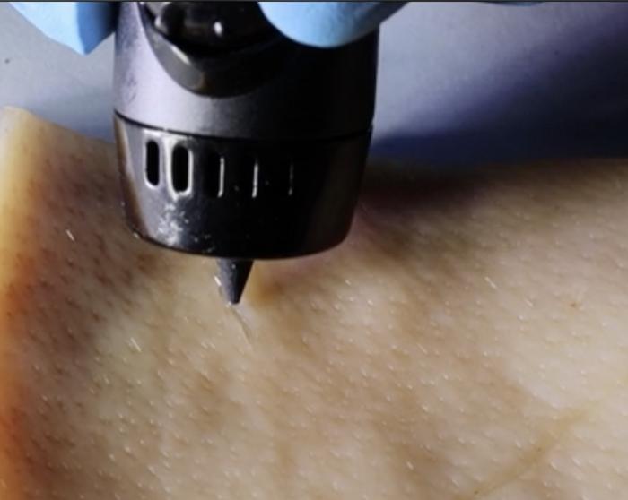
A June 1, 2023 American Chemical Society (ACS) news release (also on EurekAlert), announces a new approach to wound healing,

The body is pretty good at healing itself, though more severe wounds can require bandages or stitches. But researchers publishing in ACS Applied Materials & Interfaces have developed a wound-healing ink that can actively encourage the body to heal by exposing the cut to immune-system vesicles. The ink can be spread into a cut of any shape using a 3D-printing pen, and in mice, the technology nearly completely repaired wounds in just 12 days.

When the skin is cut or torn, the body’s natural “construction crew” kicks in to fix it back up — clearing out any bacterial invaders, regrowing broken blood vessels and eventually forming a scar. Many techniques used to heal wounds can’t do much beyond helping the body do its job better. Bandages or stitches are used to prevent further bleeding, while antibiotics work to prevent complications from infections. But by adding members of the construction crew to a wound-healing treatment or bandage, it could actually accelerate the natural healing process. Specifically, white blood cells or the extracellular vesicles (EVs) secreted from them play important roles in promoting blood vessel formation and reducing inflammation during healing. So, Dan Li, Xianguang Ding and Lianhui Wang wanted to incorporate these EVs into a hydrogel-based wound healing ink that could be painted into cuts of any shape.

The team developed a system called PAINT, or “portable bioactive ink for tissue healing,” using EVs secreted from macrophages combined with sodium alginate. These components were combined in a 3D-printing pen, where they mixed at the pen’s tip and formed a sturdy gel at the site of injury within three minutes. The EVs promoted blood vessel formation and reduced inflammatory markers in human epithelial cells, shifting them into the “proliferative,” or growth, phase of healing. PAINT was also tested on injured mice, where it promoted collagen fiber formation. Mice treated with PAINT had almost healed completely from a large wound after 12 days, compared to mice that didn’t receive the treatment, who were not nearly as far along in the healing process at this time point. The researchers say that this work could help heal a wide variety of cuts quickly and easily, without the need for complex procedures.

The authors acknowledge funding from the Leading-Edge Technology Programme of Jiangsu Natural Science Foundation, the Natural Science Foundation, the Natural Science Foundation of Jiangsu Province, the CAS [Chinese Academy of Sciences] Key Laboratory of Nano-Bio Interface, the Key Laboratory of Nanodevices and Applications, and the Postgraduate Research & Practice Innovation Program of Jiangsu Province.

The American Chemical Society (ACS) is a nonprofit organization chartered by the U.S. Congress. ACS’ mission is to advance the broader chemistry enterprise and its practitioners for the benefit of Earth and all its people. The Society is a global leader in promoting excellence in science education and providing access to chemistry-related information and research through its multiple research solutions, peer-reviewed journals, scientific conferences, eBooks and weekly news periodical Chemical & Engineering News. ACS journals are among the most cited, most trusted and most read within the scientific literature; however, ACS itself does not conduct chemical research. As a leader in scientific information solutions, its CAS division partners with global innovators to accelerate breakthroughs by curating, connecting and analyzing the world’s scientific knowledge. ACS’ main offices are in Washington, D.C., and Columbus, Ohio.

Here’s a link to and a citation for the paper,

Paintable Bioactive Extracellular Vesicle Ink for Wound Healing by Li Li, Zhiyu Wang, Kepeng Wang, Siyuan Fu, Dan Li, Mao Wang, Yi Cao, Houjuan Zhu, Ziyan Li, Lixing Weng, Zhiyang Li, Xianguang Ding, and Lianhui Wang. ACS Appl. Mater. Interfaces 2023, 15, 21, 25427–25436 DOI: https://doi.org/10.1021/acsami.3c03630 Publication Date:May 19, 2023 Copyright © 2023 American Chemical Society

This paper is behind a paywall.

Treating traumatic muscle loss with tissue nanotransfection

A November 9, 2022 news item on ScienceDaily announces some work from Indiana University (US),

Technology developed by researchers at the Indiana University School of Medicine that can change skin tissue into blood vessels and nerve cells has also shown promise as a treatment for traumatic muscle loss.

Tissue nanotransfection is a minimally invasive nanochip device that can reprogram tissue function by applying a harmless electric spark to deliver specific genes in a fraction of a second.

A November 9, 2022 Indiana University news release (also on EurekAlert), which originated the news item, provides additional technical details, Note: Links have been removed,

A new study, published in Nature Partner Journals Regenerative Medicine, tested tissue nanotransfection-based gene therapy as a treatment, with the goal of delivering a gene known to be a major driver of muscle repair and regeneration. They found that muscle function improved when tissue nanotransfection was used as a therapy for seven days following volumetric muscle loss in rats. It is the first study to report that tissue nanotransfection technology can be used to generate muscle tissue and demonstrates its benefit in addressing volumetric muscle loss.

Volumetric muscle loss is the traumatic or surgical loss of skeletal muscle that results in compromised muscle strength and mobility. Incapable of regenerating the amount of lost tissue, the affected muscle undergoes substantial loss of function, thus compromising quality of life. A 20 percent loss in mass can result in an up to 90 percent loss in muscle function.

Current clinical treatments for volumetric muscle loss are physical therapy or autologous tissue transfer (using a person’s own tissue), the outcomes of which are promising but call for improved treatment regimens.

“We are encouraged that tissue nanotransfection is emerging as a versatile platform technology for gene delivery, gene editing and in vivo tissue reprogramming,” said Chandan Sen, director of the Indiana Center for Regenerative Medicine and Engineering, associate vice president for research and Distinguished Professor at the IU School of Medicine. “This work proves the potential of tissue nanotransfection in muscle tissue, opening up a new avenue of investigational pursuit that should help in addressing traumatic muscle loss. Importantly, it demonstrates the versatility of the tissue nanotransfection technology platform in regenerative medicine.”

Sen also leads the regenerative medicine and engineering scientific pillar of the IU Precision Health Initiative and is lead author on the new publication.

The Indiana Center for Regenerative Medicine and Engineering is home to the tissue nanotransfection technology for in vivo tissue reprogramming, gene delivery and gene editing. So far, tissue nanotransfection has also been achieved in blood vessel and nerve tissue. In addition, recent work has shown that topical tissue nanotransfection can achieve cell-specific gene editing of skin wound tissue to improve wound closure.

Here’s a link to and a citation for the paper,

Myogenic tissue nanotransfection improves muscle torque recovery following volumetric muscle loss by Andrew Clark, Subhadip Ghatak, Poornachander Reddy Guda, Mohamed S. El Masry, Yi Xuan, Amy Y. Sato, Teresita Bellido & Chandan K. Sen. npj Regenerative Medicine volume 7, Article number: 63 (2022) DOI: https://doi.org/10.1038/s41536-022-00259-y Published: 20 October 2022

This paper is open access.

This is a very nice image of a delighted Dr. Sen,

Caption Chandan Sen Credit: Photo by Liz Kaye, Indiana University

Tissue nanotransfection

I’m wondering how I missed the research from last year (2021) which foregrounds this latest work. Ah well. It happens. Making up for lost time, here’s a July 18, 2022 news item on phys.org about tissue nanotransfection, Note: Links have been removed,

The Indiana Center for Regenerative Medicine and Engineering (ICRME) at Indiana University School of Medicine is home to tissue nanotransfection (TNT) regenerative medicine technology that achieves functional tissue reprogramming in the live body. Last year, ICRME researchers published on how to manufacture the TNT 2.0 silicon chip hardware in Nature Protocol. Now, their research demonstrates for the first time that TNT can serve as a non-viral, topical gene-editing delivery device.

TNT is a minimally invasive device that can reprogram tissue function in the live body by applying pulses of harmless, electric sparks to deliver specific genes of interest to the skin.

“TNT-based delivery can achieve cell-specific gene editing,” said corresponding author Chandan K. Sen, Ph.D., the J. Stanley Battersby Chair and distinguished professor of surgery, director of the ICRME at IU School of Medicine and executive director of the Indiana University Health Comprehensive Wound Care Center. “Your skin has thousands of genes and in chronic wounds many key genes are silenced by DNA methylation. TNT-based gene editing technology can remove that barrier.”

A July 18, 2022 Indiana University School of Medicine news release (also on EurekAlert), which originated the news item, updates the information with some of the latest research, Note: Links have been removed,

In this study, genome-wide methylation was observed in the chronic wound tissue of patients. This was reproduced in an experimental murine model. TNT-based, cell-specific gene editing rescued wound healing. Results were published recently [July 12, 2022] in the Journal of Clinical Investigation.

Previous TNT application studies reported on the rescue of injured legs, diabetic neuropathy, crushed nerve and the stroke-affected brain. This is the first time promoter methylation of genes is recognized as a critical barrier to wound healing. In this study, ICRME investigators found that P53 methylation and gene silencing as a critical barrier to cutaneous wound epithelial-to-mesenchymal transition (EMT), a mechanism that is necessary to close skin wounds. TNT based non-viral keratinocyte-specific demethylation of P53 gene rescued EMT and achieved wound closure.

Chronic wounds can result in serious and sometimes life-threatening complications from an abundance of dying and necrotic tissue, such as cellulitis, lower-extremity amputation and sepsis. Treating chronic wounds is estimated to cost the United States health care system $28 billion annually, which amplifies the need to test novel treatments to prevent amputation, save lives and lower health care costs.

“Inspired by observations in chronic wound patients, this work has achieved an important milestone highlighting the need to de-silence genes at the wound-site,” said first author Kanhaiya Singh, PhD, assistant professor of surgery and an investigator at the ICRME.

Here are two links and citations. First, the earlier work,

Fabrication and use of silicon hollow-needle arrays to achieve tissue nanotransfection in mouse tissue in vivo by Yi Xuan, Subhadip Ghatak, Andrew Clark, Zhigang Li, Savita Khanna, Dongmin Pak, Mangilal Agarwal, Sashwati Roy, Peter Duda & Chandan K. Sen. Nature Protocols volume 16, pages 5707–5738 (2021) DOI: https://doi.org/10.1038/s41596-021-00631-0 Published: 26 November 2021 Issue Date: December 2021

This paper is behind a paywall.

Now, the latest work

Genome-wide DNA hypermethylation opposes healing in chronic wound patients by impairing epithelial-to-mesenchymal transition by Kanhaiya Singh, Yashika Rustagi, Ahmed S. Abouhashem, Saba Tabasum, Priyanka Verma, Edward Hernandez, Durba Pal, Dolly K. Khona, Sujit K. Mohanty, Manishekhar Kumar, Rajneesh Srivastava, Poornachander R Guda, Sumit S. Verma, Sanskruti Mahajan, Jackson A. Killian, Logan A. Walker, Subhadip Ghatak, Shomita S. Mathew-Steiner, Kristen Wanczyk, Sheng Liu, Jun Wan, Pearlly Yan, Ralf Bundschuh, Savita Khanna, Gayle M. Gordillo, Michael P. Murphy, Sashwati Roy, and Chandan K. Sen. J Clin Invest. DOI: https://doi.org/10.1172/JCI157279 Published: July 12, 2022 Version 1 (In-Press Preview) Version 2: J Clin Invest. 2022;132(17):e157279. https://doi.org/10.1172/JCI157279. Volume 132, Issue 17 Published September 1, 2022

This paper is open access.