Tag Archives: Wyss Institute for Biologically Inspired Engineering at Harvard

Answer to why Roman concrete was so durable

Roman concrete lasts for millenia while our ‘modern’ concrete doesn’t and that’s what makes the Roman stuff so fascinating. There’s a very good January 6, 2023 Massachusetts Institute of Technology (MIT) news release (also on EurekAlert) which may provide an answer the mystery of the this material’s longevity,

The ancient Romans were masters of engineering, constructing vast networks of roads, aqueducts, ports, and massive buildings, whose remains have survived for two millennia. Many of these structures were built with concrete: Rome’s famed Pantheon, which has the world’s largest unreinforced concrete dome and was dedicated in A.D. 128, is still intact, and some ancient Roman aqueducts still deliver water to Rome today. Meanwhile, many modern concrete structures have crumbled after a few decades.

Researchers have spent decades trying to figure out the secret of this ultradurable ancient construction material, particularly in structures that endured especially harsh conditions, such as docks, sewers, and seawalls, or those constructed in seismically active locations.

Now, a team of investigators from MIT, Harvard University, and laboratories in Italy and Switzerland, has made progress in this field, discovering ancient concrete-manufacturing strategies that incorporated several key self-healing functionalities. The findings are published in the journal Science Advances, in a paper by MIT professor of civil and environmental engineering Admir Masic, former doctoral student Linda Seymour, and four others.

For many years, researchers have assumed that the key to the ancient concrete’s durability was based on one ingredient: pozzolanic material such as volcanic ash from the area of Pozzuoli, on the Bay of Naples. [emphasis mine] This specific kind of ash was even shipped all across the vast Roman empire to be used in construction, and was described as a key ingredient for concrete in accounts by architects and historians at the time.

Under closer examination, these ancient samples also contain small, distinctive, millimeter-scale bright white mineral features, which have been long recognized as a ubiquitous component of Roman concretes. These white chunks, often referred to as “lime clasts,” originate from lime, another key component of the ancient concrete mix. “Ever since I first began working with ancient Roman concrete, I’ve always been fascinated by these features,” says Masic. “These are not found in modern concrete formulations, so why are they present in these ancient materials?”

Previously disregarded as merely evidence of sloppy mixing practices, or poor-quality raw materials, the new study suggests that these tiny lime clasts gave the concrete a previously unrecognized self-healing capability. [emphasis mine] “The idea that the presence of these lime clasts was simply attributed to low quality control always bothered me,” says Masic. “If the Romans put so much effort into making an outstanding construction material, following all of the detailed recipes that had been optimized over the course of many centuries, why would they put so little effort into ensuring the production of a well-mixed final product? There has to be more to this story.”

Upon further characterization of these lime clasts, using high-resolution multiscale imaging and chemical mapping techniques pioneered in Masic’s research lab, the researchers gained new insights into the potential functionality of these lime clasts.

Historically, it had been assumed that when lime was incorporated into Roman concrete, it was first combined with water to form a highly reactive paste-like material, in a process known as slaking. But this process alone could not account for the presence of the lime clasts. Masic wondered: “Was it possible that the Romans might have actually directly used lime in its more reactive form, known as quicklime?”

Studying samples of this ancient concrete, he and his team determined that the white inclusions were, indeed, made out of various forms of calcium carbonate. And spectroscopic examination provided clues that these had been formed at extreme temperatures, as would be expected from the exothermic reaction produced by using quicklime instead of, or in addition to, the slaked lime in the mixture. Hot mixing, the team has now concluded, was actually the key to the super-durable nature.

“The benefits of hot mixing are twofold,” Masic says. “First, when the overall concrete is heated to high temperatures, it allows chemistries that are not possible if you only used slaked lime, producing high-temperature-associated compounds that would not otherwise form. Second, this increased temperature significantly reduces curing and setting times since all the reactions are accelerated, allowing for much faster construction.”

During the hot mixing process, the lime clasts develop a characteristically brittle nanoparticulate architecture, creating an easily fractured and reactive calcium source, which, as the team proposed, could provide a critical self-healing functionality. As soon as tiny cracks start to form within the concrete, they can preferentially travel through the high-surface-area lime clasts. This material can then react with water, creating a calcium-saturated solution, which can recrystallize as calcium carbonate and quickly fill the crack, or react with pozzolanic materials to further strengthen the composite material. These reactions take place spontaneously and therefore automatically heal the cracks before they spread. Previous support for this hypothesis was found through the examination of other Roman concrete samples that exhibited calcite-filled cracks.

To prove that this was indeed the mechanism responsible for the durability of the Roman concrete, the team produced samples of hot-mixed concrete that incorporated both ancient and modern formulations, deliberately cracked them, and then ran water through the cracks. Sure enough: Within two weeks the cracks had completely healed and the water could no longer flow. An identical chunk of concrete made without quicklime never healed, and the water just kept flowing through the sample. As a result of these successful tests, the team is working to commercialize this modified cement material.

“It’s exciting to think about how these more durable concrete formulations could expand not only the service life of these materials, but also how it could improve the durability of 3D-printed concrete formulations,” says Masic.

Through the extended functional lifespan and the development of lighter-weight concrete forms, he hopes that these efforts could help reduce the environmental impact of cement production, which currently accounts for about 8 percent of global greenhouse gas emissions. Along with other new formulations, such as concrete that can actually absorb carbon dioxide from the air, another current research focus of the Masic lab, these improvements could help to reduce concrete’s global climate impact.

The research team included Janille Maragh at MIT, Paolo Sabatini at DMAT in Italy, Michel Di Tommaso at the Instituto Meccanica dei Materiali, in Switzerland, and James Weaver at the Wyss Institute for Biologically Inspired Engineering at Harvard University. The work was carried out with the assistance of the archeological museum of Priverno, Italy.

I remember the excitement over volcanic ash (it’s mentioned in my June 3, 2016 posting titled: “Making better concrete by looking to nature for inspiration” and my February 17, 2021 posting “Nuclear power plants take a cue from Roman concrete“). As for something being ignored as unimportant or being a result poor practice when it’s not, that’s one of my favourite kinds of science story.

For the really curious, Jennifer Ouellette’s January 6, 2023 article (Ancient Roman concrete could self-heal thanks to “hot mixing” with quicklime) for Ars Technica provides a little more detail.

Here’s a link to and a citation for the latest paper,

Hot mixing: Mechanistic insights into the durability of ancient Roman concrete by Linda M. Seymour, Janille Maragh, Paolo Sabatini, Michel Di Tommaso, James C. Weaver, and Admir Masic. Science Advances 6 Jan 2023 Vol 9, Issue 1 DOI: 10.1126/sciadv.add1602

This paper is open access.

One last note, DMAT is listed as Paolo Sabatini’s home institution. It is a company for which Sabatini is a co-founder and CEO (chief executive officer). DMAT has this on its About page, “Our mission is to develop breakthrough innovations in construction materials at a global scale. DMAT is at the helm of concrete’s innovation.”
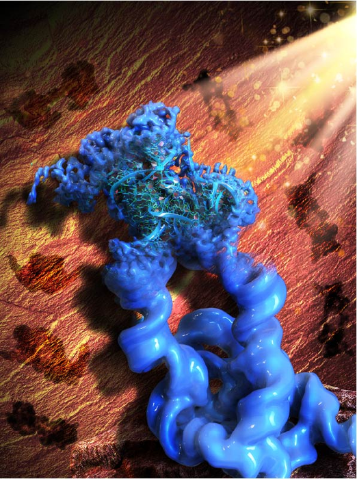
Visualization of RNA structures at near-atomic resolution enabled by nanotechnology

The illustration that accompanies the research is both fascinating and baffling as its caption,

Caption: This illustration is inspired by the Paleolithic rock painting in the Lascaux cave, signifying the acronym of our method, ROCK. Figuratively, the patterns of the rock art in the background (brown) are the 2D projections of the engineered dimeric construct of the Tetrahymena group I intron, while the main object in the front (blue) is the reconstructed 3D cryo-EM map of the dimer, with one monomer in focus and refined to the high resolution that allowed the collaborators to build an atomic model of the RNA. Credit: Wyss Institute at Harvard University

This May 2, 2022 news item on ScienceDaily announces the research into RNA molecules made possible by ROCK (the technology being illustrated in the above),

We live in a world made and run by RNA [ribonucleic acid], the equally important sibling of the genetic molecule DNA. In fact, evolutionary biologists hypothesize that RNA existed and self-replicated even before the appearance of DNA and the proteins encoded by it. Fast forward to modern day humans: science has revealed that less than 3% of the human genome is transcribed into messenger RNA (mRNA) molecules that in turn are translated into proteins. In contrast, 82% of it is transcribed into RNA molecules with other functions many of which still remain enigmatic.

To understand what an individual RNA molecule does, its 3D structure needs to be deciphered at the level of its constituent atoms and molecular bonds. Researchers have routinely studied DNA and protein molecules by turning them into regularly packed crystals that can be examined with an X-ray beam (X-ray crystallography) or radio waves (nuclear magnetic resonance). However, these techniques cannot be applied to RNA molecules with nearly the same effectiveness because their molecular composition and structural flexibility prevent them from easily forming crystals.

Now, a research collaboration led by Wyss Core Faculty member Peng Yin, Ph.D. at the Wyss Institute for Biologically Inspired Engineering at Harvard University, and Maofu Liao, Ph.D. at Harvard Medical School (HMS), has reported a fundamentally new approach to the structural investigation of RNA molecules. ROCK, as it is called, uses an RNA nanotechnological technique that allows it to assemble multiple identical RNA molecules into a highly organized structure, which significantly reduces the flexibility of individual RNA molecules and multiplies their molecular weight. Applied to well-known model RNAs with different sizes and functions as benchmarks, the team showed that their method enables the structural analysis of the contained RNA subunits with a technique known as cryo-electron microscopy (cryo-EM). Their advance is reported in Nature Methods.

A May 2, 2022 Wyss Institute for Biologically Inspired Engineering at Harvard University news release (also on EurekAlert) by Benjamin Boettner, which originated the news item, delves further into the imaging technology, Note: Links have been removed,

“ROCK is breaking the current limits of RNA structural investigations and enables 3D structures of RNA molecules to be unlocked that are difficult or impossible to access with existing methods, and at near-atomic resolution,” said Yin, who together with Liao led the study. “We expect this advance to invigorate many areas of fundamental research and drug development, including the burgeoning field of RNA therapeutics.” Yin also is a leader of the Wyss Institute’s Molecular Robotics Initiative and Professor in the Department of Systems Biology at HMS.

Gaining control over RNA

Yin’s team at the Wyss Institute has pioneered various approaches that enable DNA and RNA molecules to self-assemble into large structures based on different principles and requirements, including DNA bricks and DNA origami. They hypothesized that such strategies could also be used to assemble naturally occurring RNA molecules into highly ordered circular complexes in which their freedom to flex and move is highly restricted by specifically linking them together. Many RNAs fold in complex yet predictable ways, with small segments base-pairing with each other. The result often is a stabilized “core” and “stem-loops” bulging out into the periphery. 

“In our approach we install ‘kissing loops’ that link different peripheral stem-loops belonging to two copies of an identical RNA in a way that allows a overall stabilized ring to be formed, containing multiple copies of the RNA of interest,” said Di Liu, Ph.D., one of two first-authors and a Postdoctoral Fellow in Yin’s group. “We speculated that these higher-order rings could be analyzed with high resolution by cryo-EM, which had been applied to RNA molecules with first success.”

Picturing stabilized RNA

In cryo-EM, many single particles are flash-frozen at cryogenic temperatures to prevent any further movements, and then visualized with an electron microscope and the help of computational algorithms that compare the various aspects of a particle’s 2D surface projections and reconstruct its 3D architecture. Peng and Liu teamed up with Liao and his former graduate student François Thélot, Ph.D., the other co-first author of the study. Liao with his group has made important contributions to the rapidly advancing cryo-EM field and the experimental and computational analysis of single particles formed by specific proteins.

“Cryo-EM has great advantages over traditional methods in seeing high-resolution details of biological molecules including proteins, DNAs and RNAs, but the small size and moving tendency of most RNAs prevent successful determination of RNA structures. Our novel method of assembling RNA multimers solves these two problems at the same time, by increasing the size of RNA and reducing its movement,” said Liao, who also is Associate Professor of Cell Biology at HMS. “Our approach has opened the door to rapid structure determination of many RNAs by cryo-EM.” The integration of RNA nanotechnology and cryo-EM approaches led the team to name their method “RNA oligomerization-enabled cryo-EM via installing kissing loops” (ROCK).

To provide proof-of-principle for ROCK, the team focused on a large intron RNA from Tetrahymena, a single-celled organism, and a small intron RNA from Azoarcus, a nitrogen-fixing bacterium, as well as the so-called FMN riboswitch. Intron RNAs are non-coding RNA sequences scattered throughout the sequences of freshly-transcribed RNAs and have to be “spliced” out in order for the mature RNA to be generated. The FMN riboswitch is found in bacterial RNAs involved in the biosynthesis of flavin metabolites derived from vitamin B2. Upon binding one of them, flavin mononucleotide (FMN), it switches its 3D conformation and suppresses the synthesis of its mother RNA.  

“The assembly of the Tetrahymena group I intron into a ring-like structure made the samples more homogenous, and enabled the use of computational tools leveraging the symmetry of the assembled structure. While our dataset is relatively modest in size, ROCK’s innate advantages allowed us to resolve the structure at an unprecedented resolution,” said Thélot. “The RNA’s core is resolved at 2.85 Å [one Ångström is one ten-billions (US) of a meter and the preferred metric used by structural biologists], revealing detailed features of the nucleotide bases and sugar backbone. I don’t think we could have gotten there without ROCK – or at least not without considerably more resources.” 

Cryo-EM also is able to capture molecules in different states if they, for example, change their 3D conformation as part of their function. Applying ROCK to the Azoarcus intron RNA and the FMN riboswitch, the team managed to identify the different conformations that the Azoarcus intron transitions through during its self-splicing process, and to reveal the relative conformational rigidity of the ligand-binding site of the FMN riboswitch.

“This study by Peng Yin and his collaborators elegantly shows how RNA nanotechnology can work as an accelerator to advance other disciplines. Being able to visualize and understand the structures of many naturally occurring RNA molecules could have tremendous impact on our understanding of many biological and pathological processes across different cell types, tissues, and organisms, and even enable new drug development approaches,” said Wyss Founding Director Donald Ingber, M.D., Ph.D., who is also the Judah Folkman Professor of Vascular Biology at Harvard Medical School and Boston Children’s Hospital, and Professor of Bioengineering at the Harvard John A. Paulson School of Engineering and Applied Sciences.

The study was also authored by Joseph Piccirilli, Ph.D., an expert in RNA chemistry and biochemistry and Professor at The University of Chicago. It was supported by the National Science Foundation (NSF; grant# CMMI-1333215, CCMI-1344915, and CBET-1729397), Air Force Office of Scientific Research (AFOSR; grant MURI FATE, #FA9550-15-1-0514), National Institutes of Health (NIH; grant# 5DP1GM133052, R01GM122797, and R01GM102489), and the Wyss Institute’s Molecular Robotics Initiative.

Here’s a link to and a citation for the paper,

Sub-3-Å cryo-EM structure of RNA enabled by engineered homomeric self-assembly by Di Liu, François A. Thélot, Joseph A. Piccirilli, Maofu Liao & Peng Yin. Nature Methods (2022) DOI: https://doi.org/10.1038/s41592-022-01455-w Published: 02 May 2022

This paper is behind a paywall.

4D printing: a hydrogel orchid

In 2013, the 4th dimension for printing was self-assembly according to a March 1, 2013 article by Tuan Nguyen for ZDNET. A Jan. 25, 2016 Wyss Institute for Biologically Inspired Engineering at Harvard University news release (also on EurekAlert) points to time as the fourth dimension in a description of the Wyss Institute’s latest 4D printed object,

A team of scientists at the Wyss Institute for Biologically Inspired Engineering at Harvard University and the Harvard John A. Paulson School of Engineering and Applied Sciences has evolved their microscale 3D printing technology to the fourth dimension, time. Inspired by natural structures like plants, which respond and change their form over time according to environmental stimuli, the team has unveiled 4D-printed hydrogel composite structures that change shape upon immersion in water.

“This work represents an elegant advance in programmable materials assembly, made possible by a multidisciplinary approach,” said Jennifer Lewis, Sc.D., senior author on the new study. “We have now gone beyond integrating form and function to create transformable architectures.”

In nature, flowers and plants have tissue composition and microstructures that result in dynamic morphologies that change according to their environments. Mimicking the variety of shape changes undergone by plant organs such as tendrils, leaves, and flowers in response to environmental stimuli like humidity and/or temperature, the 4D-printed hydrogel composites developed by Lewis and her team are programmed to contain precise, localized swelling behaviors. Importantly, the hydrogel composites contain cellulose fibrils that are derived from wood and are similar to the microstructures that enable shape changes in plants.

By aligning cellulose fibrils (also known as, cellulose nanofibrils or nanofibrillated cellulose) during printing, the hydrogel composite ink is encoded with anisotropic swelling and stiffness, which can be patterned to produce intricate shape changes. The anisotropic nature of the cellulose fibrils gives rise to varied directional properties that can be predicted and controlled. Just like wood, which can be split easier along the grain rather than across it. Likewise, when immersed in water, the hydrogel-cellulose fibril ink undergoes differential swelling behavior along and orthogonal to the printing path. Combined with a proprietary mathematical model developed by the team that predicts how a 4D object must be printed to achieve prescribed transformable shapes, the new method opens up many new and exciting potential applications for 4D printing technology including smart textiles, soft electronics, biomedical devices, and tissue engineering.

“Using one composite ink printed in a single step, we can achieve shape-changing hydrogel geometries containing more complexity than any other technique, and we can do so simply by modifying the print path,” said Gladman [A. Sydney Gladman, Wyss Institute a graduate research assistant]. “What’s more, we can interchange different materials to tune for properties such as conductivity or biocompatibility.”

The composite ink that the team uses flows like liquid through the printhead, yet rapidly solidifies once printed. A variety of hydrogel materials can be used interchangeably resulting in different stimuli-responsive behavior, while the cellulose fibrils can be replaced with other anisotropic fillers of choice, including conductive fillers.

“Our mathematical model prescribes the printing pathways required to achieve the desired shape-transforming response,” said Matsumoto [Elisabetta Matsumoto, Ph.D., a postdoctoral fellow at the Wyss]. “We can control the curvature both discretely and continuously using our entirely tunable and programmable method.”

Specifically, the mathematical modeling solves the “inverse problem”, which is the challenge of being able to predict what the printing toolpath must be in order to encode swelling behaviors toward achieving a specific desired target shape.

“It is wonderful to be able to design and realize, in an engineered structure, some of nature’s solutions,” said Mahadevan [L. Mahadevan, Ph.D., a Wyss Core Faculty member] , who has studied phenomena such as how botanical tendrils coil, how flowers bloom, and how pine cones open and close. “By solving the inverse problem, we are now able to reverse-engineer the problem and determine how to vary local inhomogeneity, i.e. the spacing between the printed ink filaments, and the anisotropy, i.e. the direction of these filaments, to control the spatiotemporal response of these shapeshifting sheets. ”

“What’s remarkable about this 4D printing advance made by Jennifer and her team is that it enables the design of almost any arbitrary, transformable shape from a wide range of available materials with different properties and potential applications, truly establishing a new platform for printing self-assembling, dynamic microscale structures that could be applied to a broad range of industrial and medical applications,” said Wyss Institute Founding Director Donald Ingber, M.D., Ph.D., who is also the Judah Folkman Professor of Vascular Biology at Harvard Medical School and the Vascular Biology Program at Boston Children’s Hospital and Professor of Bioengineering at Harvard SEAS [School of Engineering and Applied Science’.

Here’s an animation from the Wyss Institute illustrating the process,

And, here’s a link to and a citation for the paper,

Biomimetic 4D printing by A. Sydney Gladman, Elisabetta A. Matsumoto, Ralph G. Nuzzo, L. Mahadevan, & Jennifer A. Lewis. Nature Materials (2016) doi:10.1038/nmat4544 Published online 25 January 2016

This paper is behind a paywall.