Tag Archives: Xin Qu

3D printed biomimetic blood vessel networks

An artificial blood vessel network that could lead the way to regenerating biologically-based blood vessel networks has been printed in 3D at the University of California at San Diego (UCSD) according to a March 2, 2017 news item on ScienceDaily,

Nanoengineers at the University of California San Diego have 3D printed a lifelike, functional blood vessel network that could pave the way toward artificial organs and regenerative therapies.

The new research, led by nanoengineering professor Shaochen Chen, addresses one of the biggest challenges in tissue engineering: creating lifelike tissues and organs with functioning vasculature — networks of blood vessels that can transport blood, nutrients, waste and other biological materials — and do so safely when implanted inside the body.

A March 2, 2017 UCSD news release (also on EurekAlert), which originated the news item, explains why this is an important development,

Researchers from other labs have used different 3D printing technologies to create artificial blood vessels. But existing technologies are slow, costly and mainly produce simple structures, such as a single blood vessel — a tube, basically. These blood vessels also are not capable of integrating with the body’s own vascular system.

“Almost all tissues and organs need blood vessels to survive and work properly. This is a big bottleneck in making organ transplants, which are in high demand but in short supply,” said Chen, who leads the Nanobiomaterials, Bioprinting, and Tissue Engineering Lab at UC San Diego. “3D bioprinting organs can help bridge this gap, and our lab has taken a big step toward that goal.”

Chen’s lab has 3D printed a vasculature network that can safely integrate with the body’s own network to circulate blood. These blood vessels branch out into many series of smaller vessels, similar to the blood vessel structures found in the body. The work was published in Biomaterials.

Chen’s team developed an innovative bioprinting technology, using their own homemade 3D printers, to rapidly produce intricate 3D microstructures that mimic the sophisticated designs and functions of biological tissues. Chen’s lab has used this technology in the past to create liver tissue and microscopic fish that can swim in the body to detect and remove toxins.

Researchers first create a 3D model of the biological structure on a computer. The computer then transfers 2D snapshots of the model to millions of microscopic-sized mirrors, which are each digitally controlled to project patterns of UV light in the form of these snapshots. The UV patterns are shined onto a solution containing live cells and light-sensitive polymers that solidify upon exposure to UV light. The structure is rapidly printed one layer at a time, in a continuous fashion, creating a 3D solid polymer scaffold encapsulating live cells that will grow and become biological tissue.

“We can directly print detailed microvasculature structures in extremely high resolution. Other 3D printing technologies produce the equivalent of ‘pixelated’ structures in comparison and usually require sacrificial materials and additional steps to create the vessels,” said Wei Zhu, a postdoctoral scholar in Chen’s lab and a lead researcher on the project.

And this entire process takes just a few seconds — a vast improvement over competing bioprinting methods, which normally take hours just to print simple structures. The process also uses materials that are inexpensive and biocompatible.

Chen’s team used medical imaging to create a digital pattern of a blood vessel network found in the body. Using their technology, they printed a structure containing endothelial cells, which are cells that form the inner lining of blood vessels.

The entire structure fits onto a small area measuring 4 millimeters × 5 millimeters, 600 micrometers thick (as thick as a stack containing 12 strands of human hair).

Researchers cultured several structures in vitro for one day, then grafted the resulting tissues into skin wounds of mice. After two weeks, the researchers examined the implants and found that they had successfully grown into and merged with the host blood vessel network, allowing blood to circulate normally.

Chen noted that the implanted blood vessels are not yet capable of other functions, such as transporting nutrients and waste. “We still have a lot of work to do to improve these materials. This is a promising step toward the future of tissue regeneration and repair,” he said.

Moving forward, Chen and his team are working on building patient-specific tissues using human induced pluripotent stem cells, which would prevent transplants from being attacked by a patient’s immune system. And since these cells are derived from a patient’s skin cells, researchers won’t need to extract any cells from inside the body to build new tissue. The team’s ultimate goal is to move their work to clinical trials. “It will take at least several years before we reach that goal,” Chen said.

Here’s a link to and a citation for the paper,

Direct 3D bioprinting of prevascularized tissue constructs with complex microarchitecture by Wei Zhu, Xin Qu, Jie Zhu, Xuanyi Ma, Sherrina Patel, Justin Liu, Pengrui Wang, Cheuk Sun Edwin Lai, Maling Gou, Yang Xu, Kang Zhang, Shaochen Chen. Biomaterials 124 (April 2017) 106-15 http://dx.doi.org/10.1016/j.biomaterials.2017.01.042

This paper is behind a paywall.

There is also an open access copy here on the university website but I cannot confirm that it is identical to the version in the journal.
Synthetic microfish (nanoengineered and 3D printed) to inspire ‘smart’ microbots

An August 26, 2015 news item on Nanowerk features some microfish (they look like sharks to me) fabricated in University of California at San Diego (UCSD) laboratories,

Nanoengineers at the University of California, San Diego used an innovative 3D printing technology they developed to manufacture multipurpose fish-shaped microrobots — called microfish — that swim around efficiently in liquids, are chemically powered by hydrogen peroxide and magnetically controlled. These proof-of-concept synthetic microfish will inspire a new generation of “smart” microrobots that have diverse capabilities such as detoxification, sensing and directed drug delivery, researchers said.

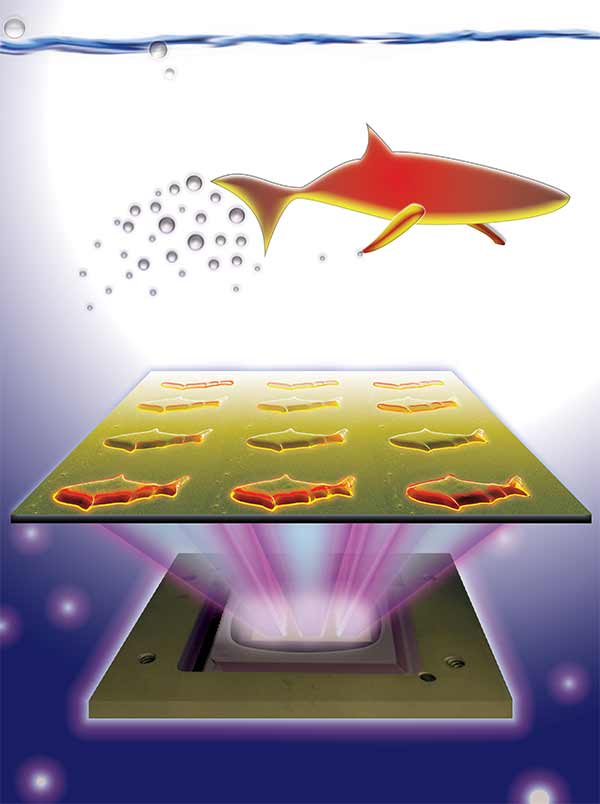
3D-printed microfish contain functional nanoparticles that enable them to be self-propelled, chemically powered and magnetically steered. The microfish are also capable of removing and sensing toxins. Image credit: J. Warner, UC San Diego Jacobs School of Engineering.

3D-printed microfish contain functional nanoparticles that enable them to be self-propelled, chemically powered and magnetically steered. The microfish are also capable of removing and sensing toxins. Image credit: J. Warner, UC San Diego Jacobs School of Engineering.

An August 25, 2015 UCSD news release (also on EurekAlert) by Liezel Labios, which originated the news item, provides more detail,

The technique used to fabricate the microfish provides numerous improvements over other methods traditionally employed to create microrobots with various locomotion mechanisms, such as microjet engines, microdrillers and microrockets. Most of these microrobots are incapable of performing more sophisticated tasks because they feature simple designs — such as spherical or cylindrical structures — and are made of homogeneous inorganic materials. In this new study, researchers demonstrated a simple way to create more complex microrobots.

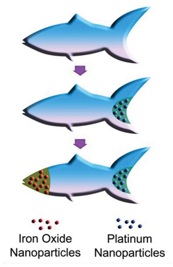
By combining Chen’s 3D printing technology with Wang’s expertise in microrobots, the team was able to custom-build microfish that can do more than simply swim around when placed in a solution containing hydrogen peroxide. Nanoengineers were able to easily add functional nanoparticles into certain parts of the microfish bodies. They installed platinum nanoparticles in the tails, which react with hydrogen peroxide to propel the microfish forward, and magnetic iron oxide nanoparticles in the heads, which allowed them to be steered with magnets.

Here’s an illustration of the platinum and iron oxide microfish,

Schematic illustration of the process of functionalizing the microfish. Platinum nanoparticles are first loaded into the tail of the fish for propulsion via reaction with hydrogen peroxide. Next, iron oxide nanoparticles are loaded into the head of the fish for magnetic control. Image credit: W. Zhu and J. Li, UC San Diego Jacobs School of Engineering.

Schematic illustration of the process of functionalizing the microfish. Platinum nanoparticles are first loaded into the tail of the fish for propulsion via reaction with hydrogen peroxide. Next, iron oxide nanoparticles are loaded into the head of the fish for magnetic control. Image credit: W. Zhu and J. Li, UC San Diego Jacobs School of Engineering.

Back to the news release,

“We have developed an entirely new method to engineer nature-inspired microscopic swimmers that have complex geometric structures and are smaller than the width of a human hair. With this method, we can easily integrate different functions inside these tiny robotic swimmers for a broad spectrum of applications,” said the co-first author Wei Zhu, a nanoengineering Ph.D. student in Chen’s research group at the Jacobs School of Engineering at UC San Diego.

As a proof-of-concept demonstration, the researchers incorporated toxin-neutralizing nanoparticles throughout the bodies of the microfish. Specifically, the researchers mixed in polydiacetylene (PDA) nanoparticles, which capture harmful pore-forming toxins such as the ones found in bee venom. The researchers noted that the powerful swimming of the microfish in solution greatly enhanced their ability to clean up toxins. When the PDA nanoparticles bind with toxin molecules, they become fluorescent and emit red-colored light. The team was able to monitor the detoxification ability of the microfish by the intensity of their red glow.

“The neat thing about this experiment is that it shows how the microfish can doubly serve as detoxification systems and as toxin sensors,” said Zhu.

“Another exciting possibility we could explore is to encapsulate medicines inside the microfish and use them for directed drug delivery,” said Jinxing Li, the other co-first author of the study and a nanoengineering Ph.D. student in Wang’s research group.

For anyone curious about the new 3D printing technique, the news release provides more information about that too,

The new microfish fabrication method is based on a rapid, high-resolution 3D printing technology called microscale continuous optical printing (μCOP), which was developed in Chen’s lab. Some of the benefits of the μCOP technology are speed, scalability, precision and flexibility. Within seconds, the researchers can print an array containing hundreds of microfish, each measuring 120 microns long and 30 microns thick. This process also does not require the use of harsh chemicals. Because the μCOP technology is digitized, the researchers could easily experiment with different designs for their microfish, including shark and manta ray shapes. [emphasis mine] “With our 3D printing technology, we are not limited to just fish shapes. We can rapidly build microrobots inspired by other biological organisms such as birds,” said Zhu.

The key component of the μCOP technology is a digital micromirror array device (DMD) chip, which contains approximately two million micromirrors. Each micromirror is individually controlled to project UV light in the desired pattern (in this case, a fish shape) onto a photosensitive material, which solidifies upon exposure to UV light. The microfish are built using a photosensitive material and are constructed one layer at a time, allowing each set of functional nanoparticles to be “printed” into specific parts of the fish bodies.

“This method has made it easier for us to test different designs for these microrobots and to test different nanoparticles to insert new functional elements into these tiny structures. It’s my personal hope to further this research to eventually develop surgical microrobots that operate safer and with more precision,” said Li.

Nice to see I can recognize a shark shape when I see one. Getting back to the research, yet again, here’s a link to and a citation for the paper.

3D-Printed Artificial Microfish by Wei Zhu, Jinxing Li, Yew J. Leong, Isaac Rozen, Xin Qu, Renfeng Dong, Zhiguang Wu, Wei Gao, Peter H. Chung, Joseph Wang, and Shaochen Chen. Advanced Materials Volume 27, Issue 30, pages 4411–4417, August 12, 2015 DOI: 10.1002/adma.201501372 Article first published online: 29 JUN 2015

© 2015 WILEY-VCH Verlag GmbH & Co. KGaA, Weinheim

This paper is behind a paywall.