Monthly Archives: July 2017

Surgery on nanoparticles?

Chemists performed “surgery” on a 23-gold-atom nanoparticle according to a June 12, 2017 news item on Nanowerk (Note: A link has been removed),

A team of chemists led by Carnegie Mellon University’s [CMU] Rongchao Jin has for the first time conducted site-specific surgery on a nanoparticle. The procedure, which allows for the precise tailoring of nanoparticles, stands to advance the field of nanochemistry.

The surgical technique developed by Qi Li, the study’s lead author and a 3rd year graduate student in the Jin group, will allow researchers to enhance nanoparticles’ functional properties, such as catalytic activity and photoluminescence, increasing their usefulness in a wide variety of fields including health care, electronics and manufacturing. The findings were published in Science Advances (“Molecular “surgery” on a 23-gold-atom nanoparticle”).

Here’s an image the researchers have provided,

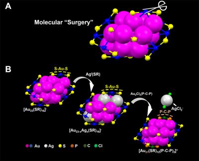
Caption: Carnegie Mellon chemists used a two-step metal exchange method to remove two S-Au-S staples from the surface of a nanoparticle. Credit: Carnegie Mellon University

A June 12, 2017 CMU press release (also on EurekAlert), which originated the news item, provides more details about the research,

“Nanochemistry is a relatively new field, it’s only about 20 years old. We’ve been racing to catch up to fields like organic chemistry that are more than 100 years old,” said Jin, a chemistry professor in the Mellon College of Science. “Organic chemists have been able to tailor the functional groups of molecules for quite some time, like tailoring penicillin for better medical functions, for example. We dreamed that we could do something similar in nanoscience. Developing atomically precise nanoparticles has allowed us to make this dream come true.”

In order to make this “nano-surgery” a reality, researchers needed to begin with atomically precise nanoparticles that could be reliably produced time after time. Jin’s lab has been at the forefront of this research. Working with gold nanoparticles, he and his team have developed methods to precisely control the number of atoms in each nanoparticle, resulting in uniformly-sized nanoparticles with every batch. With reliably precise particles, Jin and colleagues were able to identify the particles’ structures, and begin to tease out how that structure impacted the particles’ properties and functionality.

With these well-defined nanoparticles in hand, Jin’s next step was to find a way to surgically tailor the particles in order to learn more about­ – and hopefully enhance – their functionality.

In their recent study, Jin and colleagues performed nano-surgery on a gold nanoparticle made up of 23 gold atoms surrounded by a protective surface of ligands in staple-like motifs. Using a two-step metal exchange method, they removed two S-Au-S staples from the particle’s surface. In doing this they revealed the structural factors that determine the particle’s optical properties and established the role that the surface plays in photoluminescence. Significantly, the surgery increased the particle’s photoluminescence by about 10-fold. Photoluminescence plays a critical role in biological imaging, cancer diagnosis and LED technology, among other applications.

Jin and coworkers are now trying to generalize this site-specific surgery method to other nanoparticles.

Here’s a link to and a citation for the paper,

Molecular “surgery” on a 23-gold-atom nanoparticle by Qi Li, Tian-Yi Luo, Michael G. Taylor, Shuxin Wang, Xiaofan Zhu, Yongbo Song, Giannis Mpourmpakis, Nathaniel L. Rosi, and Rongchao Jin. Science Advances 19 May 2017: Vol. 3, no. 5, e1603193 DOI: 10.1126/sciadv.1603193

This paper is open access.

Mussels to muscles for biocompatible fibres

Mussels and barnacles in the intertidal near Newquay, Cornwall, England.
Wilson44691 at English Wikipedia – Photograph taken by Mark A. Wilson (Department of Geology, The College of Wooster

I love puns and word play so I was happy to see this in a June 9, 2017 news item on ScienceDaily,

Rice University chemists can thank the mussel for putting the muscle into their new macroscale scaffold fibers.

A June 9, 2017 Rice University news release (also on EurekAlert), which originated the news item, provides more details about the research,

The Rice lab of chemist Jeffrey Hartgerink had already figured out how to make biocompatible nanofibers out of synthetic peptides. In new work, the lab is using an amino acid found in the sticky feet of mussels to make those fibers line up into strong hydrogel strings.

Hartgerink and Rice graduate student I-Che Li introduced their room-temperature method this month in an open-access paper in the Journal of the American Chemical Society.

The hydrogel strings can be picked up and moved with tweezers, and Li said he expects they will help labs gain better control over the growth of cell cultures.

“Usually when cells grow on a surface, they spread randomly,” he said. “There are a lot of biomaterials we want to grow in a specific direction. With the hydrogel scaffold aligned, we can expect cells to grow the way we want them to. One example would be neuron cells, which we want to grow head-to-tail to aid nerve regeneration.

“Basically, this could allow us to direct cell growth from here to there,” he said. “That’s why this material is so exciting.”

In previous research Hartgerink’s lab had developed synthetic hydrogels that could be injected into the body to serve as scaffolds for tissue growth. The hydrogels contained hydrophobic peptides that self-assembled into fibers about 6 nanometers wide and up to several microns long. However, because the fibers did not interact with one other, they generally appeared in microscope images as a tangled mass.

Experiments showed the fibers could be coaxed into alignment with the application of shear forces, in the same way that playing cards are aligned during shuffling by pushing on both the top and bottom of the deck.

Hartgerink and Li decided to try pushing the fibers through a needle to force them into alignment, a process that would be easier if the material was water soluble. So they added a chain of amino acids known as DOPA to the sides of the fibers to allow them to remain water-soluble in the syringe, Li said.

DOPA — short for 3,4-dihydroxyphenylalanine — is the compound that lets mussels stick to just about anything. Hartgerink and Li found that the combination of DOPA and shear stress from passing through the needle prompted the fibers to form visible, rope-like bundles.

They also found that DOPA promoted chemical cross-linking reactions that helped the bundles hold their shape. “DOPA is really sensitive to oxidizing agents,” Li said. “Even exposing DOPA to air oxidizes it, and that aids in cross-linking the fibers.”

As a bonus, the aligned fibers also proved to have a curious and useful optical property called “uniform birefringence,” or double-refraction. Li said this could allow researchers to use polarized light to see exactly where the aligned fibers are, even if they’re covered by cells.

“This will be an important technique for us to make sure of the long-range order of fiber alignment when we are testing directed cell growth,” he said.

The researchers expect the aligned fibers can be used for macroscale medical applications but with nanoscale control over the structures.

“Self-assembly is basically the ability of a molecule to make ordered structure from chaos, and what I-Che has done is push this organization to a new level with his aligned strings,” said Hartgerink, a professor of chemistry and of bioengineering. “With this material, we are excited to see if we can impose this organization onto the growth of cells that interact with it.”

Here’s a link to and a citation for the paper,

Covalent Capture of Aligned Self-Assembling Nanofibers by I-Che Li and Jeffrey D. Hartgerink. J. Am. Chem. Soc., Article ASAP DOI: 10.1021/jacs.7b04655 Publication Date (Web): June 5, 2017

Copyright © 2017 American Chemical Society

This paper is open access.