Monthly Archives: June 2016

“Brute force” technique for biomolecular information processing

The research is being announced by the University of Tokyo but there is definitely a French flavour to this project. From a June 20, 2016 news item on ScienceDaily,

A Franco-Japanese research group at the University of Tokyo has developed a new “brute force” technique to test thousands of biochemical reactions at once and quickly home in on the range of conditions where they work best. Until now, optimizing such biomolecular systems, which can be applied for example to diagnostics, would have required months or years of trial and error experiments, but with this new technique that could be shortened to days.

A June 20, 2016 University of Tokyo news release on EurekAlert, which originated the news item, describes the project in more detail,

“We are interested in programming complex biochemical systems so that they can process information in a way that is analogous to electronic devices. If you could obtain a high-resolution map of all possible combinations of reaction conditions and their corresponding outcomes, the development of such reactions for specific purposes like diagnostic tests would be quicker than it is today,” explains Centre National de la Recherche Scientifique (CNRS) researcher Yannick Rondelez at the Institute of Industrial Science (IIS) [located at the University of Tokyo].

“Currently researchers use a combination of computer simulations and painstaking experiments. However, while simulations can test millions of conditions, they are based on assumptions about how molecules behave and may not reflect the full detail of reality. On the other hand, testing all possible conditions, even for a relatively simple design, is a daunting job.”

Rondelez and his colleagues at the Laboratory for Integrated Micro-Mechanical Systems (LIMMS), a 20-year collaboration between the IIS and the French CNRS, demonstrated a system that can test ten thousand different biochemical reaction conditions at once. Working with the IIS Applied Microfluidic Laboratory of Professor Teruo Fujii, they developed a platform to generate a myriad of micrometer-sized droplets containing random concentrations of reagents and then sandwich a single layer of them between glass slides. Fluorescent markers combined with the reagents are automatically read by a microscope to determine the precise concentrations in each droplet and also observe how the reaction proceeds.

“It was difficult to fine-tune the device at first,” explains Dr Anthony Genot, a CNRS researcher at LIMMS. “We needed to create generate thousands of droplets containing reagents within a precise range of concentrations to produce high resolution maps of the reactions we were studying. We expected that this would be challenging. But one unanticipated difficulty was immobilizing the droplets for the several days it took for some reactions to unfold. It took a lot of testing to create a glass chamber design that was airtight and firmly held the droplets in place.” Overall, it took nearly two years to fine-tune the device until the researchers could get their droplet experiment to run smoothly.

Seeing the new system producing results was revelatory. “You start with a screen full of randomly-colored dots, and then suddenly the computer rearranges them into a beautiful high-resolution map, revealing hidden information about the reaction dynamics. Seeing them all slide into place to produce something that had only ever been seen before through simulation was almost magical,” enthuses Rondelez.

“The map can tell us not only about the best conditions of biochemical reactions, it can also tell us about how the molecules behave in certain conditions. Using this map we’ve already found a molecular behavior that had been predicted theoretically, but had not been shown experimentally. With our technique we can explore how molecules talk to each other in test tube conditions. Ultimately, we hope to illuminate the intimate machinery of living molecular systems like ourselves,” says Rondelez.

Here’s a link to and a citation for the paper,

High-resolution mapping of bifurcations in nonlinear biochemical circuits by A. J. Genot, A. Baccouche, R. Sieskind, N. Aubert-Kato, N. Bredeche, J. F. Bartolo, V. Taly, T. Fujii, & Y. Rondelez. Nature Chemistry (2016)
doi:10.1038/nchem.2544 Published online 20 June 2016

This paper is behind a paywall.

Combat cells (Robot Wars for cells) and a plea from Concordia University

Students at Concordia University (located in Montréal, Québec, Canada) are requesting help (financial or laboratory supplies) for their submission to  the 2016 iGEM (International Genetically Engineered Machine) competition.

Here’s a little about their entry (from a June 16, 2016 request received via email),

For this year’s project, we plan to design a biological system that mimics the concept of the popular TV series Robot Wars. We will be engineering cellular species to wear nanoparticles as battle shields and then use microfluidics to guide them through an obstacle course leading to a battledome, where both cells will engage into a duel. Essentially, we want to test the interactions between nanoparticles and cell membranes, as well as their protective abilities against varying environmental conditions and other equipped cells. The method in which we will adapt Robot Wars for synthetic biology is by creating a web series that will visualize the cell battle and communicate the research behind it. This web series will serve as an entertaining  medium to educate and inspire the audience to develop an interest in science. We are incorporating the emerging fields of  synthetic biology, nanotechnology and microfluidics to make this process possible.  Furthermore, this study will contribute to the advancement of nanotechnology, an interdisciplinary field aiming to make applicable improvements in other fields such as medicine, optics and cosmetics.

Here’s a little more about iGEM (from the organization’s homepage),

The iGEM Foundation is dedicated to education and competition, advancement of synthetic biology, and the development of open community and collaboration.

The main program at the iGEM Foundation is the International Genetically Engineered Machine (iGEM) Competition. The iGEM Competition is the premiere student competition in Synthetic Biology. Since 2004, participants of the competition have experienced education, teamwork, sharing, and more in a unique competition setting.

The deadline for donations/sponsorships is the end of September 2016 and sponsors/donors will be acknowledged on “our website, all of our social media accounts (Facebook, Instagram, Twitter), at our community outreach events and at the competition [from the June 16, 2016 email].”

For more information contact:

Maria Salouros
iGEM Concordia
igem.concordia@gmail.com

Finally, there’s this:

We are excited to make this year’s project a reality and we are determined to win gold. Any help, either financially or by the donation of laboratory supplies, would contribute to the development of our project and would be greatly appreciated.

Good luck to the students! Hopefully one or more of my readers will be able to help. In which case, thank you!

Prosthetics in North Carolina and in Vancouver, Canada

North Carolina

This is the first time I’ve seen any kind of hand prosthestic offering finger control. From a May 31, 2016 OrthoCarolina news release (received via email),

Two OrthoCarolina hand surgeons have successfully completed the first surgery to allow for a prosthetic hand with individual finger control on an amputee patient. Partnering with OrthoCarolina Research Institute (OCRI) in pursuit of medical breakthroughs through orthopedic research, Drs. Glenn Gaston and Bryan Loeffler conceptualized and performed the procedure involving transferring existing muscle from the fingers to the back of the hand and wrist without damaging the nerves and blood vessels to the muscles. The patient who underwent the test surgery is now able to control individual prosthetic fingers using the same muscles that controlled his fingers pre-amputation, making him the first person in the world to have individual digit control in a functioning myoelectric prosthesis.

“Patients who have sustained full or partial hand amputations obviously have significant morbidity and limited function, which is a challenge. Because of the limited number of muscles available after a hand amputation, prostheses have previously allowed only control of the thumb and fingers as a group and single finger control was never possible,” said Dr. Glenn Gaston.  “The severity of this patient’s injury was so great that replanting the lost fingers was not possible, so we collaborated on a new surgery that would allow him to have individual digital control.”

Hypothesizing that existing muscle in the back of human fingers could be transferred to the back of the hand and wrist without damaging the nerves and blood vessels to those muscles, Drs. Gaston and Loeffler first performed cadaveric testing to ensure feasibility. The goal of the initial project was for the small muscles that control individual fingers to regain control of prosthetic fingers by maintaining enough blood and nerve supply to allow the prosthetic limb to recognize individual digits.

With successful research completed, they collaborated with the Hanger Clinic to determine how much bone would be required to be removed from the hand, allowing the prosthetic componentry enough space to maintain a normal hand length.

The two surgeons jointly performed the surgery as a pilot case on a partial hand amputee, moving the muscles while still allowing the prosthesis to detect signals from the transferred muscles; a procedure never before reported in orthopedic literature.

“Imagine the limitations you would have if all of your fingers had to move as one unit, and then suddenly you were able to move individual fingers to perform specific actions,” said Dr. Bryan Loeffler. “This muscle transfer is a breakthrough that could impact how upper extremity amputees are managed and specific amputations are done in the future.”

Drs. Loeffler and Gaston have completed a cadaver model demonstrating the capability of the same type of surgery for a more proximal level total hand amputation. They presented their research at a podium presentation to the First International Symposium on Innovations in Amputation Surgery and Prosthetic Technologies (IASPT) May 12-13, 2016 in Chicago.

OrthoCarolina Research Institute is an independent non-profit committed to the advancement of orthopedic practice through clinical research. OCRI will continue to support this ground-breaking research and the manufacturing of this cutting edge prosthesis. “This is a tremendous example of the life-changing impact that orthopedic research plays in advancing patient outcomes,” said Christi Cadd, Executive Director of OCRI.

You can find out more about OrthoCarolina here.

Vancouver, Canada

While they celebrate exciting prosthetic news in North Carolina, those of us in Vancouver have been given the opportunity to view an unusual display of vintage artificial limbs (prosthetics) in an exhibition, All Together Now, featuring a number of rarely seen private collections including corsets, Chinese restaurant menus, and pinball machines. From a June 22, 2016 article by Janet Smith for the Georgia Straight, here’s more about the prosthetic collection,

For those unfamiliar, the lifelike artificial legs and arms that hang on the Museum of Vancouver’s wall might seem like medical oddities from a less advanced era.

But for collector David Moe, a certified prosthetist, they are integral, inspiring pieces for his career, his teaching, and his workspace.

“I love them all,” he says with enthusiasm, standing in the museum’s giant new exhibit All Together Now: Vancouver Collectors and Their World, in a corner of an expansive, cabinet-of-curiosities-styled room that houses everything from scores of local Chinese-restaurant menus to rows of 19th-century corsets and a glass case full of hundreds of action figures. “It’s very strange because they have been all around me for so long and they have sat in predominant spaces at work—they sit on the top of a shelf. So when I walk back in there right now there are these kinds of empty holes.

“But I’m happy to have them on display and to let people think about what they see and have the opportunity to have them think about prosthetics. Because nobody ever thinks about them until they need one.”

Moe began collecting almost from his start, at the age of 14, when he worked sweeping floors and pouring plaster at Northern Alberta Prosthetic & Orthotic Services, his family’s business in Edmonton. One of his first big finds was a leg that sits in the exhibit today—a meticulously carved wooden limb covered in smooth skin-tone leather, dating back to the 1930s. At the time, he recognized the craftsmanship and tucked it away where it wouldn’t disappear; today he still marvels at the anatomical design, with a hinged knee that bends with the use of straps.

“… . The math is the math. But we’ve moved so far. I really love where we’ve come from,” says Moe, gesturing to the vintage pieces he uses regularly to teach students at BCIT [British Columbia Institute of Technology]. He says he can appreciate the human touch and deep care that went into each one, then adds: “All of these were used by people, so the energy of these people is in these. I feel that responsibility of these people in here.”

To show how far his specialty has come, though, Moe has juxtaposed the historic limbs with modern-day advances—decorative limb coverings with fashionable latticework, or a kids’ shin piece that’s been emblazoned with a comic-book image of Superman. Now, instead of trying to just mimic natural limbs, some people are opting for statement pieces that actually draw attention to their prosthetic. “This empowers them in this powerless situation where someone has amputated your leg,” he notes.

As with other exhibits in All Together Now, there are audiovisuals that accompany his collection—in this case showing people using the advanced limbs of today, from a female triathlete carrying her baby to another client playing competitive volleyball.

“When someone does the Grouse Grind or, hell, just walks their child down the street, that’s when they come alive. We’re rebuilding lives, not pieces,” Moe says.

You can find out more about All Together Now here,

All Together Now: Vancouver Collectors and Their Worlds features 20 beautiful, rare, and unconventional collections, with something for everyone including corsets, prosthetics, pinball machines, taxidermy, toys, and much more. In this exhibition both collector and collected are objects of study, interaction, and delight.

The exhibition runs until January 8, 2017. The last Thursday of the month is by donation from 5 pm to 8 pm. More information about admission can be found here and you might also want to check out the exhibition’s Events page.
Springer Nature and its ‘nano research tool’

There’s news from Springer Nature. From a June 17, 2016 announcement by Benedicte Page for The Bookseller,

Springer Nature has launched its first non-journal product since the merged company was formed last year.

Nano, which will sit within the Nature Research portfolio, pulls together structured information on nanotechnology – the manipulation of matter at the level of atoms or molecules – from high-impact journals and patents, spanning disciplines and combining key features of a database and an indexing discovery tool.

Bettina Goerner, Springer Nature m.d. for corporate markets and databases, told The Bookseller that nanoscience and nanotechnology was “so new and growing so fast” that research is spread across a wide range of literature and applications, with Nano bringing together all the relevant information.

Goerner said it was a truly “joint product development” between the two halves of the merged company. “I come from the former Springer side, and we started developing this two years ago, a serious effort on our side,” she said. “The moment we merged it was clear we now had access to experts we didn’t have before, so we reached out to the editor-in-chief of Nature Nanotechnology, and to other relevant journals in the Nature portfolio, and they got very involved and made recommendations on content.”

Goerner added: “We also have a database called SpringerMaterials and we are definitely looking at this as a portfolio and have lots of ideas. We do see finding the right information is vital, especially in fast-growing fields such as this.”

Access to Nano will be via subscription.

A June 15, 2016 Springer Nature press release provides more details,

Developed to meet the needs of researchers in academic, corporate and government environments, Nano combines the key features of a database and an abstracting and indexing discovery tool. Over 200,000 manually-curated profiles of nanomaterials and devices are available, containing information on properties, synthesis and applications. Trials are available from today.

Growing public and private investment into nanotechnology has led to an increase in research outputs, with related articles more than doubling over the past ten years. Nanotechnology is also of growing importance for a vast range of industries – from medicine to aerospace – in developing new products and applications. With nanotechnology becoming an inescapable part of people’s day-to-day lives, policymakers are paying closer attention to it, too.

This area of research, however, presents challenges. Dr. Omid Farokhzad, Associate Professor, Harvard Medical School, said: “Nanotechnology research and development has been rising on a sharp slope across virtually all scientific disciplines and industries. The result has been a rapidly growing body of information in disparate places that is not readily and efficiently accessible. Researchers need a multidisciplinary database that brings this vast body of data together in an organized and usable way in one place. Working together with other scientists to develop a research solution that can meet this need, through Nano’s External Advisory Board, has made me confident that this is a product that can deliver huge value to the research community.”

Partnership and consultation have been at the heart of Nano’s creation. William Chiuman, Director of Product Management, Nanoscience and Technology, Springer Nature, said: “We have worked closely with academia and industry throughout Nano’s development, and we’ll continue to be guided by these external experts to ensure that Nano keeps pace with this dynamic field, and provides up-to-date, curated content, that will ultimately save researchers time and significantly extend their knowledge base.”

Nano is the first non-journal product to be launched by Springer Nature since it was formed in May 2015 by the merger of Springer Science+Business Media and the majority of Macmillan Science and Education, and will be part of the Nature Research portfolio. Steven Inchcoombe, Chief Publishing Officer, Springer Nature, said: “Nano is a product of the combined skills and talents of our new organisation. It exemplifies our ethos and ultimate aim of putting the needs of the researcher at the heart of everything that we do.”

More information about Nano is available at nano.nature.com.

I haven’t been able to find a subscription rate but you can sign up for a trial (presumably free); scroll down about 80% of the way.

I have some information about the May 2015 merger of Springer with Nature in my Dec. 2, 2015 posting (scroll down about 90% of the way).

Frankenturtles released

This really is a ‘Frankenstein’ story complete with turtle cadavers. From a June 14, 2016 news item on ScienceDaily,

It was a dark and stormy night in the laboratory, and jagged bolts of lightning lit the sky as Dr. Kaplan and his assistant Bianca stitched the pieces of the lifeless creature back together.

Actually, it was a sunny day on the shores of Chesapeake Bay, but recent sea turtle research by Assistant Professor David Kaplan of the Virginia Institute of Marine Science and graduate student Bianca Santos easily brings to mind the classic tale of Dr. Frankenstein and his makeshift monster.

Santos, a master’s student in William & Mary’s School of Marine Science at VIMS, is working with Kaplan to reduce sea turtle mortality by trying to pinpoint where the hundreds of dead loggerhead sea turtles that wash up on Chesapeake Bay beaches each summer may have succumbed. With that knowledge, researchers could hone in on likely causes of sea-turtle death, while wildlife authorities could map out safe zones for these imperiled marine reptiles. One of Kaplan’s research specialties is the spatial management of marine ecosystems.


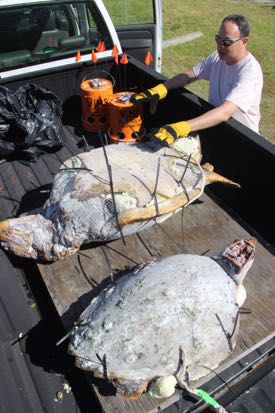
David Kaplan examines the Frankenturtles before their deployment. Also visible are the bucket drifters that more closely follow Bay currents. Courtesy: VIMS

David Kaplan examines the Frankenturtles before their deployment. Also visible are the bucket drifters that more closely follow Bay currents. Courtesy: VIMS

A June 14, 2016 Virginia Institute of Marine Science news release by David Malmquist, which originated the news item, expands on the theme,

The pair’s approach to the problem is ingenious if somewhat morbid: obtain dead sea turtles (from the Virginia Aquarium’s Stranding Response Program), replace the turtles’ inner organs with buoyant Styrofoam, “sew” their shells back together with zip ties, and then attach GPS units to track the path of the “Frankenturtles” as winds and currents disperse them from a mid-Bay release site.

“It might seem sort of gross, but it’s a good way to reuse a dead turtle that would otherwise be buried,” says Kaplan. “And hopefully, the deployment of our two Frankenturtles will ultimately help lower the number of turtle deaths in the future.”

Santos explains that the team is actually releasing three different types of drifters: the two Frankenturtles, two wooden-Styrofoam turtle models, and a pair of bucket drifters. By observing how the wind differentially affects the highly buoyant, sail-like wooden models; the partly emergent Frankenturtles; and the mostly submerged buckets, the researchers hope to better understand how a wind-driven carcass might deviate from the more predictable current patterns traced by the Bay’s surface waters. Sea turtles initially sink after dying, but quickly float back to the surface buoyed by gases from decomposing tissues.

“Our plan is to deploy the drifters on several different occasions—under a variety of wind and wave conditions—and in locations where mortality events could occur during the spring peak in strandings,” says Santos. “We’ll then use the separation rate between our bucket drifters, which closely track water movement, and our turtle carcasses to determine the amount of wind forcing to apply to simulated carcasses in our computer model.”

They initiated their field trials on June 13th [2016], deploying the drifters in open Bay waters about halfway between the mouth of the York River and Cape Charles on Virginia’s bayside Eastern Shore. One Frankenturtle comprises the remains of a 15-20 year old loggerhead killed by a boat strike. The other is a younger turtle whose mode of death remains a mystery despite a necropsy. Deploying these creatures wasn’t an easy job: in addition to the unforgettable and growing aroma of thawing turtle, the creatures are both heavy and unwieldy. The larger Frankenturtle weighs in at 150 pounds, the smaller at 70 pounds.

Modeling turtle movement

Once data from the Frankenturtle trials have allowed the researchers to properly configure their “turtle carcass drift model,” they’ll feed the model with historical records of stranding locations provided by the Virginia Aquarium’s Stranding Response Team. The team is the Commonwealth’s official entity for responding to reports of dead and injured sea turtles and other marine life in Bay and nearby coastal waters.

“If our model can accurately simulate how winds and currents act on a dead sea turtle, we should be able to backtrack from a stranding site to the place where the turtle likely died,” says Santos. “By knowing the ‘where,’” she adds, “we can better look at the ‘why.’”

The researchers plan to track the Frankenturtles and other drifters released on June 13th for 3-4 days before retrieving the GPS units for future use. Earlier experiments by Santos show that’s about how long dead turtles remain intact before they are dismembered and consumed by waves, birds, crabs, and fish. The public can view the motion of the drifters in real-time via the VIMS website at www.vims.edu/frankenturtle.

Sea turtle mortality

Mortality of loggerhead turtles in Chesapeake Bay is of continuing concern. “Strandings peaked in the early 2000s at around 200-400 per year,” says Kaplan. “Modifications to the pound-net fishery likely reduced the number to the current 100-300 per year, and it is these we’re trying to understand.” He adds that scientists don’t really don’t have a good idea what percentage of dead turtles these strandings represent. “The actual number could be much higher,” Kaplan says.

Evidence that strandings may represent only a small percentage of actual deaths comes from Santos’ decay experiments as well as the low odds of finding every dead turtle. “Bianca’s decay study shows that turtles remain intact for only 3-5 days after death, decreasing the likelihood that they might last long enough to wash up on a beach,” says Kaplan. “And of those that do wash ashore, many probably strand in remote or marshy areas where they are unlikely to be observed and reported by a beachgoer.”

Potential sources of mortality in the Bay include accidental capture in fishing gear, strikes by boat propellers, entanglement in plastic trash, and sudden drops in temperature.

Although loggerheads are the most common sea turtles in the Chesapeake, with 5,000-10,000 entering Bay waters each summer to feed, they are listed as “threatened” in U.S. waters under the Endangered Species Act due to the perils they face across their range, including loss of nesting habitat, disorientation of hatchlings by beachfront lighting, nest predation, and incidental capture in dredges and coastal fisheries. Measures to protect against these threats are enforced by NOAA Fisheries and the U.S. Fish and Wildlife Service.

Korea Advanced Institute of Science and Technology (KAIST) at summer 2016 World Economic Forum in China

From the Ideas Lab at the 2016 World Economic Forum at Davos to offering expertise at the 2016 World Economic Forum in Tanjin, China that is taking place from June 26 – 28, 2016.

Here’s more from a June 24, 2016 KAIST news release on EurekAlert,

Scientific and technological breakthroughs are more important than ever as a key agent to drive social, economic, and political changes and advancements in today’s world. The World Economic Forum (WEF), an international organization that provides one of the broadest engagement platforms to address issues of major concern to the global community, will discuss the effects of these breakthroughs at its 10th Annual Meeting of the New Champions, a.k.a., the Summer Davos Forum, in Tianjin, China, June 26-28, 2016.

Three professors from the Korea Advanced Institute of Science and Technology (KAIST) will join the Annual Meeting and offer their expertise in the fields of biotechnology, artificial intelligence, and robotics to explore the conference theme, “The Fourth Industrial Revolution and Its Transformational Impact.” The Fourth Industrial Revolution, a term coined by WEF founder, Klaus Schwab, is characterized by a range of new technologies that fuse the physical, digital, and biological worlds, such as the Internet of Things, cloud computing, and automation.

Distinguished Professor Sang Yup Lee of the Chemical and Biomolecular Engineering Department will speak at the Experts Reception to be held on June 25, 2016 on the topic of “The Summer Davos Forum and Science and Technology in Asia.” On June 27, 2016, he will participate in two separate discussion sessions.

In the first session entitled “What If Drugs Are Printed from the Internet?” Professor Lee will discuss the future of medicine being impacted by advancements in biotechnology and 3D printing technology with Nita A. Farahany, a Duke University professor, under the moderation of Clare Matterson, the Director of Strategy at Wellcome Trust in the United Kingdom. The discussants will note recent developments made in the way patients receive their medicine, for example, downloading drugs directly from the internet and the production of yeast strains to make opioids for pain treatment through systems metabolic engineering, and predicting how these emerging technologies will transform the landscape of the pharmaceutical industry in the years to come.

In the second session, “Lessons for Life,” Professor Lee will talk about how to nurture life-long learning and creativity to support personal and professional growth necessary in an era of the new industrial revolution.

During the Annual Meeting, Professors Jong-Hwan Kim of the Electrical Engineering School and David Hyunchul Shim of the Aerospace Department will host, together with researchers from Carnegie Mellon University and AnthroTronix, an engineering research and development company, a technological exhibition on robotics. Professor Kim, the founder of the internally renowned Robot World Cup, will showcase his humanoid micro-robots that play soccer, displaying their various cutting-edge technologies such as imaging processing, artificial intelligence, walking, and balancing. Professor Shim will present a human-like robotic piloting system, PIBOT, which autonomously operates a simulated flight program, grabbing control sticks and guiding an airplane from take offs to landings.

In addition, the two professors will join Professor Lee, who is also a moderator, to host a KAIST-led session on June 26, 2016, entitled “Science in Depth: From Deep Learning to Autonomous Machines.” Professors Kim and Shim will explore new opportunities and challenges in their fields from machine learning to autonomous robotics including unmanned vehicles and drones.

Since 2011, KAIST has been participating in the World Economic Forum’s two flagship conferences, the January and June Davos Forums, to introduce outstanding talents, share their latest research achievements, and interact with global leaders.

KAIST President Steve Kang said, “It is important for KAIST to be involved in global talks that identify issues critical to humanity and seek answers to solve them, where our skills and knowledge in science and technology could play a meaningful role. The Annual Meeting in China will become another venue to accomplish this.”

I mentioned KAIST and the Ideas Lab at the 2016 Davos meeting in this Nov. 20, 2015 posting and was able to clear up my (and possible other people’s) confusion as to what the Fourth Industrial revolution might be in my Dec. 3, 2015 posting.

Artificial synapse rivals biological synapse in energy consumption

How can we make computers be like biological brains which do so much work and use so little power? It’s a question scientists from many countries are trying to answer and it seems South Korean scientists are proposing an answer. From a June 20, 2016 news item on Nanowerk,

News) Creation of an artificial intelligence system that fully emulates the functions of a human brain has long been a dream of scientists. A brain has many superior functions as compared with super computers, even though it has light weight, small volume, and consumes extremely low energy. This is required to construct an artificial neural network, in which a huge amount (1014)) of synapses is needed.

Most recently, great efforts have been made to realize synaptic functions in single electronic devices, such as using resistive random access memory (RRAM), phase change memory (PCM), conductive bridges, and synaptic transistors. Artificial synapses based on highly aligned nanostructures are still desired for the construction of a highly-integrated artificial neural network.

Prof. Tae-Woo Lee, research professor Wentao Xu, and Dr. Sung-Yong Min with the Dept. of Materials Science and Engineering at POSTECH [Pohang University of Science & Technology, South Korea] have succeeded in fabricating an organic nanofiber (ONF) electronic device that emulates not only the important working principles and energy consumption of biological synapses but also the morphology. …

A June 20, 2016 Pohang University of Science & Technology (POSTECH) news release on EurekAlert, which originated the news item, describes the work in more detail,

The morphology of ONFs is very similar to that of nerve fibers, which form crisscrossing grids to enable the high memory density of a human brain. Especially, based on the e-Nanowire printing technique, highly-aligned ONFs can be massively produced with precise control over alignment and dimension. This morphology potentially enables the future construction of high-density memory of a neuromorphic system.

Important working principles of a biological synapse have been emulated, such as paired-pulse facilitation (PPF), short-term plasticity (STP), long-term plasticity (LTP), spike-timing dependent plasticity (STDP), and spike-rate dependent plasticity (SRDP). Most amazingly, energy consumption of the device can be reduced to a femtojoule level per synaptic event, which is a value magnitudes lower than previous reports. It rivals that of a biological synapse. In addition, the organic artificial synapse devices not only provide a new research direction in neuromorphic electronics but even open a new era of organic electronics.

This technology will lead to the leap of brain-inspired electronics in both memory density and energy consumption aspects. The artificial synapse developed by Prof. Lee’s research team will provide important potential applications to neuromorphic computing systems and artificial intelligence systems for autonomous cars (or self-driving cars), analysis of big data, cognitive systems, robot control, medical diagnosis, stock trading analysis, remote sensing, and other smart human-interactive systems and machines in the future.

Here’s a link to and a citation for the paper,

Organic core-sheath nanowire artificial synapses with femtojoule energy consumption by Wentao Xu, Sung-Yong Min, Hyunsang Hwang, and Tae-Woo Lee. Science Advances  17 Jun 2016: Vol. 2, no. 6, e1501326 DOI: 10.1126/sciadv.1501326

This paper is open access.

Carrying a solar cell on a pencil or glass slide?

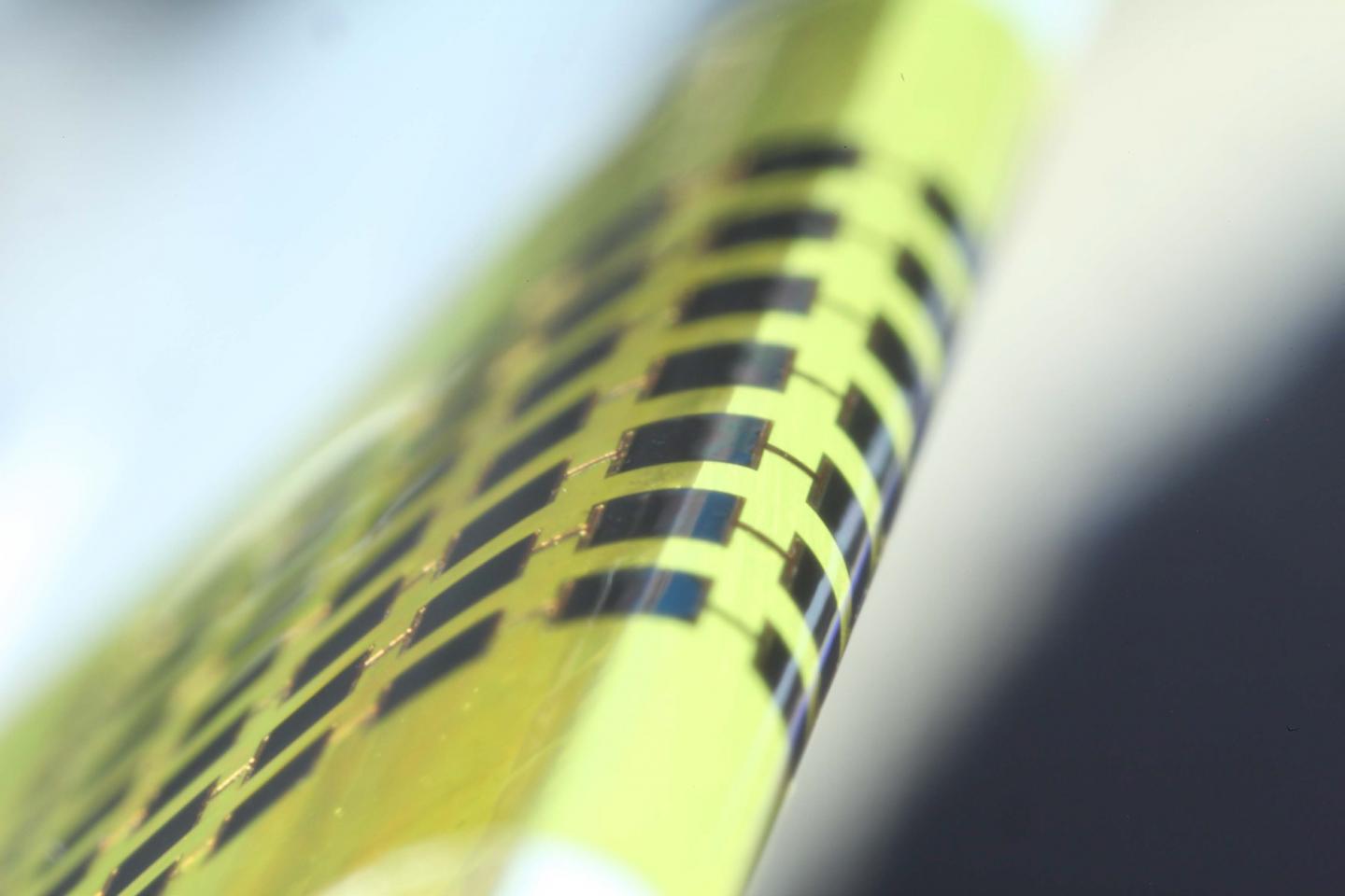
Caption: Ultra-thin solar cells are flexible enough to bend around small objects, such as the 1mm-thick edge of a glass slide, as shown here. Credit: Juho Kim, et al/ APL

Caption: Ultra-thin solar cells are flexible enough to bend around small objects, such as the 1mm-thick edge of a glass slide, as shown here. Credit: Juho Kim, et al/ APL

Yes, this is another wearable electronics story and this time, it’s from South Korea. A June 20, 2016 news item on ScienceDaily announces remarkably thin and flexible photovoltaics,

Scientists in South Korea have made ultra-thin photovoltaics flexible enough to wrap around the average pencil. The bendy solar cells could power wearable electronics like fitness trackers and smart glasses. …

A June 20, 2016 American Institute of Physics news release on EurekAlert, which originated the news item, expands on the theme,

Thin materials flex more easily than thick ones – think a piece of paper versus a cardboard shipping box. The reason for the difference: The stress in a material while it’s being bent increases farther out from the central plane. Because thick sheets have more material farther out they are harder to bend.

“Our photovoltaic is about 1 micrometer thick,” said Jongho Lee, an engineer at the Gwangju Institute of Science and Technology in South Korea. One micrometer is much thinner than an average human hair. Standard photovoltaics are usually hundreds of times thicker, and even most other thin photovoltaics are 2 to 4 times thicker.

The researchers made the ultra-thin solar cells from the semiconductor gallium arsenide. They stamped the cells directly onto a flexible substrate without using an adhesive that would add to the material’s thickness. The cells were then “cold welded” to the electrode on the substrate by applying pressure at 170 degrees Celcius and melting a top layer of material called photoresist that acted as a temporary adhesive. The photoresist was later peeled away, leaving the direct metal to metal bond.

The metal bottom layer also served as a reflector to direct stray photons back to the solar cells. The researchers tested the efficiency of the device at converting sunlight to electricity and found that it was comparable to similar thicker photovoltaics. They performed bending tests and found the cells could wrap around a radius as small as 1.4 millimeters.

The team also performed numerical analysis of the cells, finding that they experience one-fourth the amount of strain of similar cells that are 3.5 micrometers thick.

“The thinner cells are less fragile under bending, but perform similarly or even slightly better,” Lee said.

A few other groups have reported solar cells with thicknesses of around 1 micrometer, but have produced the cells in different ways, for example by removing the whole substract by etching.

By transfer printing instead of etching, the new method developed by Lee and his colleagues may be used to make very flexible photovoltaics with a smaller amount of materials.

The thin cells can be integrated onto glasses frames or fabric and might power the next wave of wearable electronics, Lee said.

Here’s a link to and a citation for the paper,

Ultra-thin flexible GaAs photovoltaics in vertical forms printed on metal surfaces without interlayer adhesives by Juho Kim, Jeongwoo Hwang, Kwangsun Song, Namyun Kim, Jae Cheol Shin, and Jongho Lee. Appl. Phys. Lett. 108, 253101 (2016); http://dx.doi.org/10.1063/1.4954039

This paper is open access.