Monthly Archives: April 2015

Water-soluble 3D filament from Graphene 3D Lab

This is almost a Canadian nanotech story since one of the company’s chief investors is Canadian and the announcement is being made at an event held at Mohawk College in Hamilton, Ontario. From an April 30, 2015 news item on Azonano,

… Graphene 3D Lab Inc. will announce details of a newly-developed water-soluble 3D filament at the Canadian Manufacturers & Exporters’ (CME) Canada Makes: Additive Manufacturing Forum at Mohawk College in Hamilton, Ontario on April 30, 2015. The water-soluble filament is the latest in a line of specialty-functional filaments that Graphene 3D is introducing to the 3D printing industry.

An April 30, 2015 Graphene 3D Lab news release provides more detail,

Elena Polyakova, Chief Operating Officer of Graphene 3D, will present details on the new water-soluble filament and discuss the effect of graphene-enhanced materials for the 3D printing industry.

Water-soluble filaments are primarily used to occupy negative space during a 3D print. These filaments are essentially used to fill gaps designed in objects and allow the print to be suspended over air. Following the completion of the printing process, the object can be placed in water, and the water-soluble material will completely dissolve, leaving empty space.

Daniel Stolyarov, President and Chief Executive Officer, stated, “We are very pleased with performance characteristics that our R&D team designed into this new industry leading filament. This filament is completely environmentally-friendly, non-toxic, dissolves completely in water in approximately 1 hour, and leaves no residual material.” The water-soluble filament will work in most commercially available 3D printers and Graphene 3D expects to have them to market within 3 months. Daniel continues, “This filament reflects the core objectives of Graphene 3D. We are creating products that allow innovative designers to do more with 3D printing and are expanding the possibilities within the 3D printing environment.”

About Graphene 3D
Graphene 3D is in the business of developing, manufacturing and marketing specialty 3D printer materials, including proprietary nanocomposite materials for various types of 3D printing. The Company is also involved in the design, manufacture and marketing of 3D printers and related products for domestic and international customers.

The Graphene 3D research and production facility is located in Calverton, NY and is equipped with material processing and analytical equipment. The company has four US patent applications pending for its technology. For more information on Graphene 3D Lab Inc., visit www.graphene3dlab.com.

As for Canadian content (beyond the announcement location), the news item on Azonano notes,

Lomiko Metals 100%-owned subsidiary Lomiko Technologies Inc. has and will continue to hold its 4,396,100 share investment in Graphene 3D Lab for the long term.

Lomiko Metals is located in the Vancouver metro region of Canadian province British Columbia.

Kenya and a draft nanotechnology policy

I don’t often stumble across information about Kenya’s nanotechnology efforts (my last was in a Sept. 1, 2011 posting) but I’m going include my latest find here even though I can’t track down the original source for the information. From an April 29, 2015 news item on SpyGhana (original source: Xinhua News Agency,  official press agency of the People’s Republic of China),

The Kenyan government will soon adopt a comprehensive policy to promote use of nanotechnology in diverse fields like medicine, agriculture, manufacturing and environment.

“Nanotechnology as a science promises more for less. The competitive edge for Kenya as a developing nation lies in robust investments in this technology,” Njeri Wamae, chairman of National Commission for Science, Technology and Innovation (NACOSTI), said in Nairobi.

Nanotechnology is relatively new in Kenya though the government has prioritized its development through research, training and setting up of supportive infrastructure.

Wamae noted that enactment of a nanotechnology policy will position Kenya as a hub for emerging technologies that would revolutionalize key sectors of the economy.

Policy briefs from Kenya’s scientific research body indicates that globally, nanotechnology was incorporated into manufacturing goods worth over 30 billion U.S. dollars in 2005.

The briefs added that nanotechnology related business was worth 2.6 trillion dollars by 2015. Kenya has borrowed best practices from industrialized countries and emerging economies to develop nanotechnology.

Professor Erastus Gatebe, an official at Kenya Industrial Research and Development Institute (KIRDI), noted that China and India offers vital lessons on harnessing nanotechnology to propel industrial growth.

This draft policy seems to be the outcome of a number of initiatives including Nanotechnologies for Development in India, Kenya and the Netherlands: Towards a Framework for Democratic Governance of Risks in Developing Countries, WOTRO (2010 – 2014) from the African Technology Policy Studies (ATPS) Network,

The ATPS has secured funding for a new Integrated Program (IP) on “Nanotechnologies for Development in India, Kenya and the Netherlands: Towards a Framework for Democratic Governance of Risks in Developing Countries, January 2010 – 2014, in liaison with partners in Europe and India. This IP which is led by Prof. Wiebe Bijker of the University of Maastricht, the Netherlands addresses the inevitable risks and benefits associated with emerging technologies, such as nanotechnology through a triangulation of PhD and Post-Doctoral positions drawn from Africa (2), India (1) and the Netherlands (2) based at the University of Maastricht but address core areas of the nanotechnology governance in Africa, India and the Netherlands. The program will be coordinated by Prof. Wiebe Bijker, the University of Maastricht, in the Netherlands; with the University of Hyderabad, India; the ATPS and the University of Nairobi, Kenya as partners.

Nanotechnology events and discussions played in important role in Kenya’s 2013 National Science, Technology and Innovation (ST&I) Week by Daphne Molewa (on the South African Agency for Science and Technology Advancement [SAASTA] website),

The National Science, Technology and Innovation (ST&I) Week, organised by the Ministry of Higher Education, Science and Technology, is a major event on the annual calendar of the Kenyan Government.

The theme for 2013, “Science, Technology and Innovation for the realisation of Kenya’s Vision 2030 and beyond” is aligned with the national vision to transform Kenya into a newly industrialised, middle-income country providing a high-quality life to all its citizens in a safe and secure environment by the year 2030. pemphasis mine]

Nanotechnology, the science of the future

SAASTA representatives Mthuthuzeli Zamxaka and Sizwe Khoza were invited to participate in this year’s festival in Nairobi [Kenya] on behalf of the Nanotechnology Public Engagement Programme (NPEP).

Zamxaka delivered a stirring presentation titled Nanotechnology Public Engagement: The Case of South Africa. He introduced the topic of nanotechnology, focusing on engagement, outreach and awareness. …

Zamxaka touched on a number of nanotechnologies that are currently being applied, such as the research conducted by the Johns Hopkins University in Maryland on biodegradable nano-sized particles that can easily slip through the body’s sticky and viscous mucus secretions to deliver a sustained-release medication cargo. It is believed that these nanoparticles, which degrade over time into harmless components, could one day carry life-saving drugs to patients suffering from dozens of health conditions, including diseases of the eye, lung, gut or female reproductive tract.

For anyone interested , look here for Kenya’s Vision 2030. Harkening back to the first news item and the mention of NACOSTI, Kenya’s National Commission for Science, Technology and Innovation, it can be found here.

Motor proteins have a stiff-legged walk

An April 23, 2015 news item on Nanowerk calls to mind Monty Python and its Ministry of Silly Walks,

The ‘stiff-legged’ walk of a motor protein along a tightrope-like filament has been captured for the first time.

Because cells are divided in many parts that serve different functions some cellular goodies need to be transported from one part of the cell to another for it to function smoothly. There is an entire class of proteins called ‘molecular motors’, such as myosin 5, that specialise in transporting cargo using chemical energy as fuel.

Remarkably, these proteins not only function like nano-scale lorries, they also look like a two-legged creature that takes very small steps. But exactly how Myosin 5 did this was unclear.

For anyone unfamiliar with The Ministry of Silly Walks (from its Wikipedia entry; Note: Links have been removed),

“The Ministry of Silly Walks” is a sketch from the Monty Python comedy troupe’s television show Monty Python’s Flying Circus, season 2, episode 14, which is entitled “Face the Press”.

Here’s an image from the sketch, which perfectly illustrates a stiff-legged walk,

John Cleese as a Civil Servant in the Ministry of Silly Walks. Screenshot from Monty Python's Flying Circus episode, Dinsdale (Alternate episode title: Face the Press). Ministry_of_Silly_Walks.jpg ‎(300 × 237 pixels, file size: 14 KB, MIME type: image/jpeg) [downloaded from http://en.wikipedia.org/wiki/File:Ministry_of_Silly_Walks.jpg]

John Cleese as a Civil Servant in the Ministry of Silly Walks. Screenshot from Monty Python’s Flying Circus episode, Dinsdale (Alternate episode title: Face the Press). Ministry_of_Silly_Walks.jpg ‎(300 × 237 pixels, file size: 14 KB, MIME type: image/jpeg) [downloaded from http://en.wikipedia.org/wiki/File:Ministry_of_Silly_Walks.jpg]

As far as I can tell, the use of this image would fall under the notion of ‘fair dealing‘ as it’s called in Canada.

Getting back to the Nanowerk news item, it started life as a University of Oxford Science blog April 23, 2015 posting  by Pete Wilton (Note: A link has been removed),

The motion of myosin 5 has now been recorded by a team led by Oxford University scientists using a new microscopy technique that can ‘see’ tiny steps of tens of nanometres captured at up to 1000 frames per second. The findings are of interest for anyone trying to understand the basis of cellular function but could also help efforts aimed at designing efficient nanomachines.

‘Until now, we believed that the sort of movements or steps these proteins made were random and free-flowing because none of the experiments suggested otherwise,’ said Philipp Kukura of Oxford University’s Department of Chemistry who led the research recently reported in the journal eLife. ‘However, what we have shown is that the movements only appeared random; if you have the capability to watch the motion with sufficient speed and precision, a rigid walking pattern emerges.’

One of the key problems for those trying to capture proteins on a walkabout is that not only are these molecules small – with steps much smaller than the wavelength of light and therefore the resolution of most optical microscopes – but they are also move very quickly.

Philipp describes how the team had to move from the microscope equivalent of an iPhone camera to something more like the high speed cameras used to snap speeding bullets. Even with such precise equipment the team had to tag the ‘feet’ of the protein in order to precisely image its gait: one foot was tagged with a quantum dot, the other with a gold particle just 20 nanometres across. (Confusingly, technically speaking, these ‘feet’ are termed the ‘heads’ of the protein because they bind to the actin filament).

I recommend reading Wilton’s post in its entirety. Meanwhile, here’s a 12 secs. video illustrating the motor protein’s stiff-legged walk,

Here’s a link to and a citation for the paper,

Structural dynamics of myosin 5 during processive motion revealed by interferometric scattering microscopy by Joanna Andrecka, Jaime Ortega Arroyo, Yasuharu Takagi, Gabrielle de Wit, Adam Fineberg, Lachlan MacKinnon, Gavin Young, James R Sellers, & Philipp Kukura. eLife 2015;4:e05413 DOI: http://dx.doi.org/10.7554/eLife.05413Published March 6, 2015

This paper is open access.

As for silly walks, there is more than one version of the sketch with John Cleese on YouTube but I was particularly taken with this public homage which took place in Brno (Czech Republic) in Jan. 2013,

Enjoy!

Gold nanoparticle clusters: four new models

This research is being done at the University of Nebraska-Lincoln (UNL) which seems to be on a publishing tear lately. From an April 27, 2015 news item on Nanowerk, here’s the latest,

They may deal in gold, atomic staples and electron volts rather than cement, support beams and kilowatt-hours, but chemists have drafted new nanoscale blueprints for low-energy structures capable of housing pharmaceuticals and oxygen atoms.

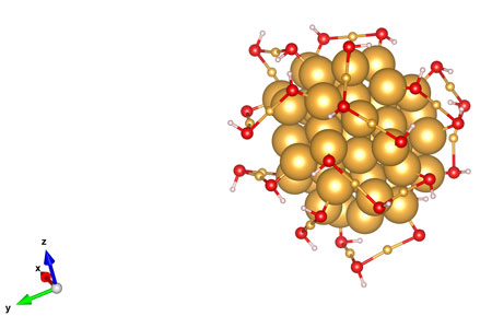
Led by UNL’s Xiao Cheng Zeng and former visiting professor Yi Gao, new research has revealed four atomic arrangements of a gold nanoparticle cluster. The arrangements exhibit much lower potential energy and greater stability than a standard-setting configuration reported last year by a Nobel Prize-winning team from Stanford University.

The modeling of these arrangements could inform the cluster’s use as a transporter of pharmaceutical drugs and as a catalyst for removing pollutants from vehicular emissions or other industrial byproducts, Zeng said.

An April 24, 2015 UNL news release (also on EurekAlert), which originated the news item, provides more technical details about the work,

Led by UNL’s Xiao Cheng Zeng and former visiting professor Yi Gao, new research has revealed four atomic arrangements of a gold nanoparticle cluster. The arrangements exhibit much lower potential energy and greater stability than a standard-setting configuration reported last year by a Nobel Prize-winning team from Stanford University.

The modeling of these arrangements could inform the cluster’s use as a transporter of pharmaceutical drugs and as a catalyst for removing pollutants from vehicular emissions or other industrial byproducts, Zeng said.

Zeng and his colleagues unveiled the arrangements for a molecule featuring 68 gold atoms and 32 pairs of bonded sulfur-hydrogen atoms. Sixteen of the gold atoms form the molecule’s core; the remainder bond with the sulfur and hydrogen to form a protective coating that stems from the core.

Differences in atomic arrangements can alter molecular energy and stability, with less potential energy making for a more stable molecule. The team calculates that one of the arrangements may represent the most stable possible structure in a molecule with its composition.

“Our group has helped lead the front on nano-gold research over the past 10 years,” said Zeng, an Ameritas University Professor of chemistry. “We’ve now found new coating structures of much lower energy, meaning they are closer to the reality than (previous) analyses. So the deciphering of this coating structure is major progress.”

The structure of the molecule’s gold core was previously detailed by the Stanford team. Building on this, Zeng and his colleagues used a computational framework dubbed “divide-and-protect” to configure potential arrangements of the remaining gold atoms and sulfur-hydrogen pairs surrounding the core.

The researchers already knew that the atomic coating features staple-shaped linkages of various lengths. They also knew the potential atomic composition of each short, medium and long staple — such as the fact that a short staple consists of two sulfur atoms bonded with one gold.

By combining this information with their knowledge of how many atoms reside outside the core, the team reduced the number of potential arrangements from millions to mere hundreds.

“We divided 32 into the short, middle and long (permutations),” said Zeng, who helped develop the divide-and-protect approach in 2008. “We lined up all those possible arrangements, and then we computed their energies to find the most stable ones.

“Without those rules, it’s like finding a needle in the Platte River. With them, it’s like finding a needle in the fountain outside the Nebraska Union. It’s still hard, but it’s much more manageable. You have a much narrower range.”

The researchers resorted to the computational approach because of the difficulty of capturing the structure via X-ray crystallography or single-particle transmission electron microscopy, two of the most common imaging methods at the atomic scale.

Knowing the nanoparticle’s most stable configurations, Zeng said, could allow biomedical engineers to identify appropriate binding sites for drugs used to treat cancer and other diseases. The findings could also optimize the use of gold nanoparticles in catalyzing the oxidation process that transforms dangerous carbon monoxide emissions into the less noxious carbon dioxide, he said.

Here’s an image illustrating the work,

This rendering shows the atomic arrangements of a gold nanocluster as reported in a new study led by UNL chemist Xiao Cheng Zeng. The cluster measures about 1.7 nanometers long -- roughly the same length that a human fingernail grows in two seconds. (Joel Brehm/Office of Research and Economic Development)

This rendering shows the atomic arrangements of a gold nanocluster as reported in a new study led by UNL chemist Xiao Cheng Zeng. The cluster measures about 1.7 nanometers long — roughly the same length that a human fingernail grows in two seconds. (Joel Brehm/Office of Research and Economic Development)

Here’s a link to and a citation for the paper,

Unraveling structures of protection ligands on gold nanoparticle Au68(SH)32 by Wen Wu Xu, Yi Gao, and Xiao Cheng Zeng. Science Advances 24 Apr 2015: Vol. 1 no. 3 e1400211 DOI: 10.1126/sciadv.1400211

This is an open access article.

As for the Stanford University team’s work mentioned in the news release, I believe it’s from the Roger Kornberg (Nobel laureate) Laboratory. There’s more about that team’s work in an Aug. 21, 2014 article (A new gold standard for nano; Note: A link has been removed) by David Bradley for Chemistry World,

Characterising gold nanoparticles at atomic resolution might improve our understanding of the catalytic activity of these materials, according to an international team. These researchers have now demonstrated that it is possible to use electron microscopy to obtain data on at least one gold cluster of greater than 1nm diameter and to validate the results by comparison with small-angle x-ray scattering data, infrared absorption spectra and density functional theory calculations.

Here’s a link to and a citation for the 2014 paper,

Electron microscopy of gold nanoparticles at atomic resolution by Maia Azubel, Jaakko Koivisto, Sami Malola, David Bushnell, Greg L. Hura, Ai Leen Koh, Hironori Tsunoyama, Tatsuya Tsukuda, Mika Pettersson, Hannu Häkkinen, & Roger D. Kornberg. Science 22 August 2014: Vol. 345 no. 6199 pp. 909-912 DOI: 10.1126/science.1251959

This paper is behind a paywall.

The most recent posting here about gold nanoparticles is an April 14, 2015 piece titled: Gold atoms: sometimes they’re a metal and sometimes they’re a molecule.

The science of the Avengers: Age of Ultron

The American Chemical Society (ACS) has produced a video (almost 4 mins.) in their Reactions Science Video Series of podcasts focusing on the Avengers, super heroes, as portrayed in Avengers: Age of Ultron and science. From an April 29, 2015 ACS news release on EurekAlert,

Science fans, assemble! On May 1, the world’s top superhero team is back to save the day in “Avengers: Age of Ultron.” This week, Reactions looks at the chemistry behind these iconic heroes’ gear and superpowers, including Tony Stark’s suit, Captain America’s shield and more.

Here’s the video,


While the chemists are interested in the metal alloys, there is more ‘super hero science’ writing out there. Given my interests, I found the ‘Captain America’s shield as supercapacitor theory’ as described in Matt Shipman’s April 15, 2014 post on The Abstract (North Carolina State University’s official newsroom blog quite interesting. I featured Shipman’s ‘super hero and science’ series of posts in my April 28, 2014 posting.

Liquid nanolaser: the first one

According to an April 24, 2015 news item on Nanowerk, there has been a big discovery at Northwestern University (located in Chicago, Illinois, US),

Northwestern University scientists have developed the first liquid nanoscale laser. And it’s tunable in real time, meaning you can quickly and simply produce different colors, a unique and useful feature. The laser technology could lead to practical applications, such as a new form of a “lab on a chip” for medical diagnostics.

To understand the concept, imagine a laser pointer whose color can be changed simply by changing the liquid inside it, instead of needing a different laser pointer for every desired color.

In addition to changing color in real time, the liquid nanolaser has additional advantages over other nanolasers: it is simple to make, inexpensive to produce and operates at room temperature.

An April 24, 2015 Northwestern University news release by Megan Fellman (also on EurekAlert), which originated the news item, offers a little history buttressed by some technical details (Note: Links have been removed),

Nanoscopic lasers — first demonstrated in 2009 — are only found in research labs today. They are, however, of great interest for advances in technology and for military applications.

“Our study allows us to think about new laser designs and what could be possible if they could actually be made,” said Teri W. Odom, who led the research. “My lab likes to go after new materials, new structures and new ways of putting them together to achieve things not yet imagined. We believe this work represents a conceptual and practical engineering advance for on-demand, reversible control of light from nanoscopic sources.”

The liquid nanolaser in this study is not a laser pointer but a laser device on a chip, Odom explained. The laser’s color can be changed in real time when the liquid dye in the microfluidic channel above the laser’s cavity is changed.

The laser’s cavity is made up of an array of reflective gold nanoparticles, where the light is concentrated around each nanoparticle and then amplified. (In contrast to conventional laser cavities, no mirrors are required for the light to bounce back and forth.) Notably, as the laser color is tuned, the nanoparticle cavity stays fixed and does not change; only the liquid gain around the nanoparticles changes.

The main advantages of very small lasers are:

• They can be used as on-chip light sources for optoelectronic integrated circuits;

• They can be used in optical data storage and lithography;

• They can operate reliably at one wavelength; and

• They should be able to operate much faster than conventional lasers because they are made from metals.

Some technical background

Plasmon lasers are promising nanoscale coherent sources of optical fields because they support ultra-small sizes and show ultra-fast dynamics. Although plasmon lasers have been demonstrated at different spectral ranges, from the ultraviolet to near-infrared, a systematic approach to manipulate the lasing emission wavelength in real time has not been possible.

The main limitation is that only solid gain materials have been used in previous work on plasmon nanolasers; hence, fixed wavelengths were shown because solid materials cannot easily be modified. Odom’s research team has found a way to integrate liquid gain materials with gold nanoparticle arrays to achieve nanoscale plasmon lasing that can be tuned dynamical, reversibly and in real time.

The use of liquid gain materials has two significant benefits:

• The organic dye molecules can be readily dissolved in solvents with different refractive indices. Thus, the dielectric environment around the nanoparticle arrays can be tuned, which also tunes the lasing wavelength.

• The liquid form of gain materials enables the fluid to be manipulated within a microfluidic channel. Thus, dynamic tuning of the lasing emission is possible simply by flowing liquid with different refractive indices. Moreover, as an added benefit of the liquid environment, the lasing-on-chip devices can show long-term stability because the gain molecules can be constantly refreshed.

These nanoscale lasers can be mass-produced with emission wavelengths over the entire gain bandwidth of the dye. Thus, the same fixed nanocavity structure (the same gold nanoparticle array) can exhibit lasing wavelengths that can be tuned over 50 nanometers, from 860 to 910 nanometers, simply by changing the solvent the dye is dissolved in.

Here’s a link to and a citation for the paper,

Real-time tunable lasing from plasmonic nanocavity arrays by Ankun Yang, Thang B. Hoang, Montacer Dridi, Claire Deeb, Maiken H. Mikkelsen, George C. Schatz, & Teri W. Odom. Nature Communications 6, Article number: 6939 doi:10.1038/ncomms7939 Published 20 April 2015

This paper is open access.

Science Hack Day (May 16 – May 17, 2015) in Washington, DC

I received an April 28, 2015 announcement from the Wilson Center’s (aka Wilson International Center for Scholars) Commons Lab about the first ever and upcoming Science Hack Day in Washington, DC (May 16 – 17, 2015),

The Wilson Center and ARTSEDGE from the Kennedy Center are proud to host the first-ever in Washington, D.C., Science Hack Day! Science Hack Day is a 48-hour-all-night event where anyone excited about making weird, silly or serious things with science comes together in the same physical space to see what they can prototype over a weekend.

Designers, artists, developers, hardware enthusiasts, scientists and anyone who is excited about making things with science are welcome to attend – no experience in science or hacking is necessary, just an insatiable curiosity. Food is provided both days to fuel hackers during the day and throughout the night. The event is completely free and open to the public (pre-registration required).

The event will kick off with a series of lightning talks from a diverse group of people in the civic sector. Participants will hack through the night and on Sunday they will demo their projects to a DC Tech panel.

For more detailed information, logistics and updated speaker list please visit: http://dc.sciencehackday.org/

Sponsors & Collaborators

Thomson Reuters End Note

GitHub

ARTSEDGE Kennedy Center for the Arts

For anyone who might need a little more information as to exactly what a ‘science hack’ might be, there’s this description from the Wilson Center’s DC Science Hack Day 2015 event page,

What’s a Hack?
A hack is a quick solution to a problem – maybe not the most elegant solution, but often the cleverest. On the web, mashups are a common example of hacking: mixing up data from different sources in new and interesting ways.

There’s also a video interview where Elizabeth Tyson, one of the organizers, describes it. First some text from an April 13, 2015 Wilson Center Science Hack Day news article,

Elizabeth Tyson is a New Projects Manager/Researcher for the Wilson Center’s Science and Technology Innovation Program. She co-directs the Commons Lab and scouts and maintains new collaborations. Additionally, she conducts original research exploring the uses of citizen science in industrializing nations. Currently she is coordinating Washington, DC’s first ever Science Hack Day.  Elizabeth reviews and edits publications on citizen science and crowdsourcing including Citizen Science and Policy: A European Perspective and a Typology of Citizen Science from an Intellectual Property Perspective.

Now the video,

You can also a Science Hack Day DC 2015 wiki here. Here are some logistics,

May 16, 2015 at 9:00am to May 17, 2015 at 5:00pm
The Wilson Center
Ronald Reagan Building
1300 Pennsylvania Avenue Northwest
Washington, D.C. 20004

Enjoy!

3D imaging biological cells with picosecond ultrasonics (acoustic imaging)

An April 22, 2015 news item on Nanowerk describes an acoustic imaging technique that’s been newly applied to biological cells,

Much like magnetic resonance imaging (MRI) is able to scan the interior of the human body, the emerging technique of “picosecond ultrasonics,” a type of acoustic imaging, can be used to make virtual slices of biological tissues without destroying them.

Now a team of researchers in Japan and Thailand has shown that picosecond ultrasonics can achieve micron resolution of single cells, imaging their interiors in slices separated by 150 nanometers — in stark contrast to the typical 0.5-millimeter spatial resolution of a standard medical MRI scan. This work is a proof-of-principle that may open the door to new ways of studying the physical properties of living cells by imaging them in vivo.

An April 20, 2015 American Institute of Physics news release, which originated the news item, provides a description of picosecond ultrasonics and more details about the research,

Picosecond ultrasonics has been used for decades as a method to explore the mechanical and thermal properties of materials like metals and semiconductors at submicron scales, and in recent years it has been applied to biological systems as well. The technique is suited for biology because it’s sensitive to sound velocity, density, acoustic impedance and the bulk modulus of cells.

This week, in a story appearing on the cover of the journal Applied Physics Letters, from AIP Publishing, researchers from Walailak University in Thailand and Hokkaido University in Japan describe the first known demonstration of 3-D cell imaging using picosecond ultrasonics.

Their work centers on imaging two types of mammalian biological tissue — a bovine aortic endothelial cell, a type of cell that lines a cow’s main artery blood vessel, and a mouse “adipose” fat cell. Endothelial cells were chosen because they play a key role in the physiology of blood cells and are useful in the study of biomechanics. Fat cells, on the other hand, were studied to provide an interesting comparison with varying cell geometries and contents.

How the Work was Done

The team accomplished the imaging by first placing a cell in solution on a titanium-coated sapphire substrate and then scanning a point source of high-frequency sound generated by using a beam of focused ultrashort laser pulses over the titanium film. This was followed by focusing another beam of laser pulses on the same point to pick up tiny changes in optical reflectance caused by the sound traveling through the cell tissue.

“By scanning both beams together, we’re able to build up an acoustic image of the cell that represents one slice of it,” explained co-author Professor Oliver B. Wright, who teaches in the Division of Applied Physics, Faculty of Engineering at Hokkaido University. “We can view a selected slice of the cell at a given depth by changing the timing between the two beams of laser pulses.”

The team’s work is particularly noteworthy because “in spite of much work imaging cells with more conventional acoustic microscopes, the time required for 3-D imaging probably remains too long to be practical,” Wright said. “Building up a 3-D acoustic image, in principle, allows you to see the 3-D relative positions of cell organelles without killing the cell. In our experiments in vitro, while we haven’t yet resolved the cell contents — possibly because cell nuclei weren’t contained within the slices we viewed — it should be possible in the future with various improvements to the technique.”

: Fluorescence micrographs of fat and endothelial cells superimposed on differential-interference and phase-contrast images, respectively.

Fluorescence micrographs of fat and endothelial cells superimposed on differential-interference and phase-contrast images, respectively. The nuclei are stained blue in the micrographs. The image on the right is a picosecond-ultrasonic image of a single endothelial cell with approximately 1-micron lateral and 150-nanometer depth resolutions. Deep blue corresponds to the lowest ultrasonic amplitude.
CREDIT: O. Wright/Hokkaido University

So far, the team has used infrared light to generate sound waves within the cell, “limiting the lateral spatial resolution to about one micron,” Wright explains. “By using an ultraviolet-pulsed laser, we could improve the lateral resolution by about a factor of three — and greatly improve the image quality. And, switching to a diamond substrate instead of sapphire would allow better heat conduction away from the probed area, which, in turn, would enable us to increase the laser power and image quality.”

So lowering the laser power or using substrates with higher thermal conductivity may soon open the door to in vivo imaging, which would be invaluable for investigating the mechanical properties of cell organelles within both vegetal and animal cells.

What’s next for the team? “The method we use to image the cells now actually involves a combination of optical and elastic parameters of the cell, which can’t be easily distinguished,” Wright said. “But we’ve thought of a way to separate them, which will allow us to measure the cell mechanical properties more accurately. So we’ll try this method in the near future, and we’d also like to try our method on single-celled organisms or even bacteria.”

Here’s a link to and a citation for the paper,

Three-dimensional imaging of biological cells with picosecond ultrasonics by Sorasak Danworaphong, Motonobu Tomoda, Yuki Matsumoto, Osamu Matsuda, Toshiro Ohashi, Hiromu Watanabe, Masafumi Nagayama, Kazutoshi Gohara, Paul H. Otsuka, and Oliver B. Wright. Appl. Phys. Lett. 106, 163701 (2015); http://dx.doi.org/10.1063/1.4918275

This paper is open access.

This research reminded me of a data sonification project that I featured in a Feb. 7, 2014 post which includes an embedded sound file of symphonic music based on data from NASA’s (US National Aeronautics and Space Administration) Voyager spacecraft.

2015 Canadian federal budget and science

Think of this post as a digest of responses to and analyses of the ‘science component’ of the Canadian federal government’s 2015 budget announcement made on April 21, 2015 by Minister of Finance, Joe Oliver. First off the mark, the Canadian Science Policy Centre (CSPC) has featured some opinions about the budget and its impact on Canadian science in an April 27, 2015 posting,

Jim Woodgett
Director, Lunenfeld-Tanenbaum Research Institute of Sinai Health System

Where’s the Science Beef in Canadian Budget 2015?

Andrew Casey
President and CEO, BIOTECanada

Budget 2015: With the fiscal balance restored where to next?

Russ Roberts
Senior Vice President – Tax & Finance, CATA Alliance

Opinion on 2015 Federal Budget

Ron Freeman
CEO of Innovation Atlas Inc. and Research Infosource Inc. formerly co-publisher of RE$EARCH MONEY and co-founder of The Impact Group

Workman-Like Budget Preserves Key National Programs

Paul Davidson
President, Universities Canada

A Reality Check on Budget 2015

Dr. Kamiel Gabriel
Associate Provost of Research and Graduate Programs at the University of Ontario Institute of Technology (UOIT), Science Adviser and Assistant Deputy Minister (ADM) of Research at the Ontario Ministry of Research & Innovation

The 2015 Federal Budget Targets Key Segments of Voters

I suggest starting with Woodgett’s piece as he points out something none of the others who chose to comment on the amount of money dedicated to the tricouncil funding agencies (Canadian Institutes of Health Research [CIHR], Natural Sciences and Engineering Research Council [NSERC], and Social Sciences and Humanities Research Council [SSHRC]) seemed to have noticed or deemed important,

The primary source of science operating funds are provided by the tricouncils, CIHR/NSERC and SSHRC, which, when indirect costs and other flow through dollars (e.g. CRCs) are included, accounts for about $2.5 billion in annual funding. There are no new dollars added to the tricouncil budgets this year (2015/16) but there is a modest $46 million to be added in 2016/17 – $15 million to CIHR and NSERC, $7.5 million to SSHRC and the rest in indirects. [emphases mine] This new money, though, is largely ear-marked for new initiatives, such as the CIHR Strategy on Patient Oriented Research ($13 million) and an anti-microbial resistant infection program ($2 million). Likewise for NSERC and SSHRC although NSERC enjoys around $16 million relief in not needing to support industrial postgraduate scholarships as this responsibility moves to MITACS with no funding loss at NSERC. Alex Usher of Higher Education Strategy Associates, estimates that, taking inflation into account, tricouncil funding will be down 9% since 2008. [emphasis mine] It is hardly surprising that funding applications to these agencies are under enormous competitive pressure. At CIHR, the last open operating grant competition yielded unprecedented low success rates of ~14% along with across-the-board budget cuts of grants that were funded of 26%. This agency is in year 1 of major program reforms and has very little wiggle-room with its frozen budget.

To be fair, there are sources other than the tricouncil for science funding although their mandate is for ‘basic’ science, more or less. Over the last few years, there’s been a greater emphasis on tricouncil funding that produces economic results and this is in line international trends.

Getting back to the CSPC’s opinions, Davidson’s piece, notes some of that additional funding,

With $1.33 billion earmarked for the Canada Foundation for Innovation [CFI], Budget 2015 marks the largest single announcement of Canadian research infrastructure funding. This is something the community prioritized, given the need for state-of-the-art equipment, labs, digital tools and high-speed technology to conduct, partner and share research results. This renewed commitment to CFI builds on the globally competitive research infrastructure that Canadians have built over the last 15 years and enables our researchers to collaborate with the very best in the world. Its benefits will be seen in universities across the country and across disciplines. Key research infrastructure investments – from digital to major science infrastructure – support the broad spectrum of university research, from theoretical and discovery to pre-competitive and applied.

The $45 million announced for TRIUMF will support the laboratory’s role in accelerating science in Canada, an important investment in discovery research.

While the news about the CFI seems to have delighted a number of observers, it should be noted (as per Woodgett’s piece) that the $1.3B is to be paid out over six years ($220M per year, more or less) and the money won’t be disbursed until the 2017/18 fiscal year. As for the $45M designated for TRIUMF (Canada’s National Laboratory for Particle and Nuclear Physics), this is exciting news for the lab which seems to have bypassed the usual channels, as it has before, to receive its funding directly from the federal government.

Another agency which seems to have received its funding directly from the federal government is the Council of Canadian Academies (CCA), From an April 22, 2015 news release,

The Council of Canadian Academies welcomes the federal government’s announcement of new funding for in-depth, authoritative, evidence-based assessments. Economic Action Plan 2015 allocated $15 million over five years [$3M per year] for the Council of Canadian Academies.

“This is welcome news for the Council and we would like to thank the Government for this commitment. Over the past 10 years the Council has worked diligently to produce high quality reports that support policy and decision-making in numerous areas,” said Janet Bax, Interim President. “We appreciate the support from Minister Holder and his predecessors, Minsters Goodyear and Rickford, for ensuring meaningful questions have been referred to the Council for assessment.” [For anyone unfamiliar with the Canadian science minister scene, Ed Holder, current Minister of State for Science and Technology, and previous Conservative government ministers, Greg Rickford and Gary Goodyear]

As of March 31st, 2015 the Council has published 31 reports on topics as diverse as business innovation, the future of Canadian policing models, and improving medicines for children. The Council has worked with over 800 expert volunteers from across Canada and abroad. These individuals have given generously of their time and as a result more than $16 million has been leveraged in volunteer support. The Council’s work has been used in many ways and had an impact on national policy agendas and strategies, research programs, and supported stakeholders and industry groups with forward looking action plans.

“On behalf of the Board of Governors I would like to extend our thanks to the Government,” said Margaret Bloodworth, Chair of the Board of Governors.  “The Board is now well positioned to consider future strategic directions for the organization and how best to further expand on the Council’s client base.”

The CCA news is one of the few item about social science funding, most observers such as Ivan Semeniuk in an April 27, 2015 article for the Globe and Mail, are largely focused on the other sciences,

Last year [2014], that funding [for the tricouncil agencies] amounted to about$2.7-billion, and this year’s budget maintains that. Because of inflation and increasing competition, that is actually a tightening of resources for rank-and-file scientists at Canada’s universities and hospitals. At the same time, those institutions are vying for a share of a $1.5-billion pot of money called the Canada First Research Excellence Fund, which the government unveiled last year and is aimed at helping push selected projects to a globally competitive level.

“This is all about creating an environment where our research community can grow,” Ed Holder, Minister of State for Science and Technology, told The Globe and Mail.

One extra bonus for science in this year’s budget is a $243.5-million commitment to secure Canada’s partnership in the Thirty Meter Telescope, a huge international observatory that is slated for construction on a Hawaiian mountain top. Given its high price-tag, many thought it unlikely that the Harper government would go for the project. In the end, the telescope likely benefited from the fact that had the Canada committed less money, most of the economic returns associated with building it would flow elsewhere.

The budget also reflects the Harper government’s preference for tying funding to partnerships with industry. A promised increase of $46-million for the granting councils next year will be largely for spurring collaborations between academic researchers and industrial partners rather than for basic research.

Whether or not science becomes an issue in the upcoming election campaign, some research advocates say the budget shows that the government’s approach to science is still too narrow. While it renews necessary commitments to research infrastructure, they fear not enough money will be left for people doing the kind of work that expands knowledge but does not always produce an immediate economic return.

An independent analysis of the 2015 budget prepared by Higher Education Strategy Associates, a Toronto based consulting firm, shows that when inflation is factored in, the money available for researchers through the granting councils has been in decline since 2009.

Canadian scientists are the not only ones feeling a pinch. Neal V. Patel’s April 27, 2015 article (originally published on Wired) on the Slate website discusses US government funding in an attempt to contextualize science research crowdfunding (Note: A link has been removed),

In the U.S., most scientific funding comes from the government, distributed in grants awarded by an assortment of federal science, health, and defense agencies. So it’s a bit disconcerting that some scientists find it necessary to fund their research the same way dudebros raise money for a potato salad. Does that migration suggest the current grant system is broken? If it is, how can we ensure that funding goes to legitimate science working toward meaningful discoveries?

On its own, the fact that scientists are seeking new sources of funding isn’t so weird. In the view of David Kaiser, a science historian at MIT, crowdfunding is simply the latest “pendulum swing” in how scientists and research institutions fund their work. Once upon a time, research at MIT and other universities was funded primarily by student tuition and private philanthropists. In 1919, however, with philanthropic investment drying up, MIT launched an ambitious plan that allowed local companies to sponsor specific labs and projects.

Critics complained the university had allowed corporate interests to dig their claws into scientific endeavors and befoul intellectual autonomy. (Sound familiar?) But once WWII began, the U.S. government became a force for funding, giving huge wartime grants to research groups nationwide. Federal patronage continued expanding in the decades after the war.

Seventy years later, that trend has reversed: As the federal budget shrinks, government investment in scientific research has reached new lows. The conventional models for federal grants, explains University of Iowa immunologist Gail Bishop, “were designed to work such that 25 to 30 percent of studies were funded. Now it’s around 10 percent.”

I’m not sure how to interpret the Canadian situation in light of other jurisdictions. It seems clear that within the Canadian context for government science funding that research funding is on a downward trend and has been going down for a few years (my June 2, 2014 posting). That said, we have another problem and that’s industrial research and development funding (my Oct. 30, 2013 posting about the 2013 OECD scorecard for science and technology; Note: the scorecard is biannual and should be issued again in 2015). Businesses don’t pay for research in Canada and it appears the Conservative and previous governments have not been successful in reversing that situation even marginally.

Queen’s University (Canada) opens Kingston Nano-Fabrication Lab (KNFL)

First, there’s the opening (from an April 24, 2015 Queen’s University news release; Note: A link has been removed),

Queen’s University has secured its place at the forefront of transforming innovative research with the opening of the Kingston Nano-Fabrication Laboratory (KNFL).

The laboratory, located at Innovation Park, represents a milestone in the 30-year collaboration between Queen’s and CMC Microsystems for advancing Canadian strength in micro-nano innovation.

Some interesting details about the deal and the proposed uses for KNFL can be found in an April 24, 2015 story by Colleen Seto for Canada Foundation for Innovation (CFI),

… a brand-new, 3,000-square-foot, $5 million research facility [KNFL] located at the Queen’s University Innovation Park. The lab includes $2.5 million in new CFI-funded custom equipment for fabricating and prototyping new nano-scale inventions to get them to market quicker.

“We’re making devices, films, coatings, and materials, and examining their properties at the nanoscale,” says Ian McWalter, President and CEO of CMC Microsystems, which manages the operations of KNFL. “This fundamental materials research spills over into experiments of great use to industry, which then looks at how to commercialize he research results.”

The Queen’s University news release describes the longstanding relationship between the company managing the KNFL and the university,

“This facility is the latest manifestation of a long and productive relationship between Queen’s and CMC Microsystems,” says Ian McWalter, president and CEO of CMC. “For more than three decades, this partnership has enabled research and advanced training activities nationwide that would not have otherwise occurred. The KNFL is a significant enhancement, and we look forward to exploring the expanded opportunities that it offers us for building Canadian strength in micro-nano research and innovation.”

The CFI story provides more specifics about the potential workings of the facility,

Take, for example, the possibilities presented by KNFL’s laser micromachining system. “This new tool could be used to engrave channels into a piece of glass or polymer to produce a microfluidic device,” says Andrew Fung, Client Technology Advisor for Microsystems and Nanotechnology at CMC. Microfluidic devices take advantage of the behaviour of fluids at a very small scale to create things like “lab-on-a-chip” technologies that can be used to cheaply and quickly diagnose diseases in developing countries, among many other things. “Microfluidics grew out of silicon-based fabrication, which costs a lot of money,” explains Fung. “These other materials are lower cost, and can be single use, consumable, and disposable for a medical device.”

Much of KNFL’s new equipment was selected to enable rapid prototyping of new nanotechnologies. “Prototypes can be ready within hours or a day, instead of days or weeks. It shortens the whole innovation process so researchers can design, make, test, and get the information they need much faster,” says Fung.

The CFI story also contextualizes this project by noting that it’s part of a larger initiative,

The KNFL is also part of Embedded Systems Canada (emSYSCAN), a $50-million, five-year project aimed at shortening the microsystems development cycle. It involves more than 350 university researchers at 37 institutions across Canada’s National Design Network (NDN), which enables multidisciplinary research and collaboration through shared technologies and expertise.

The KNFL’s open-access model is aimed specifically at supporting the NDN. “The idea is to make [expertise and tools] more available to non-experts and to overcome barriers such as lab training to access this equipment,” says McWalter. “Through the service aspect of our lab, you wouldn’t necessarily twiddle the knobs yourself, but you would contract the lab to do things for you.” This provides vital learning opportunities for students while giving researchers a more efficient means to an end — accessing the equipment they need without having to invest the time and effort to learn how to use it.

Congratulations to the folks at Queen’s University!