Category Archives: synthetic biology

Biobots (also known as biohybrid robots) occupy a third state between life and death?

I got a bit of a jolt from this September 12, 2024 essay by Peter A Noble, affiliate professor of microbiology at the University of Washington, and Alex Pozhitkov, senior technical lead of bioinformatics, Irell & Manella Graduate School of Biological Sciences at City of Hope, for The Conversation (h/t Sept. 12, 2024 item on phys.org), Note: Links have been removed,

Life and death are traditionally viewed as opposites. But the emergence of new multicellular life-forms from the cells of a dead organism introduces a “third state” that lies beyond the traditional boundaries of life and death.

Usually, scientists consider death to be the irreversible halt of functioning of an organism as a whole. However, practices such as organ donation highlight how organs, tissues and cells can continue to function even after an organism’s demise. This resilience raises the question: What mechanisms allow certain cells to keep working after an organism has died?

We are researchers who investigate what happens within organisms after they die. In our recently published review, we describe how certain cells – when provided with nutrients, oxygen, bioelectricity or biochemical cues – have the capacity to transform into multicellular organisms with new functions after death.

Life, death and emergence of something new

The third state challenges how scientists typically understand cell behavior. While caterpillars metamorphosing into butterflies, or tadpoles evolving into frogs, may be familiar developmental transformations, there are few instances where organisms change in ways that are not predetermined. Tumors, organoids and cell lines that can indefinitely divide in a petri dish, like HeLa cells [cervical cancer cells taken from Henrietta Lacks without her knowledge], are not considered part of the third state because they do not develop new functions.

However, researchers found that skin cells extracted from deceased frog embryos were able to adapt to the new conditions of a petri dish in a lab, spontaneously reorganizing into multicellular organisms called xenobots [emphasis mine]. These organisms exhibited behaviors that extend far beyond their original biological roles. Specifically, these xenobots use their cilia – small, hair-like structures – to navigate and move through their surroundings, whereas in a living frog embryo, cilia are typically used to move mucus.

Xenobots are also able to perform kinematic self-replication, meaning they can physically replicate their structure and function without growing. This differs from more common replication processes that involve growth within or on the organism’s body.

Researchers have also found that solitary human lung cells can self-assemble into miniature multicellular organisms that can move around. These anthrobots [emphasis mine] behave and are structured in new ways. They are not only able to navigate their surroundings but also repair both themselves and injured neuron cells placed nearby.

Taken together, these findings demonstrate the inherent plasticity of cellular systems and challenge the idea that cells and organisms can evolve only in predetermined ways. The third state suggests that organismal death may play a significant role in how life transforms over time.

I had not realized that xenobots are derived from dead frog embryos something I missed when mentioning or featuring them in previous stories, the latest in a September 13, 2024 posting, which also mentions anthrobots. Previous stories were published in a June 21, 2021 posting about xenobots 2.0 and their ability to move and a June 8, 2022 posting about their ability to reproduce. Thank you to the authors for relieving me of some of my ignorance.

For some reason I was expecting mention, brief or otherwise, of ethical or social implications but the authors offered this instead, from their September 12, 2024 essay, Note: Links have been removed,

Implications for biology and medicine

The third state not only offers new insights into the adaptability of cells. It also offers prospects for new treatments.

For example, anthrobots could be sourced from an individual’s living tissue to deliver drugs without triggering an unwanted immune response. Engineered anthrobots injected into the body could potentially dissolve arterial plaque in atherosclerosis patients and remove excess mucus in cystic fibrosis patients.

Importantly, these multicellular organisms have a finite life span, naturally degrading after four to six weeks. This “kill switch” prevents the growth of potentially invasive cells.

A better understanding of how some cells continue to function and metamorphose into multicellular entities some time after an organism’s demise holds promise for advancing personalized and preventive medicine.

I look forward to hearing about the third state and about any ethical or social issues that may arise from it.

Moving past xenobots (living robots based on frog stem cells)

Laura Tran’s June 14, 2024 article for The Scientist gives both a brief history of Michael Levin’s and his team’s work on developing living robots using stem cells from an African clawed frog (known as Xenopus laevis) and offers an update on the team’s work into synthetic lifeforms. First, the xenobots, Note 1: This could be difficult for people with issues regarding animal experimentation Note 1: Links have been removed,

Ibegan with little pieces of embryos scooting around in a dish. In 1998, these unassuming cells caught the attention of Michael Levin, then a postdoctoral researcher studying cell biology at Harvard University. He recalled simply recording a video before tucking the memory away. Nearly two decades later, Levin, now a developmental and synthetic biologist at Tufts University, experienced a sense of déjà vu. He observed that as a student transplanted tissues from one embryo to another, some loose cells swam free in the dish. 

Levin had a keen interest in the collective intelligence of cells, tissues, organs, and artificial constructs within regenerative medicine, and he wondered if he could explore the plasticity and harness the untapped capabilities of these swirling embryonic stem cells. “At that point, I started thinking that this is probably an amazing biorobotics platform,” recalled Levin. He rushed to describe this idea to Douglas Blackiston, a developmental and synthetic biologist at Tufts University who worked alongside Levin. 

At the time, Blackiston was conducting plasticity research to restore vision in blind African clawed frog tadpoles, Xenopus laevis, a model organism used to understand development. Blackiston transplanted the eyes to unusual places, such as the back of the head or even the tail, to test the integration of transplanted sensory organs.1 The eye axons extended to either the gut or spinal cord. In a display of dynamic plasticity, transplanted eyes on the tail that extended an optic nerve into the spinal cord restored the tadpoles’ vision.2 

In a similar vein, Josh Bongard, an evolutionary roboticist at the University of Vermont and Levin’s longtime colleague, pondered how robots could evolve like animals. He wanted to apply biological evolution to a machine by tinkering with the brains and bodies of robots and explored this idea with Sam Kriegman, then a graduate student in Bongard’s group and now an assistant professor at Northwestern University. Kriegman used evolutionary algorithms and artificial intelligence (AI) to simulate biological evolution in a virtual creature before teaming up with engineers to construct a physical version. 

i have two stories about the Xenobots. I was a little late to the party, so, the June 21, 2021 posting is about xenobots 2.0 and their ability to move and the June 8, 2022 posting is about their ability to reproduce.

Tran’s June 14, 2024 article provides the latest update, Note: Links have been removed,

Evolving Beyond the Xenobot

“People thought this was a one-off froggy-specific result, but this is a very profound thing,” emphasized Levin. To demonstrate its translatability in a non-frog model, he wondered, “What’s the furthest from an embryonic frog? Well, that would be an adult human.”

He enlisted the help of Gizem Gumuskaya, a synthetic biologist with an architectural background in Levin’s group, to tackle this challenge of creating biological robots using human cells to create anthrobots.8 While Gumuskaya was not involved with the development of xenobots, she drew inspiration from their design. By using adult human tracheal cells, she found that adult cells still displayed morphologic plasticity.

There are several key differences between xenobots and anthrobots: species, cell source (embryonic or adult), and the anthrobots’ ability to self-assemble without manipulation. “When considering applications, as a rule of thumb, xenobots are better suited to the environment. They exhibit higher durability, require less maintenance, and can coexist within the environment,” said Gumuskaya.

Meanwhile, there is greater potential for the use of mammalian-derived biobots in biomedical applications. This could include localized drug delivery, deposition into the arteries to break up plaque buildup, or deploying anthrobots into tissue to act as biosensors. “[Anthrobots] are poised as a personalized agent with the same DNA but new functionality,” remarked Gumuskaya.

Here’s a link to and a citation for the team’s latest paper,

Motile Living Biobots Self-Construct from Adult Human Somatic Progenitor Seed Cells by Gizem Gumuskaya, Pranjal Srivastava, Ben G. Cooper, Hannah Lesser, Ben Semegran, Simon Garnier, Michael Levin. Advanced Science Volume 11, Issue 4 January 26, 2024 2303575 DOI: https://doi.org/10.1002/advs.202303575 First published: 30 November 2023

This paper is open access.

Bio-hybrid robotics (living robots) needs public debate and regulation

A July 23, 2024 University of Southampton (UK) press release (also on EurekAlert but published July 22, 2024) describes the emerging science/technology of bio-hybrid robotics and a recent study about the ethical issues raised, Note 1: bio-hybrid may also be written as biohybrid; Note 2: Links have been removed,

Development of ‘living robots’ needs regulation and public debate

Researchers are calling for regulation to guide the responsible and ethical development of bio-hybrid robotics – a ground-breaking science which fuses artificial components with living tissue and cells.

In a paper published in Proceedings of the National Academy of Sciences [PNAS] a multidisciplinary team from the University of Southampton and universities in the US and Spain set out the unique ethical issues this technology presents and the need for proper governance.

Combining living materials and organisms with synthetic robotic components might sound like something out of science fiction, but this emerging field is advancing rapidly. Bio-hybrid robots using living muscles can crawl, swim, grip, pump, and sense their surroundings. Sensors made from sensory cells or insect antennae have improved chemical sensing. Living neurons have even been used to control mobile robots.

Dr Rafael Mestre from the University of Southampton, who specialises in emergent technologies and is co-lead author of the paper, said: “The challenges in overseeing bio-hybrid robotics are not dissimilar to those encountered in the regulation of biomedical devices, stem cells and other disruptive technologies. But unlike purely mechanical or digital technologies, bio-hybrid robots blend biological and synthetic components in unprecedented ways. This presents unique possible benefits but also potential dangers.”

Research publications relating to bio-hybrid robotics have increased continuously over the last decade. But the authors found that of the more than 1,500 publications on the subject at the time, only five considered its ethical implications in depth.

The paper’s authors identified three areas where bio-hybrid robotics present unique ethical issues: Interactivity – how bio-robots interact with humans and the environment, Integrability – how and whether humans might assimilate bio-robots (such as bio-robotic organs or limbs), and Moral status.

In a series of thought experiments, they describe how a bio-robot for cleaning our oceans could disrupt the food chain, how a bio-hybrid robotic arm might exacerbate inequalities [emphasis mine], and how increasing sophisticated bio-hybrid assistants could raise questions about sentience and moral value.

“Bio-hybrid robots create unique ethical dilemmas,” says Aníbal M. Astobiza, an ethicist from the University of the Basque Country in Spain and co-lead author of the paper. “The living tissue used in their fabrication, potential for sentience, distinct environmental impact, unusual moral status, and capacity for biological evolution or adaptation create unique ethical dilemmas that extend beyond those of wholly artificial or biological technologies.”

The paper is the first from the Biohybrid Futures project led by Dr Rafael Mestre, in collaboration with the Rebooting Democracy project. Biohybrid Futures is setting out to develop a framework for the responsible research, application, and governance of bio-hybrid robotics.

The paper proposes several requirements for such a framework, including risk assessments, consideration of social implications, and increasing public awareness and understanding.

Dr Matt Ryan, a political scientist from the University of Southampton and a co-author on the paper, said: “If debates around embryonic stem cells, human cloning or artificial intelligence have taught us something, it is that humans rarely agree on the correct resolution of the moral dilemmas of emergent technologies.

“Compared to related technologies such as embryonic stem cells or artificial intelligence, bio-hybrid robotics has developed relatively unattended by the media, the public and policymakers, but it is no less significant. We want the public to be included in this conversation to ensure a democratic approach to the development and ethical evaluation of this technology.”

In addition to the need for a governance framework, the authors set out actions that the research community can take now to guide their research.

“Taking these steps should not be seen as prescriptive in any way, but as an opportunity to share responsibility, taking a heavy weight away from the researcher’s shoulders,” says Dr Victoria Webster-Wood, a biomechanical engineer from Carnegie Mellon University in the US and co-author on the paper.

“Research in bio-hybrid robotics has evolved in various directions. We need to align our efforts to fully unlock its potential.”

Here’s a link to and a citation for the paper,

Ethics and responsibility in biohybrid robotics research by Rafael Mestre, Aníbal M. Astobiza, Victoria A. Webster-Wood, Matt Ryan, and M. Taher A. Saif. PNAS 121 (31) e2310458121 July 23, 2024 DOI: https://doi.org/10.1073/pnas.2310458121

This paper is open access.

Cyborg or biohybrid robot?

Earlier, I highlighted “… how a bio-hybrid robotic arm might exacerbate inequalities …” because it suggests cyborgs, which are not mentioned in the press release or in the paper, This seems like an odd omission but, over the years, terminology does change although it’s not clear that’s the situation here.

I have two ‘definitions’, the first is from an October 21, 2019 article by Javier Yanes for OpenMind BBVA, Note: More about BBVA later,

The fusion between living organisms and artificial devices has become familiar to us through the concept of the cyborg (cybernetic organism). This approach consists of restoring or improving the capacities of the organic being, usually a human being, by means of technological devices. On the other hand, biohybrid robots are in some ways the opposite idea: using living tissues or cells to provide the machine with functions that would be difficult to achieve otherwise. The idea is that if soft robots seek to achieve this through synthetic materials, why not do so directly with living materials?

In contrast, there’s this from “Biohybrid robots: recent progress, challenges, and perspectives,” Note 1: Full citation for paper follows excerpt; Note 2: Links have been removed,

2.3. Cyborgs

Another approach to building biohybrid robots is the artificial enhancement of animals or using an entire animal body as a scaffold to manipulate robotically. The locomotion of these augmented animals can then be externally controlled, spanning three modes of locomotion: walking/running, flying, and swimming. Notably, these capabilities have been demonstrated in jellyfish (figure 4(A)) [139, 140], clams (figure 4(B)) [141], turtles (figure 4(C)) [142, 143], and insects, including locusts (figure 4(D)) [27, 144], beetles (figure 4(E)) [28, 145–158], cockroaches (figure 4(F)) [159–165], and moths [166–170].

….

The advantages of using entire animals as cyborgs are multifold. For robotics, augmented animals possess inherent features that address some of the long-standing challenges within the field, including power consumption and damage tolerance, by taking advantage of animal metabolism [172], tissue healing, and other adaptive behaviors. In particular, biohybrid robotic jellyfish, composed of a self-contained microelectronic swim controller embedded into live Aurelia aurita moon jellyfish, consumed one to three orders of magnitude less power per mass than existing swimming robots [172], and cyborg insects can make use of the insect’s hemolymph directly as a fuel source [173].

So, sometimes there’s a distinction and sometimes there’s not. I take this to mean that the field is still emerging and that’s reflected in evolving terminology.

Here’s a link to and a citation for the paper,

Biohybrid robots: recent progress, challenges, and perspectives by Victoria A Webster-Wood, Maria Guix, Nicole W Xu, Bahareh Behkam, Hirotaka Sato, Deblina Sarkar, Samuel Sanchez, Masahiro Shimizu and Kevin Kit Parker. Bioinspiration & Biomimetics, Volume 18, Number 1 015001 DOI 10.1088/1748-3190/ac9c3b Published 8 November 2022 • © 2022 The Author(s). Published by IOP Publishing Ltd

This paper is open access.

A few notes about BBVA and other items

BBVA is Banco Bilbao Vizcaya Argentaria according to its Wikipedia entry, Note: Links have been removed,

Banco Bilbao Vizcaya Argentaria, S.A. (Spanish pronunciation: [ˈbaŋko βilˈβao βiθˈkaʝa aɾxenˈtaɾja]), better known by its initialism BBVA, is a Spanish multinational financial services company based in Madrid and Bilbao, Spain. It is one of the largest financial institutions in the world, and is present mainly in Spain, Portugal, Mexico, South America, Turkey, Italy and Romania.[2]

BBVA’s OpenMind is, from their About us page,

OpenMind: BBVA’s knowledge community

OpenMind is a non-profit project run by BBVA that aims to contribute to the generation and dissemination of knowledge about fundamental issues of our time, in an open and free way. The project is materialized in an online dissemination community.

Sharing knowledge for a better future.

At OpenMind we want to help people understand the main phenomena affecting our lives; the opportunities and challenges that we face in areas such as science, technology, humanities or economics. Analyzing the impact of scientific and technological advances on the future of the economy, society and our daily lives is the project’s main objective, which always starts on the premise that a broader and greater quality knowledge will help us to make better individual and collective decisions.

As for other items, you can find my latest (biorobotic, cyborg, or bionic depending what terminology you what to use) jellyfish story in this June 6, 2024 posting, the Biohybrid Futures project mentioned in the press release here, and also mentioned in the Rebooting Democracy project (unexpected in the context of an emerging science/technology) can be found here on this University of Southampton website.

Finally, you can find more on these stories (science/technology announcements and/or ethics research/issues) here by searching for ‘robots’ (tag and category), ‘cyborgs’ (tag), ‘machine/flesh’ (tag), ‘neuroprosthetic’ (tag), and human enhancement (category).

Fashion, sustainability, and the protein threads that bind textiles and cosmetics

I’m starting with a somewhat enthusiastic overview of the role synthetic biology is playing in the world of clothing and cosmetics in The Scientist and following it up with some stories about fish leather, no synthetic biology involved but all of these stories are about sustainability and fashion and, in one case, cosmetics.

Fashionable synthetic biology

Meenakshi Prabhune’s June 14, 2024 article in The Scientist, in addition to the overview, provides information that explains how some of the work on textiles and leather is being used in the production of cosmetics. She starts with a little history/mythology and then launches into the synthetic biology efforts to produce silk and leather suitable for consumer use, Note: Links have been removed,

Once upon a time, circa 2700 BC in China, empress Xi Ling Shi was enjoying her afternoon tea under a mulberry tree, when a silkworm cocoon fell from the tree into her tea. She noticed that on contact with the hot beverage, the cocoon unraveled into a long silky thread. This happy accident inspired her to acquire these threads in abundance and fashion them into an elegant fabric. 

So goes the legend, according to the writings of Confucius, about the discovery of silk and the development of sericulture in ancient China. Although archaeological evidence from Chinese ruins dates the presence of silk to 8500 years ago, hinting that the royal discovery story was spun just like the silk fabric, one part of the legend rings true.1 The Chinese royals played a pivotal role in popularizing silk as a symbol of status and wealth. By 130 BC, emperors in the Ancient Civilizations across the world desired to be clad in silken garments, paving the Silk Road that opened trade routes from China to the West. 

While silk maintained its high-society status over the next thousands of years, the demand for easy-to-use materials grew among mass consumers. In the early 20th century, textile developers applied their new-found technological prowess to make synthetic materials: petrochemical-based polymer blended textiles with improved durability, strength, and convenience. 

In their quest to make silk powerful again, not by status but rather by thread strength, scientists turned to an arachnoid. Dragline silk, the thread by which the spider hangs itself from the web, is one of the strongest fibers; its tensile strength—a measure of how much a polymer deforms when strained—is almost thrice that of silkworm silk.2 

Beyond durable fashion garments, tough silk fibers are coveted in parachutes, military protective gear, and automobile safety belts, among other applications, so scientists are keen to pull on these threads. While traditional silk production relies on sericulture, arachnophobes can relax: spider farms are not a thing.

“Spiders make very little silk and are quite territorial. So, the only way to do it is to make microbes that make the protein,” said David Breslauer, cofounder and chief technology officer at Bolt Threads, a bio apparel company. 

For decades, researchers have coaxed microbes into churning their metabolites in large fermentation tanks, which they have harvested to solve dire crises in many areas. For instance, when pharmaceuticals struggled to meet the growing demand for insulin through the traditional methods of extraction from animal pancreas, researchers at Genentech sought the aid of E. coli to generate recombinant insulin for mass production in 1978.3  [emphases mine]

Prabhune’s June 14, 2024 article notes some difficulties with spider silk, Note: Links have been removed,

… researchers soon realized that producing spider silk in microbes was no easy feat. The spider silk protein, spidroin, is larger than 300 kDa in size—a huge jump from the small 6 kDa recombinant insulin. Bulky proteins impose a heavy metabolic load on the microbes and their production yield tanks. Also, spidroin consists of repeating regions of glycine and alanine amino acids that impart strength and elasticity to the material, but the host microbes struggle with protein folding and overexpression of the corresponding tRNA molecules.4  

… researchers had gotten close, but they hadn’t been able to synthesize the full spidroin protein. Since the molecular weight of the silk protein correlates with the strength of the silk thread, Zhang [Fuzhong Zhang, a synthetic biologist at Washington University in St. Louis] was determined to produce the entire protein to mimic the silk’s natural properties.5

To achieve this goal without pushing the metabolic limits of the bacteria, Zhang and his team literally broke down the problem. In 2018, they devised a recombinant spidroin by constructing two protein halves with split inteins—peptides known to catalyze ligation between proteins while splicing out their own residues—tagged at their ends. They synthesized the halves in separate E. coli cultures, mixed the two cultures, and ligated the proteins to yielded a recombinant spidroin of 556 kDa—a size that was previously considered unobtainable.6 The resulting silk fiber made from these recombinant spidroins matched the mechanical properties of natural spider silk fiber.

While synthesizing the high molecular weight protein validated their technical prowess and strategy, Zhang knew that the yield with this approach was going to be unavoidably low. “It was not even enough to make a simple shirt,” he said.

Zhang and his team did solve the problem of getting a higher yield but that led to another problem, from Prabhune’s June 14, 2024 article,

Breslauer echoed the importance of this step. He recalled how scaling up was the biggest challenge when he and his cofounder Dan Widmaier, chief executive officer at Bolt Threads, first set up shop in 2009. The duo met during their graduate studies. Breslauer, a material science student at the University of California, Berkeley, was fascinated by spider silk and sought help for synthesizing the protein in microbes. Luckily, he met Widmaier, a synthetic biology graduate student who was optimizing systems to study complex proteins.

When their collaboration to produce recombinant spider silk proteins in yeast yielded promising results, the duo decided to challenge the status quo in the textile industry by commercially producing bio-silk apparel, and Bolt Threads was born. The market transition, however, was not as smooth as the threads they produced. 

“There was so little innovation in the textile space, and brands were really eager to talk about innovation. It felt like there was demand there. Turns out, the desire for storytelling outweighed the desire for actual innovation with those brands,” Breslauer said. “We didn’t realize how adverse [sic] people were going to be to the idea because it was so unfamiliar.”

Prabhune’s June 14, 2024 article also covers leather and cosmetics, Note: Links have been removed,

David Williamson, a chemist and the chief operations officer at Modern Meadow and his team wanted to separate themselves from the herd. In their quest for sustainable alternatives, they went back to the basic biology and chemistry of the material. As leather is made from animal skin, it is rich in collagen, a structural protein abundant in the extracellular matrix of connective tissues. If the team could produce this primary component protein at scale, they would be able to process it into leather downstream. 

In about 2017, Williamson and his team developed a fermentation-based approach to produce collagen from yeast. While they achieved scalable production, there was one small hiccup. The protein properties of collagen alone did not yield the mechanical properties they needed for their leather-like material. 

The team went to the drawing board and analyzed the amino acid residues that contributed to collagen’s characteristics to look for a substitute protein. They found an alternative that had the desirable functional elements of collagen but was also sustainable and cost effective for industrial scale up: soy protein isolate. While tinkering with their recipes, they found the perfect combination for material strength by mixing in a bio-based polyurethane polymer with the protein to yield a refined bioalloy called Bio-VERA. 

As natural textiles are derived from animal skin, hair, or proteins, it is no surprise that many synthetic biologists in the textile space have also found a niche in cosmetics. Even as the Modern Meadow team transitioned away from their protein fermentation strategies to innovate Bio-VERA, they realized that they could still apply their expertise in skincare. While leathery is not an adjective one desires to associate with skin, collagen is an integral component in both. “When our bodies make collagen and build our extracellular matrices, one of the first proteins that they deposit is type three collagen. So, you can think of type three collagen almost like the structure or scaffold of a building,” explained Williamson.

To cater to the increasing demand for solutions to achieve younger looking skin, Williamson and his team engineered a recombinant collagen type three protein containing part of the protein sequence that is rich in binding domains for fibroblast interactions.9,10  “After you expose the extracellular matrix to this protein, it stimulates the fibroblasts to make more type three collagen. That type three collagen lays down type one collagen and elastin and fibronectin in a way that actually helps to turn back time, so to speak, to increase the ratio of type three collagen relative to type one collagen,” Williamson said. 

The Modern Meadow team are not the only ones to weave their textile strands into cosmetic applications. When Artur Cavaco-Paulo, a biological engineer at the University of Minho [Portugal], was studying wool fibers, he was struck by their structural similarities to human hair. “We decided that it would be a really good idea to transfer some of the knowledge that we had in wool textiles to human hair,” said Cavaco-Paulo. Particularly, he was interested in investigating solutions to fix hair strands damaged by highly alkaline chemical products. 

Over the next few years, Cavaco-Paulo developed […] shortlisted peptides into the K18 peptide product, which is now part of a commercially available leave-in conditioner. Cavaco-Paulo serves as the chief scientific officer at the biotech company K18. 

Although he started his career with textile research, Cavaco-Paulo favors the cosmetics sector with regards to returns on research and technology investment. “The personal care market is much more accustomed to innovation and has a much better and more fluid pipeline on innovation,” seconded Breslauer. “Whereas, [in] apparel, you really have to twist arms to get people to work with your material.” Bolt Threads ventured into the personal care space when Breslauer and his team serendipitously stumbled upon an alternative use for one of their textile proteins. 

While it’s not mentioned in Prabhune’s June 14, 2024 article, sustainability is mentioned on two of the company websites,

Bolt Threads

Bolt Threads is a material solutions company. With nature as our inspiration, we invent cutting-edge materials for the fashion and beauty industries to put us on a path toward a more sustainable future.

Through innovative collaborations with world-class brands and supply chain partners, we are on a mission to create way better materials for a way better world. Join us.

Modern Meadow

Modern Meadow is a climate-tech pioneer creating the future of materials through innovations in biology and material science.

​Our bio-materials technology platform with nature-inspired protein solutions delivers better performance, sustainability, scalability, and cost while reducing reliance on petrochemical and animal-based inputs.​

K18 has not adopted a ‘sustainability’ approach to marketing its hair care products.

Sustainability without synthetic biology: fish leather

In a January 3, 2022 posting I featured fish leather/skin in a story about the “Futures exhibition/festival” held at the Smithsonian Institute from November 20, 2021 to July 6, 2022.

Before getting to Futures, here’s a brief excerpt from a June 11, 2021 Smithsonian Magazine exhibition preview article by Gia Yetikyel about one of the contributors, Elisa Palomino-Perez (Note: A link has been removed),

Elisa Palomino-Perez sheepishly admits to believing she was a mermaid as a child. Growing up in Cuenca, Spain in the 1970s and ‘80s, she practiced synchronized swimming and was deeply fascinated with fish. Now, the designer’s love for shiny fish scales and majestic oceans has evolved into an empowering mission, to challenge today’s fashion industry to be more sustainable, by using fish skin as a material.

Luxury fashion is no stranger to the artist, who has worked with designers like Christian Dior, John Galliano and Moschino in her 30-year career. For five seasons in the early 2000s, Palomino-Perez had her own fashion brand, inspired by Asian culture and full of color and embroidery. It was while heading a studio for Galliano in 2002 that she first encountered fish leather: a material made when the skin of tuna, cod, carp, catfish, salmon, sturgeon, tilapia or pirarucu gets stretched, dried and tanned.

The history of using fish leather in fashion is a bit murky. The material does not preserve well in the archeological record, and it’s been often overlooked as a “poor person’s” material due to the abundance of fish as a resource. But Indigenous groups living on coasts and rivers from Alaska to Scandinavia to Asia have used fish leather for centuries. Icelandic fishing traditions can even be traced back to the ninth century. While assimilation policies, like banning native fishing rights, forced Indigenous groups to change their lifestyle, the use of fish skin is seeing a resurgence. Its rise in popularity in the world of sustainable fashion has led to an overdue reclamation of tradition for Indigenous peoples.

Brendan Jones provides an update of sorts in his Alaska-forward take in his February 22, 2024 article “Fish Leather Is Incredibly Strong and Beautiful. Can Makers ‘Scale Up’? Meet artisans in Alaska and BC who are sustaining, and advancing, an ancient art.” for The Tyee,

Fish leather artist June Pardue began her journey into the craft not knowing where to start. Which was a problem, considering that she had been given the job of demonstrating for tourists how to tan fish skin at the Alaska Native Heritage Center in Anchorage. “I couldn’t find anyone to teach me,” Pardue said with a laugh.

“One day a guy from Mississippi noticed me fumbling around. He kindly waited until everyone had left. Then he said, ‘Do you want me to share my grandpappy’s recipe for tanning snake skins?’”

His cocktail of alcohol and glycerin allowed her to soften the skins — as tourists looked on — for future use in clothing and bags. This worked fine until she began to grow uncomfortable dumping toxins down the drain. Now she uses plant-based tannins like those found in willow branches after the season’s first snowmelt. She harvests the branches gingerly, allowing the trees to survive for the next generation of fish tanners.

Pardue, who teaches at the University of Alaska, was born on Kodiak Island, off the southern coast of the state, in Old Harbor village. Alutiiq and Iñupiaq, she was raised in Akhiok, population about 50, and Old Harbor.

Following her bumpy start at the heritage center, Pardue has since gone on to become one of Alaska’s and Canada’s most celebrated instructors and practitioners in the field of fish leather, lighting the way for others in Alaska and Canada.

Among the people Pardue has advised is CEO and founder of 7 Leagues tannery Tasha Nathanson, who is based in Vancouver. She met with Pardue to share her idea of creating a business built on making fish leather into boots and other items for a large customer base.

Before making her move to open a business, Nathanson spent a year running the numbers, she said. In 2022, the global fish leather market was valued at US$36.22 million. As fish tanneries open their doors and fashion houses take notice, the number is expected to grow 16 per cent annually, topping $100 million by 2030.

“Salmon certainly don’t come to mind when you think of tanning, but people are catching on,” said Judith Lehmann, a Sitka-based expert in fish leather, who took Pardue’s class. (The Tyee reached Lehmann in Panama, where she was experimenting with skins of bonito and mahi mahi.)

Growing numbers of buyers are willing to pay for not only the beauty but also the remarkable durability fish leather can offer. California-based eco-fashion designer Hailey Harmon’s company Aitch Aitch sells the Amelia, a teal backpack made of panelled salmon leather, for $795.

One company in France has started to collect fish skins from restaurants — material that would otherwise end up in trash cans — to make luxury watch bands and accessories. Designers like Prada, Louis Vuitton and Christian Dior have incorporated fish leather into their lines. Even Nike introduced running shoes made of perch skin.

Whether they know it or not, today’s trendsetters are rooted in ancient history. “People have been working with fish skins for thousands of years,” Pardue said. “Ireland, Iceland, Norway, China, Japan — it’s an age-old practice.”

“On a molecular level, fibres in fish leather are cross-hatched, as opposed to cow leather, which is just parallel,” Nathanson explained. “So, pound for pound, this leather is stronger, which is great for shoes. And it’s more available, and eco-conscious. It’s a win across the board.”

Jones’s February 22, 2024 article has some wonderful embedded pictures and Beth Timmins’s May 1, 2019 article for the BBC (British Broadcasting Corporation), while a little dated, offers more information about the international scene.

Synthetic biology is a scientific practice that I find disconcerting at times. That said, I’m glad to see more work on sustainable products however they are derived. On that note I have a couple of recent stories:

  • “Three century long development of a scientific idea: body armor made from silk” is the title of my July 11, 2024 posting
  • “Grown from bacteria: plastic-free vegan leather that dyes itself” is the title of my June 26, 2024 posting

Enjoy!

After sugar-free meals, soil bacteria respire more CO2

Scientists have found out more about how carbon cycles through the environment in a June 11, 2024 news item on ScienceDaily,

When soil microbes eat plant matter, the digested food follows one of two pathways. Either the microbe uses the food to build its own body, or it respires its meal as carbon dioxide (CO2) into the atmosphere.

Now, a Northwestern University [Illinois, US]-led research team has, for the first time, tracked the pathways of a mixture of plant waste as it moves through bacteria’s metabolism to contribute to atmospheric CO2. The researchers discovered that microbes respire three times as much CO2 from lignin carbons (non-sugar aromatic units) compared to cellulose carbons (glucose sugar units), which both add structure and support to plants’ cellular walls.

These findings help disentangle the role of microbes in soil carbon cycling — information that could help improve predictions of how carbon in soil will affect climate change.

Caption: Image of soil with a close-up of a bacterium and the cellular pathways involved in carbon dioxide productions. Available substrates from soil organic matter are processed through specific pathways with different amount of carbon dioxide output flux.. Credit: Aristilde Lab/Northwestern University

A June 11, 2024 Northwestern University news release (also received via email and on EurekAlert), which originated the news item, explains what this research means, Note: Links have been removed,

“The carbon pool that’s stored in soil is about 10 times the amount that’s in the atmosphere,” said Northwestern University’s Ludmilla Aristilde, who led the study. “What happens to this reservoir will have an enormous impact on the planet. Because microbes can unlock this carbon and turn it into atmospheric CO2, there is a huge interest in understanding how they metabolize plant waste. As temperatures rise, more organic matter of different types will become available in soil. That will affect the amount of CO2 that is emitted from microbial activities.”

An expert in the dynamics of organics in environmental processes, Aristilde is an associate professor of civil and environmental engineering at Northwestern’s McCormick School of Engineering and is a member of the Center for Synthetic Biology and of the Paula M. Trienens Institute for Sustainability and Energy. Caroll Mendonca, a former Ph.D. candidate in Aristilde’s laboratory, is the paper’s first author. The study includes collaborators from the University of Chicago.

‘Not all pathways are created equally’

The new study builds upon ongoing work in Aristilde’s laboratory to understand how soil stores — or releases — carbon. Although previous researchers typically tracked how broken-down compounds from plant matter move individually through bacteria, Aristilde’s team instead used a mixture of these compounds to represent what bacteria are exposed to in the natural environment. Then, to track how different plant derivatives moved through a bacterium’s metabolism, the researchers tagged individual carbon atoms with isotope labels.

“Isotope labeling allowed us to track carbon atoms specific to each compound type inside the cell,” Aristilde said. “By tracking the carbon routes, we were able to capture their paths in the metabolism. That is important because not all pathways are created equally in terms of producing carbon dioxide.”

Sugar carbons in cellulose, for example, traveled through glycolytic and pentose-phosphate pathways. These pathways lead to metabolic reactions that convert digested matter into carbons to make DNA and proteins, which build the microbe’s own biomass. But aromatic, non-sugar carbons from lignin traveled a different route — through the tricarboxylic acid cycle.

“The tricarboxylic acid cycle exists in all forms of life,” Aristilde said. “It exists in plants, microbes, animals and humans. While this cycle also produces precursors for proteins, it contains several reactions that produce CO2. Most of the CO2 that gets respired from metabolism comes from this pathway.”

Expanding the findings

After tracking the routes of metabolism, Aristilde and her team performed quantitative analysis to determine the amount of CO2 produced from different types of plant matter. After consuming a mixture of plant matter, microbes respired three times as much CO2 from carbons derived from lignin compared to carbons derived from cellulose.

“Even though microbes consume these carbons at the same time, the amount of CO2 generated from each carbon type is disproportionate,” Aristilde said. “That’s because the carbon is processed via two different metabolic pathways.”

In the initial experiments, Aristilde and her team used Pseudomonas putida, a common soil bacterium with a versatile metabolism. Curious to see if their findings applied to other bacteria, the researchers studied data from previous experiments in scientific literature. They found the same relationship they discovered among plant matter, metabolism and CO2 manifested in other soil bacteria.

“We propose a new metabolism-guided perspective for thinking about how different carbon structures accessible to soil microbes are processed,” Aristilde said. “That will be key in helping us predict what will happen with the soil carbon cycle with a changing climate.”

The study, “Disproportionate carbon dioxide efflux in bacterial metabolic pathways for different organic substrates leads to variable contribution to carbon use efficiency,” was supported by the National Science Foundation (grant numbers CBET-1653092 and CBET-2022854).

Here’s a link to and a citation for the paper,

Disproportionate Carbon Dioxide Efflux in Bacterial Metabolic Pathways for Different Organic Substrates Leads to Variable Contribution to Carbon-Use Efficiency by Caroll M. Mendonca, Lichun Zhang, Jacob R. Waldbauer, and Ludmilla Aristilde. Environ. Sci. Technol. 2024, 58, 25, 11041–11052 DOI: https://doi.org/10.1021/acs.est.4c01328 Publication Date:June 11, 2024 Copyright © 2024 The Authors. Published by American Chemical Society.

This paper is open access and has a Creative Commons licence: CC-BY-NC-ND 4.0..

A new strategy for creating hybrid bacteria and incorporatiing nanoparticles into living nanomedicine

A May 27, 2024 Nanowerk Spotlight article by Michael Berger features research into using bacteria as a delivery device for medical treatment, Note: Links have been removed,

Researchers have long sought to harness bacteria as a Trojan horse to deliver therapeutic payloads deep into tumors. Certain species of bacteria preferentially grow in the hypoxic cores of solid tumors, enabling much deeper penetration than possible with standard nanomedicine drug delivery approaches that rely on passive accumulation. Additionally, some bacteria naturally produce substances toxic to cancer cells. However, maintaining control over bacterial replication and toxicity while achieving a meaningful anti-tumor effect has proven challenging.

Now, scientists from Shanghai University in China report (Advanced Functional Materials, “Engineering Photothermal and H2S-Producing Living Nanomedicine by Bacteria-Enabled Self-Mineralization”) an innovative strategy to engineer a hybrid bacterial-nanoparticle system dubbed “Sa@FeS” to launch a multi-pronged attack against tumors from within.

They start with an attenuated strain of Salmonella typhimurium bacteria, which is drawn to the hypoxic regions in tumors. By feeding the Salmonella specific nutrients, they coax it to biomineralize its cell surface with photothermal iron sulfide nanoparticles without impairing bacterial viability and mobility.

The resulting nanomedicine platform enables three distinct but synergistic therapeutic mechanisms. First, the Salmonella bacteria naturally produce hydrogen sulfide gas, which recent studies show can be directly toxic to cancer cells by damaging DNA, disrupting mitochondrial function, and inhibiting cellular metabolism. Second, upon exposure to near-infrared laser light, the iron sulfide nanoparticles efficiently convert the light to heat, subjecting tumor cells to photothermal ablation.

Most powerfully, the released hydrogen sulfide gas, mildly acidic tumor microenvironment, and photothermal heating work in concert to dramatically amplify the effectiveness of chemodynamic therapy. In this therapy, iron-based nanoparticles convert hydrogen peroxide into highly toxic hydroxyl radicals.

While promising, chemodynamic therapy is often limited by insufficient hydrogen peroxide in tumors. The Sa@FeS therapy overcomes this by using the released hydrogen sulfide to suppress tumor cells’ enzymes that break down hydrogen peroxide, causing its levels to build up. Simultaneously, the heating and acidosis accelerate the iron-catalyzed conversion of hydrogen peroxide to hydroxyl radicals.

Berger’s May 27, 2024 article goes on to describe this new treatment’s advantages and finishes the article with scientists’ hopes that other microorganisms could be harnessed for treatments in the future, Note: Links have been removed,

Moreover, the researchers suggest that beyond bacteria, other diverse microorganisms such as fungi and viruses could potentially be engineered for similar therapeutic applications, opening up an even broader horizon for ‘living medicines’. Nevertheless, this impressive study lights the way for a new generation of bio-inspired therapies that merge the tools of synthetic biology and nanotechnology to open new fronts in the war against cancer.

On that note, my July 2, 2024 post about a new approach to ending the global amphibian pandemic, features the proposed use of a virus to kill off the fungal infection affecting frogs.

Getting back to nanomedicine and synthetic biology, here’s a link to and a citation for the paper featured in Berger’s article.,

Engineering Photothermal and H2S-Producing Living Nanomedicine by Bacteria-Enabled Self-Mineralization by Weiyi Wang, Jun Song, Weijie Yu, Meng Chen, Guangru Li, Jinli Chen, Liang Chen, Luodan Yu, Yu Chen. Advanced Functional Materials DOI: https://doi.org/10.1002/adfm.202400929 First published: 14 May 2024

This paper is behind a paywall.

Grow inorganic functional nanomaterials—quantum dots—in the nucleus of live cells

I’m not sure that “transform[ing] cells into super cells, enabling them to do unimaginable thing,” as research Pang Dai-Wen says, is something that is necessary but he and at least one of his colleagues seem quite enthused by the prospect (you’ll find Pang’s quote in the press release which follows the news item).

An April 3, 2024 news item on phys.org announces the work, Note: Links have been removed,

National Science Review recently published research on the synthesis of quantum dots (QDs) in the nucleus of live cells by Dr. Hu Yusi, Associate Professor Wang Zhi-Gang, and Professor Pang Dai-Wen from Nankai University.

During the study of QDs synthesis in mammalian cells, it was found that the treatment with glutathione (GSH) enhanced the cell’s reducing capacity. The generated QDs were not uniformly distributed within the cell but concentrated in a specific area.

Through a series of experiments, it was confirmed that this area is indeed the cell nucleus. Dr. Hu said, “This is truly amazing, almost unbelievable.”

An April 3, 2024 Science China Press press release on EurekAlert, which originated the news item, fills in a few details,

Dr. Hu and his mentor Professor Pang attempted to elucidate the molecular mechanism of quantum dot synthesis in the cell nucleus. It was found that GSH plays a significant role. There is a GSH transport protein, Bcl-2, on the nucleus, which transports GSH into the nucleus in large quantities, enhancing the reducing ability within the nucleus, promoting the generation of Se precursors. At the same time, GSH can also expose thiol groups on proteins, creating conditions for the generation of Cd precursors. The combination of these factors ultimately enables the abundant synthesis of quantum dots in the cell nucleus.

Professor Pang stated, “This is an exciting result; this work achieves the precise synthesis of QDs in live cells at the subcellular level.” He continued, “Research in the field of synthetic biology mostly focuses on live cell synthesis of organic molecules through reverse genetics. Rarely do we see the live cell synthesis of inorganic functional materials. Our study doesn’t involve complex genetic modifications; it achieves the target synthesis of inorganic fluorescent nanomaterials in cellular organelles simply by regulating the content and distribution of GSH within the cell. This addresses the deficiency in synthetic biology for the synthesis of inorganic materials.”

While the synthesis of organic materials in cells remains predominant in the field of biosynthesis, this research undoubtedly paves the way for the synthesis of inorganic materials in synthetic biology. Professor Pang expressed, “Each of our advancements is a new starting point. We firmly believe that in the near future, we can use cell synthesis to produce nanodrugs, or even nanorobots in specified organelles. Moreover, we can transform cells into super cells, enabling them to do unimaginable things.” [emphasis mine]

Here’s a link to and a citation for the paper,

In-situ synthesis of quantum dots in the nucleus of live cells by Yusi Hu, Zhi-Gang Wang, Haohao Fu, Chuanzheng Zhou, Wensheng Cai, Xueguang Shao, Shu-Lin Liu , Dai-Wen Pang. National Science Review, Volume 11, Issue 3, March 2024, nwae021, DOI: https://doi.org/10.1093/nsr/nwae021 Published:: 12 January 2024

This paper appears to be open access.

Grown from bacteria: plastic-free vegan leather that dyes itself

Interesting rather than aesthetiically pleasing,

Caption: Bacteria grown and dyed shoe. Credit; Tom Ellis/Marcus Walker/Imperial College London

An April 3, 2024 news item on phys.org announces this latest example of bacterial footwear,

Researchers at Imperial College London have genetically engineered bacteria to grow animal- and plastic-free leather that dyes itself.

In recent years, scientists and companies have started using microbes to grow sustainable textiles or to make dyes for industry—but this is the first time bacteria have been engineered to produce a material and its own pigment simultaneously.

An April 3, 2024 Imperial College London (ICL) press release (also on EurekAlert) by Caroline Brogan, which originated the news item, delves further into the research, Note: Links have been removed,

Synthetic chemical dyeing is one of the most environmentally toxic processes in fashion, and black dyes – especially those used in colouring leather – are particularly harmful. The researchers at Imperial set out to use biology to solve this.

In tackling the problem, the researchers say their self-dyeing vegan, plastic-free leather, which has been fashioned into shoe and wallet prototypes, represents a step forward in the quest for more sustainable fashion.

Their new process, which has been published in the journal Nature Biotechnology, could also theoretically be adapted to have bacteria grow materials with various vibrant colours and patterns, and to make more sustainable alternatives to other textiles such as cotton and cashmere.

Lead author Professor Tom Ellis, from Imperial College London’s Department of Bioengineering, said: “Inventing a new, faster way to produce sustainable, self-dyed leather alternatives is a major achievement for synthetic biology and sustainable fashion.

“Bacterial cellulose is inherently vegan, and its growth requires a tiny fraction of the carbon emissions, water, land use and time of farming cows for leather.

“Unlike plastic-based leather alternatives, bacterial cellulose can also be made without petrochemicals, and will biodegrade safely and non-toxically in the environment.”

Designer collaboration

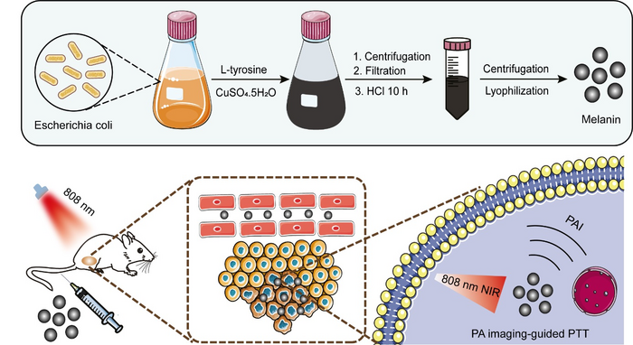
The researchers created the self-dyeing leather alternative by modifying the genes of a bacteria species that produces sheets of microbial cellulose – a strong, flexible and malleable material that is already commonly used in food, cosmetics and textiles. The genetic modifications ‘instructed’ the same microbes that were growing the material to also produce the dark black pigment, eumelanin.

They worked with designers to grow the upper part of a shoe (without the sole) by growing a sheet of bacterial cellulose in a bespoke, shoe-shaped vessel. After 14 days of growth wherein the cellulose took on the correct shape, they subjected the shoe to two days of gentle shaking at 30°C to activate the production of black pigment from the bacteria so that it dyed the material from the inside.

They also made a black wallet by growing two separate cellulose sheets, cutting them to size, and sewing them together.

As well as the prototypes, the researchers demonstrated that the bacteria can be engineered using genes from other microbes to produce colours in response to blue light. By projecting a pattern, or logo, onto the sheets using blue light, the bacteria respond by producing coloured proteins which then glow.

This allows them to project patterns and logos onto the bacterial cultures as the material grows, resulting in patterns and logos forming from within the material. 

Co-author Dr Kenneth Walker, who conducted the work at Imperial College London’s Department of Bioengineering and now works in industry, said: “Our technique works at large enough scales to create real-life products, as shown by our prototypes. From here, we can consider aesthetics as well as alternative shapes, patterns, textiles, and colours.

“The work also shows the impact that can happen when scientists and designers work together. As current and future users of new bacteria-grown textiles, designers have a key role in championing exciting new materials and giving expert feedback to improve form, function, and the switch to sustainable fashion.”

Greener clothes

The research team are now experimenting with a variety of coloured pigments to use those that can also be produced by the material-growing microbes.

The researchers and collaborators have also just won £2 million in funding from Biotechnology and Biological Sciences Research Council (BBSRC), part of UK Research and Innovation (UKRI), to use engineering biology and bacterial cellulose to solve more of fashion’s problems, such as the use of toxic chromium in leather’s production lines.

Professor Ellis said: “Microbes are already directly addressing many of the problems of animal and plastic-based leather, and we plan to get them ready to expand into new colours, materials and maybe patterns too.

“We look forward to working with the fashion industry to make the clothes we wear greener throughout the whole production line.”

The authors worked closely with Modern Synthesis, a London-based biodesign and materials company, who specialise in innovative microbial cellulose products.

This work was funded by Engineering and Physical Sciences Research Council and BBSRC, both part of UKRI.

Here’s a link to and a citation for the paper,

Self-pigmenting textiles grown from cellulose-producing bacteria with engineered tyrosinase expression by Kenneth T. Walker, Ivy S. Li, Jennifer Keane, Vivianne J. Goosens, Wenzhe Song, Koon-Yang Lee & Tom Ellis. Nature Biotechnology (2024) DOI: https://doi.org/10.1038/s41587-024-02194-3 Published: 02 April 2024

This paper is open access.

Modern Synthesis, the company with which the researchers collaborated, can be found here.

Synthetic human embryos—what now? (2 of 2)

The term they’re using in the Weizmann Institute of Science’s (Israel) announcement is “a generally accurate human embryo model.” This is in contrast to previous announcements including the one from the University of Cambridge team highlighted in Part 1.

From a September 6, 2023 news item on phys.org, Note: A link has been removed,

A research team headed by Prof. Jacob Hanna at the Weizmann Institute of Science has created complete models of human embryos from stem cells cultured in the lab—and managed to grow them outside the womb up to day 14. As reported today [September 6, 2023] in Nature, these synthetic embryo models had all the structures and compartments characteristic of this stage, including the placenta, yolk sac, chorionic sac and other external tissues that ensure the models’ dynamic and adequate growth.

Cellular aggregates derived from human stem cells in previous studies could not be considered genuinely accurate human embryo models, because they lacked nearly all the defining hallmarks of a post-implantation embryo. In particular, they failed to contain several cell types that are essential to the embryo’s development, such as those that form the placenta and the chorionic sac. In addition, they did not have the structural organization characteristic of the embryo and revealed no dynamic ability to progress to the next developmental stage.

Given their authentic complexity, the human embryo models obtained by Hanna’s group may provide an unprecedented opportunity to shed new light on the embryo’s mysterious beginnings. Little is known about the early embryo because it is so difficult to study, for both ethical and technical reasons, yet its initial stages are crucial to its future development. During these stages, the clump of cells that implants itself in the womb on the seventh day of its existence becomes, within three to four weeks, a well-structured embryo that already contains all the body organs.

“The drama is in the first month, the remaining eight months of pregnancy are mainly lots of growth,” Hanna says. “But that first month is still largely a black box. Our stem cell–derived human embryo model offers an ethical and accessible way of peering into this box. It closely mimics the development of a real human embryo, particularly the emergence of its exquisitely fine architecture.”

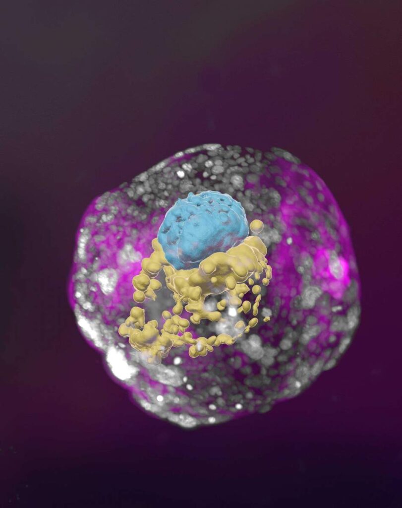
A stem cell–derived human embryo model at a developmental stage equivalent to that of a day 14 embryo. The model has all the compartments that define this stage: the yolk sac (yellow) and the part that will become the embryo itself, topped by the amnion (blue) – all enveloped by cells that will become the placenta (pink) Courtesy: Weizmann Institute of Science

A September 6, 2023 Weizmann Institute of Science press release, which originated the news item, offers a wealth of detail, Note: Links have been removed,

Letting the embryo model say “Go!”

Hanna’s team built on their previous experience in creating synthetic stem cell–based models of mouse embryos. As in that research, the scientists made no use of fertilized eggs or a womb. Rather, they started out with human cells known as pluripotent stem cells, which have the potential to differentiate into many, though not all, cell types. Some were derived from adult skin cells that had been reverted to “stemness.” Others were the progeny of human stem cell lines that had been cultured for years in the lab.

The researchers then used Hanna’s recently developed method to reprogram pluripotent stem cells so as to turn the clock further back: to revert these cells to an even earlier state – known as the naïve state – in which they are capable of becoming anything, that is, specializing into any type of cell. This stage corresponds to day 7 of the natural human embryo, around the time it implants itself in the womb. Hanna’s team had in fact been the first to start describing methods to generate human naïve stem cells, back in 2013; they continued to improve these methods, which stand at the heart of the current project, over the years.

The scientists divided the cells into three groups. The cells intended to develop into the embryo were left as is. The cells in each of the other groups were treated only with chemicals, without any need for genetic modification, so as to turn on certain genes, which was intended to cause these cells to differentiate toward one of three tissue types needed to sustain the embryo: placenta, yolk sac or the extraembryonic mesoderm membrane that ultimately creates the chorionic sac.

Soon after being mixed together under optimized, specifically developed conditions, the cells formed clumps, about 1 percent of which self-organized into complete embryo-like structures. “An embryo is self-driven by definition; we don’t need to tell it what to do – we must only unleash its internally encoded potential,” Hanna says. “It’s critical to mix in the right kinds of cells at the beginning, which can only be derived from naïve stem cells that have no developmental restrictions. Once you do that, the embryo-like model itself says, ‘Go!’”

The stem cell–based embryo-like structures (termed SEMs) developed normally outside the womb for 8 days, reaching a developmental stage equivalent to day 14 in human embryonic development. That’s the point at which natural embryos acquire the internal structures that enable them to proceed to the next stage: developing the progenitors of body organs.

Complete human embryo models match classic diagrams in terms of structure and cell identity

When the researchers compared the inner organization of their stem cell–derived embryo models with illustrations and microscopic anatomy sections in classical embryology atlases from the 1960s, they found an uncanny structural resemblance between the models and the natural human embryos at the corresponding stage. Every compartment and supporting structure was not only there, but in the right place, size and shape. Even the cells that make the hormone used in pregnancy testing were there and active: When the scientists applied secretions from these cells to a commercial pregnancy test, it came out positive.

In fact, the study has already produced a finding that may open a new direction of research into early pregnancy failure. The researchers discovered that if the embryo is not enveloped by placenta-forming cells in the right manner at day 3 of the protocol (corresponding to day 10 in natural embryonic development), its internal structures, such as the yolk sac, fail to properly develop.

“An embryo is not static. It must have the right cells in the right organization, and it must be able to progress – it’s about being and becoming,” Hanna says. “Our complete embryo models will help researchers address the most basic questions about what determines its proper growth.”

This ethical approach to unlocking the mysteries of the very first stages of embryonic development could open numerous research paths. It might help reveal the causes of many birth defects and types of infertility. It could also lead to new technologies for growing transplant tissues and organs. And it could offer a way around experiments that cannot be performed on live embryos – for example, determining the effects of exposure to drugs or other substances on fetal development.

For people who are visually inclined, there are two videos embedded in the September 6, 2023 Weizmann Institute of Science press release.

Here’s a link to and a citation for the paper,

Complete human day 14 post-implantation embryo models from naïve ES cells by Bernardo Oldak, Emilie Wildschutz, Vladyslav Bondarenko, Mehmet-Yunus Comar, Cheng Zhao, Alejandro Aguilera-Castrejon, Shadi Tarazi, Sergey Viukov, Thi Xuan Ai Pham, Shahd Ashouokhi, Dmitry Lokshtanov, Francesco Roncato, Eitan Ariel, Max Rose, Nir Livnat, Tom Shani, Carine Joubran, Roni Cohen, Yoseph Addadi, Muriel Chemla, Merav Kedmi, Hadas Keren-Shaul, Vincent Pasque, Sophie Petropoulos, Fredrik Lanner, Noa Novershtern & Jacob H. Hanna. Nature (2023) DOI: https://doi.org/10.1038/s41586-023-06604-5 Published: 06 September 2023

This paper is behind a paywall.

As for the question I asked in the head “what now?” I have absolutely no idea.

Synthetic human embryos—what now? (1 of 2)

Usually, there’s a rough chronological order to how I introduce the research, but this time I’m looking at the term used to describe it, following up with the various news releases and commentaries about the research, and finishing with a Canadian perspective.

After writing this post (but before it was published), the Weizmann Institute of Science (Israel) made their September 6, 2023 announcement and things changed a bit. That’s in Part two.

Say what you really mean (a terminology issue)

First, it might be useful to investigate the term, ‘synthetic human embryos’ as Julian Hitchcock does in his June 29, 2023 article on Bristows website (h/t Mondaq’s July 5, 2023 news item), Note: Links have been removed,

Synthetic Embryos” are neither Synthetic nor Embryos. So why are editors giving that name to stem cell-based models of human development?

One of the less convincing aspects of the last fortnight’s flurry of announcements about advances in simulating early human development (see here) concerned their name. Headlines galore (in newspapers and scientific journals) referred to “synthetic embryos“.

But embryo models, however impressive, are not embryos. To claim that the fundamental stages of embryo development that we learnt at school – fertilisation, cleavage and compaction – could now be bypassed to achieve the same result would be wrong. Nor are these objects “synthesised”: indeed, their interest to us lies in the ways in which they organise themselves. The researchers merely place the stem cells in a matrix in appropriate conditions, then stand back and watch them do it. Scientists were therefore unhappy about this use of the term in news media, and relieved when the International Society for Stem Cell Research (ISSCR) stepped in with a press release:

“Unlike some recent media reports describing this research, the ISSCR advises against using the term “synthetic embryo” to describe embryo models, because it is inaccurate and can create confusion. Integrated embryo models are neither synthetic nor embryos. While these models can replicate aspects of the early-stage development of human embryos, they cannot and will not develop to the equivalent of postnatal stage humans. Further, the ISSCR Guidelines prohibit the transfer of any embryo model to the uterus of a human or an animal.”

Although this was the ISSCR’s first attempt to put that position to the public, it had already made that recommendation to the research community two years previously. Its 2021 Guidelines for Stem Cell Research and Clinical Translation had recommended researchers to “promote accurate, current, balanced, and responsive public representations of stem cell research”. In particular:

“While organoids, chimeras, embryo models, and other stem cell-based models are useful research tools offering possibilities for further scientific progress, limitations on the current state of scientific knowledge and regulatory constraints must be clearly explained in any communications with the public or media. Suggestions that any of the current in vitro models can recapitulate an intact embryo, human sentience or integrated brain function are unfounded overstatements that should be avoided and contradicted with more precise characterizations of current understanding.”

Here’s a little bit about Hitchcock from his Bristows profile page,

  • Diploma Medical School, University of Birmingham (1975-78)
  • LLB, University of Wolverhampton
  • Diploma in Intellectual Property Law & Practice, University of Bristol
  • Qualified 1998

Following an education in medicine at the University of Birmingham and a career as a BBC science producer, Julian has focused on the law and regulation of life science technologies since 1997, practising in England and Australia. He joined Bristows with Alex Denoon in 2018.

Hitchcock’s June 29, 2023 article comments on why this term is being used,

I have a lot of sympathy with the position of the science writers and editors incurring the scientists’ ire. First, why should journalists have known of the ISSCR’s recommendations on the use of the term “synthetic embryo”? A journalist who found Recommendation 4.1 of the ISSCR Guidelines would probably not have found them specific enough to address the point, and the academic introduction containing the missing detail is hard to find. …

My second reason for being sympathetic to the use of the terrible term is that no suitable alternative has been provided, other than in the Stem Cell Reports paper, which recommends the umbrella terms “embryo models” or “stem cell based embryo models”. …

When asked why she had used the term “synthetic embryo”, the journalist I contacted remarked that, “We’re still working out the right language and it’s something we’re discussing and will no doubt evolve along with the science”.

It is absolutely in the public’s interest (and in the interest of science), that scientific research is explained in terms that the public understands. There is, therefore, a need, I think, for the scientific community to supply a name to the media or endure the penalties of misinformation …

In such an intensely competitive field of research, disagreement among researchers, even as to names, is inevitable. In consequence, however, journalists and their audiences are confronted by a slew of terms which may or may not be synonymous or overlapping, with no agreed term [emphasis mine] for the overall class of stem cell based embryo models. We cannot blame them if they make up snappy titles of their own [emphasis mine]. …

The announcement

The earliest date for the announcement at the International Society for Stem Cell Researh meeting that I can find is Hannah Devlin’s June 14, 2023 article in The Guardian newspaper, Note: A link has been removed,

Scientists have created synthetic human embryos using stem cells, in a groundbreaking advance that sidesteps the need for eggs or sperm.

Scientists say these model embryos, which resemble those in the earliest stages of human development, could provide a crucial window on the impact of genetic disorders and the biological causes of recurrent miscarriage.

However, the work also raises serious ethical and legal issues as the lab-grown entities fall outside current legislation in the UK and most other countries.

The structures do not have a beating heart or the beginnings of a brain, but include cells that would typically go on to form the placenta, yolk sac and the embryo itself.

Prof Magdalena Żernicka-Goetz, of the University of Cambridge and the California Institute of Technology, described the work in a plenary address on Wednesday [June 14, 2023] at the International Society for Stem Cell Research’s annual meeting in Boston.

The (UK) Science Media Centre made expert comments available in a June 14, 2023 posting “expert reaction to Guardian reporting news of creation of synthetic embryos using stem cells.”

Two days later, this June 16, 2023 essay by Kathryn MacKay, Senior Lecturer in Bioethics, University of Sydney (Australia), appeared on The Conversation (h/t June 16, 2023 news item on phys.org), Note: Links have been removed,

Researchers have created synthetic human embryos using stem cells, according to media reports. Remarkably, these embryos have reportedly been created from embryonic stem cells, meaning they do not require sperm and ova.

This development, widely described as a breakthrough that could help scientists learn more about human development and genetic disorders, was revealed this week in Boston at the annual meeting of the International Society for Stem Cell Research.

The research, announced by Professor Magdalena Żernicka-Goetz of the University of Cambridge and the California Institute of Technology, has not yet been published in a peer-reviewed journal. But Żernicka-Goetz told the meeting these human-like embryos had been made by reprogramming human embryonic stem cells.

So what does all this mean for science, and what ethical issues does it present?

MacKay goes on to answer her own questions, from the June 16, 2023 essay, Note: A link has been removed,

One of these quandaries arises around whether their creation really gets us away from the use of human embryos.

Robin Lovell-Badge, the head of stem cell biology and developmental genetics at the Francis Crick Institute in London UK, reportedly said that if these human-like embryos can really model human development in the early stages of pregnancy, then we will not have to use human embryos for research.

At the moment, it is unclear if this is the case for two reasons.

First, the embryos were created from human embryonic stem cells, so it seems they do still need human embryos for their creation. Perhaps more light will be shed on this when Żernicka-Goetz’s research is published.

Second, there are questions about the extent to which these human-like embryos really can model human development.

Professor Magdalena Żernicka-Goetz’s research is published

Almost two weeks later the research from the Cambridge team (there are other teams and countries also racing; see Part two for the news from Sept. 6, 2023) was published, from a June 27, 2023 news item on ScienceDaily,

Cambridge scientists have created a stem cell-derived model of the human embryo in the lab by reprogramming human stem cells. The breakthrough could help research into genetic disorders and in understanding why and how pregnancies fail.

Published today [Tuesday, June 27, 2023] in the journal Nature, this embryo model is an organised three-dimensional structure derived from pluripotent stem cells that replicate some developmental processes that occur in early human embryos.

Use of such models allows experimental modelling of embryonic development during the second week of pregnancy. They can help researchers gain basic knowledge of the developmental origins of organs and specialised cells such as sperm and eggs, and facilitate understanding of early pregnancy loss.

A June 27, 2023 University of Cambridge press release (also on EurekAlert), which originated the news item, provides more detail about the work,

“Our human embryo-like model, created entirely from human stem cells, gives us access to the developing structure at a stage that is normally hidden from us due to the implantation of the tiny embryo into the mother’s womb,” said Professor Magdalena Zernicka-Goetz in the University of Cambridge’s Department of Physiology, Development and Neuroscience, who led the work.

She added: “This exciting development allows us to manipulate genes to understand their developmental roles in a model system. This will let us test the function of specific factors, which is difficult to do in the natural embryo.”

In natural human development, the second week of development is an important time when the embryo implants into the uterus. This is the time when many pregnancies are lost.

The new advance enables scientists to peer into the mysterious ‘black box’ period of human development – usually following implantation of the embryo in the uterus – to observe processes never directly observed before.

Understanding these early developmental processes holds the potential to reveal some of the causes of human birth defects and diseases, and to develop tests for these in pregnant women.

Until now, the processes could only be observed in animal models, using cells from zebrafish and mice, for example.

Legal restrictions in the UK currently prevent the culture of natural human embryos in the lab beyond day 14 of development: this time limit was set to correspond to the stage where the embryo can no longer form a twin. [emphasis mine]

Until now, scientists have only been able to study this period of human development using donated human embryos. This advance could reduce the need for donated human embryos in research.

Zernicka-Goetz says the while these models can mimic aspects of the development of human embryos, they cannot and will not develop to the equivalent of postnatal stage humans.

Over the past decade, Zernicka-Goetz’s group in Cambridge has been studying the earliest stages of pregnancy, in order to understand why some pregnancies fail and some succeed.

In 2021 and then in 2022 her team announced in Developmental Cell, Nature and Cell Stem Cell journals that they had finally created model embryos from mouse stem cells that can develop to form a brain-like structure, a beating heart, and the foundations of all other organs of the body.

The new models derived from human stem cells do not have a brain or beating heart, but they include cells that would typically go on to form the embryo, placenta and yolk sac, and develop to form the precursors of germ cells (that will form sperm and eggs).

Many pregnancies fail at the point when these three types of cells orchestrate implantation into the uterus begin to send mechanical and chemical signals to each other, which tell the embryo how to develop properly.

There are clear regulations governing stem cell-based models of human embryos and all researchers doing embryo modelling work must first be approved by ethics committees. Journals require proof of this ethics review before they accept scientific papers for publication. Zernicka-Goetz’s laboratory holds these approvals.

“It is against the law and FDA regulations to transfer any embryo-like models into a woman for reproductive aims. These are highly manipulated human cells and their attempted reproductive use would be extremely dangerous,” said Dr Insoo Hyun, Director of the Center for Life Sciences and Public Learning at Boston’s Museum of Science and a member of Harvard Medical School’s Center for Bioethics.

Zernicka-Goetz also holds position at the California Institute of Technology and is NOMIS Distinguished Scientist and Scholar Awardee.

The research was funded by the Wellcome Trust and Open Philanthropy.

(There’s more about legal concerns further down in this post.)

Here’s a link to and a citation for the paper,

Pluripotent stem cell-derived model of the post-implantation human embryo by Bailey A. T. Weatherbee, Carlos W. Gantner, Lisa K. Iwamoto-Stohl, Riza M. Daza, Nobuhiko Hamazaki, Jay Shendure & Magdalena Zernicka-Goetz. Nature (2023) DOI: https://doi.org/10.1038/s41586-023-06368-y Published: 27 June 2023

This paper is open access.

Published the same day (June 27, 2023) is a paper (citation and link follow) also focused on studying human embryonic development using stem cells. First, there’s this from the Abstract,

Investigating human development is a substantial scientific challenge due to the technical and ethical limitations of working with embryonic samples. In the face of these difficulties, stem cells have provided an alternative to experimentally model inaccessible stages of human development in vitro …

This time the work is from a US/German team,

Self-patterning of human stem cells into post-implantation lineages by Monique Pedroza, Seher Ipek Gassaloglu, Nicolas Dias, Liangwen Zhong, Tien-Chi Jason Hou, Helene Kretzmer, Zachary D. Smith & Berna Sozen. Nature (2023) DOI: https://doi.org/10.1038/s41586-023-06354-4 Published: 27 June 2023

The paper is open access.

Legal concerns and a Canadian focus

A July 25, 2023 essay by Françoise Baylis and Jocelyn Downie of Dalhousie University (Nova Scotia, Canada) for The Conversation (h/t July 25, 2023 article on phys.org) covers the advantages of doing this work before launching into a discussion of legislation and limits in the UK and, more extensively, in Canada, Note: Links have been removed,

This research could increase our understanding of human development and genetic disorders, help us learn how to prevent early miscarriages, lead to improvements in fertility treatment, and — perhaps — eventually allow for reproduction without using sperm and eggs.

Synthetic human embryos — also called embryoid bodies, embryo-like structures or embryo models — mimic the development of “natural human embryos,” those created by fertilization. Synthetic human embryos include the “cells that would typically go on to form the embryo, placenta and yolk sac, and develop to form the precursors of germ cells (that will form sperm and eggs).”

Though research involving natural human embryos is legal in many jurisdictions, it remains controversial. For some people, research involving synthetic human embryos is less controversial because these embryos cannot “develop to the equivalent of postnatal stage humans.” In other words, these embryos are non-viable and cannot result in live births.

Now, for a closer look at the legalities in the UK and in Canada, from the July 25, 2023 essay, Note: Links have been removed,

The research presented by Żernicka-Goetz at the ISSCR meeting took place in the United Kingdom. It was conducted in accordance with the Human Fertilization and Embryology Act, 1990, with the approval of the U.K. Stem Cell Bank Steering Committee.

U.K. law limits the research use of human embryos to 14 days of development. An embryo is defined as “a live human embryo where fertilisation is complete, and references to an embryo include an egg in the process of fertilisation.”

Synthetic embryos are not created by fertilization and therefore, by definition, the 14-day limit on human embryo research does not apply to them. This means that synthetic human embryo research beyond 14 days can proceed in the U.K.

The door to the touted potential benefits — and ethical controversies — seems wide open in the U.K.

While the law in the U.K. does not apply to synthetic human embryos, the law in Canada clearly does. This is because the legal definition of an embryo in Canada is not limited to embryos created by fertilization [emphasis mine].

The Assisted Human Reproduction Act (the AHR Act) defines an embryo as “a human organism during the first 56 days of its development following fertilization or creation, excluding any time during which its development has been suspended.”

Based on this definition, the AHR Act applies to embryos created by reprogramming human embryonic stem cells — in other words, synthetic human embryos — provided such embryos qualify as human organisms.

A synthetic human embryo is a human organism. It is of the species Homo sapiens, and is thus human. It also qualifies as an organism — a life form — alongside other organisms created by means of fertilization, asexual reproduction, parthenogenesis or cloning.

Given that the AHR Act applies to synthetic human embryos, there are legal limits on their creation and use in Canada.

First, human embryos — including synthetic human embryos – can only be created for the purposes of “creating a human being, improving or providing instruction in assisted reproduction procedures.”

Given the state of the science, it follows that synthetic human embryos could legally be created for the purpose of improving assisted reproduction procedures.

Second, “spare” or “excess” human embryos — including synthetic human embryos — originally created for one of the permitted purposes, but no longer wanted for this purpose, can be used for research. This research must be done in accordance with the consent regulations which specify that consent must be for a “specific research project.”

Finally, all research involving human embryos — including synthetic human embryos — is subject to the 14-day rule. The law stipulates that: “No person shall knowingly… maintain an embryo outside the body of a female person after the fourteenth day of its development following fertilization or creation, excluding any time during which its development has been suspended.”

Putting this all together, the creation of synthetic embryos for improving assisted human reproduction procedures is permitted, as is research using “spare” or “excess” synthetic embryos originally created for this purpose — provided there is specific consent and the research does not exceed 14 days.

This means that while synthetic human embryos may be useful for limited research on pre-implantation embryo development, they are not available in Canada for research on post-implantation embryo development beyond 14 days.

The authors close with this comment about the prospects for expanding Canada’s14-day limit, from the July 25, 2023 essay,

… any argument will have to overcome the political reality that the federal government is unlikely to open up the Pandora’s box of amending the AHR Act.

It therefore seems likely that synthetic human embryo research will remain limited in Canada for the foreseeable future.

As mentioned, in September 2023 there was a new development. See: Part two.