Tag Archives: George G. Malliaras

‘Jelly’ batteries

Caption: Researchers have developed soft, stretchable ‘jelly batteries’ that could be used for wearable devices or soft robotics, or even implanted in the brain to deliver drugs or treat conditions such as epilepsy. Credit: University of Cambridge

A July 18, 2024 news item on Nanowerk announces bioinspried stretchy batteries from the University of Cambridge,

Researchers have developed soft, stretchable ‘jelly batteries’ that could be used for wearable devices or soft robotics, or even implanted in the brain to deliver drugs or treat conditions such as epilepsy.

The researchers, from the University of Cambridge, took their inspiration from electric eels, which stun their prey with modified muscle cells called electrocytes.

Like electrocytes, the jelly-like materials developed by the Cambridge researchers have a layered structure, like sticky Lego, that makes them capable of delivering an electric current.

A July 17, 2024 University of Cambridge press release (also on EurekAlert), which originated the news item, offers more details,

The self-healing jelly batteries can stretch to over ten times their original length without affecting their conductivity – the first time that such stretchability and conductivity has been combined in a single material. The results are reported in the journal Science Advances.

The jelly batteries are made from hydrogels: 3D networks of polymers that contain over 60% water. The polymers are held together by reversible on/off interactions that control the jelly’s mechanical properties.

The ability to precisely control mechanical properties and mimic the characteristics of human tissue makes hydrogels ideal candidates for soft robotics and bioelectronics; however, they need to be both conductive and stretchy for such applications.

“It’s difficult to design a material that is both highly stretchable and highly conductive, since those two properties are normally at odds with one another,” said first author Stephen O’Neill, from Cambridge’s Yusuf Hamied Department of Chemistry. “Typically, conductivity decreases when a material is stretched.”

“Normally, hydrogels are made of polymers that have a neutral charge, but if we charge them, they can become conductive,” said co-author Dr Jade McCune, also from the Department of Chemistry. “And by changing the salt component of each gel, we can make them sticky and squish them together in multiple layers, so we can build up a larger energy potential.”

Conventional electronics use rigid metallic materials with electrons as charge carriers, while the jelly batteries use ions to carry charge, like electric eels.

The hydrogels stick strongly to each other because of reversible bonds that can form between the different layers, using barrel-shaped molecules called cucurbiturils that are like molecular handcuffs. The strong adhesion between layers provided by the molecular handcuffs allows for the jelly batteries to be stretched, without the layers coming apart and crucially, without any loss of conductivity.

The properties of the jelly batteries make them promising for future use in biomedical implants, since they are soft and mould to human tissue. “We can customise the mechanical properties of the hydrogels so they match human tissue,” said Professor Oren Scherman, Director of the Melville Laboratory for Polymer Synthesis, who led the research in collaboration with Professor George Malliaras from the Department of Engineering. “Since they contain no rigid components such as metal, a hydrogel implant would be much less likely to be rejected by the body or cause the build-up of scar tissue.”

In addition to their softness, the hydrogels are also surprisingly tough. They can withstand being squashed without permanently losing their original shape, and can self-heal when damaged.

The researchers are planning future experiments to test the hydrogels in living organisms to assess their suitability for a range of medical applications.

The research was funded by the European Research Council and the Engineering and Physical Sciences Research Council (EPSRC), part of UK Research and Innovation (UKRI). Oren Scherman is a Fellow of Jesus College, Cambridge.

Here’s a link to and a citation for the paper,

Highly stretchable dynamic hydrogels for soft multilayer electronics by Stephen J. K. O’Neill, Zehuan Huang, Xiaoyi Chen, Renata L. Sala, Jade A. McCune, George G. Malliaras, and Oren A. Scherman. Science Advances 17 Jul 2024 Vol 10, Issue 29 DOI: 10.1126/sciadv.adn5142

This paper appears to be open access.

Biohybrid device (a new type of neural implant) could restore limb function

A March 23, 2023 news item on ScienceDaily announces a neural implant that addresses failures due to scarring issues,

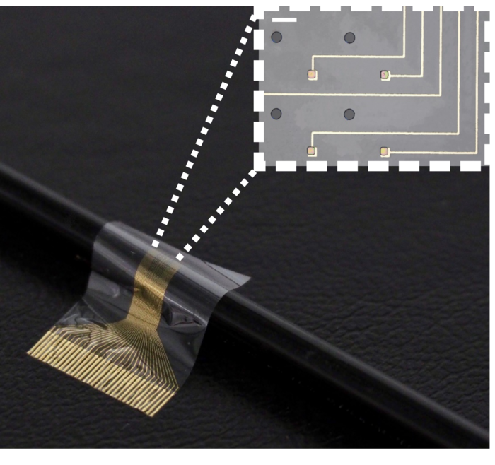
Researchers have developed a new type of neural implant that could restore limb function to amputees and others who have lost the use of their arms or legs.

In a study carried out in rats, researchers from the University of Cambridge used the device to improve the connection between the brain and paralysed limbs. The device combines flexible electronics and human stem cells — the body’s ‘reprogrammable’ master cells — to better integrate with the nerve and drive limb function.

Previous attempts at using neural implants to restore limb function have mostly failed, as scar tissue tends to form around the electrodes over time, impeding the connection between the device and the nerve. By sandwiching a layer of muscle cells reprogrammed from stem cells between the electrodes and the living tissue, the researchers found that the device integrated with the host’s body and the formation of scar tissue was prevented. The cells survived on the electrode for the duration of the 28-day experiment, the first time this has been monitored over such a long period.

A March 22, 2023 University of Cambridge press release (also on EurekAlert but published March 23, 2023) by Sarah Collins, delves further into the topic,

The researchers say that by combining two advanced therapies for nerve regeneration – cell therapy and bioelectronics – into a single device, they can overcome the shortcomings of both approaches, improving functionality and sensitivity.

While extensive research and testing will be needed before it can be used in humans, the device is a promising development for amputees or those who have lost function of a limb or limbs. The results are reported in the journal Science Advances.

A huge challenge when attempting to reverse injuries that result in the loss of a limb or the loss of function of a limb is the inability of neurons to regenerate and rebuild disrupted neural circuits.

“If someone has an arm or a leg amputated, for example, all the signals in the nervous system are still there, even though the physical limb is gone,” said Dr Damiano Barone from Cambridge’s Department of Clinical Neurosciences, who co-led the research. “The challenge with integrating artificial limbs, or restoring function to arms or legs, is extracting the information from the nerve and getting it to the limb so that function is restored.”

One way of addressing this problem is implanting a nerve in the large muscles of the shoulder and attaching electrodes to it. The problem with this approach is scar tissue forms around the electrode, plus it is only possible to extract surface-level information from the electrode.

To get better resolution, any implant for restoring function would need to extract much more information from the electrodes. And to improve sensitivity, the researchers wanted to design something that could work on the scale of a single nerve fibre, or axon.

“An axon itself has a tiny voltage,” said Barone. “But once it connects with a muscle cell, which has a much higher voltage, the signal from the muscle cell is easier to extract. That’s where you can increase the sensitivity of the implant.”

The researchers designed a biocompatible flexible electronic device that is thin enough to be attached to the end of a nerve. A layer of stem cells, reprogrammed into muscle cells, was then placed on the electrode. This is the first time that this type of stem cell, called an induced pluripotent stem cell, has been used in a living organism in this way.

“These cells give us an enormous degree of control,” said Barone. “We can tell them how to behave and check on them throughout the experiment. By putting cells in between the electronics and the living body, the body doesn’t see the electrodes, it just sees the cells, so scar tissue isn’t generated.”

The Cambridge biohybrid device was implanted into the paralysed forearm of the rats. The stem cells, which had been transformed into muscle cells prior to implantation, integrated with the nerves in the rat’s forearm. While the rats did not have movement restored to their forearms, the device was able to pick up the signals from the brain that control movement. If connected to the rest of the nerve or a prosthetic limb, the device could help restore movement.

The cell layer also improved the function of the device, by improving resolution and allowing long-term monitoring inside a living organism. The cells survived through the 28-day experiment: the first time that cells have been shown to survive an extended experiment of this kind.

The researchers say that their approach has multiple advantages over other attempts to restore function in amputees. In addition to its easier integration and long-term stability, the device is small enough that its implantation would only require keyhole surgery. Other neural interfacing technologies for the restoration of function in amputees require complex patient-specific interpretations of cortical activity to be associated with muscle movements, while the Cambridge-developed device is a highly scalable solution since it uses ‘off the shelf’ cells.

In addition to its potential for the restoration of function in people who have lost the use of a limb or limbs, the researchers say their device could also be used to control prosthetic limbs by interacting with specific axons responsible for motor control.

“This interface could revolutionise the way we interact with technology,” said co-first author Amy Rochford, from the Department of Engineering. “By combining living human cells with bioelectronic materials, we’ve created a system that can communicate with the brain in a more natural and intuitive way, opening up new possibilities for prosthetics, brain-machine interfaces, and even enhancing cognitive abilities.”

“This technology represents an exciting new approach to neural implants, which we hope will unlock new treatments for patients in need,” said co-first author Dr Alejandro Carnicer-Lombarte, also from the Department of Engineering.

“This was a high-risk endeavour, and I’m so pleased that it worked,” said Professor George Malliaras from Cambridge’s Department of Engineering, who co-led the research. “It’s one of those things that you don’t know whether it will take two years or ten before it works, and it ended up happening very efficiently.”

The researchers are now working to further optimise the devices and improve their scalability. The team have filed a patent application on the technology with the support of Cambridge Enterprise, the University’s technology transfer arm.

The technology relies on opti-oxTM enabled muscle cells. opti-ox is a precision cellular reprogramming technology that enables faithful execution of genetic programmes in cells allowing them to be manufactured consistently at scale. The opti-ox enabled muscle iPSC cell lines used in the experiment were supplied by the Kotter lab [Mark Kotter] from the University of Cambridge. The opti-ox reprogramming technology is owned by synthetic biology company bit.bio.

The research was supported in part by the Engineering and Physical Sciences Research Council (EPSRC), part of UK Research and Innovation (UKRI), Wellcome, and the European Union’s Horizon 2020 Research and Innovation Programme.
Caption: In a study carried out in rats, researchers from the University of Cambridge used a biohybrid device to improve the connection between the brain and paralysed limbs. The device combines flexible electronics and human stem cells – the body’s ‘reprogrammable’ master cells – to better integrate with the nerve and drive limb function. Credit: University of Cambirdge

Here’s a link to and a citation for the paper,

Functional neurological restoration of amputated peripheral nerve using biohybrid regenerative bioelectronics by Amy E. Rochford, Alejandro Carnicer-Lombarte, Malak Kawan, Amy Jin, Sam Hilton, Vincenzo F. Curto, Alexandra L. Rutz, Thomas Moreau, Mark R. N. Kotter, George G. Malliaras, and Damiano G. Barone. Science Advances 22 Mar 2023 Vol 9, Issue 12 DOI: 10.1126/sciadv.add8162

This paper is open access.

The synthetic biology company mentioned in the press release, bit.bio is here

Improving neuromorphic devices with ion conducting polymer

A July 1, 2020 news item on ScienceDaily announces work which researchers are hopeful will allow them exert more control over neuromorphic devices’ speed of response,

“Neuromorphic” refers to mimicking the behavior of brain neural cells. When one speaks of neuromorphic computers, they are talking about making computers think and process more like human brains-operating at high-speed with low energy consumption.

Despite a growing interest in polymer-based neuromorphic devices, researchers have yet to establish an effective method for controlling the response speed of devices. Researchers from Tohoku University and the University of Cambridge, however, have overcome this obstacle through mixing the polymers PSS-Na and PEDOT:PSS, discovering that adding an ion conducting polymer enhances neuromorphic device response time.

A June 24, 2020 Tohoku University press release (also on EurekAlert), which originated the news item, provides a few more technical details,

Polymers are materials composed of long molecular chains and play a fundamental aspect in modern life from the rubber in tires, to water bottles, to polystyrene. Mixing polymers together results in the creation of new materials with their own distinct physical properties.

Most studies on neuromorphic devices based on polymer focus exclusively on the application of PEDOT: PSS, a mixed conductor that transports both electrons and ions. PSS-Na, on the other hand, transports ions only. By blending these two polymers, the researchers could enhance the ion diffusivity in the active layer of the device. Their measurements confirmed an increase in device response time, achieving a 5-time shorting at maximum. The results also proved how closely related response time is to the diffusivity of ions in the active layer.

“Our study paves the way for a deeper understanding behind the science of conducting polymers.” explains co-author Shunsuke Yamamoto from the Department of Biomolecular Engineering at Tohoku University’s Graduate School of Engineering. “Moving forward, it may be possible to create artificial neural networks composed of multiple neuromorphic devices,” he adds.

Here’s a link to and a citation for the paper,

Controlling the Neuromorphic Behavior of Organic Electrochemical Transistors by Blending Mixed and Ion Conductors by Shunsuke Yamamoto and George G. Malliaras. ACS [American Chemical Society] Appl. Electron. Mater. 2020, XXXX, XXX, XXX-XXX DOI: https://doi.org/10.1021/acsaelm.0c00203 Publication Date:June 15, 2020 Copyright © 2020 American Chemical Society

This paper is behind a paywall.

Electronic organic micropump for direct drug delivery to the brain

I can understand the appeal but have some questions about this micropump in the brain concept. First, here’s more about the research from an April 16, 2015 news item on Nanowerk,

Many potentially efficient drugs have been created to treat neurological disorders, but they cannot be used in practice. Typically, for a condition such as epilepsy, it is essential to act at exactly the right time and place in the brain. For this reason, the team of researchers led by Christophe Bernard at Inserm Unit 1106, “Institute of Systems Neuroscience” (INS), with the help of scientists at the École des Mines de Saint-Étienne and Linköping University (Sweden) have developed an organic electronic micropump which, when combined with an anticonvulsant drug, enables localised inhibition of epileptic seizure in brain tissue in vitro.

An April 16, 2015 INSERM (Institut national de la santé et de la recherche médicale) press release on EurekAlert, which originated the news item, goes on to describe the problem the researchers are attempting to solve and their solution to it,

Drugs constitute the most widely used approach for treating brain disorders. However, many promising drugs failed during clinical testing for several reasons:

  • they are diluted in potentially toxic solutions,
  • they may themselves be toxic when they reach organs to which they were not initially directed,
  • the blood-brain barrier, which separates the brain from the blood circulation, prevents most drugs from reaching their targets in the brain,
  • drugs that succeed in penetrating the brain will act in a non-specific manner, i.e. on healthy regions of the brain, altering their functions.

Epilepsy is a typical example of a condition for which many drugs could not be commercialised because of their harmful effects, when they might have been effective for treating patients resistant to conventional treatments [1].

During an epileptic seizure, the nerve cells in a specific area of the brain are suddenly activated in an excessive manner. How can this phenomenon be controlled without affecting healthy brain regions? To answer this question, Christophe Bernard’s team, in collaboration with a team led by George Malliaras at the Georges Charpak-Provence Campus of the École des Mines of Saint-Étienne and Swedish scientists led by Magnus Berggren from Linköping University, have developed a biocompatible micropump that makes it possible to deliver therapeutic substances directly to the relevant areas of the brain.

The micropump (20 times thinner than a hair) is composed of a membrane known as “cation exchange,” i.e., it has negative ions attached to its surface. It thus attracts small positively charged molecules, whether these are ions or drugs. When an electrical current is applied to it, the flow of electrons generated projects the molecules of interest toward the target area.

To enable validation of this new technique, the researchers reproduced the hyperexcitability of epileptic neurons in mouse brains in vitro. They then injected GABA, a compound naturally produced in the brain and that inhibits neurons, into this hyperactive region using the micropump. The scientists then observed that the compound not only stopped this abnormal activity in the target region, but, most importantly, did not interfere with the functioning of the neighbouring regions.

This technology may thus resolve all the above-mentioned problems, by allowing very localised action, directly in the brain and without peripheral toxicity.

“By combining electrodes, such as those used to treat Parkinson’s disease, with this micropump, it may be possible to use this technology to treat patients with epilepsy who are resistant to conventional treatments, and those for whom the side-effects are too great,” explains Christophe Bernard, Inserm Research Director.

Based on these initial results, the researchers are now working to move on to an in vivo animal model and the possibility of combining this high-technology system with the microchip they previously developed in 2013. The device could be embedded and autonomous. The chip would be used to detect the imminent occurrence of a seizure, in order to activate the pump to inject the drug at just the right moment. It may therefore be possible to control brain activity where and when it is needed.

In addition to epilepsy, this state-of-the-art technology, combined with existing drugs, offers new opportunities for many brain diseases that remain difficult to treat at this time.

###

[1] Epilepsy in brief

This disease, which affects nearly 50 million people in the world, is the most common neurological disorder after migraine.

The neuronal dysfunctions associated with epilepsy lead to attacks with variable symptoms, from loss of consciousness to disorders of movement, sensation or mood.

Despite advances in medicine, 30% of those affected are resistant to all treatments.

Here’s a link to and a citation for the paper,

Controlling Epileptiform Activity with Organic Electronic Ion Pumps by Adam Williamson, Jonathan Rivnay, Loïg Kergoat, Amanda Jonsson, Sahika Inal, Ilke Uguz, Marc Ferro, Anton Ivanov, Theresia Arbring-Sjöström, Daniel T. Simon, Magnus Berggren, George G. Malliaras, and Christophe Bernardi. Advanced Materials First published: 11 April 2015Full publication history DOI: 10.1002/adma.201500482

This paper is behind a paywall.

Finally, my questions. How does the pump get refilled once the drugs are used up? Do you get a warning when the drug supply is almost nil? How does that warning work? Does implanting the pump require brain surgery or is there a less intrusive fashion of placing this pump exactly where you want it to be? Once it’s been implanted, how do you find a pump  20 times thinner than a human hair?

For some reason this micropump brought back memories of working in high tech environments where developers would come up with all kinds of nifty ideas but put absolutely no thought into how these ideas might actually work once human human beings got their hands on the product. In any event, the micropump seems exciting and I hope researchers work out the kinks, implementationwise, before they’re implanted.