I love the fact that ‘frozen smoke’ is another term for aerogel (which has multiple alternative terms) and the latest work on this interesting material is from the University of Cambridge (UK) according to a February 9, 2023 news item on ScienceDaily,
Researchers have developed a sensor made from ‘frozen smoke’ that uses artificial intelligence techniques to detect formaldehyde in real time at concentrations as low as eight parts per billion, far beyond the sensitivity of most indoor air quality sensors.
The researchers, from the University of Cambridge, developed sensors made from highly porous materials known as aerogels. By precisely engineering the shape of the holes in the aerogels, the sensors were able to detect the fingerprint of formaldehyde, a common indoor air pollutant, at room temperature.
The proof-of-concept sensors, which require minimal power, could be adapted to detect a wide range of hazardous gases, and could also be miniaturised for wearable and healthcare applications. The results are reported in the journal Science Advances.
Volatile organic compounds (VOCs) are a major source of indoor air pollution, causing watery eyes, burning in the eyes and throat, and difficulty breathing at elevated levels. High concentrations can trigger attacks in people with asthma, and prolonged exposure may cause certain cancers.
Formaldehyde is a common VOC and is emitted by household items including pressed wood products (such as MDF), wallpapers and paints, and some synthetic fabrics. For the most part, the levels of formaldehyde emitted by these items are low, but levels can build up over time, especially in garages where paints and other formaldehyde-emitting products are more likely to be stored.
According to a 2019 report from the campaign group Clean Air Day, a fifth of households in the UK showed notable concentrations of formaldehyde, with 13% of residences surpassing the recommended limit set by the World Health Organization (WHO).
“VOCs such as formaldehyde can lead to serious health problems with prolonged exposure even at low concentrations, but current sensors don’t have the sensitivity or selectivity to distinguish between VOCs that have different impacts on health,” said Professor Tawfique Hasan from the Cambridge Graphene Centre, who led the research.
“We wanted to develop a sensor that is small and doesn’t use much power, but can selectively detect formaldehyde at low concentrations,” said Zhuo Chen, the paper’s first author.
The researchers based their sensors on aerogels: ultra-light materials sometimes referred to as ‘liquid smoke’, since they are more than 99% air by volume. The open structure of aerogels allows gases to easily move in and out. By precisely engineering the shape, or morphology, of the holes, the aerogels can act as highly effective sensors.
Working with colleagues at Warwick University, the Cambridge researchers optimised the composition and structure of the aerogels to increase their sensitivity to formaldehyde, making them into filaments about three times the width of a human hair. The researchers 3D printed lines of a paste made from graphene, a two-dimensional form of carbon, and then freeze-dried the graphene paste to form the holes in the final aerogel structure. The aerogels also incorporate tiny semiconductors known as quantum dots.
The sensors they developed were able to detect formaldehyde at concentrations as low as eight parts per billion, which is 0.4 percent of the level deemed safe in UK workplaces. The sensors also work at room temperature, consuming very low power.
“Traditional gas sensors need to be heated up, but because of the way we’ve engineered the materials, our sensors work incredibly well at room temperature, so they use between 10 and 100 times less power than other sensors,” said Chen.
To improve selectivity, the researchers then incorporated machine learning algorithms into the sensors. The algorithms were trained to detect the ‘fingerprint’ of different gases, so that the sensor was able to distinguish the fingerprint of formaldehyde from other VOCs.
“Existing VOC detectors are blunt instruments – you only get one number for the overall concentration in the air,” said Hasan. “By building a sensor that is able to detect specific VOCs at very low concentrations in real time, it can give home and business owners a more accurate picture of air quality and any potential health risks.”
The researchers say that the same technique could be used to develop sensors to detect other VOCs. In theory, a device the size of a standard household carbon monoxide detector could incorporate multiple different sensors within it, providing real-time information about a range of different hazardous gases. The team at Warwick are developing a low-cost multi-sensor platform that will incorporate these new aerogel materials and, coupled with AI algorithms, detect different VOCs.
“By using highly porous materials as the sensing element, we’re opening up whole new ways of detecting hazardous materials in our environment,” said Chen.
The research was supported in part by the Henry Royce Institute, and the Engineering and Physical Sciences Research Council (EPSRC), part of UK Research and Innovation (UKRI). Tawfique Hasan is a Fellow of Churchill College, Cambridge.
This work is not about drinking tea with silver nanoparticles in it or ingesting colloidal silver by any means, a dangerous practice as Nicole Karlis’s January 7, 2024 article for Salon highlights, Note: Links have been removed,
The HBO docuseries “Love Has Won: The Cult of Mother God” begins with a jarring image. The corpse of the cult leader, Amy Carlson, laying in a bed, wrapped in blankets and string lights. She is noticeably gaunt and her face is a very blue color. When Carlson died in 2021 at the age of 45, a coroner’s report deemed her cause of death to be “alcohol abuse, anorexia and chronic colloidal silver ingestion.”
…
Most medical experts advise against ingesting silver — especially in large amounts. That’s because too much of it can build up in a person’s body and lead to argyria, which is the condition that Carlson and Stan Jones both had that turned them a blue. While argyria alone isn’t a serious health condition, it doesn’t go away when a person stops ingesting silver. Plus, too much silver can be fatal. [emphasis mine]
…
A November 17, 2023 news item on phys.org announced research from the Polish Academy of Sciences into improving antimicrobial activity, Note: A link has been removed,
Researchers at the Institute of Physical Chemistry of the Polish Academy of Sciences (IPC PAS) have demonstrated that green tea–silver nanoparticles as a powerful tool against pathogens such as bacteria and yeast. Their work is published in Nanoscale Advances.
Once upon a time, people believed to be invincible against bacterial diseases, thanks to the antibiotics. Does this sound like a fairy tale? By all means! Nothing could be further from the truth. Despite widespread access to antibiotic therapy, many lives are lost due to pathogens invisible to the eye. The ability to develop drugs that can combat resistant strains of bacteria has not kept pace with the spread of resistance. So far, innovations to defeat antimicrobial-resistant strains of bacteria are in high demand. Recently, researchers at the Institute of Physical Chemistry of the Polish Academy of Sciences (IPC PAS) demonstrated green tea-silver nanoparticles as a powerful tool against pathogens such as bacteria and yeast. Their goal was to develop an efficient method to combat bacteria that are otherwise unaffected by antimicrobial agents, such as antibiotics.
Following the discovery of antibiotics, there came a change in the curse of mankind by accelerating the development of medicine and extending human life expectancy. Their successful implementation led to the rapid development of pharmacy, providing more and more drugs against many pathogens. Nevertheless, the overuse of antibiotics has led to the emergence of resistance to these compounds, becoming one of the biggest health threats worldwide. As a result, antibiotic resistance has emerged faster than the advancement of antibiotics . The appearance of new drugs on the horizon to combat these pathogens is a short-lasting spark. Even if we seem to be on the losing end, there is still a chance to defeat an invisible enemy.
This hitch was researched by the team of scientists from the IPC PAS under the supervision of Prof. Jan Paczesny, who proposed new nanoformulations for use against widespread and challenging pathogens such as ESKAPE bacteria (Enterococcus faecium, Staphylococcus aureus, Klebsiella pneumoniae, Acinetobacter baumannii, Pseudomonas aeruginosa and Enterobacter spp.) and other problematic yeast pathogens such as Candida auris or Cryptococcus neoformans. These microorganisms, treated with commercially available antibiotics, rapidly develop antibiotic resistance. Researchers chose ESKAPE as the target group since these pathogens lead to serious diseases, from sepsis to even cancer. How? This is where the story begins.
A few months ago, Paczesny’s team decided to try combining silver nanoparticles, which are known for their antimicrobial and antifungal properties, and tea extracts rich in polyphenols additionally possessing antioxidant properties. The concept was built to enhance broad-spectrum efficacy against pathogens using green hybrid silver nanoparticles (AgNPs), which are significantly more effective than all ingredients and even more effective than certain antibiotics. Why are these hybrid particles so special? In their work, three well-known tea varieties: black tea (B-Tea), green tea (G-Tea) and Pu-erh tea (R-Tea) were used as a capping agent, which acts as a stabilizer to protect the synthesized particles from aggregation. In this way, the particles offer a high active surface area compared to other formulations. Additionally, such synthesis is eco-friendly for the use of natural ingredients during precipitation. The structures produced vary in shape and size from 34 to 65 nm, depending on the type of tea used during synthesis, and show different reactivity towards microorganisms.
Initially, silver nanoparticles produced in the presence of tea extracts (B-TeaNPs, G-TeaNPs and R-TeaNPs) were used to treat Gram-negative (E. coli) and Gram-positive (E. faecium) bacterial strains to test the effect on strains with different cell envelope morphologies. They looked at the interactions between the manufactured nanoparticles and the pathogens to determine efficacy, comparing the results with commercially available antibiotics. The ESKAPE pathogens were then tested according to a protocol for the most effective concentration and composition of the particles, revealing up to a 25% decrease in the number of bacterial cells in E. faecium and a 90% decrease in the case of E. cloacae. Interestingly, the green silver nanoparticles also showed antifungal activity, leading to an 80% decrease in the number of viable cells of C. auris and about a 90% decrease for C. neoformans.
The first author, Sada Raza claims “What is more, the size of nanoparticles is usually related to the cytotoxic effect of nanomaterials, with smaller particles being more cytotoxic. This should favor control AgNPs and R-TeaNPs over G-TeaNPs and B-TeaNPs in our experiments. This was not the case. In most experiments, C-AgNPs and R-TeaNPs showed the lowest antimicrobial efficacy. This is in line with other studies, which demonstrated that size is not a primary factor affecting the antimicrobial activity of AgNPs.”
The antibacterial and antifungal properties of silver nanoparticles made with tea extracts are greater than those of silver nanoparticles alone due to their high content of phenolic compounds, isoflavonoids (especially catechins such as epigallocatechin (EGC) and epigallocatechin gallate (EGCG)). These combinations, using biologically active tea extracts and smaller amounts of silver nanoparticles, appear to be a potential way to combat a range of infections and even replace antibiotics in some applications.
“We established that silver nanoparticles synthesized with tea extracts have higher antibacterial properties than silver nanoparticles alone. Therefore, lower dosages of TeaNPs could be used (0.1 mg mL−1). We confirmed that in some cases, the synergistic effect of tea extracts and silver nanoparticles allowed for efficacy higher than that of antibiotics (ampicillin) when tested at the same concentrations (0.1 mg mL−1) and after a relatively short exposure time of three hours.” – remarks Mateusz Wdowiak, co-author of this work.
The researchers found that the antimicrobial hybrid nanoparticles resulted in a significant reduction in bacteria compared to antibiotics or compounds separately. Although not all bacteria were killed, this is a significant improvement that could aid the treatment of superbugs using much lower doses than other commercially available compounds. The amount of hybrid silver nanoparticles needed to overcome bacteria or fungal infections is extremely low, making them cost-effective, so the key to using them well is not only functionality, but also the low cost of application.
It is an approach that can also be adapted to combat other difficult-to-treat bacterial infections. The new nanoparticles developed by researchers at the IPC PAS could bring us one step closer to effectively killing deadly drug-resistant superbugs, providing an alternative to antibiotics against Gram-negative and Gram-positive bacteria. This study also shows how much more work there is to be done in this field. Compounds used separately were much less effective than the green hybrid.
In the future, the researchers’ main goal is to use nanoparticles in everyday life, starting with agricultural applications, replacing harmful compounds used in fields to overcome infestations in plants and bring us closer to organic farming. On a larger scale, the proposed material could also be used in biomedical applications, such as an additive for wound dressings to protect against Gram-negative and Gram-positive bacteria. They hope to use nanotechnology to develop more targeted treatments for drug-resistant superbugs.
Their work was published in Nanoscale Advances journal and was financed by the National Science Centre, Poland, within the SONATA BIS grant number 2017/26/E/ST4/00041 and Foundation for Polish Science from the European Regional Development Fund within the project POIR.04.04.00-00-14D6/18-00 ‘Hybrid sensor platforms for integrated photonic systems based on ceramic and polymer materials (HYPHa)’ (TEAM-NET program).
This paper is licensed under a Creative Commons Attribution 3.0 Unported Licence. “You can use material from this article in other publications without requesting further permissions from the RSC [Royal Society of Chemistry], provided that the correct acknowledgement is given.” Or, consider it an open access paper.
Finally, this is not a recommendation not is it an endorsement for the ingestion of colloidal silver.
This news comes from the University of Edinburgh (Scotland). From an October 10, 2023 news item on phys.org, Note: A link has been removed,
Scientists have used gene editing techniques to identify and change parts of chicken DNA that could limit the spread of the bird flu virus in the animals.
Researchers were able to restrict—but not completely block—the virus from infecting chickens by altering a small section of their DNA.
The birds showed no signs that the change in their DNA had any impact on their health or well-being.
The findings are an encouraging step forward, but experts highlight that further gene edits would be needed to produce a chicken population which cannot be infected by bird flu—one of the world’s most costly animal diseases.
Scientists from University of Edinburgh, Imperial College London and the Pirbright Institute bred the chickens using gene editing techniques to alter the section of DNA responsible for producing the protein ANP32A. During an infection, flu viruses hijack this molecule to help replicate themselves.
When the ANP32A gene-edited chickens were exposed to a normal dose of the H9N2-UDL strain of avian influenza virus – commonly known as bird flu – 9 out of 10 birds remained uninfected and there was no spread to other chickens.
Partial protection
The research team then exposed the gene-edited birds to an artificially high dose of avian influenza virus to further test their resilience.
When exposed to the high dose, half of the group – 5 out of 10 birds – became infected. However, the gene edit did provide some protection, with the amount of virus in the infected gene-edited chickens much lower than the level typically seen during infection in non-gene-edited chickens.
The gene edit also helped to limit onward spread of the virus to just one of four non-gene-edited chickens placed in the same incubator. There was no transmission to gene-edited birds.
Viral evolution
Scientists found that in the ANP32A gene-edited birds, the virus had adapted to enlist the support of two related proteins – ANP32B and ANP32E – to replicate.
Following lab tests, scientists found that some of the mutations enabled the virus to utilise the human version of ANP32, but its replication remained low in cell cultures from the human airway.
Experts say that additional genetic changes would be needed for the virus to infect and spread effectively in humans.
However, the findings demonstrate that the single ANP32A gene edit is not robust enough for application in the production of chickens, according to the team.
Gene editing
Scientists from University of Edinburgh, Imperial College London and the Pirbright Institute bred the chickens using gene editing techniques to alter the section of DNA responsible for producing the protein ANP32A. During an infection, flu viruses hijack this molecule to help replicate themselves.
When the ANP32A gene-edited chickens were exposed to a normal dose of the H9N2-UDL strain of avian influenza virus – commonly known as bird flu – 9 out of 10 birds remained uninfected and there was no spread to other chickens.
Partial protection
The research team then exposed the gene-edited birds to an artificially high dose of avian influenza virus to further test their resilience.
When exposed to the high dose, half of the group – 5 out of 10 birds – became infected. However, the gene edit did provide some protection, with the amount of virus in the infected gene-edited chickens much lower than the level typically seen during infection in non-gene-edited chickens.
The gene edit also helped to limit onward spread of the virus to just one of four non-gene-edited chickens placed in the same incubator. There was no transmission to gene-edited birds.
Viral evolution
Scientists found that in the ANP32A gene-edited birds, the virus had adapted to enlist the support of two related proteins – ANP32B and ANP32E – to replicate.
Following lab tests, scientists found that some of the mutations enabled the virus to utilise the human version of ANP32, but its replication remained low in cell cultures from the human airway.
Experts say that additional genetic changes would be needed for the virus to infect and spread effectively in humans.
However, the findings demonstrate that the single ANP32A gene edit is not robust enough for application in the production of chickens, according to the team.
Further edits
To prevent the emergence of escape viruses – viruses that adapt to evade the gene edit and cause infection – the research team next targeted additional sections of DNA responsible for producing all three proteins – ANP32A, ANP32B and ANP32E – inside lab-grown chicken cells.
In cell cultures in the lab, growth of the virus was successfully blocked in cells with the three gene edits.
The next step will be to try to develop chickens with edits to all three genes. No birds have been produced yet.
The study highlights the importance of responsible gene editing and the need to be alert to the risks of driving viral evolution in unwanted directions if complete resistance is not achieved, experts say.
Bird flu is a major global threat, with a devastating impact in both farmed and wild bird populations. In the UK alone, the current outbreak of H5N1 bird flu has decimated seabird populations and cost the poultry industry more than £100 million in losses.
In rare instances, mutations in the bird flu virus allow it to infect people and cause serious illness. Efforts to control the spread of the disease are urgently needed.
“Bird flu is a great threat to bird populations. Vaccination against the virus poses a number of challenges, with significant practical and cost issues associated with vaccine deployment. Gene-editing offers a promising route towards permanent disease resistance, which could be passed down through generations, protecting poultry and reducing the risks to humans and wild birds. Our work shows that stopping the spread of avian influenza in chickens will need several simultaneous genetic changes.” Professor Mike McGrew, The study’s principal investigator, from the University of Edinburgh’s Roslin Institute
“This work is an exciting collaboration that fuses our expertise in virology with the world-leading genetic capability at the Roslin Institute. Although we haven’t yet got the perfect combination of gene edits to take this approach into the field, the results have told us a lot about how influenza virus functions inside the infected cell and how to slow its replication.” Professor Wendy Barclay, Imperial College London
The research was funded by UKRI-BBSRC, which also provides strategic funding to The Roslin Institute, and was supported by Edinburgh Innovations, the University’s commercialisation service.
Scientists have successfully used gene editing techniques to limit the spread of bird flu in chickens.
In a UK first, researchers have been able to restrict, but not completely block, the avian influenza virus from infecting the birds by precisely altering a small section of their DNA.
The modified birds showed no signs that the change had any impact on the animals’ health or well-being.
But the researchers say that while the findings are encouraging, further gene edits would be needed to produce chickens which cannot be infected by bird flu.
The study, carried out by researchers from the University of Edinburgh, Imperial College London and the Pirbright Institute, is published in the journal Nature Communications.
Professor Wendy Barclay, Head of the Department of Infectious Disease at Imperial College London, said: “This work is an exciting collaboration that fuses our expertise in virology with the world world-leading genetic capability at the Roslin Institute.
“Although we haven’t yet got the perfect combination of gene edits to take this approach into the field, the results have told us a lot about how influenza virus functions inside the infected cell and how to slow its replication.”
Global Threat
Bird flu is a major global threat, with a devastating impact in both farmed and wild bird populations. In the UK alone, the current outbreak of H5N1 bird flu has decimated seabird populations and cost the poultry industry more than £100 million in losses.
In the latest study, researchers aimed to test whether precise edits to the chicken’s genome could potentially generate birds which are resistant to the virus.
The team bred chickens with small edits to a gene called ANP32A. During an infection, influenza viruses hijack the ANP32A protein to help replicate themselves.
But when the gene-edited birds were exposed to a normal dose of virus (the H9N2 strain of avian influenza), 9 out of 10 birds remained uninfected and there was no spread to other chickens.
When the birds were exposed to an artificially high dose of virus, only half of them became infected. The single gene edit also provided some protection against transmission, with a much lower amount of virus in infected gene-edited birds compared to non-edited birds.
In addition, the edit also helped to limit onward spread of the virus to just one of four non-edited chickens placed in the same incubator. There was no transmission to gene-edited birds.
Triple edits
Analysis revealed that in the edited birds, the virus adapted to enlist the support of two related proteins to replicate – ANP32B and ANP32E.
Following lab tests, the researchers found some of the mutations may enable the virus to utilise the human version of ANP32, but replication remained low in cell cultures from the human airway. The researchers stress that additional genetic changes would be needed for the virus to have the potential to infect and spread effectively in humans.
According to the team, the findings demonstrate that a single gene edit is not robust enough to produce resistant chickens. To prevent the emergence of viruses able to adapt to the single edit, the team next used a triple edit to target additional proteins (ANP32A, ANP32B and ANP32E) in lab-grown chicken cells.
In cell cultures in the lab, growth of the virus was successfully blocked in cells with edits to all three genes. In future, researchers hope to develop chickens with this triple edit, but no birds have been produced at this stage.
According to the researchers, the study highlights the importance of responsible gene editing and the need to be alert to the risks of driving viral evolution in unwanted directions if complete resistance is not achieved, experts say.
Professor Mike McGrew, from the University of Edinburgh’s Roslin Institute and principal investigator of the study, said: “Bird flu is a great threat to bird populations. Vaccination against the virus poses a number of challenges, with significant practical and cost issues associated with vaccine deployment.
“Gene-editing offers a promising route towards permanent disease resistance, which could be passed down through generations, protecting poultry and reducing the risks to humans and wild birds. Our work shows that stopping the spread of avian influenza in chickens will need several simultaneous genetic changes.”
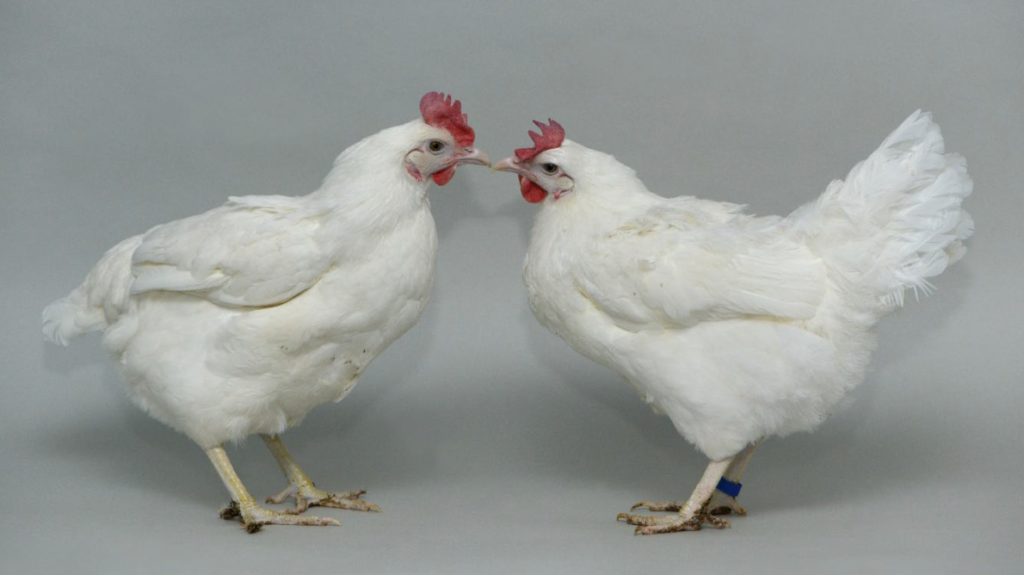
A non-gene-edited chicken (left) pictured next to an ANP32A gene-edited chicken (right). Image credit: Norrie Russell Courtesy: University of Edinburgh
There’s also an October 10, 2023 article by Jon Cohen for Science.org, which gives some idea of how much work it took to get to this point, Note: Links have been removed,
For 3 decades, Helen Sang has tinkered with the genomes of chickens to try to make the birds resistant to the flu viruses that periodically devastate flocks and raise fears of a human pandemic. Now, as an especially virulent strain of avian influenza sweeps through poultry and wild birds around the world, the geneticist at the University of Edinburgh’s Roslin Institute has her first solid success. Using the CRISPR gene editor and recent findings about what makes poultry vulnerable to flu, Sang and colleagues from three other institutions have created chickens that can resist real-life doses of avian flu viruses. “Sticking to it gets you somewhere in the end,” she says.
The result, published today [October 5, 2023] in Nature Communications, is “a long-awaited achievement,” says Jiří Hejnar, a virologist at the Czech Academy of Sciences’s Institute of Molecular Genetics whose group showed in 2020 that CRISPR-edited chickens could resist a cancer-causing virus. But farmers won’t be raising flu-proof chickens anytime soon. The edited birds still became infected when exposed to larger amounts of the flu virus. And the strategy raises a safety concern: chickens edited this way could, in theory, drive the evolution of flu variants better at infecting people. “What this showed is a proof of concept,” says Wendy Barclay, a virologist at Imperial College London who worked on the new study. “But we’re not there yet.”
…
Here’s a link to and a citation for the paper,
Creating resistance to avian influenza infection through genome editing of the ANP32 gene family by Alewo Idoko-Akoh, Daniel H. Goldhill, Carol M. Sheppard, Dagmara Bialy, Jessica L. Quantrill, Ksenia Sukhova, Jonathan C. Brown, Samuel Richardson, Ciara Campbell, Lorna Taylor, Adrian Sherman, Salik Nazki, Jason S. Long, Michael A. Skinner, Holly Shelton, Helen M. Sang, Wendy S. Barclay & Mike J. McGrew. Nature Communications volume 14, Article number: 6136 (2023) DOI: https://doi.org/10.1038/s41467-023-41476-3 Published: 10 October 2023
Before getting to the latest about carbon dots, there’s something to be clarified (and it was news to me), a carbon dot is not a quantum dot. So says this 2020 paper, “Advances in carbon dots: from the perspective of traditional quantum dots” by Yanhong Liu, Hui Huang, Weijing Cao, Baodong Mao, Yang Liu, and Zhenhui Kang. Mater. Chem. Front., 2020,4, 1586-1613 First published March 17, 2020.
Abstract
Quantum dots (QDs) have been the core concept of nanoscience and nanotechnology since their inception, and play a dominant role in the development of the nano-field. Carbon dots (CDots), defined by a feature size of <10 nm, have become a rising star in the crossover field of carbon materials and traditional QDs (TQDs). CDots possess many unique structural, physicochemical and photochemical properties that render them a promising platform for biology, devices, catalysis and other applications. …
This story is about carbon dots but you can find out more about quantum dots in my October 6, 2023 posting concerning the 2023 Nobel prizes; scroll down to the ‘Chemistry’ subhead.
An August 30, 2023 news item on phys.org describes work from Concordia University (Montréal, Canada) on carbon dots,
Researchers at Concordia have developed a new system using tiny nanosensors called carbon dots to detect the presence of the widely used chemical glyphosate. Their research, titled “Ratiometric Sensing of Glyphosate in Water Using Dual Fluorescent Carbon Dots,” is published in Sensors.
Glyphosate is a pesticide found in more than 750 agricultural, forestry, urban and home products, including Monsanto’s popular weed-killer Roundup. It is also controversial: studies have linked its overuse to environmental pollution and cancer in humans. Its sale is banned or restricted in dozens of countries and jurisdictions, including Canada.
The researchers’ system relies on the carbon dots’ chemical interaction with glyphosate to detect its presence. Carbon dots are exceedingly small fluorescent particles, usually no more than 10 or 15 nanometres in size (a human hair is between 80,000 and 100,000 nanometres). But when they are added to water solutions, these nanomaterials emit blue and red fluorescence.
The researchers employed an analysis technique called a ratiometric self-referencing assay to determine glyphosate levels in a solution. The red fluorescence emitted by the carbon dots when exposed to varying concentrations of the chemical and different pH levels is compared with a control in which no glyphosate is present. In all the tests, the blue fluorescence remained unchanged, giving the researchers a common reference point across the different tests.
They observed that higher levels of glyphosate quenched the red fluorescence, which they accredited to the interaction of the pesticide with the carbon dots’ surface.
“Our system differs from others because we are measuring the area between two peaks—two fluorescent signatures—on the visible spectrum,” says Adryanne Clermont-Paquette, a PhD candidate in biology and the paper’s lead author. “This is the integrated area between the two curves. Ratiometric measurement allows us to ignore variables such as temperature, pH levels or other environmental factors. That allows us to just only look at the levels of glyphosate and carbon dots that are in the system.”
“By understanding the chemistry at the surface of these very small dots and by knowing their optical properties, we can use them to our advantage for many different applications,” says Rafik Naccache, an associate professor of chemistry and biochemistry and the paper’s supervising author.
Research assistants Diego-Andrés Mendoza and Amir Sadeghi, along with associate professor of biology Alisa Piekny, are co-authors.
Starting small
Naccache says the technique is designed to detect minute amounts of the pesticide. The technique they developed is sensitive enough to be able to detect the presence of pesticide at levels as low as 0.03 parts per million.
“The challenge is always in the other direction, to see how low we can go in terms of sensitivity and selectivity,” he says.
There remains much work to be done before this technology can be used widely. But as Clermont-Paquette notes, this paper represents an important beginning.
“Understanding the interaction between glyphosate and carbon dots is a first step. If we are to move this along further, and develop it into a real-life application, we have to start with the fundamentals.”
The researchers are supported by funding from the Natural Sciences and Engineering Research Council of Canada.
By combining seaweed and graphene, scientists have been able to create sensors that can be worn like a ‘second skin’ and outperform other similar biosensors, according to a March 3, 2023 news item on ScienceDaily,
Scientists at the University of Sussex have successfully trialed new biodegradable health sensors that could change the way we experience personal healthcare and fitness monitoring technology.
The team at Sussex have developed the new health sensors — such as those worn by runners or patients to monitor heart rate and temperature — using natural elements like rock salt, water and seaweed, combined with graphene. Because they are solely made with ingredients found in nature, the sensors are fully biodegradable, making them more environmentally friendly than commonly used rubber and plastic-based alternatives. Their natural composition also places them within the emerging scientific field of edible electronics — electronic devices that are safe for a person to consume.
Better still, the researchers found that their sustainable seaweed-based sensors actually outperform existing synthetic based hydrogels and nanomaterials, used in wearable health monitors, in terms of sensitivity. Therefore, improving the accuracy, as the more sensitive a sensor, the more accurately it will record a person’s vital signs.
Dr Conor Boland, a materials physics lecturer in the School of Mathematical and Physical Sciences, said: “I was first inspired to use seaweed in the lab after watching MasterChef during lockdown. Seaweed, when used to thicken deserts, gives them a soft and bouncy structure – favored by vegans and vegetarians as an alternative to gelatin. It got me thinking: “what if we could do that with sensing technology?”.
“For me, one of the most exciting aspects to this development is that we have a sensor that is both fully biodegradable and highly effective. The mass production of unsustainable rubber and plastic based health technology could, ironically, pose a risk to human health through microplastics leeching into water sources as they degrade.
“As a new parent, I see it as my responsibility to ensure my research enables the realisation of a cleaner world for all our children.”
Seaweed is first and foremost an insulator, but by adding a critical amount of graphene to a seaweed mixture the scientists were able to create an electrically conductive film. When soaked in a salt bath, the film rapidly absorbs water, resulting in a soft, spongy, electrically conductive hydrogel.
The development has the potential to revolutionise health monitoring technology, as future applications of the clinical grade wearable sensors would look something like a second skin or a temporary tattoo: lightweight, easy to apply, and safe, as they are made with all natural ingredients. This would significantly improve the overall patient experience, without the need for more commonly used and potentially invasive hospital instruments, wires and leads.
Dr Sue Baxter, Director of Innovation and Business Partnerships at the University of Sussex, is excited about the potential benefits of this technology: “At the University of Sussex, we are committed to protecting the future of the planet through sustainability research, expertise and innovation. What’s so exciting about this development from Dr Conor Boland and his team is that it manages to be all at once truly sustainable, affordable, and highly effective – out-performing synthetic alternatives.
“What’s also remarkable for this stage of research – and I think this speaks to the meticulous ground-work that Dr Boland and his team put in when they created their blueprint – is that it’s more than a proof of principle development. Our Sussex scientists have created a device that has real potential for industry development into a product from which you or I could benefit in the relatively near future.”
This latest research breakthrough follows the publication of a blueprint for nanomaterial development from the Sussex scientists in 2019, which presented a method for researchers to follow in order to optimise the development of nanomaterial sensors.
…
Kevin Doty, a Masters student in the School of Mathematical and Physical Sciences, at the University of Sussex, said: “I taught chemistry previously, but decided I wanted to learn more about nanoscience. My gamble paid off, and not only did I enjoy it more than I expected, but I also ended up with an opportunity to utilize the information I had learned to work on a novel idea that has evolved into a first author publication as an MSc student. Learning about nanoscience showed me just how varied and multidisciplinary the field is. Any science background can bring knowledge that can be applied to this field in a unique way. This has led to further studies in a PhD studentship, opening up an all new career path I could not have previously considered.”
This environmental monitoring story focused on the roly-poly was announced in an April 18, 2023 news item on Statnano,
The woodlouse goes by many names: roly-poly, pill bug, potato bug, tomato bug, butchy boy, cheesy bob, and chiggy pig, to name just a few. It is best known for contracting into a ball when agitated. This crustacean (yes, it’s a crustacean, not an insect) thrives in heavily metal-contaminated areas due to its specialized digestive organ, called a hepatopancreas, that stores and expels unwanted metals.
Metal nanoparticles are common in industrial and research plants. However, they can leach into the surrounding environment. Currently, little is known about the toxicity of metal nanoparticles for nearby organisms because detecting metal nanoparticles, particularly gold, requires microscopic, 3D imaging that cannot be done in the field
….
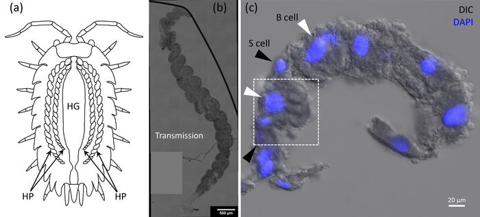
Caption: (a) Cartoon of a woodlouse depicting the hepatopancreas (HP) and the hind gut (HG). (b) Transmission overview of a single HP tubule, showing the helical structure. (c) Section from a HP tubule with the nuclei fluorescently labeled in blue. Credit: Iestyn Pope, Nuno G.C. Ferreira, Peter Kille, Wolfgang Langbein, and Paola Borri
In Applied Physics Letters, by AIP Publishing, researchers from Cardiff University in the U.K. introduce a novel imaging method to detect gold nanoparticles in woodlice. With information about the quantity, location, and impact of gold nanoparticles within the organism, scientists can better understand the potential harm other metals may have on nature.
“Gold nanoparticles are used extensively for biological research applications owing to their biocompatibility and photostability and are available in a large range of shapes and sizes,” said author Wolfgang Langbein. “By using gold nanoparticles, which would not normally be present in the woodlice diet, we can study the journey of nanoparticles inside complex biological systems.”
The researchers developed an imaging method known as four-wave mixing microscopy, which flashes light that the gold nanoparticles absorb. The light flashes again and the subsequent scattering reveals the nanoparticles’ locations. Using this state-of-the-art technique, they locate the individual gold nanoparticles in the 3D cellular environment.
“By precisely pinpointing the fate of individual gold nanoparticles in the hepatopancreas of woodlice, we can gain a better understanding of how these organisms sequester and respond to metals ingested from the environment,” said Langbein. “Tracking this metal within these organisms is the first step enabling further study to determine, for example, if gold is collected within specific cells, or if it can interfere with the metabolisms in high doses.”
The use of gold nanoparticles in medical devices is increasing and with it, their abundance in the environment. This imaging technique will provide clarity into the little-understood mechanisms in the woodlice hepatopancreas and will also provide helpful environmental monitoring.
In the future, background-free four-wave mixing microscopy could be used to detect other metal nanoparticles and may be applied to organisms like fish larvae and even human cell cultures.
I imagine this wearable technology would also be useful for the military too. However, the focus for these researchers from China is firefighting. (Given the situation with the Canadian wildfires in June 2023, we have 10x more than the average at this time in the season over the last 10 years, it’s good to see some work focused on safety for firefighters.) From a January 17, 2023 news item on phys.org,
Firefighting may look vastly different in the future thanks to intelligent fire suits and masks developed by multiple research institutions in China.
Researchers published results showing breathable electrodes woven into fabric used in fire suits have proven to be stable at temperatures over 520ºC. At these temperatures, the fabric is found to be essentially non-combustible with high rates of thermal protection time.
…
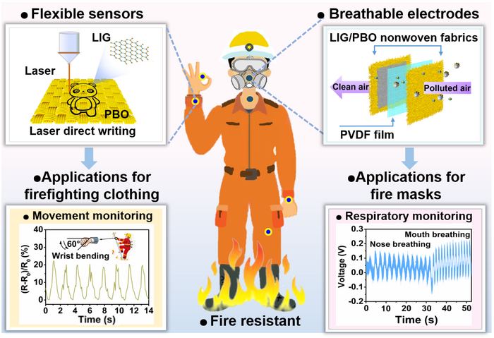
Caption: Scientists from multiple institutions address the challenges and limitations of current fire-fighting gear by introducing wearable, breathable sensors and electrodes to better serve firefighters. Credit: Nano Research, Tsinghua University Press
The results show the efficacy and practicality of Janus graphene/poly(p-phenylene benzobisoxazole), or PBO, woven fabric in making firefighting “smarter” with the main goal being to manufacture products on an industrial scale that are flame-retardant but also intelligent enough to warn the firefighter of increased risks while traversing the flames.
“Conventional firefighting clothing and fire masks can ensure firemen’s safety to a certain extent,” said Wei Fan, professor at the School of Textile Science and Engineering at Xi’an Polytechnic University. “However, the fire scene often changes quickly, sometimes making firefighters trapped in the fire for failing to judge the risks in time. At this situation, firefighters also need to be rescued.”
The key here is the use of Janus graphene/PBO, woven fabrics. While not the first of its kind, the introduction of PBO fibers offers better strength and fire protection than other similar fibers, such as Kevlar. The PBO fibers are first woven into a fabric that is then irradiated using a CO2 infrared laser. From here, the fabric becomes the Janus graphene/PBO hybrid that is the focus of the study.
The mask also utilizes a top and bottom layer of Janus graphene/PBO with a piezoelectric layer in between that acts as a way to convert mechanical pressures to electricity.
“The mask has a good smoke particle filtration effect, and the filtration efficiency of PM2.5 and PM3.0 reaches 95% and 100%, respectively. Meanwhile, the mask has good wearing comfort as its respiratory resistance (46.8 Pa) is lower than 49 Pa of commercial masks. Besides, the mask is sensitive to the speed and intensity of human breathing, which can dynamically monitor the health of the firemen” said Fan.
Flame-retardant electronics featured in these fire suits are flexible, heat resistant, quick to make and low-cost which makes scaling for industrial production a tangible achievement. This makes it more likely that the future of firefighting suits and masks will be able to effectively use this technology. Quick, effective responses can also reduce economic losses attributed to fires.
“The graphene/PBO woven fabrics-based sensors exhibit good repeatability and stability in human motion monitoring and NO2 gas detection, the main toxic gas in fires, which can be applied to firefighting suits to help firefighters effectively avoiding danger” Fan said. Being able to detect sharp increases in NO2 gas can help firefighters change course in an instant if needed and could be a lifesaving addition to firefighter gear.
Major improvements can be made in the firefighting field to better protect the firefighters by taking advantage of graphene/PBO woven and nonwoven fabrics. Widescale use of this technology can help the researchers reach their ultimate goal of reducing mortality and injury to those who risk their lives fighting fires.
Yu Luo and Yaping Miao of the School of Textile Science and Engineering at Xi’an Polytechnic University contributed equally to this work. Professor Wei Fan is the corresponding author. Yingying Zhang and Huimin Wang of the Department of Chemistry at Tsinghua University, Kai Dong of the Beijing Institute of Nanoenergy and Nanosystems at the Chinese Academy of Sciences, and Lin Hou and Yanyan Xu of Shaanxi Textile Research Institute Co., LTD, Weichun Chen and Yao Zhang of the School of Textile Science and Engineering at Xi’an Polytechnic University contributed to this research.
This work was supported by the National Natural Science Foundation of China, Textile Vision Basic Research Program of China, Key Research and Development Program of Xianyang Science and Technology Bureau, Key Research and Development Program of Shaanxi Province, Natural Science Foundation of Shaanxi Province, and Scientific Research Project of Shaanxi Provincial Education Department.
Here are two links and a citation for the same paper,
This April 19, 2022 news item on Nanowerk announces mosquito research from Ohio State University (OSU), Note: A link has been removed,
Before being accidentally introduced to the New World by the 16th century slave trade, the yellow fever mosquito was a species native only to Africa. Highly adaptable, it has since become an invasive species in North America, but researchers at The Ohio State University may have found a way to squash the pesky population in its juvenile stages.
Recently published in the journal Insects (“Larvicidal Activity of Carbon Black against the Yellow Fever Mosquito Aedes aegypti”), a new paper describes how mosquitoes have evolved a natural resistance to some chemical insecticides, and offers an alternative called carbon black, a type of carbon-based nanoparticles, or CNPs [when it’s specifically carbon black nanoparticles, it may sometimes be abbreviated to CBNPs; more about that at the end of this post].
Study co-author and an associate professor of entomology at Ohio State, Peter Piermarini described CNPs as “microscopic” materials made out of organic elements. The study used a modified version of carbon black called Emperor 1800, which is often used to coat automobiles black. While CNPs are a relatively new scientific development, they have been considered as new tools to control various insect and pest infestations, he said.
“If we can learn more about how carbon black works and how to use it safely, we could design a commercially available nanoparticle that is highly effective against insecticide-resistant mosquitoes,” Piermarini said.
The yellow fever mosquito, or Aedes aegypti, is a species of mosquito known for spreading not just yellow fever, but also diseases like the Zika virus, dengue fever and chikungunya fever. Adults rarely fly more than a few hundred meters from where they emerge, but their abundance leads to steady transmission of diseases – enough to claim tens of thousands of lives every year and hospitalize hundreds of thousands more people.
Because of this, the mosquito is considered to be one of the deadliest animals on the planet. For this study, the researchers’ goal was to figure out how toxic these nanomaterials could be to mosquito larvae, or the immature form of the insect.
Contrary to popular belief, not all mosquitoes set their sights on turning our blood into their latest meal. Male mosquitoes subsist only on flower nectar; it’s the females that will consume both flower nectar and blood in a bid to provide their eggs with enough protein to grow.
When female mosquitoes are ready to lay their eggs, they return to standing pools of water, like lakes or birdbaths, to release them. After they hatch, these larvae will stay in the water for about a week until they reach adulthood, and take wing.
To test whether Emperor 1800 would be effective in stopping that process, researchers worked with two different strains of the yellow fever mosquito inside the lab, one extremely susceptible to typical chemical insecticides, and the other, extremely resistant to them.
By applying the carbon black nanomaterials to the water during the earliest stages of the mosquito’s life cycle and checking in 48 hours later, they were able to determine that CNPs kill mosquito larvae both quickly and efficiently.
“Given the properties of carbon black, it has the most potential for killing larvae because it can be suspended in water,” Piermarini said. Their findings showed that the material seemed to accumulate on the mosquito larvae’s head, abdomen, and even in its gut, meaning that at some point, the larvae were ingesting smaller particles of carbon black.
“Our hypothesis is that these materials may be physically obstructing their ability to perform basic biological functions. It could be blocking their digestion, or might be interfering with their ability to breathe,” said Piermarini.
However, there was one thing that Piermarini found particularly surprising.
When first suspended in water, carbon black appeared equally toxic to larvae of insecticide-resistant and insecticide-susceptible mosquitoes, but the longer the carbon black was suspended in water before treating them. it became more toxic to the insecticide-resistant larvae.
“When you first apply the CNP solution it has similar toxicity against both strains,” Piermarini said. “But when you let the suspension age for a few weeks, it tends to become more potent against the resistant strain of mosquitoes.”
Although they couldn’t determine the reason behind the time-lapsed deaths, the study concluded that these new nanomaterials could be extremely beneficial to controlling the species when applied as a preventive treatment to mosquito breeding grounds.
But before it can be utilized by the public, Piermarini said, carbon black needs to undergo rigorous testing to ensure it won’t harm humans and the environment as a whole.
Co-authors were Erick Martinez Rodriguez, a visiting scholar currently in the Ohio State Entomology Graduate program, Parker Evans, a previous PhD student in the Ohio State Translational Plant Sciences Graduate program, and Megha Kalsi, a previous postdoctoral researcher in entomology. This research was supported by Ohio State’s College of Food, Agricultural, and Environmental Sciences and Vaylenx LLC.
The nomenclature for carbon at the nanoscale is a little confusing to me. As best as I can determine all of the elements have multiple names at the nanoscale but it’s only with carbon that subcategories function as categories themselves. For example, fullerenes (C60s), single-walled carbon nanotubes (SWCNTs), double-walled carbon nanotubes (DWCNTs), and mulit-walled carbon nanotubes (MWCNTs) are subcategories that stand on their own but, sometimes, are referred to as carbon nanoparticles, which is the main category. I checked carbon black nanoparticles online and found a number of instances where it was abbreviated to CBNP and it can also be a CNP since it is found under the carbon nanoparticle category as per this Wikipedia entry.
A November 23, 2021 news item on phys.org announces research from Australia that may lead to pain relief for anyone with sensitive teeth,
In an Australian first, researchers from the University of Queensland have used nanotechnology to develop effective ways to manage tooth sensitivity.
Dr. Chun Xu from UQ’s [University of Queensland] School of Dentistry said the approach might provide more effective long-term pain relief for people with sensitive teeth, compared to current options.
“Dentin tubules are located in the dentin, one of the layers below the enamel surface of your teeth,” Dr Xu said.
“When tooth enamel has been worn down, and the dentin are exposed, eating or drinking something cold or hot can cause a sudden sharp flash of pain.
“The nanomaterials used in this preclinical study can rapidly block the exposed dentin tubules and prevent the unpleasant pain.
“Our approach acts faster and lasts longer than current treatment options.
“The materials could be developed into a paste, so people who have sensitive teeth could simply apply this paste to the tooth and massage for one to three minutes.
“The next step is clinical trials.”
Tooth sensitivity affects up to 74 per cent of the population, at times severely impacting quality of life and requiring expensive treatment.
“If clinical trials are successful people will benefit from this new method that can be used at home, without the need to go to a dentist in the near future,” Dr Xu said.
“We hope this study encourages more research using nanotechnology to address dental problems.”
The team also included researchers from UQ’s Australian Institute for Bioengineering and Nanotechnology (AIBN.
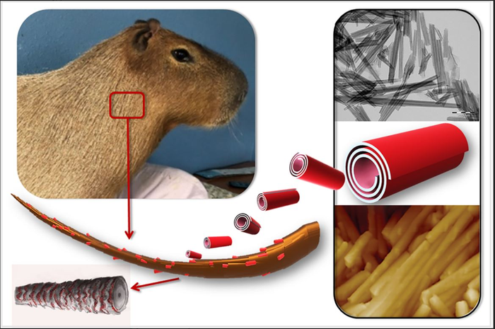
Caption Graphical abstract [the animal is a capybara, world’s largest rodent] Credit: Kazan Federal University, Louisiana Tech University, Gubkin University
An international researcher team of Louisiana Tech University, Gubkin University [also known as, Gubkin Russian State University of Oil and Gas] and Kazan Federal University reported the fabrication of nanoscale insecticidal hair coating for prolonged anti-lice protection. The study was supported by the Russian Science Foundation.
“Treating agricultural and domestic animals infected with ectoparasites (such as lice, fleas, chewing lice, etc.) is among the primary challenges of veterinary medicine and agriculture. In case of mass infestation, regular measures, such as isolation of infected animals or repeated reapplication of insecticides, are not always effective. These methods are time-limited and provide a short-term therapeutic effect,” explains co-author Rawil Fakhrullin, Head of Kazan University’s Bionanotechnology Lab. “Using an inorganic nanoscale carrier as a component of a therapeutic formulation for topical application of insecticides might be the optimal way to address this challenge.”
Halloysite, a natural nanosized tubular mineral, was used as a drug carrier capable of forming a durable and uniform coating on the surface of animal hair.
“Loading an insecticidal drug, permethrin, into halloysite nanotubes reduces the release rate, leading to fewer re-treatments and fewer side effects,” continues Dr. Fakhrullin.
The paper shows that after goat hair samples were treated with halloysite-based nanocontainers, a stable 2-3 micron waterproof coating was formed on the surface of the hair, suitable for long-term antiparasitic protection.
“Long-term insecticidal activity is the result of the gradual release of the drug from the nanotubes. A formulation based on halloysite retains its protective antiparasitic properties after washing the animal’s hair with water. This stable and water-resistant composite coating provides a drug dose effective for long-term protection of animals,” says the interviewee.
The authors also examined the hair structure of the capybara, world’s largest rodent native to South America. They found that the wax-like layer present on the hair surface of this semi-aquatic animal facilitates the formation of a denser and more durable coating of halloysite than in terrestrial animals (guinea pigs and goats). The wax helps retaining nanoclay particles on the surface of the animal’s hair.
Dr. Fakhrullin comments about the test subjects, “We studied the suppressive effects of nanocontainers on goat ectoparasites Damalinia caprae from the Trichodectidae family. At the same time, our technique can be effective towards other types of lice, since these parasites live in hair and maintain close contact with hair cuticles, regardless of the animal’s dietary preferences. We believe that this approach can be used for long-term and sustainable antiparasitic protection of farm animals, especially if other insecticidal preparations are encapsulated in addition to permethrin. In addition, similar drugs can be used for the prevention or treatment of head lice in humans.”
Furthermore, the described material can also be helpful in treating fur in zoological collections.
Here’s a link to and a citation for the paper,
Clay Nanotube Immobilization on Animal Hair for Sustained Anti-Lice Protection by Naureen Rahman, Faith Hannah Scott, Yuri Lvov, Anna Stavitskaya, Farida Akhatova, Svetlana Konnova, Gӧlnur Fakhrullina and Rawil Fakhrullin. Pharmaceutics 2021, 13(9), 1477; DOI: https://doi.org/10.3390/pharmaceutics13091477 Published: 15 September 2021