It’s fascinating to read about a technique for applying ‘tattoos’ to living cells and I have two news items and news releases with different perspectives about this same research.
Engineers have developed nanoscale tattoos — dots and wires that adhere to live cells — in a breakthrough that puts researchers one step closer to tracking the health of individual cells.
The new technology allows for the first time the placement of optical elements or electronics on live cells with tattoo-like arrays that stick on cells while flexing and conforming to the cells’ wet and fluid outer structure.
“If you imagine where this is all going in the future, we would like to have sensors to remotely monitor and control the state of individual cells and the environment surrounding those cells in real time,” said David Gracias, a professor of chemical and biomolecular engineering at Johns Hopkins University who led the development of the technology. “If we had technologies to track the health of isolated cells, we could maybe diagnose and treat diseases much earlier and not wait until the entire organ is damaged.”
Gracias, who works on developing biosensor technologies that are nontoxic and noninvasive for the body, said the tattoos bridge the gap between living cells or tissue and conventional sensors and electronic materials. They’re essentially like barcodes or QR codes, he said.
“We’re talking about putting something like an electronic tattoo on a living object tens of times smaller than the head of a pin,” Gracias said. “It’s the first step towards attaching sensors and electronics on live cells.”
The structures were able to stick to soft cells for 16 hours even as the cells moved.
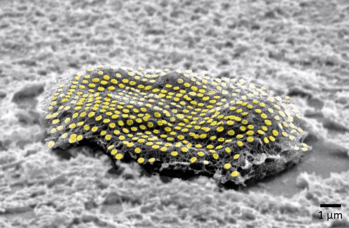
The researchers built the tattoos in the form of arrays with gold, a material known for its ability to prevent signal loss or distortion in electronic wiring. They attached the arrays to cells that make and sustain tissue in the human body, called fibroblasts. The arrays were then treated with molecular glues and transferred onto the cells using an alginate hydrogel film, a gel-like laminate that can be dissolved after the gold adheres to the cell. The molecular glue on the array bonds to a film secreted by the cells called the extracellular matrix.
Previous research has demonstrated how to use hydrogels to stick nanotechnology onto human skin and internal animal organs. By showing how to adhere nanowires and nanodots onto single cells, Gracias’ team is addressing the long-standing challenge of making optical sensors and electronics compatible with biological matter at the single cell level.
“We’ve shown we can attach complex nanopatterns to living cells, while ensuring that the cell doesn’t die,” Gracias said. “It’s a very important result that the cells can live and move with the tattoos because there’s often a significant incompatibility between living cells and the methods engineers use to fabricate electronics.”
The team’s ability to attach the dots and wires in an array form is also crucial. To use this technology to track bioinformation, researchers must be able to arrange sensors and wiring into specific patterns not unlike how they are arranged in electronic chips.
“This is an array with specific spacing,” Gracias explained, “not a haphazard bunch of dots.”
The team plans to try to attach more complex nanocircuits that can stay in place for longer periods. They also want to experiment with different types of cells.
Other Johns Hopkins authors are Kam Sang Kwok, Yi Zuo, Soo Jin Choi, Gayatri J. Pahapale, and Luo Gu.
This looks more like a sea creature to me but it’s not,
Caption: False-colored gold nanodot array on a fibroblast cell. Credit: Kam Sang Kwok and Soo Jin Choi, Gracias Lab/Johns Hopkins University.[The measurement, i.e., what looks like a ‘u’ with a preceding tail, in the lower right corner of the image is one micron/one millionth add that to the ‘m’ and you have what’s commonly described as one micrometre.]
An August 10, 2023 news item on ScienceDaily offers a different perspective from the American Chemical Society (ACS) on this research,
For now, cyborgs exist only in fiction, but the concept is becoming more plausible as science progresses. And now, researchers are reporting in ACS’ Nano Letters that they have developed a proof-of-concept technique to “tattoo” living cells and tissues with flexible arrays of gold nanodots and nanowires. With further refinement, this method could eventually be used to integrate smart devices with living tissue for biomedical applications, such as bionics and biosensing.
Advances in electronics have enabled manufacturers to make integrated circuits and sensors with nanoscale resolution. More recently, laser printing and other techniques have made it possible to assemble flexible devices that can mold to curved surfaces. But these processes often use harsh chemicals, high temperatures or pressure extremes that are incompatible with living cells. Other methods are too slow or have poor spatial resolution. To avoid these drawbacks, David Gracias, Luo Gu and colleagues wanted to develop a nontoxic, high-resolution, lithographic method to attach nanomaterials to living tissue and cells.
The team used nanoimprint lithography to print a pattern of nanoscale gold lines or dots on a polymer-coated silicon wafer. The polymer was then dissolved to free the gold nanoarray so it could be transferred to a thin piece of glass. Next, the gold was functionalized with cysteamine and covered with a hydrogel layer, which, when peeled away, removed the array from the glass. The patterned side of this flexible array/hydrogel layer was coated with gelatin and attached to individual live fibroblast cells. In the final step, the hydrogel was degraded to expose the gold pattern on the surface of the cells. The researchers used similar techniques to apply gold nanoarrays to sheets of fibroblasts or to rat brains. Experiments showed that the arrays were biocompatible and could guide cell orientation and migration.
The researchers say their cost-effective approach could be used to attach other nanoscale components, such as electrodes, antennas and circuits, to hydrogels or living organisms, thereby opening up opportunities for the development of biohybrid materials, bionic devices and biosensors.
The authors acknowledge funding from the Air Force Office of Scientific Research, the National Institute on Aging, the National Science Foundation and the Johns Hopkins University Surpass Program.
By combining seaweed and graphene, scientists have been able to create sensors that can be worn like a ‘second skin’ and outperform other similar biosensors, according to a March 3, 2023 news item on ScienceDaily,
Scientists at the University of Sussex have successfully trialed new biodegradable health sensors that could change the way we experience personal healthcare and fitness monitoring technology.
The team at Sussex have developed the new health sensors — such as those worn by runners or patients to monitor heart rate and temperature — using natural elements like rock salt, water and seaweed, combined with graphene. Because they are solely made with ingredients found in nature, the sensors are fully biodegradable, making them more environmentally friendly than commonly used rubber and plastic-based alternatives. Their natural composition also places them within the emerging scientific field of edible electronics — electronic devices that are safe for a person to consume.
Better still, the researchers found that their sustainable seaweed-based sensors actually outperform existing synthetic based hydrogels and nanomaterials, used in wearable health monitors, in terms of sensitivity. Therefore, improving the accuracy, as the more sensitive a sensor, the more accurately it will record a person’s vital signs.
Dr Conor Boland, a materials physics lecturer in the School of Mathematical and Physical Sciences, said: “I was first inspired to use seaweed in the lab after watching MasterChef during lockdown. Seaweed, when used to thicken deserts, gives them a soft and bouncy structure – favored by vegans and vegetarians as an alternative to gelatin. It got me thinking: “what if we could do that with sensing technology?”.
“For me, one of the most exciting aspects to this development is that we have a sensor that is both fully biodegradable and highly effective. The mass production of unsustainable rubber and plastic based health technology could, ironically, pose a risk to human health through microplastics leeching into water sources as they degrade.
“As a new parent, I see it as my responsibility to ensure my research enables the realisation of a cleaner world for all our children.”
Seaweed is first and foremost an insulator, but by adding a critical amount of graphene to a seaweed mixture the scientists were able to create an electrically conductive film. When soaked in a salt bath, the film rapidly absorbs water, resulting in a soft, spongy, electrically conductive hydrogel.
The development has the potential to revolutionise health monitoring technology, as future applications of the clinical grade wearable sensors would look something like a second skin or a temporary tattoo: lightweight, easy to apply, and safe, as they are made with all natural ingredients. This would significantly improve the overall patient experience, without the need for more commonly used and potentially invasive hospital instruments, wires and leads.
Dr Sue Baxter, Director of Innovation and Business Partnerships at the University of Sussex, is excited about the potential benefits of this technology: “At the University of Sussex, we are committed to protecting the future of the planet through sustainability research, expertise and innovation. What’s so exciting about this development from Dr Conor Boland and his team is that it manages to be all at once truly sustainable, affordable, and highly effective – out-performing synthetic alternatives.
“What’s also remarkable for this stage of research – and I think this speaks to the meticulous ground-work that Dr Boland and his team put in when they created their blueprint – is that it’s more than a proof of principle development. Our Sussex scientists have created a device that has real potential for industry development into a product from which you or I could benefit in the relatively near future.”
This latest research breakthrough follows the publication of a blueprint for nanomaterial development from the Sussex scientists in 2019, which presented a method for researchers to follow in order to optimise the development of nanomaterial sensors.
…
Kevin Doty, a Masters student in the School of Mathematical and Physical Sciences, at the University of Sussex, said: “I taught chemistry previously, but decided I wanted to learn more about nanoscience. My gamble paid off, and not only did I enjoy it more than I expected, but I also ended up with an opportunity to utilize the information I had learned to work on a novel idea that has evolved into a first author publication as an MSc student. Learning about nanoscience showed me just how varied and multidisciplinary the field is. Any science background can bring knowledge that can be applied to this field in a unique way. This has led to further studies in a PhD studentship, opening up an all new career path I could not have previously considered.”
This device could help people with disabilities to regain control of their limbs or provide advance warnings of seizures to people with epilepsy and it’s all based on technology that is a century old.
Scientists from Skoltech, South Ural State University, and elsewhere have developed a device for recording brain activity that is more compact and affordable than the solutions currently on the market. With its high signal quality and customizable configuration, the device could help people with restricted mobility regain control of their limbs or provide advance warnings of an impending seizure to patients with epilepsy. The article presenting the device and testing results came out in Experimental Brain Research.
Researchers and medics, as well as engineers working on futuristic gadgets, need tools that measure brain activity. Among their scientific applications are research on sleep, decision-making, memory, and attention. In a clinical setting, these tools allow doctors to assess the extent of damage to an injured brain and monitor coma patients. Further down cyberpunk lane, brain signals can be translated into commands and sent to an external or implanted device, either to make up for lost functions in the body or for plain fun. The commands could range from moving the arm of an exoskeleton worn by a paralyzed person to turning on the TV.
Invented about a century ago, electroencephalographers are devices that read the electrical activity of the brain via small electrodes placed on the scalp. The recorded signals are then used for research, diagnostics, or gadgetry. The problem with the existing systems used in labs and hospitals is they are bulky and/or expensive. And even then, the number of electrodes is limited, resulting in moderate signal quality. Amateur devices tend to be more affordable, but with even poorer sensitivity.
To fill that gap, researchers from South Ural State University, North Carolina State University, and Brainflow — led by electronic research engineer Ildar Rakhmatulin and Skoltech neuroscientist Professor Mikhail Lebedev — created a device you can build for just $350, compared with the $1,000 or more you would need for currently available analogs. Besides being less expensive, the new electroencephalographer has as many as 24 electrodes or more. Importantly, it also provides research-grade signal quality. At half a centimeter in diameter (about 1/5 inches), the processing unit is compact enough to be worn throughout the day or during the night. The entire device weighs about 150 grams (about 5 ounces).
The researchers have made the instructions for building the device and the accompanying documentation and software openly available on GitHub. The team hopes this will attract more enthusiasts involved in brain-computer interface development, giving an impetus to support and rehabilitation system development, cognitive research, and pushing the geek community to come up with new futuristic gizmos.
“The more convenient and affordable such devices become, the more chances there are this would drive the home lab movement, with some of the research on brain-computer interfaces migrating from large science centers to small-scale amateur projects,” Lebedev said.
“Or we could see people with limited mobility using do-it-yourself interfaces to train, say, a smartphone-based system that would electrically stimulate a biceps to flex the arm at the elbow,” the researcher went on. “That works on someone who has lost control over their arm due to spinal cord trauma or a stroke, where the commands are still generated in the brain — they just don’t reach the limb, and that’s where our little brain-computer interfacing comes in.”
According to the team, such interfaces could also help patients with epilepsy by detecting tell-tale brain activity patterns that indicate when a seizure is imminent, so they can prepare by lying down comfortably in a safe space or attempting to suppress the seizure via electrical stimulation.
Here’s a link to and a citation for the paper,
Low-cost brain computer interface for everyday use by Ildar Rakhmatulin, Andrey Parfenov, Zachary Traylor, Chang S. Nam & Mikhail Lebedev. Experimental Brain Research volume 239,Issue Date: December 2021, pages 3573–3583 (2021) DOI: https://doi.org/10.1007/s00221-021-06231-4 Published online: 29 September 2021
This paper is behind a paywall.
You can find Brainflow here and this description on its homepage: “BrainFlow is a library intended to obtain, parse and analyze EEG, EMG, ECG and other kinds of data from biosensors.”
While there’s a January 10, 2022 news item on Nanowerk, the research being announced was made available online in the Fall of 2021 and is now available in print,
Gold nanoclusters are groups of a few gold atoms with interesting photoluminescent properties. The features of gold nanoclusters depend not only on their structure, but their size and also by the ligands coordinated to them. These inorganic nanomaterials have been used in sensing, biomedicine and optics and their coordination with biomolecules can endow multiple capabilities in biological media.
A research collaboration between the groups of Dr. Juan Cabanillas, Research Professor at IMDEA Nanociencia and Dr. Aitziber L. Cortajarena, Ikerbasque Professor and Principal Investigator at CIC biomaGUNE have explored the use of natural proteins to grow gold nanoclusters, resulting in hybrid bionanomaterials with tunable photoluminescent properties and with a plethora of potential applications.
The nanoclusters –with less than 2 nm in size- differentiate from larger nanoparticles (plasmonic) since they present discrete energy levels coupled optically. The groups of amino acids within the proteins coordinate the gold atoms and allow the groups to be arranged around the gold nanocluster, facilitating the stabilization and adding an extra level of tailoring. These nanoclusters have interesting energy harvesting features. Since the discrete energy levels are optically coupled, the absorption of a photon leads to promotion of an electron to higher levels, which can trigger a photophysical process or a photochemical reaction.
The results by Cabanillas and Cortajarena groups, published in Advanced Optical Materials and Nano Letters, explore the origin of the photoluminescence in protein-designed gold nanoclusters and shed light into the strong influence of environmental conditions on the nature of luminescence. Nanocluster capping by two types of amino acids (histidine and cysteine) allow for changing the emission spectral range from blue to red, paving the way to tune the optical properties by an appropriate ligand choice. The nature of emission is also changed with capping, from fluorescence to phosphorescence, respectively. The synergistic protein-nanocluster effects on emission are still not clear, and the groups at IMDEA Nanociencia and CIC biomaGUNE are working to elucidate the mechanisms behind. There are potential applications for the aforementioned nanoclusters, in solid state as active medium in laser cavities. Optical gain properties from these nanoclusters are yet to be demonstrated, which could pave the way to a new generation of potentially interesting laser devices. As the combination of gold plus proteins is potentially biocompatible, many potential applications in biomedicine can also be envisaged.
A related publication of the groups in Nano Letters demonstrates that the insertion of tryptophans, amino acids with high electron density, in the vicinity of the nanocluster boosts its photoluminescence quantum efficiency up to 40% in some cases, values relevant for solid state light emission applications. Researchers also observed an antenna effect: the tryptophans can absorb light in a discrete manner and transfer the energy to the cluster. This effect has interest for energy harvesting and for sensing purposes as well.
The proteins through the biocapping enable the synthesis of the nanoclusters and largely improve their quantum efficiency. “The photoluminescence quantum efficiency is largely improved when using the biocapping” Dr. Cabanillas says. He believes this research work means “a new field opening for the tuning of optical properties of nanoclusters through protein engineering, and much work is ahead for the understanding of the amplification mechanism”. Dr. Cortajarena emphasizes “we have already demonstrated the great potential of engineered photoluminescent protein-nanocluster in biomedical and technological fields, and understanding the fundamental emission mechanisms is pivotal for future applications“. A variety of further applications include biosensors, as the protein admits functionalization with recognition molecules, energy harvesting, imaging and photodynamic therapies. Further work is ahead this opening avenue for photophysics research.
This research is a collaboration led by Dr. Juan Cabanillas and Dr. Aitziber L. Cortajarena research groups at IMDEA Nanociencia and CIC biomaGUNE, with contributions from researchers at the Diamond Light Source Ltd. [synchrotron] and DIPC. It has been cofounded by the projects AMAPOLA, NMAT2D, FULMATEN, Atracción de Talento from Comunidad de Madrid and the Severo Ochoa Centre of Excellence award to IMDEA Nanociencia. CIC biomaGUNE acknowledges support by the projects ERC-ProNANO, ERC-NIMM, ProTOOLs and the Maria de Maeztu Units of Excellence Programme.
Here are links to and citations for the papers,
Tuning the Optical Properties of Au Nanoclusters by Designed Proteins by Elena Lopez-Martinez, Diego Gianolio, Saül Garcia-Orrit, Victor Vega-Mayoral, Juan Cabanillas-Gonzalez, Carlos Sanchez-Cano, Aitziber L. Cortajarena. Advanced Optical Materials Volume 10, Issue 1 January 4, 2022 2101332 DOI: https://doi.org/10.1002/adom.202101332 First published: 31 October 2021
Not being familiar with either of the two research institutions mentioned in the press release, I did a little digging.
Here’s a little information about IMDEA Nanociencia (IMDEA Nanoscience Institute), from its Wikipedia entry, Note: All links have been removed,
IMDEA Nanoscience Institute is a private non-profit foundation within the IMDEA Institutes network, created in 2006-2007 as a result of collaboration agreement between the Community of Madrid and Spanish Ministry of Education and Science. The foundation manages IMDEA-Nanoscience Institute,[1] a scientific centre dedicated to front-line research in nanoscience, nanotechnology and molecular design and aiming at transferable innovations and close contact with industries. IMDEA Nanoscience is a member of the Campus of International excellence, a consortium of research institutes promoted by the Autonomous University of Madrid and Spanish National Research Council (UAM/CSIC).[2]
As for CIC biomaGUNE, here’s more from its institutional profile on the science.eus website,
The Centre for Cooperative Research in Biomaterials-CIC biomaGUNE, located in San Sebastian (Spain), was officially opened in December 2006. CIC biomaGUNE is a non-profit research organization created to promote scientific research and technological innovation at the highest levels in the Basque Country following the BioBasque policy in order to create a new business sector based on biosciences. Established by the Department of Industry, Technology & Innovation of the Government of the Autonomous Community of the Basque Country, CIC biomaGUNE constitutes one of the Centres of the CIC network, the largest Basque Country research network on specific strategic areas, having the mission to contribute to the economical and social development of the country through the generation of knowledge and speeding up the process that leads to technological innovation.
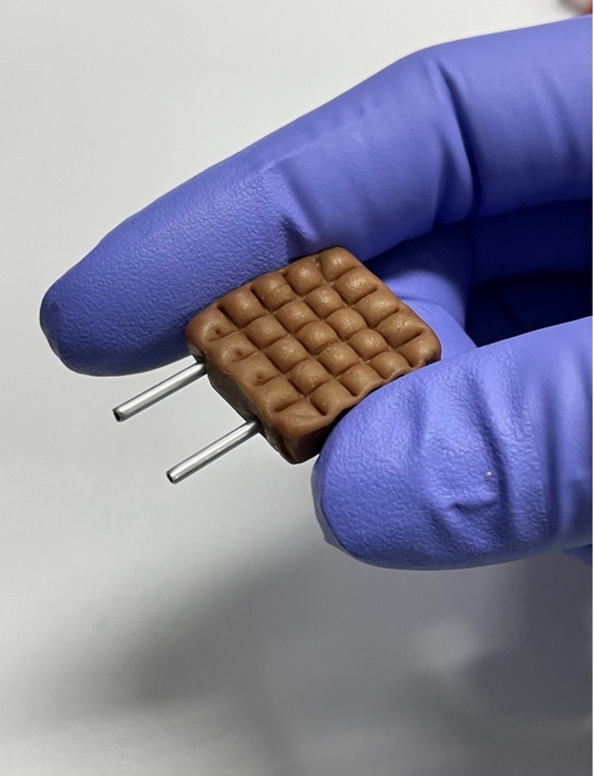
Caption: An electrode made with a molded Tootsie Roll® and aluminum tubes can help monitor ovulation status and kidney health. Credit: Adapted from ACS Applied Materials & Interfaces 2021, DOI: 10.1021/acsami.1c11306
Single-use diagnostic tests often aren’t practical for health professionals or patients in resource-limited areas, where cost and waste disposal are big concerns. So, researchers reporting in ACS Applied Materials & Interfaces have turned to a surprising material, Tootsie Roll® candy, to develop an inexpensive and low-waste device. The candy was used as an electrode, the part of the sensor that detects salt and electrolyte levels in saliva, to monitor ovulation status or kidney health.
Disposable test strips have advanced the speed and accuracy of at-home health monitoring. For example, ovulation predictor kits measure luteinizing hormone levels, and there are test strips that measure creatinine levels for patients with chronic kidney disease. However, their costs add up quickly and, between the packaging and the strips themselves, there’s a lot of waste that needs to be disposed of. Previous researchers have indicated that simple measurements of a person’s salivary salt and electrolyte content could be appropriate for managing some conditions. So, Beelee Chua and Donghyun Lee wanted to repurpose unconventional and widely available materials, including electrically conductive soft candies, into an easily accessible, low-waste sensor that could simply be licked by patients to analyze their saliva.
To make the prototype sensor, the researchers first flattened a Tootsie Roll® and pressed crevices into its surface in a crosshatched pattern to hold the saliva sample. Then, they inserted two thin, reusable aluminum tubes, which acted as electrical contacts, connecting the candy electrode into a circuit with a current source and an output voltage detector. In preliminary tests, the device could measure salt levels that were physiologically relevant for health monitoring in a salt-water solution and artificial saliva. For example, when covered in diluted artificial saliva, the sensor could reliably measure a change in voltage low enough to detect the 10-30% drop in salts that occurs when a person ovulates. While the maximum salt content in the artificial saliva samples was similar to that of a healthy adult, the researchers used calculations to estimate that conductivities three times higher, which signal a problem with the kidneys, would be within the measurable range of the device. Although testing with real human samples is still needed, the researchers say that using soft candy as electrodes opens up the possibility for low-waste, inexpensive electrochemical sensors and circuits in the future.
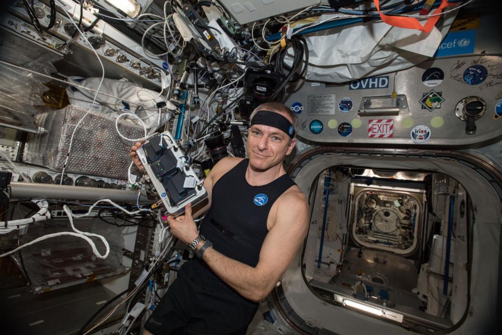
Caption: Canadian Space Agency astronaut David Saint-Jacques tries the Bio-Monitor, a new Canadian technology, for the first time in space (January 16, 2019). The innovative smart shirt system is designed to measure and record astronauts’ vital signs. Credit: Canadian Space Agency/NASA
A technology-packed tank top offers a simple, effective way to track astronauts’ vital signs and physiological changes during spaceflight, according to research being presented at the American Physiological Society annual meeting during the Experimental Biology (EB) 2021 meeting, held virtually April 27-30.
By monitoring key health markers over long periods of time with one non-intrusive device, researchers say the garment can help improve understanding of how spaceflight affects the body.
“Until now, the heart rate and activity levels of astronauts were monitored by separate devices,” said Carmelo Mastrandrea, PhD, a postdoctoral fellow at the Schlegel-University of Waterloo Research Institute for Aging in Canada, and the study’s first author. “The Bio-Monitor shirt allows simultaneous and continuous direct measurements of heart rate, breathing rate, oxygen saturation in the blood, physical activity and skin temperature, and provides a continuous estimate of arterial systolic blood pressure.”
The Bio-Monitor shirt was developed for the Canadian Space Agency by Carré Technologies based on its commercially available Hexoskin garment. In a study funded by the Canadian Space Agency, a team of researchers from the Schlegel-University of Waterloo Research Institute for Aging oversaw the first test of the shirt in space for a scientific purpose. Astronauts wore the shirt continually for 72 hours before their spaceflight and 72 hours during spaceflight, except for periods of water immersion or when the device conflicted with another activity.
The shirt’s sensors and accelerometer performed well, providing consistent results and a large amount of usable data. Based on these initial results, researchers say the shirt represents an improvement over conventional methods for monitoring astronauts’ health, which require more hands-on attention.
“By monitoring continuously and non-intrusively, we remove the psychological impacts of defined testing periods from astronaut measurements,” said Mastrandrea. “Additionally, we are able to gather information during normal activities over several days, including during daily activities and sleep, something that traditional testing cannot achieve.”
In flight, the astronauts recorded far less physical activity than the two and a half hours per day recorded in the monitoring period before takeoff, a finding that aligns with previous studies showing large reductions in physical activity during spaceflight. In addition to monitoring astronauts’ health and physical activity in space, Mastrandrea noted that the shirt could provide early warning of any health problems that occur as their bodies re-adapt to gravity back on Earth.
The commercial version of the Bio-Monitor shirt is available to the public, where it can be used for various applications including assessing athletic performance and monitoring the health of people with limited mobility. In addition to spaceflight, researchers are examining its potential use in other occupational settings that involve extreme environments, such as firefighting.
Mastrandrea will present this research in poster R2888 (abstract). Contact the media team for more information or to obtain a free press pass to access the virtual meeting.
###
About Experimental Biology 2021
Experimental Biology is an annual meeting comprised of thousands of scientists from five host societies and multiple guest societies. With a mission to share the newest scientific concepts and research findings shaping clinical advances, the meeting offers an unparalleled opportunity for exchange among scientists from across the U.S. and the world who represent dozens of scientific areas, from laboratory to translational to clinical research. http://www.experimentalbiology.org #expbio
About the American Physiological Society (APS)
Physiology is a broad area of scientific inquiry that focuses on how molecules, cells, tissues and organs function in health and disease. The American Physiological Society connects a global, multidisciplinary community of more than 10,000 biomedical scientists and educators as part of its mission to advance scientific discovery, understand life and improve health. The Society drives collaboration and spotlights scientific discoveries through its 16 scholarly journals and programming that support researchers and educators in their work. http://www.physiology.org
Carmelo Mastrandrea (Schlegel-UW Research Institute for Aging)| Danielle Greaves (Schlegel-UW Research Institute for Aging)| Richard Hughson (Schlegel-UW Research Institute for Aging)
Astronauts develop insulin resistance, and are at risk for cardiovascular deconditioning, during long-duration missions to the International Space Station (ISS) despite their daily exercise sessions (Hughson et al. Am J Physiol Heart Circ Physiol 310: H628–H638, 2016). Chronic unloading of the musculoskeletal and cardiovascular systems in microgravity dramatically reduces the challenge of daily activities, and the astronauts’ schedules limit them to approximately 30-min/day aerobic exercise. To understand the physical demands of spaceflight and how these change from daily life on Earth, the Vascular Aging experiment is equipping astronauts for 48-72h continuous recordings with the Canadian Space Agency’s Bio-Monitor wearable sensor shirt. The Bio-Monitor (Bio-M), developed from the commercial Hexoskin® device, consists of 3-lead ECG, thoracic and abdominal respiratory bands, 3-axis accelerometer, skin temperature and SpO2 sensor placed on the forehead. Our utilisation of this equipment necessitated the development of novel processing and visualisation techniques, to better interpret and guide subsequent data analyses [emphasis mine]. Here we present initial data from astronauts wearing the BioM prior to launch and aboard the ISS, demonstrating the ability to extract useful data from BioM, using software developed ‘in-house’.
Astronauts wore the Bio-M continually for 72-h except for periods of water immersion or when the device conflicted with another activity. After physical exercise, astronauts changed to a dry shirt. First, we assessed the key data-quality metrics to provide initial appraisals of acceptable recordings. Mean total recording length pre-flight (60.5 hours) was similar to that in-flight (66.5 hours), with a consistent distribution of recorded day (44% vs 45%, 6am-6pm) and night (56% vs 55%, 6pm-6am) hours (pre-flight vs in-flight respectively).
For each recording, quality assessment of ECG signals was performed for individual leads, before combining signals and cross-correlating R-waves to produce reliable heart-rate timings. Mean ECG quality for individual leads, represented here as the percentage of usable signal to total recording duration, was somewhat lower in-flight (92%) when compared to pre-flight (96%), likely caused by poor skin contact or dry shirt electrodes; combining lead signals as mentioned above improved the proportion of usable data to 97% and 98% respectively. Accelerometer recordings identified a significant reduction in high-force movements over the 72-hour recordings, with just over 2.5 hours/day of high-force activity in astronauts pre-flight vs 50 minutes/day in-flight. It should be noted however that accelerometer measurements in zero-gravity are likely to be reduced, and future refinement of activity data continues. Average heart rates in-flight showed little difference when compared to pre-flight, although future analyses will compare periods of sleep, rest, and activity to further refine this comparison.
We conclude that utilisation of the BioM hardware with our own analysis techniques produces high-quality data allowing for future interpretation and investigation of spaceflight-induced physiological adaptations.
As for Hexoskin (Carré Technologies inc.), I found out more on the About Us webpage of the Hexoskin website (Note: Links have been removed),
Hexoskin (Carré Technologies inc.)
Founded in 2006 in Montreal [Canada], Hexoskin is a growing private company, leader in non-invasive sensors, software, data science & AI services. The company headquartered in the bustling Rosemont neighborhood, provides solutions and services directly to customers & researchers; and through B2B contracts in pharmaceutical, academics, healthcare, security, defense, first responders, aerospace public & private organizations.
Hexoskin’s mission has always been to make the precise health data collected by its body-worn sensors accessible and useful for everyone. When the cofounders Pierre-Alexandre Fournier and Jean-François Roy started the company back in 2006, the existing technologies to report rich health data continuously didn’t exist. Hexoskin took a different approach to non-portable and invasive monitoring solutions by releasing in 2013 the first washable Smart Shirts that captures cardiac, respiratory, and activity body metrics. Today Hexoskin’s main R&D focus is the development of innovative body-worn sensors for health, mobile, and distributed software for health data management and analysis.
Since then, Hexoskin has designed the Hexoskin Connected Health Platform, a system to minimize user setup time and to maximize vital signs monitoring over long periods in a non-obstructive way with sensors embedded in a Smart Shirt. Data are synced to local and remote servers for health data management and analysis. The Hexoskin Smart Garments are clinically validated and are developed involving patients & clients to be comfortable and easy to use.
The system is the next evolution to improve the standard of care in the following therapeutic areas: respiratory, cardiology, mental health, behavioral and physiological psychology, somnology, aging and physical performance, physical conditioning & wellbeing etc.
…
Next Generation Biometric Smart Shirts
Hexoskin supported the evolution of its 100% washable industry-leading Hexoskin Smart Garments to offer an easy and comfortable solution for continuously monitoring precise data during daily activities and sleep. Hexoskin is a machine washable Smart garment, designed and made in Canada that allows precise long-term monitoring of respiratory, cardiac and activity functions simultaneously, as well as sleep quality.
Users are provided access the Hexoskin Connected Health Platform, an end-to-end system that supplies the tools to report and analyze precise data from the Hexoskin & third-party body-worn sensors. The platform offers apps for iOS, Android, and Watch OS devices. Users can access from anywhere an online dashboard with advanced reporting and analytics functionalities. Today, the Hexoskin Connected Platform is used worldwide and supported thousands of users and organizations to achieve their goals.
In 2019, Hexoskin launched the new Hexoskin ProShirt line for Men and Women with an all-new design to withstand the most active lifestyle and diverse daily living activities. The Hexoskin ProShirtcomes with built-in textile ECG & Respiratory sensors and a precise Activity sensor. The ProShirt works with the latest Hexoskin Smart recording device to offer uninterrupted continuous 24-hour monitoring.
Today, the Hexoskin ProShirt are used by professional athletes for performance training, police & first responders for longitudinal stress monitoring, and patients in clinical trials living with chronic cardiac & respiratory conditions.
Connected Health & Software Solutions
Hexoskin provides interoperable software solutions, secure and private infrastructure and data science services to support research and professional organizations. The system is designed to reduce the frequency of travel and allow remote communication between patients, study volunteers, caregivers, and researchers. Hexoskin is an efficient and precise solution that collects daily quantitative data from users, in their everyday lives, and over long periods of time.
Conscious of the need for its users to understand how the data is collected and interpreted, Hexoskin early took a transparent approach by opening and documenting its Application Programing Interface (API). Today, part of Hexoskin’s success can be attributed to its community of developers and scientists that are leveraging its Connected Health Platform to create new applications and interventions not possible just a few years ago.
Future Applications—remote health to space exploration
Since 2011, Hexoskin collaborated with the Canadian Space Agency on the Astroskin, a cutting edge Space Grade Smart Garment, now used in the International Space Station to monitor the astronauts’ health in Space. The Astroskin Vital Signs Monitoring Platform is also available to conduct research on earth.
Hexoskin hopes to bring the innovations developed for Space and its Hexoskin Connected Health Platform to support the growing need to provide patients’ access to affordable and adapted healthcare services remotely. Future applications include healthcare, chronic disease management, sleep medicine, aging at home, security & defense, and space exploration missions.
Thinking that Astroskin will be perfect for your next study or project? Contact us to discuss how Astroskin can support your next project. You can also request a demo of the Astroskin Vital Signs Monitoring Platform here.
Finally, I noticed that the researchers on this project were from the Schlegel-UW [University of Waterloo] Research Institute for Aging. I gather this was all about aging.
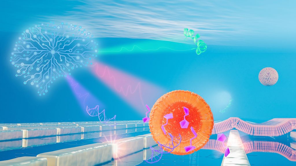
EPFL scientists have developed AI-powered nanosensors that let researchers track various kinds of biological molecules without disturbing them. Courtesy: École polytechnique fédérale de Lausanne (EPFL)
If you look at the big orange dot (representing the nanosensors?), you’ll see those purplish/fuschia objects resemble musical notes (biological molecules?). I think that brainlike object to the left and in light blue is the artificial intelligence (AI) component. (If anyone wants to correct my guesses or identify the bits I can’t, please feel free to add to the Comments for this blog.)
Getting back to my topic, keep the ‘musical notes’ in mind as you read about some of the latest research from l’École polytechnique fédérale de Lausanne (EPFL) in an April 7, 2021 news item on Nanowerk,
The tiny world of biomolecules is rich in fascinating interactions between a plethora of different agents such as intricate nanomachines (proteins), shape-shifting vessels (lipid complexes), chains of vital information (DNA) and energy fuel (carbohydrates). Yet the ways in which biomolecules meet and interact to define the symphony of life is exceedingly complex.
Scientists at the Bionanophotonic Systems Laboratory in EPFL’s School of Engineering have now developed a new biosensor that can be used to observe all major biomolecule classes of the nanoworld without disturbing them. Their innovative technique uses nanotechnology, metasurfaces, infrared light and artificial intelligence.
To each molecule its own melody
In this nano-sized symphony, perfect orchestration makes physiological wonders such as vision and taste possible, while slight dissonances can amplify into horrendous cacophonies leading to pathologies such as cancer and neurodegeneration.
“Tuning into this tiny world and being able to differentiate between proteins, lipids, nucleic acids and carbohydrates without disturbing their interactions is of fundamental importance for understanding life processes and disease mechanisms,” says Hatice Altug, the head of the Bionanophotonic Systems Laboratory.
Light, and more specifically infrared light, is at the core of the biosensor developed by Altug’s team. Humans cannot see infrared light, which is beyond the visible light spectrum that ranges from blue to red. However, we can feel it in the form of heat in our bodies, as our molecules vibrate under the infrared light excitation.
Molecules consist of atoms bonded to each other and – depending on the mass of the atoms and the arrangement and stiffness of their bonds – vibrate at specific frequencies. This is similar to the strings on a musical instrument that vibrate at specific frequencies depending on their length. These resonant frequencies are molecule-specific, and they mostly occur in the infrared frequency range of the electromagnetic spectrum.
“If you imagine audio frequencies instead of infrared frequencies, it’s as if each molecule has its own characteristic melody,” says Aurélian John-Herpin, a doctoral assistant at Altug’s lab and the first author of the publication. “However, tuning into these melodies is very challenging because without amplification, they are mere whispers in a sea of sounds. To make matters worse, their melodies can present very similar motifs making it hard to tell them apart.”
Metasurfaces and artificial intelligence
The scientists solved these two issues using metasurfaces and AI. Metasurfaces are man-made materials with outstanding light manipulation capabilities at the nano scale, thereby enabling functions beyond what is otherwise seen in nature. Here, their precisely engineered meta-atoms made out of gold nanorods act like amplifiers of light-matter interactions by tapping into the plasmonic excitations resulting from the collective oscillations of free electrons in metals. “In our analogy, these enhanced interactions make the whispered molecule melodies more audible,” says John-Herpin.
AI is a powerful tool that can be fed with more data than humans can handle in the same amount of time and that can quickly develop the ability to recognize complex patterns from the data. John-Herpin explains, “AI can be imagined as a complete beginner musician who listens to the different amplified melodies and develops a perfect ear after just a few minutes and can tell the melodies apart, even when they are played together – like in an orchestra featuring many instruments simultaneously.”
The first biosensor of its kind
When the scientists’ infrared metasurfaces are augmented with AI, the new sensor can be used to analyze biological assays featuring multiple analytes simultaneously from the major biomolecule classes and resolving their dynamic interactions.
“We looked in particular at lipid vesicle-based nanoparticles and monitored their breakage through the insertion of a toxin peptide and the subsequent release of vesicle cargos of nucleotides and carbohydrates, as well as the formation of supported lipid bilayer patches on the metasurface,” says Altug.
This pioneering AI-powered, metasurface-based biosensor will open up exciting perspectives for studying and unraveling inherently complex biological processes, such as intercellular communication via exosomesand the interaction of nucleic acids and carbohydrates with proteins in gene regulation and neurodegeneration.
“We imagine that our technology will have applications in the fields of biology, bioanalytics and pharmacology – from fundamental research and disease diagnostics to drug development,” says Altug.
A Dec. 7, 2020 news item on Nanowerk announced a new technology for rapid COVID-19 testing (Note: A link has been removed),
As the COVID-19 pandemic continues to spread across the world, testing remains a key strategy for tracking and containing the virus. Bioengineering graduate student, Maha Alafeef, has co-developed a rapid, ultrasensitive test using a paper-based electrochemical sensor that can detect the presence of the virus in less than five minutes.
The team led by professor Dipanjan Pan reported their findings in ACS Nano (“Rapid, Ultrasensitive, and Quantitative Detection of SARS-CoV-2 Using Antisense Oligonucleotides Directed Electrochemical Biosensor Chip”).
“Currently, we are experiencing a once-in-a-century life-changing event,” said Alafeef. “We are responding to this global need from a holistic approach by developing multidisciplinary tools for early detection and diagnosis and treatment for SARS-CoV-2.”
…
I wonder why they didn’t think to provide a caption for the graphene substrate (the square surface) underlying the gold electrode (the round thing) or provide a caption for the electrode. Maybe they assumed anyone knowledgeable about graphene would be able to identify it?
Caption: COVID-19 electrochemical sensing platform. Credit: University of Illinois
There are two broad categories of COVID-19 tests on the market. The first category uses reverse transcriptase real-time polymerase chain reaction (RT-PCR) and nucleic acid hybridization strategies to identify viral RNA. Current FDA [US Food and Drug Administration]-approved diagnostic tests use this technique. Some drawbacks include the amount of time it takes to complete the test, the need for specialized personnel and the availability of equipment and reagents.
The second category of tests focuses on the detection of antibodies. However, there could be a delay of a few days to a few weeks after a person has been exposed to the virus for them to produce detectable antibodies.
In recent years, researchers have had some success with creating point-of-care biosensors using 2D nanomaterials such as graphene to detect diseases. The main advantages of graphene-based biosensors are their sensitivity, low cost of production and rapid detection turnaround. “The discovery of graphene opened up a new era of sensor development due to its properties. Graphene exhibits unique mechanical and electrochemical properties that make it ideal for the development of sensitive electrochemical sensors,” said Alafeef. The team created a graphene-based electrochemical biosensor with an electrical read-out setup to selectively detect the presence of SARS-CoV-2 genetic material.
There are two components [emphasis mine] to this biosensor: a platform to measure an electrical read-out and probes to detect the presence of viral RNA. To create the platform, researchers first coated filter paper with a layer of graphene nanoplatelets to create a conductive film [emphasis mine]. Then, they placed a gold electrode with a predefined design on top of the graphene [emphasis mine] as a contact pad for electrical readout. Both gold and graphene have high sensitivity and conductivity which makes this platform ultrasensitive to detect changes in electrical signals.
Current RNA-based COVID-19 tests screen for the presence of the N-gene (nucleocapsid phosphoprotein) on the SARS-CoV-2 virus. In this research, the team designed antisense oligonucleotide (ASOs) probes to target two regions of the N-gene. Targeting two regions ensures the reliability of the senor in case one region undergoes gene mutation. Furthermore, gold nanoparticles (AuNP) are capped with these single-stranded nucleic acids (ssDNA), which represents an ultra-sensitive sensing probe for the SARS-CoV-2 RNA.
The researchers previously showed the sensitivity of the developed sensing probes in their earlier work published in ACS Nano. The hybridization of the viral RNA with these probes causes a change in the sensor electrical response. The AuNP caps accelerate the electron transfer and when broadcasted over the sensing platform, results in an increase in the output signal and indicates the presence of the virus.
The team tested the performance of this sensor by using COVID-19 positive and negative samples. The sensor showed a significant increase in the voltage of positive samples compared to the negative ones and confirmed the presence of viral genetic material in less than five minutes. Furthermore, the sensor was able to differentiate viral RNA loads in these samples. Viral load is an important quantitative indicator of the progress of infection and a challenge to measure using existing diagnostic methods.
This platform has far-reaching applications due to its portability and low cost. The sensor, when integrated with microcontrollers and LED screens or with a smartphone via Bluetooth or wifi, could be used at the point-of-care in a doctor’s office or even at home. Beyond COVID-19, the research team also foresees the system to be adaptable for the detection of many different diseases.
“The unlimited potential of bioengineering has always sparked my utmost interest with its innovative translational applications,” Alafeef said. “I am happy to see my research project has an impact on solving a real-world problem. Finally, I would like to thank my Ph.D. advisor professor Dipanjan Pan for his endless support, research scientist Dr. Parikshit Moitra, and research assistant Ketan Dighe for their help and contribution toward the success of this study.”
I’m not sure where I found this notice but it is most definitely from the American Chemical Society: “This paper is freely accessible, at this time, for unrestricted RESEARCH re-use and analyses in any form or by any means with acknowledgement of the original source. These permissions are granted for the duration of the World Health Organization (WHO) declaration of COVID-19 as a global pandemic.”
I think I can safely say that Carson J. Bruns, a Professor at the University of Colorado Boulder, is an electronic tattoo enthusiast. His Sept. 24, 2020 essay on electronic tattoos for The Conversation (also found on Fast Company) outlines a very rosy view of a future where health monitoring is constant and visible on your skin (Note: Links have been removed),
In the sci-fi novel “The Diamond Age” by Neal Stephenson, body art has evolved into “constantly shifting mediatronic tattoos” – in-skin displays powered by nanotech robopigments. In the 25 years since the novel was published, nanotechnology has had time to catch up, and the sci-fi vision of dynamic tattoos is starting to become a reality.
The first examples of color-changing nanotech tattoos have been developed over the past few years, and they’re not just for body art. They have a biomedical purpose. Imagine a tattoo that alerts you to a health problem signaled by a change in your biochemistry, or to radiation exposure that could be dangerous to your health.
You can’t walk into a doctor’s office and get a dynamic tattoo yet, but they are on the way. …
…
In 2017, researchers tattooed pigskin, which had been removed from the pig, with molecular biosensors that use color to indicate sodium, glucose or pH levels in the skin’s fluids.
In 2019, a team of researchers expanded on that study to include protein sensing and developed smartphone readouts for the tattoos. This year, they also showed that electrolyte levels could be detected with fluorescent tattoo sensors.
In 2018, a team of biologists developed a tattoo made of engineered skin cells that darken when they sense an imbalance of calcium caused by certain cancers. They demonstrated the cancer-detecting tattoo in living mice.
…
My lab is looking at tech tattoos from a different angle. We are interested in sensing external harms, such as ultraviolet radiation. UV exposure in sunlight and tanning beds is the main risk factor for all types of skin cancer. Nonmelanoma skin cancers are the most common malignancies in the U.S., Australia and Europe.
…
I served as the first human test subject for these tattoos. I created “solar freckles” on my forearm – invisible spots that turned blue under UV exposure and reminded me when to wear sunscreen. My lab is also working on invisible UV-protective tattoos that would absorb UV light penetrating through the skin, like a long-lasting sunscreen just below the surface. We’re also working on “thermometer” tattoos using temperature-sensitive inks. Ultimately, we believe tattoo inks could be used to prevent and diagnose disease.
…
Temporary transfer tattoos are also undergoing a high-tech revolution. Wearable electronic tattoos that can sense electrophysiological signals like heart rate and brain activity or monitor hydration and glucose levels from sweat are under development. They can even be used for controlling mobile devices, for example shuffling a music playlist at the touch of a tattoo, or for luminescent body art that lights up the skin.
The advantage of these wearable tattoos is that they can use battery-powered electronics. The disadvantage is that they are much less permanent and comfortable than traditional tattoos. Likewise, electronic devices that go underneath the skin are being developed by scientists, designers and biohackers alike, but they require invasive surgical procedures for implantation.
Tattoos injected into the skin offer the best of both worlds: minimally invasive, yet permanent and comfortable. [emphasis mine] New needle-free tattooing methods that fire microscopic ink droplets into the skin are now in development. Once perfected they will make tattooing quicker and less painful.
…
The color-changing tattoos in development are also going to open the door to a new kind of dynamic body art. Now that tattoo colors can be changed by an electromagnetic signal, you’ll soon be able to “program” your tattoo’s design, or switch it on and off. You can proudly display your neck tattoo at the motorcycle rally and still have clear skin in the courtroom.
As researchers develop dynamic tattoos, they’ll need to study the safety [emphasis mine] of the high-tech inks. As it is, little is known about the safety of the more than 100 different pigments used in normal tattoo inks [emphasis mine]. The U.S. Food and Drug Administration has not exercised regulatory authority over tattoo pigments, citing other competing public health priorities and a lack of evidence of safety problems with the pigments. So U.S. manufacturers can put whatever they want in tattoo inks [emphasis mine] and sell them without FDA approval.
…
A wave of high-tech tattoos is slowly upwelling, and it will probably keep rising for the foreseeable future. When it arrives, you can decide to surf or watch from the beach. If you do climb on board, you’ll be able to check your body temperature or UV exposure by simply glancing at one of your tattoos.
There are definitely some interesting possibilities, artistic, health, and medical, offered by electronic tattoos. As you may have guessed, I’m not quite the enthusiast that Dr. Bruns seems to be but I could be persuaded, assuming there’s evidence to support the claims.
The Velcro-like food sensor, made from an array of silk microneedles, can pierce through plastic packaging to sample food for signs of spoilage and bacterial contamination. Image: Felice Frankel
MIT engineers have designed a Velcro-like food sensor, made from an array of silk microneedles, that pierces through plastic packaging to sample food for signs of spoilage and bacterial contamination.
The sensor’s microneedles are molded from a solution of edible proteins found in silk cocoons, and are designed to draw fluid into the back of the sensor, which is printed with two types of specialized ink. One of these “bioinks” changes color when in contact with fluid of a certain pH range, indicating that the food has spoiled; the other turns color when it senses contaminating bacteria such as pathogenic E. coli.
The researchers attached the sensor to a fillet of raw fish that they had injected with a solution contaminated with E. coli. After less than a day, they found that the part of the sensor that was printed with bacteria-sensing bioink turned from blue to red — a clear sign that the fish was contaminated. After a few more hours, the pH-sensitive bioink also changed color, signaling that the fish had also spoiled.
The results, published today in the journal Advanced Functional Materials, are a first step toward developing a new colorimetric sensor that can detect signs of food spoilage and contamination.
Such smart food sensors might help head off outbreaks such as the recent salmonella contamination in onions and peaches. They could also prevent consumers from throwing out food that may be past a printed expiration date, but is in fact still consumable.
“There is a lot of food that’s wasted due to lack of proper labeling, and we’re throwing food away without even knowing if it’s spoiled or not,” says Benedetto Marelli, the Paul M. Cook Career Development Assistant Professor in MIT’s Department of Civil and Environmental Engineering. “People also waste a lot of food after outbreaks, because they’re not sure if the food is actually contaminated or not. A technology like this would give confidence to the end user to not waste food.”
Marelli’s co-authors on the paper are Doyoon Kim, Yunteng Cao, Dhanushkodi Mariappan, Michael S. Bono Jr., and A. John Hart.
Silk and printing
The new food sensor is the product of a collaboration between Marelli, whose lab harnesses the properties of silk to develop new technologies, and Hart, whose group develops new manufacturing processes.
Hart recently developed a high-resolution floxography technique, realizing microscopic patterns that can enable low-cost printed electronics and sensors. Meanwhile, Marelli had developed a silk-based microneedle stamp that penetrates and delivers nutrients to plants. In conversation, the researchers wondered whether their technologies could be paired to produce a printed food sensor that monitors food safety.
“Assessing the health of food by just measuring its surface is often not good enough. At some point, Benedetto mentioned his group’s microneedle work with plants, and we realized that we could combine our expertise to make a more effective sensor,” Hart recalls.
The team looked to create a sensor that could pierce through the surface of many types of food. The design they came up with consisted of an array of microneedles made from silk.
“Silk is completely edible, nontoxic, and can be used as a food ingredient, and it’s mechanically robust enough to penetrate through a large spectrum of tissue types, like meat, peaches, and lettuce,” Marelli says.
A deeper detection
To make the new sensor, Kim first made a solution of silk fibroin, a protein extracted from moth cocoons, and poured the solution into a silicone microneedle mold. After drying, he peeled away the resulting array of microneedles, each measuring about 1.6 millimeters long and 600 microns wide — about one-third the diameter of a spaghetti strand.
The team then developed solutions for two kinds of bioink — color-changing printable polymers that can be mixed with other sensing ingredients. In this case, the researchers mixed into one bioink an antibody that is sensitive to a molecule in E. coli. When the antibody comes in contact with that molecule, it changes shape and physically pushes on the surrounding polymer, which in turn changes the way the bioink absorbs light. In this way, the bioink can change color when it senses contaminating bacteria.
The researchers made a bioink containing antibodies sensitive to E. coli, and a second bioink sensitive to pH levels that are associated with spoilage. They printed the bacteria-sensing bioink on the surface of the microneedle array, in the pattern of the letter “E,” next to which they printed the pH-sensitive bioink, as a “C.” Both letters initially appeared blue in color.
Kim then embedded pores within each microneedle to increase the array’s ability to draw up fluid via capillary action. To test the new sensor, he bought several fillets of raw fish from a local grocery store and injected each fillet with a fluid containing either E. coli, Salmonella, or the fluid without any contaminants. He stuck a sensor into each fillet. Then, he waited.
After about 16 hours, the team observed that the “E” turned from blue to red, only in the fillet contaminated with E. coli, indicating that the sensor accurately detected the bacterial antigens. After several more hours, both the “C” and “E” in all samples turned red, indicating that every fillet had spoiled.
The researchers also found their new sensor indicates contamination and spoilage faster than existing sensors that only detect pathogens on the surface of foods.
“There are many cavities and holes in food where pathogens are embedded, and surface sensors cannot detect these,” Kim says. “So we have to plug in a bit deeper to improve the reliability of the detection. Using this piercing technique, we also don’t have to open a package to inspect food quality.”
The team is looking for ways to speed up the microneedles’ absorption of fluid, as well as the bioinks’ sensing of contaminants. Once the design is optimized, they envision the sensor could be used at various stages along the supply chain, from operators in processing plants, who can use the sensors to monitor products before they are shipped out, to consumers who may choose to apply the sensors on certain foods to make sure they are safe to eat.