A fascinating new use for hyaluronic acid (usually discussed in relation to cosmetic wrinkle-reduction) has been found according to a November 1, 2023 news item on ScienceDaily.
In a recent publication in the journal Nature, researchers from the Institute of Basic Science (IBS) in South Korea have made significant strides in biomaterial technology and rehabilitation medicine. They’ve developed a novel approach to healing muscle injury by employing “injectable tissue prosthesis” in the form of conductive hydrogels and combining it with a robot-assisted rehabilitation system.
Let’s imagine you are swimming in the ocean. A giant shark approaches and bites a huge chunk of meat out of your thigh, resulting in a complete loss of motor/sensor function in your leg.
If left untreated, such severe muscle damage would result in permanent loss of function and disability.
How on Earth will you be able to recover from this kind of injury?
Traditional rehabilitation methods for these kinds of muscle injuries have long sought an efficient closed-loop gait rehabilitation system that merges lightweight exoskeletons and wearable/implantable devices.
Such assistive prosthetic system is required to aid the patients through the process of recovering sensory and motor functions linked to nerve and muscle damage.
Unfortunately, the mechanical properties and rigid nature of existing electronic materials render them incompatible with soft tissues.
This leads to friction and potential inflammation, stalling patient rehabilitation.
To overcome these limitations, the IBS researchers turned to a material commonly used as a wrinkle-smoothing filler, called hyaluronic acid.
Using this substance [hyaluronic acid], an injectable hydrogel was developed for “tissue prostheses”, which can temporarily fill the gap of the missing muscle/nerve tissues while it regenerates. The injectable nature of this material gives it a significant advantage over traditional bioelectronic devices, which are unsuitable for narrow, deep, or small areas, and necessitate invasive surgeries.
Thanks to its highly “tissue-like” properties, this hydrogel seamlessly interfaces with biological tissues and can be easily administered to hard-to-reach body areas without surgery. The reversible and irreversible crosslinks within the hydrogel adapt to high shear stress during injection, ensuring excellent mechanical stability. This hydrogel also incorporates gold nanoparticles, which gives it decent electrical properties. Its conductive nature allows for the effective transmission of electrophysiological signals between the two ends of injured tissues. In addition, the hydrogel is biodegrdable, meaning that the patients do not need to get surgery again.
With mechanical properties akin to natural tissues, exceptional tissue adhesion, and injectable characteristics, researchers believe this material offers a novel approach to rehabilitation.
Next, the researchers put this novel idea to the test in rodent models. To simulate volumetric muscle loss injury, a large chunk of muscle has been removed from the hind legs of these animals. By injecting the hydrogel and implanting the two kinds of stretchable tissue-interfacing devices for electrical sensing and stimulation, the researchers were able to improve the gait in the “injured” rodents. The hydrogel prosthetics were combined with robot assistance, guided by muscle electromyography signals. Together, the two helped enhance the animal’s gait without nerve stimulation. Furthermore, muscle tissue regeneration was effectively improved over the long term after the conductive hydrogel was used to fill muscle damage.
The injectable conductive hydrogel developed in this study excels in electrophysiological signal recording and stimulation performance, offering the potential to expand its applications. It presents a fresh approach to the field of bioelectronic devices and holds promise as a soft tissue prosthesis for rehabilitation support.
Emphasizing the significance of the research, Professor SHIN Mikyung notes, “We’ve created an injectable, mechanically tough, and electrically conductive soft tissue prosthesis ideal for addressing severe muscle damage requiring neuromusculoskeletal rehabilitation. The development of this injectable hydrogel, utilizing a novel cross-linking method, is a notable achievement. We believe it will be applicable not only in muscles and peripheral nerves but also in various organs like the brain and heart.”
Professor SON Donghee added, “In this study, the closed-loop gait rehabilitation system entailing tough injectable hydrogel and stretchable and self-healing sensors could significantly enhance the rehabilitation prospects for patients with neurological and musculoskeletal challenges. It could also play a vital role in precise diagnosis and treatment across various organs in the human body.”
The research team is currently pursuing further studies to develop new materials for nerve and muscle tissue regeneration that can be implanted in a minimally invasive manner. They are also exploring the potential for recovery in various tissue damages through the injection of the conductive hydrogel, eliminating the need for open surgery.
Here’s a link to and a citation for the paper,
Injectable tissue prosthesis for instantaneous closed-loop rehabilitation by Subin Jin, Heewon Choi, Duhwan Seong, Chang-Lim You, Jong-Sun Kang, Seunghyok Rho, Won Bo Lee, Donghee Son & Mikyung Shin. Nature volume 623, pages 58–65 (2023) DOI: https://doi.org/10.1038/s41586-023-06628-x Published: 01 November 2023 Issue Date: 02 November 2023
Who doesn’t love a panda? It looks like someone is drawing on the armband with their fingers but the lines look a lot finer, more like a stylus was used.
Caption: When a person draws a panda on this touch-responsive armband that’s worn on their forearm (bottom right of photo), it shows up on a computer. Credit: Adapted from ACS Nano 2023, DOI: 10.1021/acsnano.2c12612
It’s time to roll up your sleeves for the next advance in wearable technology — a fabric armband that’s actually a touch pad. In ACS [American Chemical Society] Nano, researchers say they have devised a way to make playing video games, sketching cartoons and signing documents easier. Their proof-of-concept silk armband turns a person’s forearm into a keyboard or sketchpad. The three-layer, touch-responsive material interprets what a user draws or types and converts it into images on a computer.
Computer trackpads and electronic signature-capture devices seem to be everywhere, but they aren’t as widely used in wearables. Researchers have suggested making flexible touch-responsive panels from clear, electrically conductive hydrogels, but these substances are sticky, making them hard to write on and irritating to the skin. So, Xueji Zhang, Lijun Qu, Mingwei Tian and colleagues wanted to incorporate a similar hydrogel into a comfortable fabric sleeve for drawing or playing games on a computer.
The researchers sandwiched a pressure-sensitive hydrogel between layers of knit silk. The top piece was coated in graphene nanosheets to make the fabric electrically conductive. Attaching the sensing panel to electrodes and a data collection system produced a pressure-responsive pad with real-time, rapid sensing when a finger slid over it, writing numbers and letters. The device was then incorporated into an arm-length silk sleeve with a touch-responsive area on the forearm. In experiments, a user controlled the direction of blocks in a computer game and sketched colorful cartoons in a computer drawing program from the armband. The researchers say that their proof-of-concept wearable touch panel could inspire the next generation of flexible keyboards and wearable sketchpads.
By combining seaweed and graphene, scientists have been able to create sensors that can be worn like a ‘second skin’ and outperform other similar biosensors, according to a March 3, 2023 news item on ScienceDaily,
Scientists at the University of Sussex have successfully trialed new biodegradable health sensors that could change the way we experience personal healthcare and fitness monitoring technology.
The team at Sussex have developed the new health sensors — such as those worn by runners or patients to monitor heart rate and temperature — using natural elements like rock salt, water and seaweed, combined with graphene. Because they are solely made with ingredients found in nature, the sensors are fully biodegradable, making them more environmentally friendly than commonly used rubber and plastic-based alternatives. Their natural composition also places them within the emerging scientific field of edible electronics — electronic devices that are safe for a person to consume.
Better still, the researchers found that their sustainable seaweed-based sensors actually outperform existing synthetic based hydrogels and nanomaterials, used in wearable health monitors, in terms of sensitivity. Therefore, improving the accuracy, as the more sensitive a sensor, the more accurately it will record a person’s vital signs.
Dr Conor Boland, a materials physics lecturer in the School of Mathematical and Physical Sciences, said: “I was first inspired to use seaweed in the lab after watching MasterChef during lockdown. Seaweed, when used to thicken deserts, gives them a soft and bouncy structure – favored by vegans and vegetarians as an alternative to gelatin. It got me thinking: “what if we could do that with sensing technology?”.
“For me, one of the most exciting aspects to this development is that we have a sensor that is both fully biodegradable and highly effective. The mass production of unsustainable rubber and plastic based health technology could, ironically, pose a risk to human health through microplastics leeching into water sources as they degrade.
“As a new parent, I see it as my responsibility to ensure my research enables the realisation of a cleaner world for all our children.”
Seaweed is first and foremost an insulator, but by adding a critical amount of graphene to a seaweed mixture the scientists were able to create an electrically conductive film. When soaked in a salt bath, the film rapidly absorbs water, resulting in a soft, spongy, electrically conductive hydrogel.
The development has the potential to revolutionise health monitoring technology, as future applications of the clinical grade wearable sensors would look something like a second skin or a temporary tattoo: lightweight, easy to apply, and safe, as they are made with all natural ingredients. This would significantly improve the overall patient experience, without the need for more commonly used and potentially invasive hospital instruments, wires and leads.
Dr Sue Baxter, Director of Innovation and Business Partnerships at the University of Sussex, is excited about the potential benefits of this technology: “At the University of Sussex, we are committed to protecting the future of the planet through sustainability research, expertise and innovation. What’s so exciting about this development from Dr Conor Boland and his team is that it manages to be all at once truly sustainable, affordable, and highly effective – out-performing synthetic alternatives.
“What’s also remarkable for this stage of research – and I think this speaks to the meticulous ground-work that Dr Boland and his team put in when they created their blueprint – is that it’s more than a proof of principle development. Our Sussex scientists have created a device that has real potential for industry development into a product from which you or I could benefit in the relatively near future.”
This latest research breakthrough follows the publication of a blueprint for nanomaterial development from the Sussex scientists in 2019, which presented a method for researchers to follow in order to optimise the development of nanomaterial sensors.
…
Kevin Doty, a Masters student in the School of Mathematical and Physical Sciences, at the University of Sussex, said: “I taught chemistry previously, but decided I wanted to learn more about nanoscience. My gamble paid off, and not only did I enjoy it more than I expected, but I also ended up with an opportunity to utilize the information I had learned to work on a novel idea that has evolved into a first author publication as an MSc student. Learning about nanoscience showed me just how varied and multidisciplinary the field is. Any science background can bring knowledge that can be applied to this field in a unique way. This has led to further studies in a PhD studentship, opening up an all new career path I could not have previously considered.”
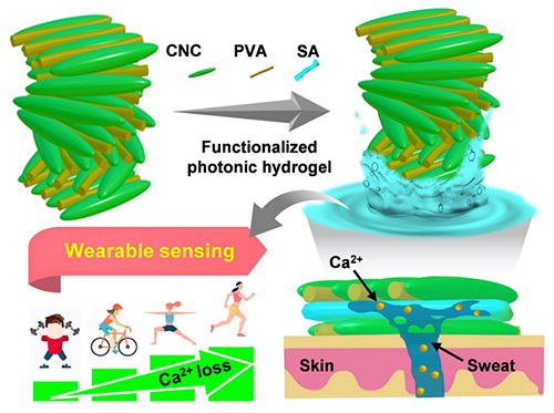
It turns out there’s also a hydrogel aspect to this story about a flexible sweat sensor. As for cellulose nanocrystals (CNC), Canada played a leading role in the development of this nanomaterial and I have a bit more about the Canadian CNC scene later in this posting following the link and citation for the research paper. On to the research,
Highly elastic hydrogels constructed by heat-induced hydrogen bond remodeling can switch between wet and dry states (Image by ZHANG Fusheng and LI Qiongya)
A May 8, 2023 news item on phys.org features this work from the Dalian Institute of Chemical Physics of the Chinese Academy Sciences,
Cellulose nanocrystal (CNC), an emerging bio-based material, has been widely applied in fields such as electronics, bioplastics and energy. However, the functional failure of such materials in wet or liquid environments inevitably impairs their development in biomedicine, membrane separation, environmental monitoring, and wearable devices.
Now, a research group led by Prof. Qing Guangyan from the Dalian Institute of Chemical Physics (DICP) of the Chinese Academy of Sciences [CAS] reported a sustainable, insoluble, and chiral photonic cellulose nanocrystal patch for calcium ion (Ca2+) sensing in sweat.
The researchers developed a simple and efficient method to fabricate insoluble CNC-based hydrogels. They found that by utilizing intermolecular hydrogen bond reconstruction, thermal dehydration enabled the optimized CNC composite photonic film to form a stable hydrogel network in an aqueous solution. Moreover, they indicated that the hydrogel could be reversibly switched between dry and wet states, which was convenient for specific functionalization.
The introduction of functionalized molecules by adsorption swelling in a liquid environment resulted in a hydrogel with freeze resistance (–20°C), strong adhesion, good biocompatibility, and high sensitivity to Ca2+.
“This work is expected to facilitate the application of sustainable cellulose sensors to monitor other metabolites (i.e., glucose, urea, and vitamins, etc.),” said Prof. QING. “It also lays foundation for digitally controlled hydrogel systems operating in environment monitoring, membrane separation, and wearable devices.”
FPInnovations is a Canadian research and development (R&D) not-for profit organization that was instrumental in the development of CNC. (If memory serves, they are a spinoff from the University of British Columbia.) There are two Canadian CNC production facilities (that I know of): CelluForce in Québec and Blue Goose Biorefineries in Saskatchewan. I get more information about research into applications for CNC from other parts of the world while the Canadian scene remains mostly silent.
Seems almost magical but it takes years to do this research. That video was posted in September 2019 and the latest research is being announced in a February 28, 2022 news item on phys.org,
Hydrogels have an astonishing ability to swell and take on water. In daily life, they are used in dressings, nappies, and more to lock moisture away. A team of researchers has now found another use: quickly extracting large amounts of freshwater from air using a specially developed hydrogel containing a hygroscopic salt. The study, published in the journal Angewandte Chemie, shows that the salt enhances the moisture uptake of the gel, making it suitable for water harvesting in dry regions.
Hydrogels can absorb and store many times their weight in water. In so doing, the underlying polymer swells considerably by incorporating water. However, to date, use of this property to produce freshwater from atmospheric water has not been feasible, since collecting moisture from the air is still too slow and inefficient.
On the other hand, moisture absorption could be enhanced by adding hygroscopic salts that can rapidly remove large amounts of moisture from the air. However, hygroscopic salts and hydrogels are usually not compatible, as a large amount of salt influences the swelling capability of the hydrogel and thus degrades its properties. In addition, the salt ions are not tightly coordinated within the gel and are easily washed away.
The materials scientist Guihua Yu and his team at the University of Texas at Austin, USA, have now overcome these issues by developing a particularly “salt-friendly” hydrogel. As their study shows, this gel gains the ability to absorb and retain water when combined with a hygroscopic salt. Using their hydrogel, the team were able to extract almost six liters of pure water per kilo of material in 24 hours, from air with 30% relative humidity.
The basis for the new hydrogel was a polymer constructed from zwitterionic molecules. Polyzwitterions carry both positive and negative charged functional groups, which helped the polymer to become more responsive to the salt in this case. Initially, the molecular strands in the polymer were tightly intermingled, but when the researchers added the lithium chloride salt, the strands relaxed and a porous, spongy hydrogel was formed. This hydrogel loaded with the hygroscopic salt was able to incorporate water molecules quickly and easily.
In fact, water incorporation was so quick and easy that the team were able to set up a cyclical system for continuous water separation. They left the hydrogel for an hour each time to absorb atmospheric moisture, then dried the gel in a condenser to collect the condensed water. They repeated this procedure multiple times without it resulting in any substantial loss of the amount of water absorbed, condensed, or collected.
Yu and the team say that the as-prepared hydrogel “should be optimal for efficient moisture harvesting for the potential daily water yield”. They add that polyzwitterionic hydrogels could play a fundamental role in the future for recovering atmospheric water in arid, drought-stricken regions.
Here’s a link to and a citation for the paper,
Polyzwitterionic Hydrogels for Efficient Atmospheric Water Harvesting by Chuxin Lei, Youhong Guo, Weixin Guan, Hengyi Lu, Wen Shi, Guihua Yu. Angewandte Chemie International Edition Volume 61, Issue1 3 March 21, 2022 e202200271 DOI: https://doi.org/10.1002/anie.202200271 First published: 28 January 2022
The word ‘living’ isn’t usually associated with optical fibers and the addition had me thinking that this October 11, 2021 Nanowerk Spotlight story by Michael Berger would be a synthetic biology story. Well, not exactly. Do read on for a good introduction describing glass, fiber optics, and optogenetics,
Glass is one of the oldest manufactured materials used by humans and glass making dates back at least 6000 years, long before humans had discovered how to smelt iron. Glasses have been based on the chemical compound silica – silicon dioxide, or quartz – the primary constituent of sand. Soda-lime glass, containing around 70% silica, accounts for around 90% of manufactured glass.
Historically, we are familiar with glasses’ decorative use or as window panes, household items, and in optics such as eyeglasses, microscopes and telescopes. More recently, starting in the 1950s, glass has been used in the manufacture of fiber optic cables, a technology that has revolutionized the communications industry and helped ring in the digital revolution.
Fiber optic cables propagate a signal as a pulse of light along a transparent medium, usually glass. This is not only used to transmit information but, for instance in many healthcare and biomedical applications, scientists use optical fibers for sensing applications by shining light into a sample and evaluating the absorbed or transmitted light.
A recent development in this field is optogenetics, a neuromodulation method that uses activation or deactivation of brain cells by illumination with different colors of light in order to treat brain disorders.
…
Berger goes on to explain the latest work and reveals what ‘living’ means where this work is concerned,
This work represents a simple and low-cost approach to fabricating optical fibers made from biological materials. These fibers can be easily modified for specific applications and don’t require sophisticated equipment to generate relevant information. This method could be used for many practical sensing and biological modeling applications.
…
“We use a natural, ionic, and biologically compatible crosslinking approach, which enables us to produce flexible hydrogel fibers in continuous multi-layered architectures, meaning they are easy to produce and can be modified after fabrication,” explains Guimarães [Carlos Guimarães, the paper’s first author]. “Similarly to silica fibers, the core hydrogel of our structures can be exposed, fused to another fiber or reassembled if they break, and efficiently guide light through the established connection.”
These flexible hydrogel fibers are made from sugars and work just like solid-state optical fibers used to transmit data. However, they are biocompatible so they can be easily integrated with biological systems.
“We could even consider them to be alive [emphasis mine] since we can use them to grow living cells inside the fiber,” says Guimarães. “As these embedded cells grow over time, we can then use light to inform on living dynamic events, for example to track cancer invasive proliferation into optical information.” [emphasis mine]
…
As to what constitutes optical information in this context,
Another intriguing aspect of these hydrogel fibers is that their permeable mesh enables the inclusion of biological targets of interest for detection. For example, the scientists observed that fibers were able to soak SARS-CoV-2 viruses, and by integrating nanoparticles for their binding and detection, shifts in visible light could be observed for detecting the accumulation of viral particles within the fiber.
“When light moving through the fiber encounters living cells, it changes its characteristics depending on cellular density, invasive proliferation, expression of molecules, etc.” Guimarães notes. “This light-cell interaction can digitize complex biological events, converting responses such as cancer cell progression in 3D environments and susceptibility to drugs into numbers and data, very fast and without the need for sample destruction.”
A May 4, 2021 news item on ScienceDaily announced work that may result the restoration of nasal cartilage for skin cancer patients,
A team of University of Alberta researchers has discovered a way to use 3-D bioprinting technology to create custom-shaped cartilage for use in surgical procedures. The work aims to make it easier for surgeons to safely restore the features of skin cancer patients living with nasal cartilage defects after surgery.
The researchers used a specially designed hydrogel — a material similar to Jell-O — that could be mixed with cells harvested from a patient and then printed in a specific shape captured through 3-D imaging. Over a matter of weeks, the material is cultured in a lab to become functional cartilage.
“It takes a lifetime to make cartilage in an individual, while this method takes about four weeks. So you still expect that there will be some degree of maturity that it has to go through, especially when implanted in the body. But functionally it’s able to do the things that cartilage does,” said Adetola Adesida, a professor of surgery in the Faculty of Medicine & Dentistry.
“It has to have certain mechanical properties and it has to have strength. This meets those requirements with a material that (at the outset) is 92 per cent water,” added Yaman Boluk, a professor in the Faculty of Engineering.
…
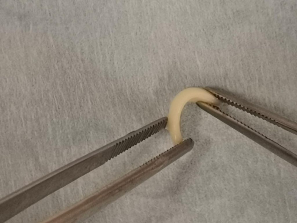
Who would have thought that nose cartilage would look like a worm?
Caption: 3-D printed cartilage is shaped into a curve suitable for use in surgery to rebuild a nose. The technology could eventually replace the traditional method of taking cartilage from the patient’s rib, a procedure that comes with complications. Credit: University of Alberta
Adesida, Boluk and graduate student Xiaoyi Lan led the project to create the 3-D printed cartilage in hopes of providing a better solution for a clinical problem facing many patients with skin cancer.
Each year upwards of three million people in North America are diagnosed with non-melanoma skin cancer. Of those, 40 per cent will have lesions on their noses, with many requiring surgery to remove them. As part of the procedure, many patients may have cartilage removed, leaving facial disfiguration.
Traditionally, surgeons would take cartilage from one of the patient’s ribs and reshape it to fit the needed size and shape for reconstructive surgery. But the procedure comes with complications.
“When the surgeons restructure the nose, it is straight. But when it adapts to its new environment, it goes through a period of remodelling where it warps, almost like the curvature of the rib,” said Adesida. “Visually on the face, that’s a problem.
“The other issue is that you’re opening the rib compartment, which protects the lungs, just to restructure the nose. It’s a very vital anatomical location. The patient could have a collapsed lung and has a much higher risk of dying,” he added.
The researchers say their work is an example of both precision medicine and regenerative medicine. Lab-grown cartilage printed specifically for the patient can remove the risk of lung collapse, infection in the lungs and severe scarring at the site of a patient’s ribs.
“This is to the benefit of the patient. They can go on the operating table, have a small biopsy taken from their nose in about 30 minutes, and from there we can build different shapes of cartilage specifically for them,” said Adesida. “We can even bank the cells and use them later to build everything needed for the surgery. This is what this technology allows you to do.”
The team is continuing its research and is now testing whether the lab-grown cartilage retains its properties after transplantation in animal models. The team hopes to move the work to a clinical trial within the next two to three years.
Here’s a link to and a citation for the paper,
Bioprinting of human nasoseptal chondrocytes-laden collagen hydrogel for cartilage tissue engineering by Xiaoyi Lan, Yan Liang, Esra J. N. Erkut, Melanie Kunze, Aillette Mulet-Sierra, Tianxing Gong, Martin Osswald, Khalid Ansari, Hadi Seikaly, Yaman Boluk, Adetola B. Adesida. The FASEB Journal Volume 35, Issue 3 March 2021 e21191 DOI: https://doi.org/10.1096/fj.202002081R First published online: 17 February 2021
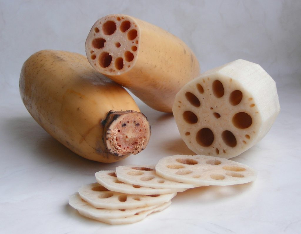
By FotoosRobin – originally posted to Flickr as Lotus root, CC BY-SA 2.0, https://commons.wikimedia.org/w/index.php?curid=4826529
The lotus (Nelumbo nucifera) rhizome (mass of roots) is not the prettiest part of the lotus but its fibers (and presumably fiber from other parts of the lotus plant) served as inspiration for a hydrogel that might be used as a surgical suture according to a Jan. 14, 2021 news item on phys.org (Note: Links have been removed),
“The lotus roots may break, but the fiber remains joined”—an old Chinese saying that reflects the unique structure and mechanical properties of the lotus fiber. The outstanding mechanical properties of lotus fibers can be attributed to their unique spiral structure, which provides an attractive model for biomimetic design of artificial fibers.
In a new study published in Nano Letters, a team led by Prof. Yu Shuhong from the University of Science and Technology of China (USTC) of the Chinese Academy of Sciences (CAS) reported a bio-inspired lotus-fiber-mimetic spiral structure bacterial cellulose (BC) hydrogel fiber with high strength, high toughness, excellent biocompatibility, good stretchability, and high energy dissipation.
Unlike polymer-based hydrogel, the newly designed biomimetic hydrogel fiber (BHF) is based on the BC hydrogel with 3D cellulose nanofiber networks produced by bacteria. The cellulose nanofibers provide the reversible hydrogen bonding network that results in unique mechanical properties.
The researchers applied a constant tangential force to the pretreated BC hydrogel along the cross-sectional direction. Then, the two sides of the hydrogel were subjected to opposite tangential forces, and local plastic deformation occurred.
The hydrogen bonds in the 3D network of cellulose nanofibers were broken by the tangential force, causing the hydrogel strip to twist spirally and the network to slip and deform. When the tangential force was removed, the hydrogen bonds reformed between the nanofibers, and the spiral structure of the fiber was fixed.
Benefited from lotus-fiber-mimetic spiral structure, the toughness of BHF can reach ?116.3 MJ m-3, which is more than nine times higher than those of non-spiralized BC hydrogel fiber. Besides, once the BHF is stretched, it is nearly non-resilient.
Combining outstanding mechanical properties with excellent biocompatibility derived from BC, BHF is a promising hydrogel fiber for biomedical material, especially for surgical suture, a commonly used structural biomedical material for wound repair.
Compared with commercial surgical suture with higher modulus, the BHF has similar modulus and strength to soft tissue, like skin. The outstanding stretchability and energy dissipation of BHF allow it to absorb energy from the tissue deformation around a wound and effectively protect the wound from rupture, which makes BHF an ideal surgical suture.
What’s more, the porous structure of BHF also allows it to adsorb functional small molecules, such as antibiotics or anti-inflammatory compounds, and sustainably release them on wounds. With an appropriate design, BHF would be a powerful platform for many medical applications.
While it’s late in the season to be thinking of frostbite in the Northern Hemisphere, there’s always next year. This research from India looks quite promising, assuming you have the gel available when you first get frostbite. From a December 11, 2019 news item on Nanowerk (Note: A link has been removed),
Mountaineers and winter sports enthusiasts know the dangers of frostbite –– the tissue damage that can occur when extremities, such as the nose, ears, fingers and toes, are exposed to very cold temperatures. However, it can be difficult to get treated quickly in remote, snowbound areas.
Now, researchers reporting in ACS Biomaterials Science & Engineering (“Heparin-Encapsulated Metered-Dose Topical “Nano-Spray Gel” Liposomal Formulation Ensures Rapid On-Site Management of Frostbite Injury by Inflammatory Cytokines Scavenging”) have developed a convenient gel that could be sprayed onto frostbite injuries when they occur, helping wounds heal.
Frostbite causes fluids in the skin and underlying tissues to freeze and crystallize, resulting in inflammation, decreased blood flow and cell death. Extremities are the most affected areas because they are farther away from the body’s core and already have reduced blood flow. If frostbite is not treated soon after the injury, it could lead to gangrene and amputation of the affected parts. Conventional treatments include immersing the body part in warm water, applying topical antibiotic creams or administering vasodilators and anti-inflammatory drugs, but many of these are unavailable in isolated snowy areas, like mountaintops. Others, such as topical medications, could end up freezing themselves. Rahul Verma and colleagues at the Institute of Nano Science and Technology [India] wanted to develop a cold-stable spray gel that could be administered on-site for the immediate treatment of frostbite injuries.
To develop their spray, the researchers packaged heparin, an anticoagulant that improves blood flow by reducing clotting and aiding in blood vessel repair, into liposomes. These lipid carriers helped deliver heparin deep inside the skin. They embedded the heparin-loaded liposomes in a sprayable hydrogel that also contained ibuprofen (a painkiller and anti-inflammatory drug) and propylene glycol, which helped keep the spray from freezing at very low temperatures. When the researchers tested the spray gel on rats with frostbite, they found that the treatment completely healed the injuries within 14 days, whereas untreated injuries were only about 40% healed, and wounds treated with an antibiotic cream were about 80% healed. The spray reduced levels of inflammatory cytokines at the wound site and in the blood circulation, which likely accelerated healing, the researchers say.
I have two stories about lungs and they are entirely different with the older one being a bioengineering story from the US and the more recent one being an artificial tissue story from the University of Toronto and the University of Ottawa (both in Canada).
Lab grown lungs
The Canadian Broadcasting Corporation’s Quirks and Quarks radio programme posted a December 29, 2018 news item (with embedded radio files) about bioengineered lunjgs,
There are two major components to building an organ: the structure and the right cells on that structure. A team led by Dr. Joan Nichols, a Professor of Internal Medicine, Microbiology and Immunology at the University of Texas Medical Branch in Galveston, were able to tackle both parts of the problem
In their experiment they used a donor organ for the structure. They took a lung from an unrelated pig, and stripped it of its cells, leaving a scaffold of collagen, a tough, flexible protein. This provided a pre-made appropriate structure, though in future they think it may be possible to use 3-D printing technology to get the same result.
They then added cultured cells from the animal who would be receiving the transplant – so the lung was made of the animal’s own cells. Cultured lung and blood vessel cells were placed on the scaffold and it was placed in a tank for 30 days with a cocktail of nutrients to help the cells stick to the scaffold and proliferate. The result was a kind of baby lung.
They then transplanted the bio-engineered, though immature, lung into the recipient animal where they hoped it would continue to develop and mature – growing to become a healthy, functioning organ.
The recipients of the bio-engineered lungs were four pigs adult pigs, which appeared to tolerate the transplants well. In order to study the development of the bio-engineered lungs, they euthanized the animals at different times: 10 hours, two weeks, one month and two months after transplantation.
They found that as early as two weeks, the bio-engineered lung had integrated into the recipient animals’ body, building a strong network of blood vessels essential for the lung to survive. There was no evidence of pulmonary edema, the build of fluid in the lungs, which is usually a sign of the blood vessels not working efficiently. There was no sign of rejection of the transplanted organs, and the pigs were healthy up to the point where they were euthanized.
One lingering concern is how well the bio-engineered lungs delivered oxygen. The four pigs who received the trasplant [sic] had one original functioning lung, so they didn’t depend on their new bio-engineered lung for breathing. The scientists were not sure that the bio-engineered lung was mature enough to handle the full load of oxygen on its own.
…
You can hear Bob McDonald’s (host of Quirks & Quarks, a Canadian Broadcasting Corporation science radio programme) interview lead scientist, Dr. Joan Nichols if you go to here. (Note: I find he overmodulates his voice but some may find he has a ‘friendly’ voice.)
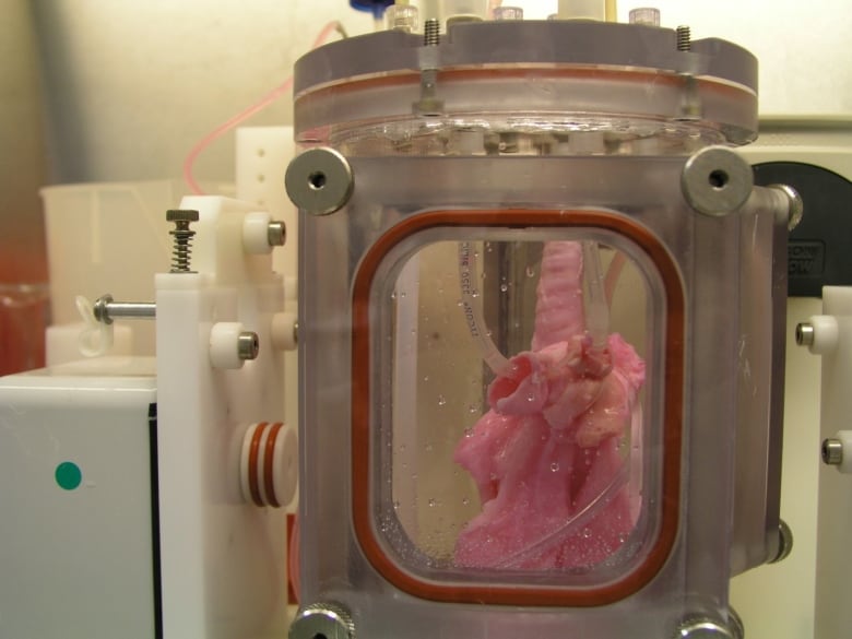
This is an image of the lung scaffold produced by the team,
Lung scaffold in the bioreactor chamber on Day 1 of the experiment, before the cells from the study pig were added. (Credit: Joan Nichols) [downloaded from https://www.cbc.ca/radio/quirks/dec-29-2018-water-on-mars-lab-grown-lungs-and-more-the-biggest-science-stories-of-2018-1.4940811/lab-grown-lungs-are-transplanted-in-pigs-today-they-may-help-humans-tomorrow-1.4940822]
In 2014, Joan Nichols and Joaquin Cortiella from The University of Texas Medical Branch at Galveston were the first research team to successfully bioengineer human lungs in a lab. In a paper now available in Science Translational Medicine, they provide details of how their work has progressed from 2014 to the point no complications have occurred in the pigs as part of standard preclinical testing.
“The number of people who have developed severe lung injuries has increased worldwide, while the number of available transplantable organs have decreased,” said Cortiella, professor of pediatric anesthesia. “Our ultimate goal is to eventually provide new options for the many people awaiting a transplant,” said Nichols, professor of internal medicine and associate director of the Galveston National Laboratory at UTMB.
To produce a bioengineered lung, a support scaffold is needed that meets the structural needs of a lung. A support scaffold was created using a lung from an unrelated animal that was treated using a special mixture of sugar and detergent to eliminate all cells and blood in the lung, leaving only the scaffolding proteins or skeleton of the lung behind. This is a lung-shaped scaffold made totally from lung proteins.
The cells used to produce each bioengineered lung came from a single lung removed from each of the study animals. This was the source of the cells used to produce a tissue-matched bioengineered lung for each animal in the study. The lung scaffold was placed into a tank filled with a carefully blended cocktail of nutrients and the animals’ own cells were added to the scaffold following a carefully designed protocol or recipe. The bioengineered lungs were grown in a bioreactor for 30 days prior to transplantation. Animal recipients were survived for 10 hours, two weeks, one month and two months after transplantation, allowing the research team to examine development of the lung tissue following transplantation and how the bioengineered lung would integrate with the body.
All of the pigs that received a bioengineered lung stayed healthy. As early as two weeks post-transplant, the bioengineered lung had established the strong network of blood vessels needed for the lung to survive.
“We saw no signs of pulmonary edema, which is usually a sign of the vasculature not being mature enough,” said Nichols and Cortiella. “The bioengineered lungs continued to develop post-transplant without any infusions of growth factors, the body provided all of the building blocks that the new lungs needed.”
Nichols said that the focus of the study was to learn how well the bioengineered lung adapted and continued to mature within a large, living body. They didn’t evaluate how much the bioengineered lung provided oxygenation to the animal.
“We do know that the animals had 100 percent oxygen saturation, as they had one normal functioning lung,” said Cortiella. “Even after two months, the bioengineered lung was not yet mature enough for us to stop the animal from breathing on the normal lung and switch to just the bioengineered lung.”
For this reason, future studies will look at long-term survival and maturation of the tissues as well as gas exchange capability.
The researchers said that with enough funding, they could grow lungs to transplant into people in compassionate use circumstances within five to 10 years.
“It has taken a lot of heart and 15 years of research to get us this far, our team has done something incredible with a ridiculously small budget and an amazingly dedicated group of people,” Nichols and Cortiella said.
Here’s a citation and another link for the paper,
Production and transplantation of bioengineered lung into a large-animal model by Joan E. Nichols, Saverio La Francesca, Jean A. Niles, Stephanie P. Vega, Lissenya B. Argueta, Luba Frank, David C. Christiani, Richard B. Pyles, Blanca E. Himes, Ruyang Zhang, Su Li, Jason Sakamoto, Jessica Rhudy, Greg Hendricks, Filippo Begarani, Xuewu Liu, Igor Patrikeev, Rahul Pal, Emiliya Usheva, Grace Vargas, Aaron Miller, Lee Woodson, Adam Wacher, Maria Grimaldo, Daniil Weaver, Ron Mlcak, and Joaquin Cortiella. Science Translational Medicine 01 Aug 2018: Vol. 10, Issue 452, eaao3926 DOI: 10.1126/scitranslmed.aao3926
This paper is behind a paywall.
Artificial lung cancer tissue
The research teams at the University of Toronto and the University of Ottawa worked on creating artificial lung tissue but other applications are possible too. First, there’s the announcement in a February 25, 2019 news item on phys.org,
A 3-D hydrogel created by researchers in U of T Engineering Professor Molly Shoichet’s lab is helping University of Ottawa researchers to quickly screen hundreds of potential drugs for their ability to fight highly invasive cancers.
Cell invasion is a critical hallmark of metastatic cancers, such as certain types of lung and brain cancer. Fighting these cancers requires therapies that can both kill cancer cells as well as prevent cell invasion of healthy tissue. Today, most cancer drugs are only screened for their ability to kill cancer cells.
“In highly invasive diseases, there is a crucial need to screen for both of these functions,” says Shoichet. “We now have a way to do this.”
In their latest research, the team used hydrogels to mimic the environment of lung cancer, selectively allowing cancer cells, and not healthy cells, to invade. In their latest research, the team used hydrogels to mimic the environment of lung cancer, selectively allowing cancer cells, and not healthy cells, to invade. This emulated environment enabled their collaborators in Professor Bill Stanford’s lab at University of Ottawa to screen for both cancer-cell growth and invasion. The study, led by Roger Y. Tam, a research associate in Shochet’s lab, was recently published in Advanced Materials.
“We can conduct this in a 384-well plate, which is no bigger than your hand. And with image-analysis software, we can automate this method to enable quick, targeted screenings for hundreds of potential cancer treatments,” says Shoichet.
One example is the researchers’ drug screening for lymphangioleiomyomatosis (LAM), a rare lung disease affecting women. Shoichet and her team were inspired by the work of Green Eggs and LAM, a Toronto-based organization raising awareness of the disease.
Using their hydrogels, they were able to automate and screen more than 800 drugs, thereby uncovering treatments that could target disease growth and invasion.
In the ongoing collaboration, the researchers plan to next screen multiple drugs at different doses to gain greater insight into new treatment methods for LAM. The strategies and insights they gain could also help identify new drugs for other invasive cancers.
Shoichet, who was recently named a Distinguished Woman in Chemistry or Chemical Engineering, also plans to patent the hydrogel technology.
“This has, and continues to be, a great collaboration that is advancing knowledge at the intersection of engineering and biology,” says Shoichet.
I note that Shoichet (pronounced ShoyKet) is getting ready to patent this work. I do have a question about this and it’s not up to Shoichet to answer as she didn’t create the system. Will the taxpayers who funded her work receive any financial benefits should the hydrogel prove to be successful or will we be paying double, both supporting her research and paying for the hydrogel through our healthcare costs?
Getting back to the research, here’s a link to and a citation for the paper,
Rationally Designed 3D Hydrogels Model Invasive Lung Diseases Enabling High‐Content Drug Screening by Roger Y. Tam, Julien Yockell‐Lelièvre, Laura J. Smith, Lisa M. Julian, Alexander E. G. Baker, Chandarong Choey, Mohamed S. Hasim, Jim Dimitroulakos, William L. Stanford, Molly S. Shoichet. Advanced Materials Volume 31, Issue 7 February 15, 2019 1806214 First published online: 27 December 2018 DOI: https://doi.org/10.1002/adma.201806214