Tag Archives: stem cells

Grow better organ-like tissues by using silkworms

A June 6, 2024 news item on ScienceDaily describes a technique, which could lead to better organ-on-a-chip (OOC) systems,

Biomedical engineers at Duke University [North Carolina, US] have developed a silk-based, ultrathin membrane that can be used in organ-on-a-chip models to better mimic the natural environment of cells and tissues within the body. When used in a kidney organ-on-a-chip platform, the membrane helped tissues grow to recreate the functionality of both healthy and diseased kidneys.

By allowing the cells to grow closer together, this new membrane helps researchers to better control the growth and function of the key cells and tissues of any organ, enabling them to more accurately model a wide range of diseases and test therapeutics.

A June 6, 2024 Duke University news release (also on EurekAlert), which originated the news item, describes the OOC system and the problem these researchers are seeking to solve,

Often no larger than a USB flash drive, organ-on-a-chip (OOC) systems have revolutionized how researchers study the underlying biology of the human body, whether it’s creating dynamic models of tissue structures, studying organ functions or modeling diseases. These platforms are designed to stimulate cell growth and differentiation in a way that best mimics the organ of interest. Researchers can even populate these tools with human stem cells to generate patient-specific organ models for pre-clinical studies.

But as the technology has evolved, problems in the chip’s design have also emerged –– most notably with the materials used to create the membranes that form the support structure for the specialized cells to grow on. These membranes are typically composed of polymers that don’t degrade, creating a permanent barrier between cells and tissues. While the extracellular membranes in human organs are often less than one micron thick, these polymer membranes are anywhere from 30 to 50 microns, hindering communication between cells and limiting cell growth.

“We want to handle the tissues in these chips just like a pathologist would handle biopsy samples or even living tissues from a patient, but this wasn’t possible with the standard polymer membranes because the extra thickness prevented the cells from forming structures that more closely resemble tissues in the human body,” said Samira Musah, an assistant professor of biomedical engineering and medicine at Duke. “We thought, ‘Wouldn’t it be nice if we could get a protein-based material that mimics the structure of these natural membranes and is thin enough for us to slice and study?’”

This question led Musah and George (Xingrui) Mou, a PhD student in Musah’s lab and first author on the paper, to silk fibroin, a protein created by silkworms that can be electronically spun into a membrane. When examined under a microscope, silk fibroin looks like spaghetti or a Jackson Pollock painting. Made out of long, intertwining fibers, the porous material better mimics the structure of the extracellular matrix found in human organs, and it has previously been used to create scaffolds for purposes like wound healing.

“The silk fibroin allowed us to bring the membrane thickness down from 50 microns to five or fewer, which gets us an order of magnitude closer to what you’d see in a living organism,” explained Mao.

To test this new membrane, Musah and Mao applied the material to their kidney chip models. Made out of a clear plastic and roughly the size of a quarter, this OOC platform is meant to resemble a cross section of a human kidney––specifically the glomerular capillary wall, a key structure in the organ made from clusters of blood vessels that is responsible for filtering blood.

Once the membrane was in place, the team added human induced pluripotent stem cell derivatives into the chip. They observed that these cells were able to send signals across the ultrathin membrane, which helped the cells differentiate into glomerular cells, podocytes and vascular endothelial cells. The platform also triggered the development of endothelial fenestrations in the growing tissue, which are holes that allow for the passage of fluid between the cellular layers.

By the end of the test, these different kidney cell types had assembled into a glomerular capillary wall and could efficiently filter molecules by size.

“The new microfluidic chip system’s ability to simulate in vivo-like tissue-tissue interfaces and induce the formation of specialized cells, such as fenestrated endothelium and mature glomerular podocytes from stem cells, holds significant potential for advancing our understanding of human organ development, disease progression, and therapeutic development,” said Musah.

As they continue to optimize their model, Musah and colleagues are hoping to use this technology to better understand the mechanisms behind kidney disease. Despite affecting more than 15 percent of American adults, researchers lack effective models for the disease. Patients are also often not diagnosed until the kidneys have been substantially damaged, and they are often required to undergo dialysis or receive a kidney transplant.

“Using this platform to develop kidney disease models could help us discover new biomarkers of the disease,” said Mao. “This could also be used to help us screen for drug candidates for several kidney disease models. The possibilities are very exciting.”

“This technology has implications for all organ-on-a-chip models,” said Musah. “Our tissues are made up of membranes and interfaces, so you can imagine using this membrane to improve models of other organs, like the brain, liver, and lungs, or other disease states. That’s where the power of our platform really lies.”

This work was supported by a Whitehead Scholarship in Biomedical Research, Chair’s Research Award from the Department of Medicine at Duke University, MEDx Pilot Grant on Biomechanics in Injury or Injury Repair, Burroughs Wellcome Fund PDEP Career Transition Ad Hoc Award, Duke Incubation Fund from the Duke Innovation & Entrepreneurship Initiative, Genetech Research Award, a George M. O’Brien Kidney Center Pilot Grant (P30 DK081943), an NIH [National Institutes of Health] Director’s New Innovator Grant (DP2DK138544).

Here’s a link to and a citation for the paper,

An Ultrathin Membrane Mediates Tissue-Specific Morphogenesis and Barrier Function in a Human Kidney Chip by Xingrui Mou, Jessica Shah, Yasmin Roye, Carolyn Du, Samira Musah. Science Advances. June 4, 2024 Vol 10, Issue 23 DOI: https://doi.org/10.1126/sciadv.adn2689

This paper is open access.

Organoids with four different types of brain cells from the University of Saskatchewan (USask)

While a USask-designed “mini-brain” synthetic organoid might look like a tiny wad of chewing gum, it could be a gamechanger for Alzheimer’s research (credit: USask/David Stobbe)

A May 14, 2024 news item on ScienceDaily announces research from the University of Saskatchewan that could improve diagnosis and treatment for Alzheimer’s disease,

Using an innovative new method, a University of Saskatchewan (USask) researcher is building tiny pseudo-organs from stem cells to help diagnose and treat Alzheimer’s.

When Dr. Tyler Wenzel (PhD) first came up with the idea of building a miniature brain from stem cells, he never could have predicted how well his creations would work.

Now, Wenzel’s “mini-brain” could revolutionize the way Alzheimer’s and other brain-related diseases are diagnosed and treated.

“Never in our wildest dreams did we think that our crazy idea would work,” he said. “These could be used as a diagnostic tool, built from blood.”

A May 14, 2024 University of Saskatchewan news release (also on EurekAlert), which originated the news item, provides more technical details, Note: A link has been removed,

Wenzel, a postdoctoral fellow in the College of Medicine’s Department of Psychiatry, developed the idea for the “mini-brain” – or more formally, a one-of-a-kind cerebral organoid model – while working under the supervision of Dr. Darrell Mousseau (PhD).

Human stem cells can be manipulated to develop into practically any other cell in the body. Using stem cells taken from human blood, Wenzel was able to create a tiny artificial organ – roughly three millimetres across and resembling visually what Wenzel described as a piece of chewed gum someone has tried to smooth out again.

These “mini-brains” are built by creating stem cells from a blood sample, and then transforming these stem cells into functioning brain cells. Using small synthetic organoids for research is not a novel concept – but the “mini-brains” developed in Wenzel’s lab are unique. As outlined in Wenzel’s recent published article in Frontiers of Cellular Neuroscience, the brains from Wenzel’s lab are comprised of four different types of brain cells while most brain organoids are comprised of only neurons.

In testing, Wenzel’s “mini-brains” more accurately reflect a fully-fledged adult human brain, so they can be used to more closely examine neurological conditions of adult patients, such as Alzheimer disease.

And for those “mini-brains” created from the stem cells of individuals who have Alzheimer’s, Wenzel determined that the artificial organ displayed the pathology of Alzheimer’s – just on a smaller scale.

“If stem cells have the capacity to become any cell in the human body, the question then came ‘could we create something that resembles an entire organ?’” Wenzel said. “While we were developing it, I had the crazy idea that if these truly are human brains, if a patient had a disease like Alzheimer’s and we grew their ‘mini-brain,’ in theory that tiny brain would have Alzheimer’s.”

Wenzel said this technology has the potential to change the way health services are provided to those with Alzheimer’s, particularly in rural and remote communities. This groundbreaking research has already received support from the Alzheimer Society of Canada.

If Wenzel and his colleagues can create a consistent way to diagnose and treat neurological conditions like Alzheimer’s using only a small blood sample – which has a relatively long shelf life and can be couriered – instead of requiring patients to travel to hospitals or specialized clinics, it could be a tremendous resource savings for the healthcare system and a burden off of patients.

“In theory, if this tool works the way we think it does, we could just get a blood sample shipped from La Loche or La Ronge to the university and diagnose you like that,” he said.

The early proof-of-concept work on the “mini-brains” has been extremely promising – which means the next step for Wenzel is expanding the testing to a larger pool of patients.

The researchers are also interested in trying to expand the scope of the “mini-brain” research. According to Wenzel, if they can confirm the “mini-brains” accurately reflect other brain diseases or neurological conditions, they could potentially be used to speed up diagnoses or test the efficacy of drugs on patients.

As an example, Wenzel pointed to the substantial wait times to see a psychiatrist in Saskatchewan. If the “mini-brains” could be used to test which antidepressant works best on a patient suffering from depression, it could dramatically reduce the time required to see a doctor and receive a prescription.

A former high school science teacher who made the move into the world of research and academia, Wenzel said it’s the “nature of research” to come up with a hypothesis and hit close to the mark in an experiment that excites him his work.

The astounding success of the early “mini-brains,” however, has been so staggering that Wenzel admitted he still struggles to wrap his own brain around it.

“I’m still in disbelief, but it’s also extremely motivating that something like this happened,” Wenzel said. “It gives me something that I think will impact society and have actual relevance and create some change … it has a strong potential to shift the landscape of medicine.”

Here’s a link to and a citation for the paper,

Brain organoids engineered to give rise to glia and neural networks after 90 days in culture exhibit human-specific proteoforms by Tyler J. Wenzel, Darrell D. Mousseau. Front. Cell. Neurosci., Volume 18 – 2024 DOI: https://doi.org/10.3389/fncel.2024.1383688 Published: 08 May 2024

This paper is open access.

Synthetic human embryos—what now? (2 of 2)

The term they’re using in the Weizmann Institute of Science’s (Israel) announcement is “a generally accurate human embryo model.” This is in contrast to previous announcements including the one from the University of Cambridge team highlighted in Part 1.

From a September 6, 2023 news item on phys.org, Note: A link has been removed,

A research team headed by Prof. Jacob Hanna at the Weizmann Institute of Science has created complete models of human embryos from stem cells cultured in the lab—and managed to grow them outside the womb up to day 14. As reported today [September 6, 2023] in Nature, these synthetic embryo models had all the structures and compartments characteristic of this stage, including the placenta, yolk sac, chorionic sac and other external tissues that ensure the models’ dynamic and adequate growth.

Cellular aggregates derived from human stem cells in previous studies could not be considered genuinely accurate human embryo models, because they lacked nearly all the defining hallmarks of a post-implantation embryo. In particular, they failed to contain several cell types that are essential to the embryo’s development, such as those that form the placenta and the chorionic sac. In addition, they did not have the structural organization characteristic of the embryo and revealed no dynamic ability to progress to the next developmental stage.

Given their authentic complexity, the human embryo models obtained by Hanna’s group may provide an unprecedented opportunity to shed new light on the embryo’s mysterious beginnings. Little is known about the early embryo because it is so difficult to study, for both ethical and technical reasons, yet its initial stages are crucial to its future development. During these stages, the clump of cells that implants itself in the womb on the seventh day of its existence becomes, within three to four weeks, a well-structured embryo that already contains all the body organs.

“The drama is in the first month, the remaining eight months of pregnancy are mainly lots of growth,” Hanna says. “But that first month is still largely a black box. Our stem cell–derived human embryo model offers an ethical and accessible way of peering into this box. It closely mimics the development of a real human embryo, particularly the emergence of its exquisitely fine architecture.”

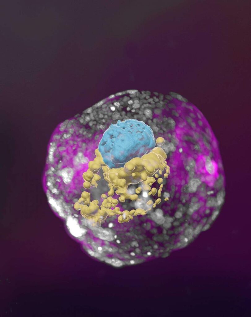
A stem cell–derived human embryo model at a developmental stage equivalent to that of a day 14 embryo. The model has all the compartments that define this stage: the yolk sac (yellow) and the part that will become the embryo itself, topped by the amnion (blue) – all enveloped by cells that will become the placenta (pink) Courtesy: Weizmann Institute of Science

A September 6, 2023 Weizmann Institute of Science press release, which originated the news item, offers a wealth of detail, Note: Links have been removed,

Letting the embryo model say “Go!”

Hanna’s team built on their previous experience in creating synthetic stem cell–based models of mouse embryos. As in that research, the scientists made no use of fertilized eggs or a womb. Rather, they started out with human cells known as pluripotent stem cells, which have the potential to differentiate into many, though not all, cell types. Some were derived from adult skin cells that had been reverted to “stemness.” Others were the progeny of human stem cell lines that had been cultured for years in the lab.

The researchers then used Hanna’s recently developed method to reprogram pluripotent stem cells so as to turn the clock further back: to revert these cells to an even earlier state – known as the naïve state – in which they are capable of becoming anything, that is, specializing into any type of cell. This stage corresponds to day 7 of the natural human embryo, around the time it implants itself in the womb. Hanna’s team had in fact been the first to start describing methods to generate human naïve stem cells, back in 2013; they continued to improve these methods, which stand at the heart of the current project, over the years.

The scientists divided the cells into three groups. The cells intended to develop into the embryo were left as is. The cells in each of the other groups were treated only with chemicals, without any need for genetic modification, so as to turn on certain genes, which was intended to cause these cells to differentiate toward one of three tissue types needed to sustain the embryo: placenta, yolk sac or the extraembryonic mesoderm membrane that ultimately creates the chorionic sac.

Soon after being mixed together under optimized, specifically developed conditions, the cells formed clumps, about 1 percent of which self-organized into complete embryo-like structures. “An embryo is self-driven by definition; we don’t need to tell it what to do – we must only unleash its internally encoded potential,” Hanna says. “It’s critical to mix in the right kinds of cells at the beginning, which can only be derived from naïve stem cells that have no developmental restrictions. Once you do that, the embryo-like model itself says, ‘Go!’”

The stem cell–based embryo-like structures (termed SEMs) developed normally outside the womb for 8 days, reaching a developmental stage equivalent to day 14 in human embryonic development. That’s the point at which natural embryos acquire the internal structures that enable them to proceed to the next stage: developing the progenitors of body organs.

Complete human embryo models match classic diagrams in terms of structure and cell identity

When the researchers compared the inner organization of their stem cell–derived embryo models with illustrations and microscopic anatomy sections in classical embryology atlases from the 1960s, they found an uncanny structural resemblance between the models and the natural human embryos at the corresponding stage. Every compartment and supporting structure was not only there, but in the right place, size and shape. Even the cells that make the hormone used in pregnancy testing were there and active: When the scientists applied secretions from these cells to a commercial pregnancy test, it came out positive.

In fact, the study has already produced a finding that may open a new direction of research into early pregnancy failure. The researchers discovered that if the embryo is not enveloped by placenta-forming cells in the right manner at day 3 of the protocol (corresponding to day 10 in natural embryonic development), its internal structures, such as the yolk sac, fail to properly develop.

“An embryo is not static. It must have the right cells in the right organization, and it must be able to progress – it’s about being and becoming,” Hanna says. “Our complete embryo models will help researchers address the most basic questions about what determines its proper growth.”

This ethical approach to unlocking the mysteries of the very first stages of embryonic development could open numerous research paths. It might help reveal the causes of many birth defects and types of infertility. It could also lead to new technologies for growing transplant tissues and organs. And it could offer a way around experiments that cannot be performed on live embryos – for example, determining the effects of exposure to drugs or other substances on fetal development.

For people who are visually inclined, there are two videos embedded in the September 6, 2023 Weizmann Institute of Science press release.

Here’s a link to and a citation for the paper,

Complete human day 14 post-implantation embryo models from naïve ES cells by Bernardo Oldak, Emilie Wildschutz, Vladyslav Bondarenko, Mehmet-Yunus Comar, Cheng Zhao, Alejandro Aguilera-Castrejon, Shadi Tarazi, Sergey Viukov, Thi Xuan Ai Pham, Shahd Ashouokhi, Dmitry Lokshtanov, Francesco Roncato, Eitan Ariel, Max Rose, Nir Livnat, Tom Shani, Carine Joubran, Roni Cohen, Yoseph Addadi, Muriel Chemla, Merav Kedmi, Hadas Keren-Shaul, Vincent Pasque, Sophie Petropoulos, Fredrik Lanner, Noa Novershtern & Jacob H. Hanna. Nature (2023) DOI: https://doi.org/10.1038/s41586-023-06604-5 Published: 06 September 2023

This paper is behind a paywall.

As for the question I asked in the head “what now?” I have absolutely no idea.

Synthetic human embryos—what now? (1 of 2)

Usually, there’s a rough chronological order to how I introduce the research, but this time I’m looking at the term used to describe it, following up with the various news releases and commentaries about the research, and finishing with a Canadian perspective.

After writing this post (but before it was published), the Weizmann Institute of Science (Israel) made their September 6, 2023 announcement and things changed a bit. That’s in Part two.

Say what you really mean (a terminology issue)

First, it might be useful to investigate the term, ‘synthetic human embryos’ as Julian Hitchcock does in his June 29, 2023 article on Bristows website (h/t Mondaq’s July 5, 2023 news item), Note: Links have been removed,

Synthetic Embryos” are neither Synthetic nor Embryos. So why are editors giving that name to stem cell-based models of human development?

One of the less convincing aspects of the last fortnight’s flurry of announcements about advances in simulating early human development (see here) concerned their name. Headlines galore (in newspapers and scientific journals) referred to “synthetic embryos“.

But embryo models, however impressive, are not embryos. To claim that the fundamental stages of embryo development that we learnt at school – fertilisation, cleavage and compaction – could now be bypassed to achieve the same result would be wrong. Nor are these objects “synthesised”: indeed, their interest to us lies in the ways in which they organise themselves. The researchers merely place the stem cells in a matrix in appropriate conditions, then stand back and watch them do it. Scientists were therefore unhappy about this use of the term in news media, and relieved when the International Society for Stem Cell Research (ISSCR) stepped in with a press release:

“Unlike some recent media reports describing this research, the ISSCR advises against using the term “synthetic embryo” to describe embryo models, because it is inaccurate and can create confusion. Integrated embryo models are neither synthetic nor embryos. While these models can replicate aspects of the early-stage development of human embryos, they cannot and will not develop to the equivalent of postnatal stage humans. Further, the ISSCR Guidelines prohibit the transfer of any embryo model to the uterus of a human or an animal.”

Although this was the ISSCR’s first attempt to put that position to the public, it had already made that recommendation to the research community two years previously. Its 2021 Guidelines for Stem Cell Research and Clinical Translation had recommended researchers to “promote accurate, current, balanced, and responsive public representations of stem cell research”. In particular:

“While organoids, chimeras, embryo models, and other stem cell-based models are useful research tools offering possibilities for further scientific progress, limitations on the current state of scientific knowledge and regulatory constraints must be clearly explained in any communications with the public or media. Suggestions that any of the current in vitro models can recapitulate an intact embryo, human sentience or integrated brain function are unfounded overstatements that should be avoided and contradicted with more precise characterizations of current understanding.”

Here’s a little bit about Hitchcock from his Bristows profile page,

  • Diploma Medical School, University of Birmingham (1975-78)
  • LLB, University of Wolverhampton
  • Diploma in Intellectual Property Law & Practice, University of Bristol
  • Qualified 1998

Following an education in medicine at the University of Birmingham and a career as a BBC science producer, Julian has focused on the law and regulation of life science technologies since 1997, practising in England and Australia. He joined Bristows with Alex Denoon in 2018.

Hitchcock’s June 29, 2023 article comments on why this term is being used,

I have a lot of sympathy with the position of the science writers and editors incurring the scientists’ ire. First, why should journalists have known of the ISSCR’s recommendations on the use of the term “synthetic embryo”? A journalist who found Recommendation 4.1 of the ISSCR Guidelines would probably not have found them specific enough to address the point, and the academic introduction containing the missing detail is hard to find. …

My second reason for being sympathetic to the use of the terrible term is that no suitable alternative has been provided, other than in the Stem Cell Reports paper, which recommends the umbrella terms “embryo models” or “stem cell based embryo models”. …

When asked why she had used the term “synthetic embryo”, the journalist I contacted remarked that, “We’re still working out the right language and it’s something we’re discussing and will no doubt evolve along with the science”.

It is absolutely in the public’s interest (and in the interest of science), that scientific research is explained in terms that the public understands. There is, therefore, a need, I think, for the scientific community to supply a name to the media or endure the penalties of misinformation …

In such an intensely competitive field of research, disagreement among researchers, even as to names, is inevitable. In consequence, however, journalists and their audiences are confronted by a slew of terms which may or may not be synonymous or overlapping, with no agreed term [emphasis mine] for the overall class of stem cell based embryo models. We cannot blame them if they make up snappy titles of their own [emphasis mine]. …

The announcement

The earliest date for the announcement at the International Society for Stem Cell Researh meeting that I can find is Hannah Devlin’s June 14, 2023 article in The Guardian newspaper, Note: A link has been removed,

Scientists have created synthetic human embryos using stem cells, in a groundbreaking advance that sidesteps the need for eggs or sperm.

Scientists say these model embryos, which resemble those in the earliest stages of human development, could provide a crucial window on the impact of genetic disorders and the biological causes of recurrent miscarriage.

However, the work also raises serious ethical and legal issues as the lab-grown entities fall outside current legislation in the UK and most other countries.

The structures do not have a beating heart or the beginnings of a brain, but include cells that would typically go on to form the placenta, yolk sac and the embryo itself.

Prof Magdalena Żernicka-Goetz, of the University of Cambridge and the California Institute of Technology, described the work in a plenary address on Wednesday [June 14, 2023] at the International Society for Stem Cell Research’s annual meeting in Boston.

The (UK) Science Media Centre made expert comments available in a June 14, 2023 posting “expert reaction to Guardian reporting news of creation of synthetic embryos using stem cells.”

Two days later, this June 16, 2023 essay by Kathryn MacKay, Senior Lecturer in Bioethics, University of Sydney (Australia), appeared on The Conversation (h/t June 16, 2023 news item on phys.org), Note: Links have been removed,

Researchers have created synthetic human embryos using stem cells, according to media reports. Remarkably, these embryos have reportedly been created from embryonic stem cells, meaning they do not require sperm and ova.

This development, widely described as a breakthrough that could help scientists learn more about human development and genetic disorders, was revealed this week in Boston at the annual meeting of the International Society for Stem Cell Research.

The research, announced by Professor Magdalena Żernicka-Goetz of the University of Cambridge and the California Institute of Technology, has not yet been published in a peer-reviewed journal. But Żernicka-Goetz told the meeting these human-like embryos had been made by reprogramming human embryonic stem cells.

So what does all this mean for science, and what ethical issues does it present?

MacKay goes on to answer her own questions, from the June 16, 2023 essay, Note: A link has been removed,

One of these quandaries arises around whether their creation really gets us away from the use of human embryos.

Robin Lovell-Badge, the head of stem cell biology and developmental genetics at the Francis Crick Institute in London UK, reportedly said that if these human-like embryos can really model human development in the early stages of pregnancy, then we will not have to use human embryos for research.

At the moment, it is unclear if this is the case for two reasons.

First, the embryos were created from human embryonic stem cells, so it seems they do still need human embryos for their creation. Perhaps more light will be shed on this when Żernicka-Goetz’s research is published.

Second, there are questions about the extent to which these human-like embryos really can model human development.

Professor Magdalena Żernicka-Goetz’s research is published

Almost two weeks later the research from the Cambridge team (there are other teams and countries also racing; see Part two for the news from Sept. 6, 2023) was published, from a June 27, 2023 news item on ScienceDaily,

Cambridge scientists have created a stem cell-derived model of the human embryo in the lab by reprogramming human stem cells. The breakthrough could help research into genetic disorders and in understanding why and how pregnancies fail.

Published today [Tuesday, June 27, 2023] in the journal Nature, this embryo model is an organised three-dimensional structure derived from pluripotent stem cells that replicate some developmental processes that occur in early human embryos.

Use of such models allows experimental modelling of embryonic development during the second week of pregnancy. They can help researchers gain basic knowledge of the developmental origins of organs and specialised cells such as sperm and eggs, and facilitate understanding of early pregnancy loss.

A June 27, 2023 University of Cambridge press release (also on EurekAlert), which originated the news item, provides more detail about the work,

“Our human embryo-like model, created entirely from human stem cells, gives us access to the developing structure at a stage that is normally hidden from us due to the implantation of the tiny embryo into the mother’s womb,” said Professor Magdalena Zernicka-Goetz in the University of Cambridge’s Department of Physiology, Development and Neuroscience, who led the work.

She added: “This exciting development allows us to manipulate genes to understand their developmental roles in a model system. This will let us test the function of specific factors, which is difficult to do in the natural embryo.”

In natural human development, the second week of development is an important time when the embryo implants into the uterus. This is the time when many pregnancies are lost.

The new advance enables scientists to peer into the mysterious ‘black box’ period of human development – usually following implantation of the embryo in the uterus – to observe processes never directly observed before.

Understanding these early developmental processes holds the potential to reveal some of the causes of human birth defects and diseases, and to develop tests for these in pregnant women.

Until now, the processes could only be observed in animal models, using cells from zebrafish and mice, for example.

Legal restrictions in the UK currently prevent the culture of natural human embryos in the lab beyond day 14 of development: this time limit was set to correspond to the stage where the embryo can no longer form a twin. [emphasis mine]

Until now, scientists have only been able to study this period of human development using donated human embryos. This advance could reduce the need for donated human embryos in research.

Zernicka-Goetz says the while these models can mimic aspects of the development of human embryos, they cannot and will not develop to the equivalent of postnatal stage humans.

Over the past decade, Zernicka-Goetz’s group in Cambridge has been studying the earliest stages of pregnancy, in order to understand why some pregnancies fail and some succeed.

In 2021 and then in 2022 her team announced in Developmental Cell, Nature and Cell Stem Cell journals that they had finally created model embryos from mouse stem cells that can develop to form a brain-like structure, a beating heart, and the foundations of all other organs of the body.

The new models derived from human stem cells do not have a brain or beating heart, but they include cells that would typically go on to form the embryo, placenta and yolk sac, and develop to form the precursors of germ cells (that will form sperm and eggs).

Many pregnancies fail at the point when these three types of cells orchestrate implantation into the uterus begin to send mechanical and chemical signals to each other, which tell the embryo how to develop properly.

There are clear regulations governing stem cell-based models of human embryos and all researchers doing embryo modelling work must first be approved by ethics committees. Journals require proof of this ethics review before they accept scientific papers for publication. Zernicka-Goetz’s laboratory holds these approvals.

“It is against the law and FDA regulations to transfer any embryo-like models into a woman for reproductive aims. These are highly manipulated human cells and their attempted reproductive use would be extremely dangerous,” said Dr Insoo Hyun, Director of the Center for Life Sciences and Public Learning at Boston’s Museum of Science and a member of Harvard Medical School’s Center for Bioethics.

Zernicka-Goetz also holds position at the California Institute of Technology and is NOMIS Distinguished Scientist and Scholar Awardee.

The research was funded by the Wellcome Trust and Open Philanthropy.

(There’s more about legal concerns further down in this post.)

Here’s a link to and a citation for the paper,

Pluripotent stem cell-derived model of the post-implantation human embryo by Bailey A. T. Weatherbee, Carlos W. Gantner, Lisa K. Iwamoto-Stohl, Riza M. Daza, Nobuhiko Hamazaki, Jay Shendure & Magdalena Zernicka-Goetz. Nature (2023) DOI: https://doi.org/10.1038/s41586-023-06368-y Published: 27 June 2023

This paper is open access.

Published the same day (June 27, 2023) is a paper (citation and link follow) also focused on studying human embryonic development using stem cells. First, there’s this from the Abstract,

Investigating human development is a substantial scientific challenge due to the technical and ethical limitations of working with embryonic samples. In the face of these difficulties, stem cells have provided an alternative to experimentally model inaccessible stages of human development in vitro …

This time the work is from a US/German team,

Self-patterning of human stem cells into post-implantation lineages by Monique Pedroza, Seher Ipek Gassaloglu, Nicolas Dias, Liangwen Zhong, Tien-Chi Jason Hou, Helene Kretzmer, Zachary D. Smith & Berna Sozen. Nature (2023) DOI: https://doi.org/10.1038/s41586-023-06354-4 Published: 27 June 2023

The paper is open access.

Legal concerns and a Canadian focus

A July 25, 2023 essay by Françoise Baylis and Jocelyn Downie of Dalhousie University (Nova Scotia, Canada) for The Conversation (h/t July 25, 2023 article on phys.org) covers the advantages of doing this work before launching into a discussion of legislation and limits in the UK and, more extensively, in Canada, Note: Links have been removed,

This research could increase our understanding of human development and genetic disorders, help us learn how to prevent early miscarriages, lead to improvements in fertility treatment, and — perhaps — eventually allow for reproduction without using sperm and eggs.

Synthetic human embryos — also called embryoid bodies, embryo-like structures or embryo models — mimic the development of “natural human embryos,” those created by fertilization. Synthetic human embryos include the “cells that would typically go on to form the embryo, placenta and yolk sac, and develop to form the precursors of germ cells (that will form sperm and eggs).”

Though research involving natural human embryos is legal in many jurisdictions, it remains controversial. For some people, research involving synthetic human embryos is less controversial because these embryos cannot “develop to the equivalent of postnatal stage humans.” In other words, these embryos are non-viable and cannot result in live births.

Now, for a closer look at the legalities in the UK and in Canada, from the July 25, 2023 essay, Note: Links have been removed,

The research presented by Żernicka-Goetz at the ISSCR meeting took place in the United Kingdom. It was conducted in accordance with the Human Fertilization and Embryology Act, 1990, with the approval of the U.K. Stem Cell Bank Steering Committee.

U.K. law limits the research use of human embryos to 14 days of development. An embryo is defined as “a live human embryo where fertilisation is complete, and references to an embryo include an egg in the process of fertilisation.”

Synthetic embryos are not created by fertilization and therefore, by definition, the 14-day limit on human embryo research does not apply to them. This means that synthetic human embryo research beyond 14 days can proceed in the U.K.

The door to the touted potential benefits — and ethical controversies — seems wide open in the U.K.

While the law in the U.K. does not apply to synthetic human embryos, the law in Canada clearly does. This is because the legal definition of an embryo in Canada is not limited to embryos created by fertilization [emphasis mine].

The Assisted Human Reproduction Act (the AHR Act) defines an embryo as “a human organism during the first 56 days of its development following fertilization or creation, excluding any time during which its development has been suspended.”

Based on this definition, the AHR Act applies to embryos created by reprogramming human embryonic stem cells — in other words, synthetic human embryos — provided such embryos qualify as human organisms.

A synthetic human embryo is a human organism. It is of the species Homo sapiens, and is thus human. It also qualifies as an organism — a life form — alongside other organisms created by means of fertilization, asexual reproduction, parthenogenesis or cloning.

Given that the AHR Act applies to synthetic human embryos, there are legal limits on their creation and use in Canada.

First, human embryos — including synthetic human embryos – can only be created for the purposes of “creating a human being, improving or providing instruction in assisted reproduction procedures.”

Given the state of the science, it follows that synthetic human embryos could legally be created for the purpose of improving assisted reproduction procedures.

Second, “spare” or “excess” human embryos — including synthetic human embryos — originally created for one of the permitted purposes, but no longer wanted for this purpose, can be used for research. This research must be done in accordance with the consent regulations which specify that consent must be for a “specific research project.”

Finally, all research involving human embryos — including synthetic human embryos — is subject to the 14-day rule. The law stipulates that: “No person shall knowingly… maintain an embryo outside the body of a female person after the fourteenth day of its development following fertilization or creation, excluding any time during which its development has been suspended.”

Putting this all together, the creation of synthetic embryos for improving assisted human reproduction procedures is permitted, as is research using “spare” or “excess” synthetic embryos originally created for this purpose — provided there is specific consent and the research does not exceed 14 days.

This means that while synthetic human embryos may be useful for limited research on pre-implantation embryo development, they are not available in Canada for research on post-implantation embryo development beyond 14 days.

The authors close with this comment about the prospects for expanding Canada’s14-day limit, from the July 25, 2023 essay,

… any argument will have to overcome the political reality that the federal government is unlikely to open up the Pandora’s box of amending the AHR Act.

It therefore seems likely that synthetic human embryo research will remain limited in Canada for the foreseeable future.

As mentioned, in September 2023 there was a new development. See: Part two.
Artificially-grown mini-brains (organoids)—without animal components— offer opportunities for neuroscience

There’s a good (brief) description of how these fibres become organoids in the photo caption,

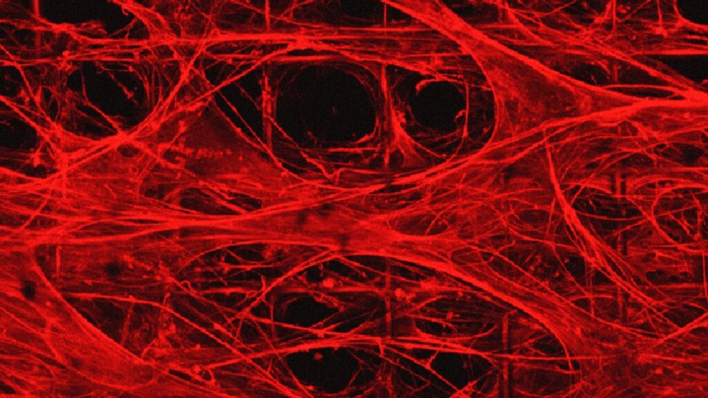
Engineered extracellular matrices composed of fibrillar fibronectin are suspended over a porous polymer framework and provide the niche for stem cells to attach, differentiate, and mature into organoids. Credit: Ayse Muñiz Courtesy: Michigan Medicine – University of Michigan

A July 13 ,2023 University of Michigan (Michigan Medicine) news release by Noah Fromson (also on EurekAlert) announces ‘kinder, gentler’ brain organoids. Coincidentally, these organoids more closely resemble human brains, Note: Links have been removed,

Researchers at University of Michigan developed a method to produce artificially grown miniature brains — called human brain organoids — free of animal cells that could greatly improve the way neurodegenerative conditions are studied and, eventually, treated.

Over the last decade of researching neurologic diseases, scientists have explored the use of human brain organoids as an alternative to mouse models. These self-assembled, 3D tissues derived from embryonic or pluripotent stem cells more closely model the complex brain structure compared to conventional two-dimensional cultures.

Until now, the engineered network of proteins and molecules that give structure to the cells in brain organoids, known as extracellular matrices, often used a substance derived from mouse sarcomas called Matrigel. That method suffers significant disadvantages, with a relatively undefined composition and batch-to-batch variability.

The latest U-M research, published in Annals of Clinical and Translational Neurology, offers a solution to overcome Matrigel’s weaknesses. Investigators created a novel culture method that uses an engineered extracellular matrix for human brain organoids — without the presence of animal components – and enhanced the neurogenesis of brain organoids compared to previous studies.

“This advancement in the development of human brain organoids free of animal components will allow for significant strides in the understanding of neurodevelopmental biology,” said senior author Joerg Lahann, Ph.D., director of the U-M Biointerfaces Institute and Wolfgang Pauli Collegiate Professor of Chemical Engineering at U-M.

“Scientists have long struggled to translate animal research into the clinical world, and this novel method will make it easier for translational research to make its way from the lab to the clinic.”

The foundational extracellular matrices of the research team’s brain organoids were comprised of human fibronectin, a protein that serves as a native structure for stem cells to adhere, differentiate and mature. They were supported by a highly porous polymer scaffold.

The organoids were cultured for months, while lab staff was unable to enter the building due to the COVID 19-pandemic.

Using proteomics, researchers found their brain organoids developed cerebral spinal fluid, a clear liquid that flows around healthy brain and spinal cords. This fluid more closely matched human adult CSF compared to a landmark study of human brain organoids developed in Matrigel.

“When our brains are naturally developing in utero, they are of course not growing on a bed of extracellular matrix produced by mouse cancer cells,” said first author Ayşe Muñiz, Ph.D., who was a graduate student in the U-M Macromolecular Science and Engineering Program at the time of the work.  

“By putting cells in an engineered niche that more closely resembles their natural environment, we predicted we would observe differences in organoid development that more faithfully mimics what we see in nature.”

The success of these xenogeneic-free human brain organoids opens the door for reprogramming with cells from patients with neurodegenerative diseases, says co-author Eva Feldman, M.D., Ph.D., director of the ALS Center of Excellence at U-M and James W. Albers Distinguished Professor of Neurology at U-M Medical School.

“There is a possibility to take the stem cells from a patient with a condition such as ALS or Alzheimer’s and, essentially, build an avatar mini brain of that patients to investigate possible treatments or model how their disease will progress,” Feldman said. “These models would create another avenue to predict disease and study treatment on a personalized level for conditions that often vary greatly from person to person.”

Here’s a link to and a citation for the paper,

Engineered extracellular matrices facilitate brain organoids from human pluripotent stem cells by Ayşe J. Muñiz, Tuğba Topal, Michael D. Brooks, Angela Sze, Do Hoon Kim, Jacob Jordahl, Joe Nguyen, Paul H. Krebsbach, Masha G. Savelieff, Eva L. Feldman, Joerg Lahann. Annals of Clinical and Translational Neurology DOI: https://doi.org/10.1002/acn3.51820 First published: 07 June 2023

This paper is open access.

Protocols for mouse-human chimeric embryos

This work on a type of species boundary-crossing could be very disturbing for some folks. That said, here’s more about the science from a July 2, 2021 news item on phys.org,

A year after University at Buffalo [in New York state] scientists demonstrated that it was possible to produce millions of mature human cells in a mouse embryo, they have published a detailed description of the method so that other laboratories can do it, too.

A July 2, 2021 University at Buffalo (UB) news release (also on EurekAlert) by Ellen Goldbaum, which originated the news item, explains why scientists have created these chimeras,

The ability to produce millions of mature human cells in a living organism, called a chimera, which contains the cells of two species, is critical if the ultimate promise of stem cells to treat or cure human disease is to be realized. But to produce those mature cells, human primed stem cells must be converted back into an earlier, less developed naive state so that the human stem cells can co-develop with the inner cell mass in a mouse blastocyst.

The protocol outlining how to do that has now been published in Nature Protocols by the UB scientists. They were invited to publish it because of the significant interest generated by the team’s initial publication describing their breakthrough last May [2020].

“This paper will enable many scientists to use this new platform to study the human disease of their interest,” said Jian Feng, PhD, professor of physiology and biophysics in the Jacobs School of Medicine and Biomedical Sciences at UB and senior author. “Over time, it will transform biomedical research toward a more effective use of the human model system to directly study virtually any inborn condition of an individual. It will stimulate unforeseen discoveries and applications that may fundamentally change our understanding of human biology and medicine.”

The protocol will allow scientists to create animal models that Feng said provide a much more realistic picture of embryonic development than has ever been possible. These more realistic animal models also will have the potential to reveal the mechaniswms behind numerous diseases, especially those that afflict individuals from birth.

Better mouse models

“This step-by-step protocol will benefit the entire field by enabling other scientists to use our methods to generate chimeras to study human diseases that they are experts in,” said Feng. “It will lead to the generation of better mouse models for various human diseases, such as sickle cell anemia, COVID-19 and many others, or various human developmental disorders.” The paper demonstrates how to generate naive human pluripotent stem cells from existing induced pluripotent stem cells that may be derived from patients with various diseases, how to generate mouse-human chimeras using these cells and how to quantify the amount of human cells in the chimeras.

“Using our method, one can now track the development of naive human pluripotent stem cells in mouse-human chimeric embryos in real-time,” said Feng. These stem cells can then be manipulated either genetically or pharmacologically, providing valuable information about human development and disease.

“For example, one can label naive human pluripotent stem cells by inserting green fluorescent protein in a hemoglobin gene to study the development of human red blood cells in mouse-human chimeras,” said Feng.

Another application is to generate humanized mouse models to study many human diseases.

“These mice contain critical human cells, tissues or even organs so that they more accurately reflect the human condition,” said Feng. “With our method, the human cells are made along with the mouse during the development of the mouse embryo. There would be better matching and no rejections, because there are ways for the human cells to be made where there is no competition from their mouse counterparts.”

Organs for transplant in the future

By allowing others to improve and adapt the method to eventually generate chimeras in larger animals, this protocol may also lead to the generation of human organs to address the dire shortage of organs available for transplant, said Feng.

“If naive human pluripotent stem cells are able to generate significant amounts of mature human cells in other larger species, it could be possible to make human tissues or even human organs in chimeric animals,” Feng explained.

This would be possible using blastocyst complementation where, Feng explained, normal pluripotent stem cells from one species can reconstitute an organ for that species in a blastocyst of another species that been genetically modified not to grow that particular organ.

Feng added: “Ultimately, a better understanding of how human cells develop and grow in chimeras may enable the generation of human cells, tissues and organs in a completely artificial system and fundamentally change how we treat many human diseases. Research using chimeras is a bridge that must be crossed to reach that possibility.”

Here’s a link to and a citation for the 2021 article,

Generation of mouse–human chimeric embryos by Boyang Zhang, Hanqin Li, Zhixing Hu, Houbo Jiang, Aimee B. Stablewski, Brandon J. Marzullo, Donald A. Yergeau & Jian Feng. Nature Protocols (2021) DOI: https://doi.org/10.1038/s41596-021-00565-7 Published 02 July 2021

This article is behind a paywall.

Here’s a link to and citation for the 2020 work, which led to the publication of the protocols,

Transient inhibition of mTOR in human pluripotent stem cells enables robust formation of mouse-human chimeric embryos by Zhixing Hu, Hanqin Li, Houbo Jiang, Yong Ren, Xinyang Yu, Jingxin Qiu, Aimee B. Stablewski, Boyang Zhang, Michael J. Buck, Jian Feng. Science Advances 13 May 2020: Vol. 6, no. 20, eaaz0298 DOI: 10.1126/sciadv.aaz0298

This paper is open access.

Cortical spheroids (like mini-brains) could unlock (larger) brain’s mysteries

A March 19, 2021 Northwestern University news release on EurekAlert announces the creation of a device designed to monitor brain organoids (for anyone unfamiliar with brain organoids there’s more information after the news),

A team of scientists, led by researchers at Northwestern University, Shirley Ryan AbilityLab and the University of Illinois at Chicago (UIC), has developed novel technology promising to increase understanding of how brains develop, and offer answers on repairing brains in the wake of neurotrauma and neurodegenerative diseases.

Their research is the first to combine the most sophisticated 3-D bioelectronic systems with highly advanced 3-D human neural cultures. The goal is to enable precise studies of how human brain circuits develop and repair themselves in vitro. The study is the cover story for the March 19 [March 17, 2021 according to the citation] issue of Science Advances.

The cortical spheroids used in the study, akin to “mini-brains,” were derived from human-induced pluripotent stem cells. Leveraging a 3-D neural interface system that the team developed, scientists were able to create a “mini laboratory in a dish” specifically tailored to study the mini-brains and collect different types of data simultaneously. Scientists incorporated electrodes to record electrical activity. They added tiny heating elements to either keep the brain cultures warm or, in some cases, intentionally overheated the cultures to stress them. They also incorporated tiny probes — such as oxygen sensors and small LED lights — to perform optogenetic experiments. For instance, they introduced genes into the cells that allowed them to control the neural activity using different-colored light pulses.

This platform then enabled scientists to perform complex studies of human tissue without directly involving humans or performing invasive testing. In theory, any person could donate a limited number of their cells (e.g., blood sample, skin biopsy). Scientists can then reprogram these cells to produce a tiny brain spheroid that shares the person’s genetic identity. The authors believe that, by combining this technology with a personalized medicine approach using human stem cell-derived brain cultures, they will be able to glean insights faster and generate better, novel interventions.

“The advances spurred by this research will offer a new frontier in the way we study and understand the brain,” said Shirley Ryan AbilityLab’s Dr. Colin Franz, co-lead author on the paper who led the testing of the cortical spheroids. “Now that the 3-D platform has been developed and validated, we will be able to perform more targeted studies on our patients recovering from neurological injury or battling a neurodegenerative disease.”

Yoonseok Park, postdoctoral fellow at Northwestern University and co-lead author, added, “This is just the beginning of an entirely new class of miniaturized, 3-D bioelectronic systems that we can construct to expand the capacity of the regenerative medicine field. For example, our next generation of device will support the formation of even more complex neural circuits from brain to muscle, and increasingly dynamic tissues like a beating heart.”

Current electrode arrays for tissue cultures are 2-D, flat and unable to match the complex structural designs found throughout nature, such as those found in the human brain. Moreover, even when a system is 3-D, it is extremely challenging to incorporate more than one type of material into a small 3-D structure. With this advance, however, an entire class of 3-D bioelectronics devices has been tailored for the field of regenerative medicine.

“Now, with our small, soft 3-D electronics, the capacity to build devices that mimic the complex biological shapes found in the human body is finally possible, providing a much more holistic understanding of a culture,” said Northwestern’s John Rogers, who led the technology development using technology similar to that found in phones and computers. “We no longer have to compromise function to achieve the optimal form for interfacing with our biology.”

As a next step, scientists will use the devices to better understand neurological disease, test drugs and therapies that have clinical potential, and compare different patient-derived cell models. This understanding will then enable a better grasp of individual differences that may account for the wide variation of outcomes seen in neurological rehabilitation.

“As scientists, our goal is to make laboratory research as clinically relevant as possible,” said Kristen Cotton, research assistant in Dr. Franz’s lab. “This 3-D platform opens the door to new experiments, discovery and scientific advances in regenerative neurorehabilitation medicine that have never been possible.”

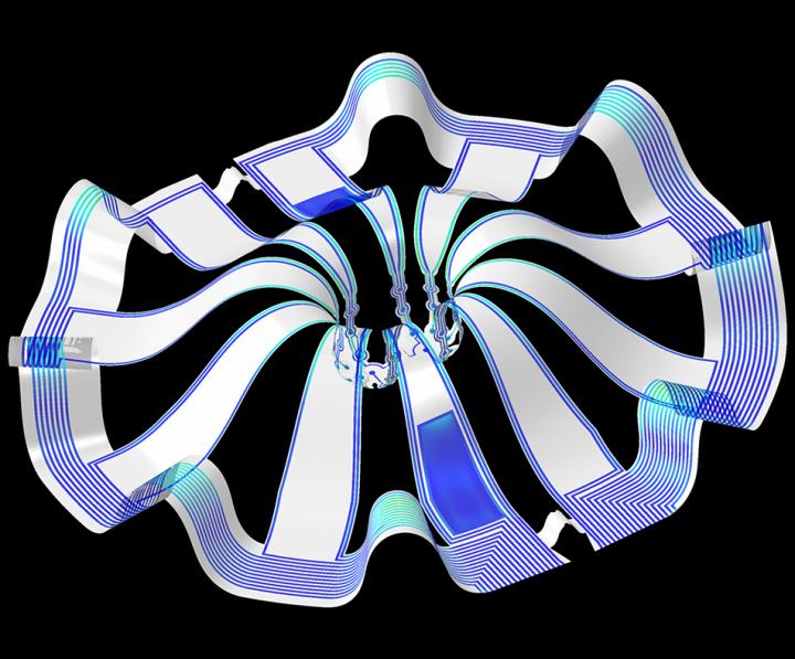
Caption: Three dimensional multifunctional neural interfaces for cortical spheroids and engineered assembloids Credit: Northwestern University

As for what brain ogranoids might be, Carl Zimmer in an Aug. 29, 2019 article for the New York Times provides an explanation,

Organoids Are Not Brains. How Are They Making Brain Waves?

Two hundred and fifty miles over Alysson Muotri’s head, a thousand tiny spheres of brain cells were sailing through space.

The clusters, called brain organoids, had been grown a few weeks earlier in the biologist’s lab here at the University of California, San Diego. He and his colleagues altered human skin cells into stem cells, then coaxed them to develop as brain cells do in an embryo.

The organoids grew into balls about the size of a pinhead, each containing hundreds of thousands of cells in a variety of types, each type producing the same chemicals and electrical signals as those cells do in our own brains.

In July, NASA packed the organoids aboard a rocket and sent them to the International Space Station to see how they develop in zero gravity.

Now the organoids were stowed inside a metal box, fed by bags of nutritious broth. “I think they are replicating like crazy at this stage, and so we’re going to have bigger organoids,” Dr. Muotri said in a recent interview in his office overlooking the Pacific.

What, exactly, are they growing into? That’s a question that has scientists and philosophers alike scratching their heads.

On Thursday, Dr. Muotri and his colleagues reported that they  have recorded simple brain waves in these organoids. In mature human brains, such waves are produced by widespread networks of neurons firing in synchrony. Particular wave patterns are linked to particular forms of brain activity, like retrieving memories and dreaming.

As the organoids mature, the researchers also found, the waves change in ways that resemble the changes in the developing brains of premature babies.

“It’s pretty amazing,” said Giorgia Quadrato, a neurobiologist at the University of Southern California who was not involved in the new study. “No one really knew if that was possible.”

But Dr. Quadrato stressed it was important not to read too much into the parallels. What she, Dr. Muotri and other brain organoid experts build are clusters of replicating brain cells, not actual brains.

If you have the time, I recommend reading Zimmer’s article in its entirety. Perhaps not coincidentally, Zimmer has an excerpt titled “Lab-Grown Brain Organoids Aren’t Alive. But They’re Not Not Alive, Either.” published in Slate.com,

From Life’s Edge: The Search For What It Means To Be Alive by Carl Zimmer, published by Dutton, an imprint of Penguin Publishing Group, a division of Penguin Random House, LLC. Copyright © 2021 by Carl Zimmer.

Cleber Trujillo led me to a windowless room banked with refrigerators, incubators, and microscopes. He extended his blue-gloved hands to either side and nearly touched the walls. “This is where we spend half our day,” he said.

In that room Trujillo and a team of graduate students raised a special kind of life. He opened an incubator and picked out a clear plastic box. Raising it above his head, he had me look up at it through its base. Inside the box were six circular wells, each the width of a cookie and filled with what looked like watered-down grape juice. In each well 100 pale globes floated, each the size of a housefly head.

Getting back to the research about monitoring brain organoids, here’s a link to and a citation for the paper about cortical spheroids,

Three-dimensional, multifunctional neural interfaces for cortical spheroids and engineered assembloids by Yoonseok Park, Colin K. Franz, Hanjun Ryu, Haiwen Luan, Kristen Y. Cotton, Jong Uk Kim, Ted S. Chung, Shiwei Zhao, Abraham Vazquez-Guardado, Da Som Yang, Kan Li, Raudel Avila, Jack K. Phillips, Maria J. Quezada, Hokyung Jang, Sung Soo Kwak, Sang Min Won, Kyeongha Kwon, Hyoyoung Jeong, Amay J. Bandodkar, Mengdi Han, Hangbo Zhao, Gabrielle R. Osher, Heling Wang, KunHyuck Lee, Yihui Zhang, Yonggang Huang, John D. Finan and John A. Rogers. Science Advances 17 Mar 2021: Vol. 7, no. 12, eabf9153 DOI: 10.1126/sciadv.abf9153

This paper appears to be open access.

According to a March 22, 2021 posting on the Shirley Riley AbilityLab website, the paper is featured on the front cover of Science Advances (vol. 7 no. 12).

First 3D heart printed using patient’s biological materials

This is very exciting news and it’s likely be at least 10 years before this technology could be made available to the public.

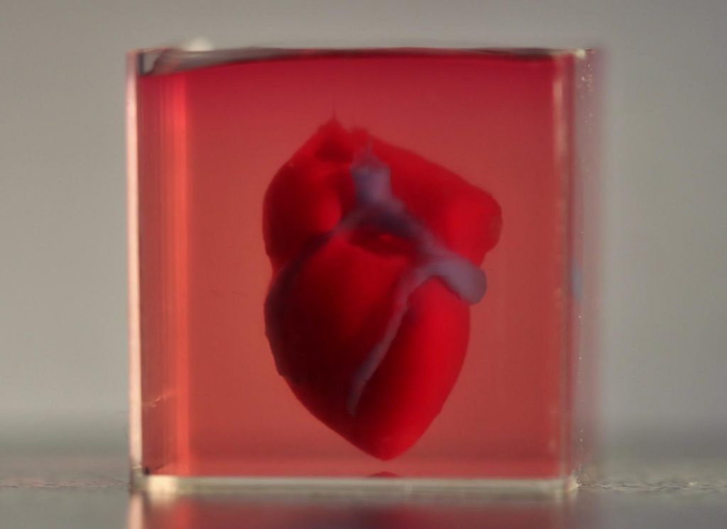
Caption: A 3D-printed, small-scaled human heart engineered from the patient’s own materials and cells. Credit: Advanced Science. © 2019 The Authors.

An April 15, 2019 news item on ScienceDaily makes a remarkable announcement,

In a major medical breakthrough, Tel Aviv University researchers have “printed” the world’s first 3D vascularised engineered heart using a patient’s own cells and biological materials. Their findings were published on April 15 [2019] in a study in Advanced Science.

Until now, scientists in regenerative medicine — a field positioned at the crossroads of biology and technology — have been successful in printing only simple tissues without blood vessels.

“This is the first time anyone anywhere has successfully engineered and printed an entire heart replete with cells, blood vessels, ventricles and chambers,” says Prof. Tal Dvir of TAU’s School of Molecular Cell Biology and Biotechnology, Department of Materials Science and Engineering, Center for Nanoscience and Nanotechnology and Sagol Center for Regenerative Biotechnology, who led the research for the study.

An April 15, 2019 Amricna Friends of Tel Aviv University (TAU) news release (also on EurekAlert), which originated the news item, provides more detail,

Heart disease is the leading cause of death among both men and women in the United States. Heart transplantation is currently the only treatment available to patients with end-stage heart failure. Given the dire shortage of heart donors, the need to develop new approaches to regenerate the diseased heart is urgent.

“This heart is made from human cells and patient-specific biological materials. In our process these materials serve as the bioinks, substances made of sugars and proteins that can be used for 3D printing of complex tissue models,” Prof. Dvir says. “People have managed to 3D-print the structure of a heart in the past, but not with cells or with blood vessels. Our results demonstrate the potential of our approach for engineering personalized tissue and organ replacement in the future.

Research for the study was conducted jointly by Prof. Dvir, Dr. Assaf Shapira of TAU’s Faculty of Life Sciences and Nadav Moor, a doctoral student in Prof. Dvir’s lab.

“At this stage, our 3D heart is small, the size of a rabbit’s heart, [emphasis mine] ” explains Prof. Dvir. “But larger human hearts require the same technology.”

For the research, a biopsy of fatty tissue was taken from patients. The cellular and a-cellular materials of the tissue were then separated. While the cells were reprogrammed to become pluripotent stem cells, the extracellular matrix (ECM), a three-dimensional network of extracellular macromolecules such as collagen and glycoproteins, were processed into a personalized hydrogel that served as the printing “ink.”

After being mixed with the hydrogel, the cells were efficiently differentiated to cardiac or endothelial cells to create patient-specific, immune-compatible cardiac patches with blood vessels and, subsequently, an entire heart.

According to Prof. Dvir, the use of “native” patient-specific materials is crucial to successfully engineering tissues and organs.

“The biocompatibility of engineered materials is crucial to eliminating the risk of implant rejection, which jeopardizes the success of such treatments,” Prof. Dvir says. “Ideally, the biomaterial should possess the same biochemical, mechanical and topographical properties of the patient’s own tissues. Here, we can report a simple approach to 3D-printed thick, vascularized and perfusable cardiac tissues that completely match the immunological, cellular, biochemical and anatomical properties of the patient.”

The researchers are now planning on culturing the printed hearts in the lab and “teaching them to behave” like hearts, Prof. Dvir says. They then plan to transplant the 3D-printed heart in animal models.

“We need to develop the printed heart further,” he concludes. “The cells need to form a pumping ability; they can currently contract, but we need them to work together. Our hope is that we will succeed and prove our method’s efficacy and usefulness.

“Maybe, in ten years, there will be organ printers in the finest hospitals around the world, and these procedures will be conducted routinely.”

Growing the heart to human size and getting the cells to work together so the heart will pump makes it seem like the 10 years Dvir imagines as the future date when there will be organ printers in hospitals routinely printing up hearts seems a bit optimistic. Regardless, I hope he’s right. Bravo to these Israeli researchers!

Here’s a link to and a citation for the paper,

3D Printing of Personalized Thick and Perfusable Cardiac Patches and Hearts by Nadav Noor, Assaf Shapira, Reuven Edri, Idan Gal, Lior Wertheim, Tal Dvir. Advanced Science DOI: https://doi.org/10.1002/advs.201900344 First published: 15 April 2019

This paper is open access.

Cyborg organoids?

Every time I think I’ve become inured to the idea of a fuzzy boundary between life and nonlife something new crosses my path such as integrating nanoelectronics with cells for cyborg organoids. An August 9, 2019 news item on ScienceDaily makes the announcement,

What happens in the early days of organ development? How do a small group of cells organize to become a heart, a brain, or a kidney? This critical period of development has long remained the black box of developmental biology, in part because no sensor was small or flexible enough to observe this process without damaging the cells.

Now, researchers from the Harvard John A. Paulson School of Engineering and Applied Sciences (SEAS) have grown simplified organs known as organoids with fully integrated sensors. These so-called cyborg organoids offer a rare glimpse into the early stages of organ development.

An August 8, 2019 Harvard John A. Paulson School of Engineering and Applied Sciences news release (also on EurekAlert but published August 9, 2019) by Leah Burrows, which originated the news item, expands on the theme,

“I was so inspired by the natural organ development process in high school, in which 3D organs start from few cells in 2D structures. I think if we can develop nanoelectronics that are so flexible, stretchable, and soft that they can grow together with developing tissue through their natural development process, the embedded sensors can measure the entire activity of this developmental process,” said Jia Liu, Assistant Professor of Bioengineering at SEAS and senior author of the study. “The end result is a piece of tissue with a nanoscale device completely distributed and integrated across the entire three-dimensional volume of the tissue.”

This type of device emerges from the work that Liu began as a graduate student in the lab of Charles M. Lieber, the Joshua and Beth Friedman University Professor. In Lieber’s lab, Liu once developed flexible, mesh-like nanoelectronics that could be injected in specific regions of tissue.

Building on that design, Liu and his team increased the stretchability of the nanoelectronics by changing the shape of the mesh from straight lines to serpentine structures (similar structures are used in wearable electronics). Then, the team transferred the mesh nanoelectronics onto a 2D sheet of stem cells, where the cells covered and interwove with the nanoelectronics via cell-cell attraction forces. As the stem cells began to morph into a 3D structure, the nanoelectronics seamlessly reconfigured themselves along with the cells, resulting in fully-grown 3D organoids with embedded sensors.

The stem cells were then differentiated into cardiomyocytes — heart cells — and the researchers were able to monitor and record the electrophysiological activity for 90 days.

“This method allows us to continuously monitor the developmental process and understand how the dynamics of individual cells start to interact and synchronize during the entire developmental process,” said Liu. “It could be used to turn any organoid into cyborg organoids, including brain and pancreas organoids.”

In addition to helping answer fundamental questions about biology, cyborg organoids could be used to test and monitor patient-specific drug treatments and potentially used for transplantations.

Here’s a link to and a citation for the paper

Cyborg Organoids: Implantation of Nanoelectronics via Organogenesis for Tissue-Wide Electrophysiology by Qiang Li, Kewang Nan, Paul Le Floch, Zuwan Lin, Hao Sheng, Thomas S. Blum, Jia Liu. Nano Lett.20191985781-5789 DOI: https://doi.org/10.1021/acs.nanolett.9b02512 Publication Date:July 26, 2019 Copyright © 2019 American Chemical Society

This paper is behind a paywall.

Cooking up a lung one way or the other

I have two stories about lungs and they are entirely different with the older one being a bioengineering story from the US and the more recent one being an artificial tissue story from the University of Toronto and the University of Ottawa (both in Canada).

Lab grown lungs

The Canadian Broadcasting Corporation’s Quirks and Quarks radio programme posted a December 29, 2018 news item (with embedded radio files) about bioengineered lunjgs,

There are two major components to building an organ: the structure and the right cells on that structure. A team led by Dr. Joan Nichols, a Professor of Internal Medicine, Microbiology and Immunology at the University of Texas Medical Branch in Galveston, were able to tackle both parts of the problem

In their experiment they used a donor organ for the structure. They took a lung from an unrelated pig, and stripped it of its cells, leaving a scaffold of collagen, a tough, flexible protein.  This provided a pre-made appropriate structure, though in future they think it may be possible to use 3-D printing technology to get the same result.

They then added cultured cells from the animal who would be receiving the transplant – so the lung was made of the animal’s own cells. Cultured lung and blood vessel cells were placed on the scaffold and it was  placed in a tank for 30 days with a cocktail of nutrients to help the cells stick to the scaffold and proliferate. The result was a kind of baby lung.

They then transplanted the bio-engineered, though immature, lung into the recipient animal where they hoped it would continue to develop and mature – growing to become a healthy, functioning organ.

The recipients of the bio-engineered lungs were four pigs adult pigs, which appeared to tolerate the transplants well. In order to study the development of the bio-engineered lungs, they euthanized the animals at different times: 10 hours, two weeks, one month and two months after transplantation.

They found that as early as two weeks, the bio-engineered lung had integrated into the recipient animals’ body, building a strong network of blood vessels essential for the lung to survive. There was no evidence of pulmonary edema, the build of fluid in the lungs, which is usually a sign of the blood vessels not working efficiently.  There was no sign of rejection of the transplanted organs, and the pigs were healthy up to the point where they were euthanized.

One lingering concern is how well the bio-engineered lungs delivered oxygen. The four pigs who received the trasplant [sic] had one original functioning lung, so they didn’t depend on their new bio-engineered lung for breathing. The scientists were not sure that the bio-engineered lung was mature enough to handle the full load of oxygen on its own.

You can hear Bob McDonald’s (host of Quirks & Quarks, a Canadian Broadcasting Corporation science radio programme) interview lead scientist, Dr. Joan Nichols if you go to here. (Note: I find he overmodulates his voice but some may find he has a ‘friendly’ voice.)

This is an image of the lung scaffold produced by the team,

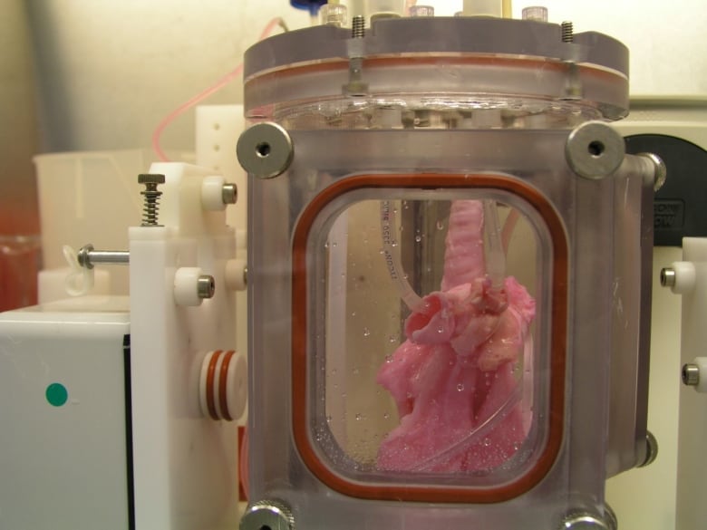
Lung scaffold in the bioreactor chamber on Day 1 of the experiment, before the cells from the study pig were added. (Credit: Joan Nichols) [downloaded from https://www.cbc.ca/radio/quirks/dec-29-2018-water-on-mars-lab-grown-lungs-and-more-the-biggest-science-stories-of-2018-1.4940811/lab-grown-lungs-are-transplanted-in-pigs-today-they-may-help-humans-tomorrow-1.4940822]

Here’s more technical detail in an August 1, 2018i University of Texas Medical Branch (UTMB) news release (also on EurekAlert), which originally announced the research,

A research team at the University of Texas Medical Branch at Galveston have bioengineered lungs and transplanted them into adult pigs with no medical complication.

In 2014, Joan Nichols and Joaquin Cortiella from The University of Texas Medical Branch at Galveston were the first research team to successfully bioengineer human lungs in a lab. In a paper now available in Science Translational Medicine, they provide details of how their work has progressed from 2014 to the point no complications have occurred in the pigs as part of standard preclinical testing.

“The number of people who have developed severe lung injuries has increased worldwide, while the number of available transplantable organs have decreased,” said Cortiella, professor of pediatric anesthesia. “Our ultimate goal is to eventually provide new options for the many people awaiting a transplant,” said Nichols, professor of internal medicine and associate director of the Galveston National Laboratory at UTMB.

To produce a bioengineered lung, a support scaffold is needed that meets the structural needs of a lung. A support scaffold was created using a lung from an unrelated animal that was treated using a special mixture of sugar and detergent to eliminate all cells and blood in the lung, leaving only the scaffolding proteins or skeleton of the lung behind. This is a lung-shaped scaffold made totally from lung proteins.

The cells used to produce each bioengineered lung came from a single lung removed from each of the study animals. This was the source of the cells used to produce a tissue-matched bioengineered lung for each animal in the study. The lung scaffold was placed into a tank filled with a carefully blended cocktail of nutrients and the animals’ own cells were added to the scaffold following a carefully designed protocol or recipe. The bioengineered lungs were grown in a bioreactor for 30 days prior to transplantation. Animal recipients were survived for 10 hours, two weeks, one month and two months after transplantation, allowing the research team to examine development of the lung tissue following transplantation and how the bioengineered lung would integrate with the body.

All of the pigs that received a bioengineered lung stayed healthy. As early as two weeks post-transplant, the bioengineered lung had established the strong network of blood vessels needed for the lung to survive.

“We saw no signs of pulmonary edema, which is usually a sign of the vasculature not being mature enough,” said Nichols and Cortiella. “The bioengineered lungs continued to develop post-transplant without any infusions of growth factors, the body provided all of the building blocks that the new lungs needed.”

Nichols said that the focus of the study was to learn how well the bioengineered lung adapted and continued to mature within a large, living body. They didn’t evaluate how much the bioengineered lung provided oxygenation to the animal.

“We do know that the animals had 100 percent oxygen saturation, as they had one normal functioning lung,” said Cortiella. “Even after two months, the bioengineered lung was not yet mature enough for us to stop the animal from breathing on the normal lung and switch to just the bioengineered lung.”

For this reason, future studies will look at long-term survival and maturation of the tissues as well as gas exchange capability.

The researchers said that with enough funding, they could grow lungs to transplant into people in compassionate use circumstances within five to 10 years.

“It has taken a lot of heart and 15 years of research to get us this far, our team has done something incredible with a ridiculously small budget and an amazingly dedicated group of people,” Nichols and Cortiella said.

Here’s a citation and another link for the paper,

Production and transplantation of bioengineered lung into a large-animal model by Joan E. Nichols, Saverio La Francesca, Jean A. Niles, Stephanie P. Vega, Lissenya B. Argueta, Luba Frank, David C. Christiani, Richard B. Pyles, Blanca E. Himes, Ruyang Zhang, Su Li, Jason Sakamoto, Jessica Rhudy, Greg Hendricks, Filippo Begarani, Xuewu Liu, Igor Patrikeev, Rahul Pal, Emiliya Usheva, Grace Vargas, Aaron Miller, Lee Woodson, Adam Wacher, Maria Grimaldo, Daniil Weaver, Ron Mlcak, and Joaquin Cortiella. Science Translational Medicine 01 Aug 2018: Vol. 10, Issue 452, eaao3926 DOI: 10.1126/scitranslmed.aao3926

This paper is behind a paywall.

Artificial lung cancer tissue

The research teams at the University of Toronto and the University of Ottawa worked on creating artificial lung tissue but other applications are possible too. First, there’s the announcement in a February 25, 2019 news item on phys.org,

A 3-D hydrogel created by researchers in U of T Engineering Professor Molly Shoichet’s lab is helping University of Ottawa researchers to quickly screen hundreds of potential drugs for their ability to fight highly invasive cancers.

Cell invasion is a critical hallmark of metastatic cancers, such as certain types of lung and brain cancer. Fighting these cancers requires therapies that can both kill cancer cells as well as prevent cell invasion of healthy tissue. Today, most cancer drugs are only screened for their ability to kill cancer cells.

“In highly invasive diseases, there is a crucial need to screen for both of these functions,” says Shoichet. “We now have a way to do this.”

A February 25, 2019 University of Toronto news release (also on EurekAlert), which originated the news item, offers more detail ,

In their latest research, the team used hydrogels to mimic the environment of lung cancer, selectively allowing cancer cells, and not healthy cells, to invade. In their latest research, the team used hydrogels to mimic the environment of lung cancer, selectively allowing cancer cells, and not healthy cells, to invade. This emulated environment enabled their collaborators in Professor Bill Stanford’s lab at University of Ottawa to screen for both cancer-cell growth and invasion. The study, led by Roger Y. Tam, a research associate in Shochet’s lab, was recently published in Advanced Materials.

“We can conduct this in a 384-well plate, which is no bigger than your hand. And with image-analysis software, we can automate this method to enable quick, targeted screenings for hundreds of potential cancer treatments,” says Shoichet.

One example is the researchers’ drug screening for lymphangioleiomyomatosis (LAM), a rare lung disease affecting women. Shoichet and her team were inspired by the work of Green Eggs and LAM, a Toronto-based organization raising awareness of the disease.

Using their hydrogels, they were able to automate and screen more than 800 drugs, thereby uncovering treatments that could target disease growth and invasion.

In the ongoing collaboration, the researchers plan to next screen multiple drugs at different doses to gain greater insight into new treatment methods for LAM. The strategies and insights they gain could also help identify new drugs for other invasive cancers.

Shoichet, who was recently named a Distinguished Woman in Chemistry or Chemical Engineering, also plans to patent the hydrogel technology.

“This has, and continues to be, a great collaboration that is advancing knowledge at the intersection of engineering and biology,” says Shoichet.

I note that Shoichet (pronounced ShoyKet) is getting ready to patent this work. I do have a question about this and it’s not up to Shoichet to answer as she didn’t create the system. Will the taxpayers who funded her work receive any financial benefits should the hydrogel prove to be successful or will we be paying double, both supporting her research and paying for the hydrogel through our healthcare costs?

Getting back to the research, here’s a link to and a citation for the paper,

Rationally Designed 3D Hydrogels Model Invasive Lung Diseases Enabling High‐Content Drug Screening by Roger Y. Tam, Julien Yockell‐Lelièvre, Laura J. Smith, Lisa M. Julian, Alexander E. G. Baker, Chandarong Choey, Mohamed S. Hasim, Jim Dimitroulakos, William L. Stanford, Molly S. Shoichet. Advanced Materials Volume 31, Issue 7 February 15, 2019 1806214 First published online: 27 December 2018 DOI: https://doi.org/10.1002/adma.201806214

This paper is behind a paywall.