Tag Archives: Newcastle University

Building materials made with knitted molds and the root network of fungi

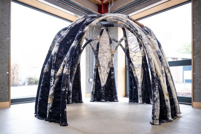
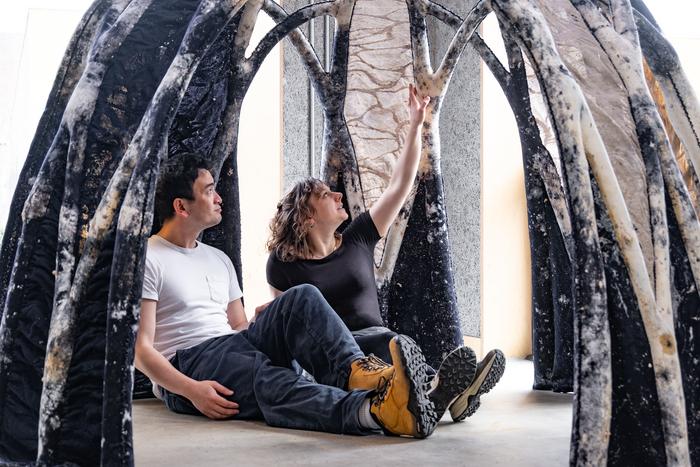
Caption: A 1.8m high, 2m diameter freestanding structure [mycelium vault], made of the BioKnit mycocrete using knitted formwork. Two people are sitting inside it. Credit: Image courtesy of the Hub for Biotechnology in the Built Environment.

The molds for the framework were knitted and filled with ‘mycocrete’ according to a July 14, 2023 Frontiers (Pub.) press release by Angharad Brewer Gillham (also on EurekAlert and published July 17, 2023 on the Newcastle University website),

Scientists hoping to reduce the environmental impact of the construction industry have developed a way to grow building materials using knitted molds and the root network of fungi. Although researchers have experimented with similar composites before, the shape and growth constraints of the organic material have made it hard to develop diverse applications that fulfil its potential. Using the knitted molds as a flexible framework or ‘formwork’, the scientists created a composite called ‘mycocrete’ which is stronger and more versatile in terms of shape and form, allowing the scientists to grow lightweight and relatively eco-friendly construction materials.

“Our ambition is to transform the look, feel and wellbeing of architectural spaces using mycelium in combination with biobased materials such as wool, sawdust and cellulose,” said Dr Jane Scott of Newcastle University [UK], corresponding author of the paper in Frontiers in Bioengineering and Biotechnology. The research was carried out by a team of designers, engineers, and scientists in the Living Textiles Research Group, part of the Hub for Biotechnology in the Built Environment at Newcastle University, which is funded by Research England.

Root networks

To make composites using mycelium, part of the root network of fungi, scientists mix mycelium spores with grains they can feed on and material that they can grow on. This mixture is packed into a mold and placed in a dark, humid, and warm environment so that the mycelium can grow, binding the substrate tightly together. Once it’s reached the right density, but before it starts to produce the fruiting bodies we call mushrooms, it is dried out. This process could provide a cheap, sustainable replacement for foam, timber, and plastic. But mycelium needs oxygen to grow, which constrains the size and shape of conventional rigid molds and limits current applications.

Knitted textiles offer a possible solution: oxygen-permeable molds that could change from flexible to stiff with the growth of the mycelium. But textiles can be too yielding, and it is difficult to pack the molds consistently. Scott and her colleagues set out to design a mycelium mixture and a production system that could exploit the potential of knitted forms.

“Knitting is an incredibly versatile 3D manufacturing system,” said Scott. “It is lightweight, flexible, and formable. The major advantage of knitting technology compared to other textile processes is the ability to knit 3D structures and forms with no seams and no waste.”

Samples of conventional mycelium composite were prepared by the scientists as controls, and grown alongside samples of mycocrete, which also contained paper powder, paper fiber clumps, water, glycerin, and xanthan gum. This paste was designed to be delivered into the knitted formwork with an injection gun to improve packing consistency: the paste needed to be liquid enough for the delivery system, but not so liquid that it failed to hold its shape.

Tubes for their planned test structure were knitted from merino yarn, sterilized, and fixed to a rigid structure while they were filled with the paste, so that changes in tension of the fabric would not affect the performance of the mycocrete.

Building the future

Once dried, samples were subjected to strength tests in tension, compression and flexion. The mycocrete samples proved to be stronger than the conventional mycelium composite samples and outperformed mycelium composites grown without knitted formwork. In addition, the porous knitted fabric of the formwork provided better oxygen availability, and the samples grown in it shrank less than most mycelium composite materials do when they are dried, suggesting more predictable and consistent manufacturing results could be achieved.

The team were also able to build a larger proof-of-concept prototype structure called BioKnit – a complex freestanding dome constructed in a single piece without joins that could prove to be weak points, thanks to the flexible knitted form.

“The mechanical performance of the mycocrete used in combination with permanent knitted formwork is a significant result, and a step towards the use of mycelium and textile biohybrids within construction,” said Scott. “In this paper we have specified particular yarns, substrates, and mycelium necessary to achieve a specific goal. However, there is extensive opportunity to adapt this formulation for different applications. Biofabricated architecture may require new machine technology to move textiles into the construction sector.”

The mycelium vault (also pictured above) is a freestanding structure,

Caption: A 1.8m high, 2m diameter freestanding structure made of the BioKnit mycocrete using knitted formwork. Credit: Courtesy of the Hub for Biotechnology in the Built Environment.

Here’s a link to and a citation for the paper,

BioKnit: development of mycellium paste for use with permanent textile formwork by Romy Kaiser, Ben Bridgens, Elise Elsacker, Jane Scott. Front. Bioeng. Biotechnol., 14 July 2023 Volume 11 – 2023 DOI: https://doi.org/10.3389/fbioe.2023.1229693

This paper appears to be open access.

Concerns about Zoom? Call for expressions of interest in “Zoom Obscura,” creative interventions for a data ethics of video conferencing

Have you wondered about Zoom video conferencing and all that data being made available? Perhaps questioned ethical issues in addition to those associated with data security? Is so and you’d like to come up with a creative intervention that delves beyond encryption issues, there’s Zoom Obscura (on the creativeinformatics.org website),

CI [Creative Informatics] researchers Pip Thornton, Chris Elsden and Chris Speed were recently awarded funding from the Human Data Interaction Network (HDI +) Ethics & Data competition. Collaborating with researchers from Durham [Durham University] and KCL [Kings College London], the Zoom Obscura project aims to investigate creative interventions for a data ethics of video conferencing beyond encryption.

The COVID-19 pandemic has gifted video conferencing companies, such as Zoom, with a vast amount of economically valuable and sensitive data such as our facial and voice biometrics, backgrounds and chat scripts. Before the pandemic, this ‘new normal’ would be subject to scrutiny, scepticism and critique. Yet, the urgent need for remote working and socialising left us with little choice but to engage with these potentially exploitative platforms.

While much of the narrative around data security revolves around technological ‘solutions’ such as encryption, we think there are other – more creative – ways to push back against the systems of digital capitalism that continue to encroach on our everyday lives.

As part of this HDI-funded project, we seek artists, hackers and creative technologists who are interested in experimenting with creative methods to join us in a series of online workshops that will explore how to restore some control and agency in how we can be seen and heard in these newly ubiquitous online spaces. Through three half-day workshops held remotely, we will bring artists and technicians together to ideate, prototype, and exhibit various interventions into the rapidly normalising culture of video-calling in ways that do not compromise our privacy and limit the sharing of our data. We invite interventions that begin at any stage of the video-calling process – from analogue obfuscation, to software manipulation or camera trickery.

Selected artists/collectives will receive a £1000 commission to take part and contribute in three workshops, in order to design and produce one or more, individual or collaborative, creative interventions developed from the workshops. These will include both technical support from a creative technologist as well as a curator for dissemination both online and in Edinburgh and London.

If you are an artist / technologist interested in disrupting/subverting the pandemic-inspired digital status quo, please send expressions of interest of no more than 500 words to pip.thornton@ed.ac.uk , andrew.dwyer@bristol.ac.uk, celsden@ed.ac.uk and michael.duggan@kcl.ac.uk by 8th October 2020. We don’t expect fully formed projects (these will come in the workshop sessions), but please indicate any broad ideas and thoughts you have, and highlight how your past and present practice might be a good fit for the project and its aims.

The Zoom Obscura project is in collaboration with Tinderbox Lab in Edinburgh and Hannah Redler-Hawes (independent curator and codirector of the Data as Culture art programme at the Open Data Institute in London). Outputs from the project will be hosted and exhibited via the Data as Culture archive site and at a Creative Informatics event at the University of Edinburgh.

Are folks outside the UK eligible?

I asked Dr. Pip Thornton about eligibility and she kindly noted this in her Sept. 25, 2020 tweet (reply copied from my Twitter feed),

Open to all, but workshop timings may be more amenable to UK working hours. Having said that, we won’t know what the critical mass is until we review all the applications, so please do apply if you’re interested!

Who are the members of the Zoom Obscura project team?

From the Zoom Obscura webpage (on the creativeinformatics.org website),

Dr. Pip Thornton is a post-doctoral research associate in Creative Informatics at the University of Edinburgh, having recently gained her PhD in Geopolitics and Cybersecurity from Royal Holloway, University of London. Her thesis, Language in the Age of Algorithmic Reproduction: A Critique of Linguistic Capitalism, included theoretical, political and artistic critiques of Google’s search and advertising platforms. She has presented in a variety of venues including the Science Museum, the Alan Turing Institute and transmediale. Her work has featured in WIRED UK and New Scientist, and a collection from her {poem}.py intervention has been displayed at Open Data Institute in London. Her Edinburgh Futures Institute (EFI) funded installation Newspeak 2019, shown at the Edinburgh Festival Fringe (2019), was recently awarded an honourable mention in the Surveillance Studies Network biennial art competition (2020) and is shortlisted for the 2020 Lumen Prize for art and technology in the AI category.

Dr. Andrew Dwyer is a research associate  in the University of Bristol’s Cyber Security Group. Andrew gained a DPhil in Cyber Security at the University of Oxford, where he studied and questioned the role of malware – commonly known as computational viruses and worms –  through its analysis, detection, and translation into international politics and its intersection with multiple ecologies. In his doctoral thesis – Malware Ecologies: A Politics of Cybersecurity – he argued for a re-evaluation of the role of computational actors in the production and negotiation of security, and what this means for human-centred notions of weapons and warfare. Previously, Andrew has been a visiting fellow at the German ‘Dynamics of Security’ collaborative research centre based between Philipps-Universität Marburg, Justus-Liebig-Universität Gießen and the Herder Institute, Marburg and is a Research Affiliate at the Centre for Technology and Global Affairs at the University of Oxford. He will soon be starting a 3-year Addison Wheeler research fellowship in the Department of Geography at the Durham University

Dr Chris Elsden is a research associate in Design Informatics at the University of Edinburgh. Chris is primarily working on the AHRC Creative Informatics project., with specific interests in FinTech and livestreaming within the Creative Industries. He is an HCI researcher, with a background in sociology, and expertise in the human experience of a data-driven life. Using and developing innovative design research methods, his work undertakes diverse, qualitative and often speculative engagements with participants to investigate emerging relationships with technology – particularly data-driven tools and financialn technologies. Chris gained his PhD in Computer Science at Open Lab, Newcastle University in 2018, and in 2019 was a recipient of a SIGCHI Outstanding Dissertation Award.

Dr Mike Duggan is a Teaching Fellow in Digital Cultures in the Department of Digital Humanities at Kings College London. He was awarded a PhD in Cultural Geography from Royal Holloway University of London in 2017, which examined everyday digital mapping practices. This project was co-funded by the Ordnance Survey and the EPSRC. He is a member of the Living Maps network, where he is an editor for the ‘navigations’ section and previously curated the seminar series. Mike’s research is broadly interested in the digital and cultural geographies that emerge from the intersections between everyday life and digital technology.

Professor Chris Speed is Chair of Design Informatics at the University of Edinburgh where his research focuses upon the Network Society, Digital Art and Technology, and The Internet of Things. Chris has sustained a critical enquiry into how network technology can engage with the fields of art, design and social experience through a variety of international digital art exhibitions, funded research projects, books journals and conferences. At present Chris is working on funded projects that engage with the social opportunities of crypto-currencies, an internet of toilet roll holders, and a persistent argument that chickens are actually robots.  Chris is co-editor of the journal Ubiquity and co-directs the Design Informatics Research Centre that is home to a combination of researchers working across the fields of interaction design, temporal design, anthropology, software engineering and digital architecture, as well as the PhD, MA/MFA and MSc and Advanced MSc programmes.

David Chatting is a designer and technologist who works in software and hardware to explore the impact of emerging technologies in everyday lives. He is currently a PhD student in the Department of Design at Goldsmiths – University of London, a Visiting Researcher at Newcastle University’s Open Lab and has his own design practice. Previously he was a Senior Researcher at BTs Broadband Applications Research Centre. David has a Masters degree in Design Interactions from the Royal College of Art (2012) and a Bachelors degree in Computer Science from the University of Birmingham (2000). He has published papers and filed patents in the fields of HCI, psychology, tangible interfaces, computer vision and computer graphics.

Hannah Redler Hawes (Data as Culture) is an independent curator and codirector of the Data as Culture art programme at the Open Data Institute in London. Hannah specialises in emerging artistic practice within the fields of art and science and technology, with an interest in participatory process. She has previously developed projects for museums, galleries, corporate contexts, digital space and the public realm including the  Institute of Physics, Tate Modern, The Lowry, Natural History Museum, FACT Liverpool, the Digital Catapult and Science Gallery London, and has provided specialist consultancy services to the Wellcome Collection, Discover South Kensington and the Horniman Museum. Hannah enjoys projects that redraw boundaries between different disciplines. Current research is around addiction, open data, networked culture and new forms of programming beyond the gallery.

Tinderbox Collective : From grass-roots youth work to award-winning music productions, Tinderbox is building a vibrant and eclectic community of young musicians and artists in Scotland. We have a number of programmes that cross over with each other and come together wherever possible.  They are open to children and young people aged 10 – 25, from complete beginners to young professionals and all levels in between. Tinderbox Lab is our digital arts programme and shared studio maker-space in Edinburgh that brings together artists across disciplines with an interest in digital media and interactive technologies. It is a new programme that started development in 2019, leading to projects and events such as Room to Play, a 10-week course for emerging artists led by Yann Seznec; various guest artist talks & workshops; digital arts exhibitions at the V&A Dundee & Edinburgh Festival of Sound; digital/electronics workshops design/development for children & young people; and research included as part of Electronic Visualisation and the Arts (EVA) London 2019 conference.

Jack Nissan (Tinderbox) is the founder and director of the Tinderbox Collective. In 2012/13, Jack took part in a fellowship programmed called International Creative Entrepreneurs and spent several months working with community activists and social enterprises in China, primarily with families and communities on the outskirts of Beijing with an organisation called Hua Dan. Following this, he set up a number of international exchanges and cross-cultural productions that formed the basis for Tinderbox’s Journey of a Thousand Wings programme, a project bringing together artists and community projects from different countries. He is also a co-director and founding member of Hidden Door, a volunteer-run multi-arts festival, and has won a number of awards for his work across creative and social enterprise sectors. He has been invited to take part in several steering committees and advisory roles, including for Creative Scotland’s new cross-cutting theme on Creative Learning and Artworks Scotland’s peer-networks for artists working in participatory settings. Previously, Jack worked as a researcher in psychology and ageing for the multidisciplinary MRC Centre for Cognitive Ageing and Cognitive Epidemiology, specialising in areas of neuropsychology and memory.

Luci Holland (Tinderbox) is a Scottish (Edinburgh-based) composer, sound artist and radio presenter who composes and produces music and audiovisual art for film, games and concert. As a games music composer Luci wrote the original dynamic/responsive music for Blazing Griffin‘s 2018 release Murderous Pursuits, and has composed and arranged for numerous video game music collaborations, such as orchestrating and producing an arrangement of Jessica Curry‘s Disappearing with label Materia Collective’s bespoke cover album Pattern: An Homage to Everybody’s Gone to the Rapture. Currently she has also been composing custom game music tracks for Skyrim mod Lordbound and a variety of other film and game music projects. Luci also builds and designs interactive sonic art installations for festivals and venues (Refraction (Cryptic), CITADEL (Hidden Door)); and in 2019 Luci joined new classical music station Scala Radio to present The Console, a weekly one-hour show dedicated to celebrating great music in games. Luci also works as a musical director and composer with the youth music charity Tinderbox Project on their Orchestra & Digital Arts programmes; classical music organisation Absolute Classics; and occasionally coordinates musical experiments and productions with her music-for-media band Mantra Sound.

Good luck to all who submit an expression of interest and good luck to Dr. Thornton (I see from her bio that she’s been shortlisted for the 2020 Lumen Prize).

A 3D printed eye cornea and a 3D printed copy of your brain (also: a Brad Pitt connection)

Sometimes it’s hard to keep up with 3D tissue printing news. I have two news bits, one concerning eyes and another concerning brains.

3D printed human corneas

A May 29, 2018 news item on ScienceDaily trumpets the news,

The first human corneas have been 3D printed by scientists at Newcastle University, UK.

It means the technique could be used in the future to ensure an unlimited supply of corneas.

As the outermost layer of the human eye, the cornea has an important role in focusing vision.

Yet there is a significant shortage of corneas available to transplant, with 10 million people worldwide requiring surgery to prevent corneal blindness as a result of diseases such as trachoma, an infectious eye disorder.

In addition, almost 5 million people suffer total blindness due to corneal scarring caused by burns, lacerations, abrasion or disease.

The proof-of-concept research, published today [May 29, 2018] in Experimental Eye Research, reports how stem cells (human corneal stromal cells) from a healthy donor cornea were mixed together with alginate and collagen to create a solution that could be printed, a ‘bio-ink’.

Here are the proud researchers with their cornea,
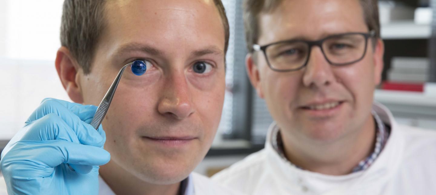
Caption: Dr. Steve Swioklo and Professor Che Connon with a dyed cornea. Credit: Newcastle University, UK

A May 30,2018 Newcastle University press release (also on EurekAlert but published on May 29, 2018), which originated the news item, adds more details,

Using a simple low-cost 3D bio-printer, the bio-ink was successfully extruded in concentric circles to form the shape of a human cornea. It took less than 10 minutes to print.

The stem cells were then shown to culture – or grow.

Che Connon, Professor of Tissue Engineering at Newcastle University, who led the work, said: “Many teams across the world have been chasing the ideal bio-ink to make this process feasible.

“Our unique gel – a combination of alginate and collagen – keeps the stem cells alive whilst producing a material which is stiff enough to hold its shape but soft enough to be squeezed out the nozzle of a 3D printer.

“This builds upon our previous work in which we kept cells alive for weeks at room temperature within a similar hydrogel. Now we have a ready to use bio-ink containing stem cells allowing users to start printing tissues without having to worry about growing the cells separately.”

The scientists, including first author and PhD student Ms Abigail Isaacson from the Institute of Genetic Medicine, Newcastle University, also demonstrated that they could build a cornea to match a patient’s unique specifications.

The dimensions of the printed tissue were originally taken from an actual cornea. By scanning a patient’s eye, they could use the data to rapidly print a cornea which matched the size and shape.

Professor Connon added: “Our 3D printed corneas will now have to undergo further testing and it will be several years before we could be in the position where we are using them for transplants.

“However, what we have shown is that it is feasible to print corneas using coordinates taken from a patient eye and that this approach has potential to combat the world-wide shortage.”

Here’s a link to and a citation for the paper,

3D bioprinting of a corneal stroma equivalent by Abigail Isaacson, Stephen Swioklo, Che J. Connon. Experimental Eye Research Volume 173, August 2018, Pages 188–193 and 2018 May 14 pii: S0014-4835(18)30212-4. doi: 10.1016/j.exer.2018.05.010. [Epub ahead of print]

This paper is behind a paywall.

A 3D printed copy of your brain

I love the title for this May 30, 2018 Wyss Institute for Biologically Inspired Engineering news release: Creating piece of mind by Lindsay Brownell (also on EurekAlert),

What if you could hold a physical model of your own brain in your hands, accurate down to its every unique fold? That’s just a normal part of life for Steven Keating, Ph.D., who had a baseball-sized tumor removed from his brain at age 26 while he was a graduate student in the MIT Media Lab’s Mediated Matter group. Curious to see what his brain actually looked like before the tumor was removed, and with the goal of better understanding his diagnosis and treatment options, Keating collected his medical data and began 3D printing his MRI [magnetic resonance imaging] and CT [computed tomography] scans, but was frustrated that existing methods were prohibitively time-intensive, cumbersome, and failed to accurately reveal important features of interest. Keating reached out to some of his group’s collaborators, including members of the Wyss Institute at Harvard University, who were exploring a new method for 3D printing biological samples.

“It never occurred to us to use this approach for human anatomy until Steve came to us and said, ‘Guys, here’s my data, what can we do?” says Ahmed Hosny, who was a Research Fellow with at the Wyss Institute at the time and is now a machine learning engineer at the Dana-Farber Cancer Institute. The result of that impromptu collaboration – which grew to involve James Weaver, Ph.D., Senior Research Scientist at the Wyss Institute; Neri Oxman, [emphasis mine] Ph.D., Director of the MIT Media Lab’s Mediated Matter group and Associate Professor of Media Arts and Sciences; and a team of researchers and physicians at several other academic and medical centers in the US and Germany – is a new technique that allows images from MRI, CT, and other medical scans to be easily and quickly converted into physical models with unprecedented detail. The research is reported in 3D Printing and Additive Manufacturing.

“I nearly jumped out of my chair when I saw what this technology is able to do,” says Beth Ripley, M.D. Ph.D., an Assistant Professor of Radiology at the University of Washington and clinical radiologist at the Seattle VA, and co-author of the paper. “It creates exquisitely detailed 3D-printed medical models with a fraction of the manual labor currently required, making 3D printing more accessible to the medical field as a tool for research and diagnosis.”

Imaging technologies like MRI and CT scans produce high-resolution images as a series of “slices” that reveal the details of structures inside the human body, making them an invaluable resource for evaluating and diagnosing medical conditions. Most 3D printers build physical models in a layer-by-layer process, so feeding them layers of medical images to create a solid structure is an obvious synergy between the two technologies.

However, there is a problem: MRI and CT scans produce images with so much detail that the object(s) of interest need to be isolated from surrounding tissue and converted into surface meshes in order to be printed. This is achieved via either a very time-intensive process called “segmentation” where a radiologist manually traces the desired object on every single image slice (sometimes hundreds of images for a single sample), or an automatic “thresholding” process in which a computer program quickly converts areas that contain grayscale pixels into either solid black or solid white pixels, based on a shade of gray that is chosen to be the threshold between black and white. However, medical imaging data sets often contain objects that are irregularly shaped and lack clear, well-defined borders; as a result, auto-thresholding (or even manual segmentation) often over- or under-exaggerates the size of a feature of interest and washes out critical detail.

The new method described by the paper’s authors gives medical professionals the best of both worlds, offering a fast and highly accurate method for converting complex images into a format that can be easily 3D printed. The key lies in printing with dithered bitmaps, a digital file format in which each pixel of a grayscale image is converted into a series of black and white pixels, and the density of the black pixels is what defines the different shades of gray rather than the pixels themselves varying in color.

Similar to the way images in black-and-white newsprint use varying sizes of black ink dots to convey shading, the more black pixels that are present in a given area, the darker it appears. By simplifying all pixels from various shades of gray into a mixture of black or white pixels, dithered bitmaps allow a 3D printer to print complex medical images using two different materials that preserve all the subtle variations of the original data with much greater accuracy and speed.

The team of researchers used bitmap-based 3D printing to create models of Keating’s brain and tumor that faithfully preserved all of the gradations of detail present in the raw MRI data down to a resolution that is on par with what the human eye can distinguish from about 9-10 inches away. Using this same approach, they were also able to print a variable stiffness model of a human heart valve using different materials for the valve tissue versus the mineral plaques that had formed within the valve, resulting in a model that exhibited mechanical property gradients and provided new insights into the actual effects of the plaques on valve function.

“Our approach not only allows for high levels of detail to be preserved and printed into medical models, but it also saves a tremendous amount of time and money,” says Weaver, who is the corresponding author of the paper. “Manually segmenting a CT scan of a healthy human foot, with all its internal bone structure, bone marrow, tendons, muscles, soft tissue, and skin, for example, can take more than 30 hours, even by a trained professional – we were able to do it in less than an hour.”

The researchers hope that their method will help make 3D printing a more viable tool for routine exams and diagnoses, patient education, and understanding the human body. “Right now, it’s just too expensive for hospitals to employ a team of specialists to go in and hand-segment image data sets for 3D printing, except in extremely high-risk or high-profile cases. We’re hoping to change that,” says Hosny.

In order for that to happen, some entrenched elements of the medical field need to change as well. Most patients’ data are compressed to save space on hospital servers, so it’s often difficult to get the raw MRI or CT scan files needed for high-resolution 3D printing. Additionally, the team’s research was facilitated through a joint collaboration with leading 3D printer manufacturer Stratasys, which allowed access to their 3D printer’s intrinsic bitmap printing capabilities. New software packages also still need to be developed to better leverage these capabilities and make them more accessible to medical professionals.

Despite these hurdles, the researchers are confident that their achievements present a significant value to the medical community. “I imagine that sometime within the next 5 years, the day could come when any patient that goes into a doctor’s office for a routine or non-routine CT or MRI scan will be able to get a 3D-printed model of their patient-specific data within a few days,” says Weaver.

Keating, who has become a passionate advocate of efforts to enable patients to access their own medical data, still 3D prints his MRI scans to see how his skull is healing post-surgery and check on his brain to make sure his tumor isn’t coming back. “The ability to understand what’s happening inside of you, to actually hold it in your hands and see the effects of treatment, is incredibly empowering,” he says.

“Curiosity is one of the biggest drivers of innovation and change for the greater good, especially when it involves exploring questions across disciplines and institutions. The Wyss Institute is proud to be a space where this kind of cross-field innovation can flourish,” says Wyss Institute Founding Director Donald Ingber, M.D., Ph.D., who is also the Judah Folkman Professor of Vascular Biology at Harvard Medical School (HMS) and the Vascular Biology Program at Boston Children’s Hospital, as well as Professor of Bioengineering at Harvard’s John A. Paulson School of Engineering and Applied Sciences (SEAS).

Here’s an image illustrating the work,

Caption: This 3D-printed model of Steven Keating’s skull and brain clearly shows his brain tumor and other fine details thanks to the new data processing method pioneered by the study’s authors. Credit: Wyss Institute at Harvard University

Here’s a link to and a citation for the paper,

From Improved Diagnostics to Presurgical Planning: High-Resolution Functionally Graded Multimaterial 3D Printing of Biomedical Tomographic Data Sets by Ahmed Hosny , Steven J. Keating, Joshua D. Dilley, Beth Ripley, Tatiana Kelil, Steve Pieper, Dominik Kolb, Christoph Bader, Anne-Marie Pobloth, Molly Griffin, Reza Nezafat, Georg Duda, Ennio A. Chiocca, James R.. Stone, James S. Michaelson, Mason N. Dean, Neri Oxman, and James C. Weaver. 3D Printing and Additive Manufacturing http://doi.org/10.1089/3dp.2017.0140 Online Ahead of Print:May 29, 2018

This paper appears to be open access.

A tangential Brad Pitt connection

It’s a bit of Hollywood gossip. There was some speculation in April 2018 that Brad Pitt was dating Dr. Neri Oxman highlighted in the Wyss Institute news release. Here’s a sample of an April 13, 2018 posting on Laineygossip (Note: A link has been removed),

It took him a long time to date, but he is now,” the insider tells PEOPLE. “He likes women who challenge him in every way, especially in the intellect department. Brad has seen how happy and different Amal has made his friend (George Clooney). It has given him something to think about.”

While a Pitt source has maintained he and Oxman are “just friends,” they’ve met up a few times since the fall and the insider notes Pitt has been flying frequently to the East Coast. He dropped by one of Oxman’s classes last fall and was spotted at MIT again a few weeks ago.

Pitt and Oxman got to know each other through an architecture project at MIT, where she works as a professor of media arts and sciences at the school’s Media Lab. Pitt has always been interested in architecture and founded the Make It Right Foundation, which builds affordable and environmentally friendly homes in New Orleans for people in need.

“One of the things Brad has said all along is that he wants to do more architecture and design work,” another source says. “He loves this, has found the furniture design and New Orleans developing work fulfilling, and knows he has a talent for it.”

It’s only been a week since Page Six first broke the news that Brad and Dr Oxman have been spending time together.

I’m fascinated by Oxman’s (and her colleagues’) furniture. Rose Brook writes about one particular Oxman piece in her March 27, 2014 posting for TCT magazine (Note: Links have been removed),

MIT Professor and 3D printing forerunner Neri Oxman has unveiled her striking acoustic chaise longue, which was made using Stratasys 3D printing technology.

Oxman collaborated with Professor W Craig Carter and Composer and fellow MIT Professor Tod Machover to explore material properties and their spatial arrangement to form the acoustic piece.

Christened Gemini, the two-part chaise was produced using a Stratasys Objet500 Connex3 multi-colour, multi-material 3D printer as well as traditional furniture-making techniques and it will be on display at the Vocal Vibrations exhibition at Le Laboratoire in Paris from March 28th 2014.

An Architect, Designer and Professor of Media, Arts and Science at MIT, Oxman’s creation aims to convey the relationship of twins in the womb through material properties and their arrangement. It was made using both subtractive and additive manufacturing and is part of Oxman’s ongoing exploration of what Stratasys’ ground-breaking multi-colour, multi-material 3D printer can do.

Brook goes on to explain how the chaise was made and the inspiration that led to it. Finally, it’s interesting to note that Oxman was working with Stratasys in 2014 and that this 2018 brain project is being developed in a joint collaboration with Statasys.

That’s it for 3D printing today.
Newcastle University (UK) has a PhD Studentship in Synthetic Biology and Nanotechnology available

Open to UK, European Union, and international students, the studentship deadline for applying is Aug. 18, 2014. Here’s more from the Newcastle University notice on the jobs.ac.uk website (Note: Links have been removed),

PhD Studentship in Synthetic Biology and Nanotechnology – Towards Algorithmic Living Manufacturing (TALIsMAN)

Value, Duration and Start Date of the Award
The Doctoral Training Award is for £20,000 per annum. This award covers fees and a contribution to an annual stipend (living expenses).

Three year PhD

Start date: 14 September 2014

Sponsor
Science Agriculture and Engineering Faculty Doctoral Training Awards

Project Description
The discipline of Synthetic Biology (SB), considers the cell to be a machine that can be built -from parts- in a manner similar to, e.g., electronic circuits, airplanes, etc. SB has sought to co-opt cells for nano-computation and nano-manufacturing purposes. During this scholarship programme of doctoral studies the student will pursue investigations at the interface of computing science (biodesign & biomodeling), chemical sciences (nanoparticle delivery systems), microbiology (bacterial genetic engineering) and nanoscience (DNA origami).

Name of the Supervisors
Professor Natalio Krasnogor (Lead Supervisor), School of Computing Science

Dr David Fulton, School of Chemistry

Dr Chien-Yi Chang, Centre for Bacterial Cell Biology

Person Specification and Eligibility Criteria
You must have an MSc in synthetic biology, microbiology, organic chemistry or computing science. You also should have demonstrable independent research skills, e.g. having completed a successful MSc dissertation or having a publication in a recognised peer reviewed conference or, ideally, journal. The candidate must have substantial laboratory experience and excellent programming and numeracy skills.

This award is available to UK/EU and International candidates. If English is not your first language, you must have IELTS 6.5.

Closing Date for Applications
Applications will be considered until Monday 18 August 2014. However, awards may be made to successful applicants before this date and early application is recommended.

So according to the line above, it’s better to apply sooner rather than later. Good luck!

Trapping gases left from nuclear fuels

A July 20, 2014 news item on ScienceDaily provides some insight into recycling nuclear fuel,

When nuclear fuel gets recycled, the process releases radioactive krypton and xenon gases. Naturally occurring uranium in rock contaminates basements with the related gas radon. A new porous material called CC3 effectively traps these gases, and research appearing July 20 in Nature Materials shows how: by breathing enough to let the gases in but not out.

The CC3 material could be helpful in removing unwanted or hazardous radioactive elements from nuclear fuel or air in buildings and also in recycling useful elements from the nuclear fuel cycle. CC3 is much more selective in trapping these gases compared to other experimental materials. Also, CC3 will likely use less energy to recover elements than conventional treatments, according to the authors.

A July 21, 2014 US Department of Energy (DOE) Pacific Northwest National Laboratory (PNNL) news release (also on EurekAlert), which originated the news item despite the difference in dates, provides more details (Note: A link has been removed),

The team made up of scientists at the University of Liverpool in the U.K., the Department of Energy’s Pacific Northwest National Laboratory, Newcastle University in the U.K., and Aix-Marseille Universite in France performed simulations and laboratory experiments to determine how — and how well — CC3 might separate these gases from exhaust or waste.

“Xenon, krypton and radon are noble gases, which are chemically inert. That makes it difficult to find materials that can trap them,” said coauthor Praveen Thallapally of PNNL. “So we were happily surprised at how easily CC3 removed them from the gas stream.”

Noble gases are rare in the atmosphere but some such as radon come in radioactive forms and can contribute to cancer. Others such as xenon are useful industrial gases in commercial lighting, medical imaging and anesthesia.

The conventional way to remove xenon from the air or recover it from nuclear fuel involves cooling the air far below where water freezes. Such cryogenic separations are energy intensive and expensive. Researchers have been exploring materials called metal-organic frameworks, also known as MOFs, that could potentially trap xenon and krypton without having to use cryogenics. Although a leading MOF could remove xenon at very low concentrations and at ambient temperatures admirably, researchers wanted to find a material that performed better.

Thallapally’s collaborator Andrew Cooper at the University of Liverpool and others had been researching materials called porous organic cages, whose molecular structures are made up of repeating units that form 3-D cages. Cages built from a molecule called CC3 are the right size to hold about three atoms of xenon, krypton or radon.

To test whether CC3 might be useful here, the team simulated on a computer CC3 interacting with atoms of xenon and other noble gases. The molecular structure of CC3 naturally expands and contracts. The researchers found this breathing created a hole in the cage that grew to 4.5 angstroms wide and shrunk to 3.6 angstroms. One atom of xenon is 4.1 angstroms wide, suggesting it could fit within the window if the cage opens long enough. (Krypton and radon are 3.69 angstroms and 4.17 angstroms wide, respectively, and it takes 10 million angstroms to span a millimeter.)

The computer simulations revealed that CC3 opens its windows big enough for xenon about 7 percent of the time, but that is enough for xenon to hop in. In addition, xenon has a higher likelihood of hopping in than hopping out, essentially trapping the noble gas inside.

The team then tested how well CC3 could pull low concentrations of xenon and krypton out of air, a mix of gases that included oxygen, argon, carbon dioxide and nitrogen. With xenon at 400 parts per million and krypton at 40 parts per million, the researchers sent the mix through a sample of CC3 and measured how long it took for the gases to come out the other side.

Oxygen, nitrogen, argon and carbon dioxide — abundant components of air — traveled through the CC3 and continued to be measured for the experiment’s full 45 minute span. Xenon however stayed within the CC3 for 15 minutes, showing that CC3 could separate xenon from air.

In addition, CC3 trapped twice as much xenon as the leading MOF material. It also caught xenon 20 times more often than it caught krypton, a characteristic known as selectivity. The leading MOF only preferred xenon 7 times as much. These experiments indicated improved performance in two important characteristics of such a material, capacity and selectivity.

“We know that CC3 does this but we’re not sure why. Once we understand why CC3 traps the noble gases so easily, we can improve on it,” said Thallapally.

To explore whether MOFs and porous organic cages offer economic advantages, the researchers estimated the cost compared to cryogenic separations and determined they would likely be less expensive.

“Because these materials function well at ambient or close to ambient temperatures, the processes based on them are less energy intensive to use,” said PNNL’s Denis Strachan.

The material might also find use in pharmaceuticals. Most molecules come in right- and left-handed forms and often only one form works in people. In additional experiments, Cooper and colleagues in the U.K. tested CC3’s ability to distinguish and separate left- and right-handed versions of an alcohol. After separating left- and right-handed forms of CC3, the team showed in biochemical experiments that each form selectively trapped only one form of the alcohol.

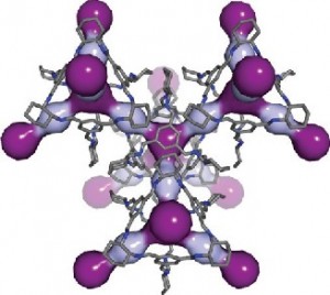
The researchers have provided an image illustrating a CC3 cage,

Breathing room: In this computer simulation, light and dark purple highlight the cavities within the 3D pore structure of CC3. Courtesy:  PNNL

Breathing room: In this computer simulation, light and dark purple highlight the cavities within the 3D pore structure of CC3. Courtesy: PNNL

Here’s a link to and a citation for the paper,

Separation of rare gases and chiral molecules by selective binding in porous organic cages by Linjiang Chen, Paul S. Reiss, Samantha Y. Chong, Daniel Holden, Kim E. Jelfs, Tom Hasell, Marc A. Little, Adam Kewley, Michael E. Briggs, Andrew Stephenson, K. Mark Thomas, Jayne A. Armstrong, Jon Bell, Jose Busto, Raymond Noel, Jian Liu, Denis M. Strachan, Praveen K. Thallapally, & Andrew I. Cooper. Nature Material (2014) doi:10.1038/nmat4035 Published online 20 July 2014

This paper is behind a paywall.

Darwin’s barnacles become unglued

The world’s strongest glue comes from barnacles and those creatures have something to teach us. From a July 18, 2014 news item on Nanowerk,

Over a 150 years since it was first described by Darwin, scientists are finally uncovering the secrets behind the super strength of barnacle glue.

Still far better than anything we have been able to develop synthetically, barnacle glue – or cement – sticks to any surface, under any conditions.

But exactly how this superglue of superglues works has remained a mystery – until now.

An international team of scientists led by Newcastle University, UK, and funded by the US Office of Naval Research, have shown for the first time that barnacle larvae release an oily droplet to clear the water from surfaces before sticking down using a phosphoprotein adhesive.

A July 18, 2014 Newcastle University (UK) press release, which originated the news item, provides some context and describes the research,

“It’s over 150 years since Darwin first described the cement glands of barnacle larvae and little work has been done since then,” says Dr Aldred, a research associate in the School of Marine Science and Technology at Newcastle University, one of the world’s leading institutions in this field of research.

“We’ve known for a while there are two components to the bioadhesive but until now, it was thought they behaved a bit like some of the synthetic glues – mixing before hardening.  But that still left the question, how does the glue contact the surface in the first place if it is already covered with water?  This is one of the key hurdles to developing glues for underwater applications.

“Advances in imaging techniques, such as 2-photon microscopy, have allowed us to observe the adhesion process and characterise the two components. We now know that these two substances play very different roles – one clearing water from the surface and the other cementing the barnacle down.

“The ocean is a complex mixture of dissolved ions, the pH varies significantly across geographical areas and, obviously, it’s wet.  Yet despite these hostile conditions, barnacle glue is able to withstand the test of time.

“It’s an incredibly clever natural solution to this problem of how to deal with a water barrier on a surface it will change the way we think about developing bio-inspired adhesives that are safe and already optimised to work in conditions similar to those in the human body, as well as marine paints that stop barnacles from sticking.”

Barnacles have two larval stages – the nauplius and the cyprid.  The nauplius, is common to most crustacea and it swims freely once it hatches out of the egg, feeding in the plankton.

The final larval stage, however, is the cyprid, which is unique to barnacles.  It investigates surfaces, selecting one that provides suitable conditions for growth. Once it has decided to attach permanently, the cyprid releases its glue and cements itself to the surface where it will live out the rest of its days.

“The key here is the technology.  With these new tools we are able to study processes in living tissues, as they happen. We can get compositional and molecular information by other methods, but they don’t explain the mechanism.  There’s no substitute for seeing things with your own eyes. ” explains Dr Aldred.

“In the past, the strong lasers used for optically sectioning biological samples have typically killed the samples, but now technology allows us to study life processes exactly as they would happen in nature.”

The press release also notes some possible applications for these research findings (Note: Links have been removed),

Publishing their findings this week in the prestigious academic journal Nature Communications, author Dr Nick Aldred says the findings could pave the way for the development of novel synthetic bioadhesives for use in medical implants and micro-electronics.  The research will also be important in the production of new anti-fouling coatings for ships.

Here’s a link to and a citation for the paper,

Synergistic roles for lipids and proteins in the permanent adhesive of barnacle larvae by Neeraj V. Gohad, Nick Aldred, Christopher M. Hartshorn, Young Jong Lee, Marcus T. Cicerone, Beatriz Orihuela, Anthony S. Clare, Dan Rittschof, & Andrew S. Mount. Nature Communications 5, Article number: 4414 doi:10.1038/ncomms5414 Published 11 July 2014

This paper is behind a paywall although a free preview is available via ReadCube Access.

Phytoremediation, clearing pollutants from industrial lands, could also be called phyto-mining

The University of Edinburgh (along with the Universities of Warwick and Birmingham, Newcastle University and Cranfield University) according to its Mar. 4, 2013 news release on EurekAlert is involved in a phytoremediation project,

Common garden plants are to be used to clean polluted land, with the extracted poisons being used to produce car parts and aid medical research.

Scientists will use plants such as alyssum, pteridaceae and a type of mustard called sinapi to soak up metals from land previously occupied by factories, mines and landfill sites.

Dangerous levels of metals such as arsenic and platinum, which can lurk in the ground and can cause harm to people and animals, will be extracted using a natural process known as phytoremediation.

A Mar. 4, 2013 news item on the BBC News Edinburgh, Fife and East Scotland site offers more details about the project and the technology,

A team of researchers from the Universities of Edinburgh, Warwick, Birmingham, Newcastle and Cranfield has developed a way of extracting the chemicals through a process called phytoremediation, and are testing its effectiveness.

Once the plants have drawn contaminated material out of the soil, they will be harvested and processed in a bio-refinery.

A specially designed bacteria will be added to the waste to transform the toxic metal ions into metallic nanoparticles.

The team said these tiny particles could then be used to develop cancer treatments, and could also be used to make catalytic converters for cars.

Dr Louise Horsfall, of Edinburgh’s University’s school of biological sciences, said: “Land is a finite resource. As the world’s population grows along with the associated demand for food and shelter, we believe that it is worth decontaminating land to unlock vast areas for better food security and housing.

“I hope to use synthetic biology to enable bacteria to produce high value nanoparticles and thereby help make land decontamination financially viable.”

The research team said the land where phytoremediation was used would also be cleared of chemicals, meaning it could be reused for new building projects.

In my Sept. 28, 2012 posting I featured an international collaboration between universities in the UK, US, Canada, and New Zealand in a ‘phyto-mining’ project bearing some resemblance to this newly announced project. In that project, announced in Fall 2012, scientists were studying how they might remove platinum for reuse from plants near the tailings of mines.

I do have one other posting about phytoremediation. I featured a previously published piece by Joe Martin in a two-part series on the topic plant (phyto) and nano soil remediation. The March 30, 2012 posting is part one, which focuses on the role of plants in soil remediation.

Sea urchins taste yummy and (might) hold key to carbon capture

A prized sushi food item, sea urchins use nickel particles to convert carbon dioxide according to a Feb. 5, 2013 news item on ScienceDaily,

The discovery that sea urchins use nickel particles to harness carbon dioxide from the sea could be the key to capturing tons of carbon dioxide (CO2) from the atmosphere.

Experts at Newcastle University, UK, have discovered that in the presence of a nickel catalyst, CO2 can be converted rapidly and cheaply into the harmless, solid mineral, calcium carbonate.

This discovery, which is published February 5 in the academic journal Catalysis Science & Technology, has the potential to revolutionize the way we capture and store carbon enabling us to significantly reduce CO2 emissions — the key greenhouse gas responsible for climate change.

The Newcastle University Feb. 5, 2013 news release, which originated the news item, details how this discovery came about,

Dr Lidija Šiller, a physicist and Reader in Nanoscale Technology at Newcastle University, says the discovery was made completely by chance.

“We had set out to understand in detail the carbonic acid reaction – which is what happens when CO2 reacts with water – and needed a catalyst to speed up the process,” she explains.

“At the same time, I was looking at how organisms absorb CO2 into their skeletons and in particular the sea urchin which converts the CO2 to calcium carbonate.

“When we analysed the surface of the urchin larvae we found a high concentration of Nickel on their exoskeleton.  Taking Nickel nanoparticles which have a large surface area, we added them to our carbonic acid test and the result was the complete removal of CO2.”

Before discussing the implications it’s useful to understand the current situation regarding carbon capture processes, from the news release,

At the moment, pilot studies for Carbon Capture and Storage (CCS) systems propose the removal of CO2 by pumping it into holes deep underground.  However, this is a costly and difficult process and carries with it a long term risk of the gas leaking back out – possibly many miles away from the original downward source.

An alternative solution is to convert the CO2 into calcium or magnesium carbonate.

“One way to do this is to use an enzyme called carbonic anhydrase,” explains Gaurav Bhaduri, lead author on the paper and a PhD student in the University’s School of Chemical Engineering and Advanced Materials.

“However, the enzyme is inactive in acid conditions and since one of the products of the reaction is carbonic acid, this means the enzyme is only effective for a very short time and also makes the process very expensive.

“The beauty of a Nickel catalyst is that it carries on working regardless of the pH and because of its magnetic properties it can be re-captured and re-used time and time again. It’s also very cheap – 1,000 times cheaper than the enzyme.  And the by-product – the carbonate – is useful and not damaging to the environment.

“What our discovery offers is a real opportunity for industries such as power stations and chemical processing plants to capture all their waste CO2 before it ever reaches the atmosphere and store it as a safe, stable and useful product.”

Each year, humans emit on average 33.4 billion metric tons of CO2 – around 45% of which remains in the atmosphere.  Typically, a petrol-driven car will produce a ton of CO2 every 4,000 miles.

Calcium carbonate, or chalk, makes up around 4% of the Earth’s crust and acts as a carbon reservoir, estimated to be equivalent to 1.5 million billion metric tons of carbon dioxide.

It is the main component of shells of marine organisms, snails, pearls, and eggshells and is a completely stable mineral, widely used in the building industry to make cement and other materials and also in hospitals to make plaster casts.

The process developed by the Newcastle team involves passing the waste gas directly from the chimney top, through a water column rich in Nickel nano-particles and recovering the solid calcium carbonate from the bottom.

Dr Šiller adds: “The capture and removal of CO2 from our atmosphere is one of the most pressing dilemmas of our time.

“Our process would not work in every situation – it couldn’t be fitted to the back of a car, for example – but it is an effective, cheap solution that could be available world-wide to some of our most polluting industries and have a significant impact on the reduction of atmospheric CO2.”

According the news release the researchers have patented the process and are looking for investors as they plan for future development.

Phyto-mining and environmental remediation flower in the United Kingdom

Researchers on a £3 million research programme called “Cleaning Land for Wealth” (CL4W) are confident they’ll be able to use flowers and plants to clean soil of poisonous materials (environmental remediation) and to recover platinum (phyto-mining). From the Nov. 21, 2012 news item on Nanowerk,

A consortium of researchers led by WMG (Warwick Manufacturing Group) at the University of Warwick are to embark on a £3 million research programme called “Cleaning Land for Wealth” (CL4W), that will use a common class of flower to restore poisoned soils while at the same time producing perfectly sized and shaped nano sized platinum and arsenic nanoparticles for use in catalytic convertors, cancer treatments and a range of other applications.

The Nov. 20, 2012 University of Warwick news release, which originated the news item, describes both how CL4W came together and how it produced an unintended project benefit,

A “Sandpit” exercise organised by the Engineering and Physical Sciences Research Council (EPSRC) allowed researchers from WMG (Warwick Manufacturing group) at the University of Warwick, Newcastle University, The University of Birmingham, Cranfield University and the University of Edinburgh to come together and share technologies and skills to come up with an innovative multidisciplinary research project that could help solve major technological and environmental challenges.

The researchers pooled their knowledge of how to use plants and bacteria to soak up particular elements and chemicals and how to subsequently harvest, process and collect that material. They have devised an approach to demonstrate the feasibility in which they are confident that they can use common classes of flower and plants (such as Alyssum), to remove poisonous chemicals such as arsenic and platinum from polluted land and water courses potentially allowing that land to be reclaimed and reused.

That in itself would be a significant achievement, but as the sandpit progressed the researchers found that jointly they had the knowledge to achieve much more than just cleaning up the land.

As lead researcher on the project Professor Kerry Kirwan from WMG at the University of Warwick explained:

“The processes we are developing will not only remove poisons such as arsenic and platinum from contaminated land and water courses, we are also confident that we can develop suitable biology and biorefining processes (or biofactories as we are calling them) that can tailor the shapes and sizes of the metallic nanoparticles they will make. This would give manufacturers of catalytic convertors, developers of cancer treatments and other applicable technologies exactly the right shape, size and functionality they need without subsequent refinement. We are also expecting to recover other high value materials such as fine chemicals, pharmaceuticals, anti-oxidants etc. from the crops during the same biorefining process.”

I last mentioned phyto-mining in my Sept. 26, 2012 post with regard to an international project being led by researchers at the University of York (UK).  The biorefining processes (biofactories) mentioned by Kirwan takes the idea of recovering platinum, etc. one step beyond phyto-mining recovery.

Here’s a picture of the flower (Alyssum) mentioned in the news release,

Alyssum montanum photographed by myself in 1988, Unterfranken, Germany [http://en.wikipedia.org/wiki/Alyssum]

From the Wikipedia essay (Note: I have removed links],

Alyssum is a genus of about 100–170 species of flowering plants in the family Brassicaceae, native to Europe, Asia, and northern Africa, with the highest species diversity in the Mediterranean region. The genus comprises annual and perennial herbaceous plants or (rarely) small shrubs, growing to 10–100 cm tall, with oblong-oval leaves and yellow or white flowers (pink to purple in a few species).