Tag Archives: Wyss Institute for Biologically Inspired Engineering

Xenobots (living robots) that can reproduce

Xenobots (living robots made from African frog (Xenopus laevis) frog cells) can now self-replicate. First mentioned here in a June 21, 2021 posting, xenobots have captured the imagination of various media outlets including the Canadian Broadcasting Corporation’s (CBC) Quirks and Quarks radio programme and blog where Amanda Buckiewicz posted a December 3, 2021 article about the latest xenobot development (Note: Links have been removed),

In a new study, Bongard [Joshua Bongard, a computer scientist at the University of Vermont] and his colleagues from Tufts University and Harvard’s Wyss Institute for Biologically Inspired Engineering found that the xenobots would autonomously collect loose single cells in their environment, gathering hundreds of cells together until new xenobots had formed.

“This took a little bit for us to wrap our minds around,” he said. “There’s no programming here. Instead, we’re designing or shaping these xenobots, and what they do, the way they behave, is based on shape.”

“We take a couple of thousand of those frog cells and we squish them together into a ball and put that in the bottom of a petri dish,” Bongard told Quirks & Quarks host Bob McDonald. 

“If you were to look into the dish, you would see some very small, what look like specks of pepper, moving about in the bottom of the petri dish.”

The xenobots initially received no instruction from humans on how to replicate. But when researchers added extra cells to the dish containing xenobots, they observed that the xenobots would assemble them into piles.

“Cells early in development are sticky,” said Bongard. “If the pile is large enough and the cells stick together, the outer ones on the surface will grow very small hairs, which are called cilia. And eventually, after four days, those cilia will start to beat back and forth like flexible oars, and the pile will start moving.”

“And that’s a child xenobot.” 

A November 29, 2021 Wyss Institute news release by Joshua Brown describes the process a little differently,

To persist, life must reproduce. Over billions of years, organisms have evolved many ways of replicating, from budding plants to sexual animals to invading viruses.

Now scientists at the University of Vermont, Tufts University, and the Wyss Institute for Biologically Inspired Engineering at Harvard University have discovered an entirely new form of biological reproduction—and applied their discovery to create the first-ever, self-replicating living robots.

The same team that built the first living robots (“Xenobots,” assembled from frog cells—reported in 2020) has discovered that these computer-designed and hand-assembled organisms can swim out into their tiny dish, find single cells, gather hundreds of them together, and assemble “baby” Xenobots inside their Pac-Man-shaped “mouth”—that, a few days later, become new Xenobots that look and move just like themselves.

And then these new Xenobots can go out, find cells, and build copies of themselves. Again and again.

In a Xenopus laevis frog, these embryonic cells would develop into skin. “They would be sitting on the outside of a tadpole, keeping out pathogens and redistributing mucus,” says Michael Levin, Ph.D., a professor of biology and director of the Allen Discovery Center at Tufts University and co-leader of the new research. “But we’re putting them into a novel context. We’re giving them a chance to reimagine their multicellularity.” Levin is also an Associate Faculty member at the Wyss Institute.

And what they imagine is something far different than skin. “People have thought for quite a long time that we’ve worked out all the ways that life can reproduce or replicate. But this is something that’s never been observed before,” says co-author Douglas Blackiston, Ph.D., the senior scientist at Tufts University and the Wyss Institute who assembled the Xenobot “parents” and developed the biological portion of the new study.

“This is profound,” says Levin. “These cells have the genome of a frog, but, freed from becoming tadpoles, they use their collective intelligence, a plasticity, to do something astounding.” In earlier experiments, the scientists were amazed that Xenobots could be designed to achieve simple tasks. Now they are stunned that these biological objects—a computer-designed collection of cells—will spontaneously replicate. “We have the full, unaltered frog genome,” says Levin, “but it gave no hint that these cells can work together on this new task,” of gathering and then compressing separated cells into working self-copies.

“These are frog cells replicating in a way that is very different from how frogs do it. No animal or plant known to science replicates in this way,” says Sam Kriegman, Ph.D.,  the lead author on the new study, who completed his Ph.D. in Bongard’s lab at UVM and is now a post-doctoral researcher at Tuft’s Allen Center and Harvard University’s Wyss Institute for Biologically Inspired Engineering.

Both Buckiewicz’s December 3, 2021 article and Brown’s November 29, 2021 Wyss Institute news release are good reads with liberal used of embedded images. If you have time, start with Buckiewicz as she provides a good introduction and follow up with Brown who gives more detail and has an embedded video of a December 1, 2021 panel discussion with the scientists behind the xenobots.

Here’s a link to and a citation for the latest paper,

Kinematic self-replication in reconfigurable organisms by Sam Kriegman, Douglas Blackiston, Michael Levin, and Josh Bongard. PNAS [Proceedings of the National Academy of Sciences] December 7, 2021 118 (49) e2112672118; https://doi.org/10.1073/pnas.2112672118

This paper appears to be open access.

Teaching molecular and synthetic biology in grades K-12

This* story actually started in 2018 with an August 1, 2018 Harvard University news release (h/t Aug. 1, 2018 news item on phys.org) by Leslie Brownell announcing molecular and synthetic biology educational kits that been tested in the classroom. (In 2019, a new kit was released but more about that later.)

As biologists have probed deeper into the molecular and genetic underpinnings of life, K-12 schools have struggled to provide a curriculum that reflects those advances. Hands-on learning is known to be more engaging and effective for teaching science to students, but even the most basic molecular and synthetic biology experiments require equipment far beyond an average classroom’s budget, and often involve the use of bacteria and other substances that can be difficult to manage outside a controlled lab setting.

Now, a collaboration between the Wyss Institute at Harvard University, MIT [Massachusetts Institute of Technology], and Northwestern University has developed BioBits, new educational biology kits that use freeze-dried cell-free (FD-CF) reactions to enable students to perform a range of simple, hands-on biological experiments. The BioBits kits introduce molecular and synthetic biology concepts without the need for specialized lab equipment, at a fraction of the cost of current standard experimental designs. The kits are described in two papers published in Science Advances [2018].

“The main motivation in developing these kits was to give students fun activities that allow them to actually see, smell, and touch the outcomes of the biological reactions they’re doing at the molecular level,” said Ally Huang, a co-first author on both papers who is an MIT graduate student in the lab of Wyss Founding Core Faculty member Jim Collins, Ph.D. “My hope is that they will inspire more kids to consider a career in STEM [science, technology, engineering, and math] and, more generally, give all students a basic understanding of how biology works, because they may one day have to make personal or policy decisions based on modern science.”

Synthetic and molecular biology frequently make use of the cellular machinery found in E. coli bacteria to produce a desired protein. But this system requires that the bacteria be kept alive and contained for an extended period of time, and involves several complicated preparation and processing steps. The FD-CF reactions pioneered in Collins’ lab for molecular manufacturing, when combined with innovations from the lab of Michael Jewett, Ph.D. at Northwestern University, offer a solution to this problem by removing bacteria from the equation altogether.

“You can think of it like opening the hood of a car and taking the engine out: we’ve taken the ‘engine’ that drives protein production out of a bacterial cell and given it the fuel it needs, including ribosomes and amino acids, to create proteins from DNA outside of the bacteria itself,” explained Jewett, who is the Charles Deering McCormick Professor of Teaching Excellence at Northwestern University’s McCormick School of Engineering and co-director of Northwestern’s Center for Synthetic Biology, and co-corresponding author of both papers. This collection of molecular machinery is then freeze-dried into pellets so that it becomes shelf-stable at room temperature. To initiate the transcription of DNA into RNA and the translation of that RNA into a protein, a student just needs to add the desired DNA and water to the freeze-dried pellets.

The researchers designed a range of molecular experiments that can be performed using this system, and coupled each of them to a signal that the students can easily detect with their sense of sight, smell, or touch. The first, called BioBits Bright, contains six different freeze-dried DNA templates that each encode a protein that fluoresces a different color when illuminated with blue light. To produce the proteins, students simply add these DNA templates and water to the FD-CF machinery and put the reactions in an inexpensive incubator (~$30) for several hours, and then view them with a blue light illuminator (~$15). The students can also design their own experiments to produce a desired collection of colors that they can then arrange into a visual image, a bit like using a Light Brite ©. “Challenging the students to build their own in vitro synthetic programs also allows educators to start to talk about how synthetic biologists might control biology to make important products, such as medicines or chemicals,” explained Jessica Stark, an NSF Graduate Research Fellow in the Jewett lab at Northwestern University who is co-first author on both papers.

An expansion of the BioBits Bright kit, called BioBits Explorer, includes experiments that engage the senses of smell and touch and allow students to probe their environment using designer synthetic biosensors. In the first experiment, the FD-CF reaction pellets contain a gene that drives the conversion of isoamyl alcohol to isoamyl acetate, a compound that produces a strong banana odor. In the second experiment, the FD-CF reactions contain a gene coding for the enzyme sortase, which recognizes and links specific segments of proteins in a liquid solution together to form a squishy, semi-solid hydrogel, which the students can touch and manipulate. The third module uses another Wyss technology, the toehold switch sensor, to identify DNA extracted from a banana or a kiwi. The sensors are hairpin-shaped RNA molecules designed such that when they bind to a “trigger” RNA, they spring open and reveal a genetic sequence that produces a fluorescent protein. When fruit DNA is added to the sensor-containing FD-CF pellets, only the sensors that are designed to open in the presence of each fruit’s RNA will produce the fluorescent protein.

The researchers tested their BioBits kits in the Chicago Public School system, and demonstrated that students and teachers were able to perform the experiments in the kits with the same success as trained synthetic biology researchers. In addition to refining the kits’ design so that they can one day provide them to classrooms around the world, the authors hope to create an open-source online database where teachers and students can share their results and ideas for ways to modify the kits to explore different biological questions.

“Synthetic biology is going to be one of the defining technologies of the century, and yet it has been challenging to teach the fundamental concepts of the field in K-12 classrooms given that such efforts often require expensive, complicated equipment,” said Collins, who is a co-corresponding author of both papers and also the Termeer Professor of Medical Engineering & Science at MIT. “We show that it is possible to use freeze-dried, cell-free extracts along with freeze-dried synthetic biology components to conduct innovative educational experiments in classrooms and other low-resource settings. The BioBits kits enable us to expose young kids, older kids, and even adults to the wonders of synthetic biology and, as a result, are poised to transform science education and society.

“All scientists are passionate about what they do, and we are frustrated by the difficulty our educational system has had in inciting a similar level of passion in young people. This BioBits project demonstrates the kind of out-of-the-box thinking and refusal to accept the status quo that we value and cultivate at the Wyss Institute, and we all hope it will stimulate young people to be intrigued by science,” said Wyss Institute Founding Director Donald Ingber, M.D., Ph.D., who is also the Judah Folkman Professor of Vascular Biology at Harvard Medical School (HMS) and the Vascular Biology Program at Boston Children’s Hospital, as well as Professor of Bioengineering at Harvard’s John A. Paulson School of Engineering and Applied Sciences (SEAS). “It’s exciting to see this project move forward and become available to biology classrooms worldwide and, hopefully some of these students will pursue a path in science because of their experience.”

Additional authors of the papers include Peter Nguyen, Ph.D., Nina Donghia, and Tom Ferrante from the Wyss Institute; Melissa Takahashi, Ph.D. and Aaron Dy from MIT; Karen Hsu and Rachel Dubner from Northwestern University; Keith Pardee, Ph.D., Assistant Professor at the University of Toronto; and a number of teachers and students in the Chicago school system including: Mary Anderson, Ada Kanapskyte, Quinn Mucha, Jessica Packett, Palak Patel, Richa Patel, Deema Qaq, Tyler Zondor, Julie Burke, Tom Martinez, Ashlee Miller-Berry, Aparna Puppala, Kara Reichert, Miriam Schmid, Lance Brand, Lander Hill, Jemima Chellaswamy, Nuhie Faheem, Suzanne Fetherling, Elissa Gong, Eddie Marie Gonzales, Teresa Granito, Jenna Koritsaris, Binh Nguyen, Sujud Ottman, Christina Palffy, Angela Patel, Sheila Skweres, Adriane Slaton, and TaRhonda Woods.

This research was supported by the Army Research Office, the National Science Foundation, the Air Force Research Laboratory Center of Excellence Grant, The Defense Threat Reduction Agency Grant, the David and Lucile Packard Foundation, the Camille Dreyfus Teacher-Scholar Program, the Wyss Institute at Harvard University, the Paul G. Allen Frontiers Group, The Air Force Office of Scientific Research, and the Natural Sciences and Engineering Council of Canada. [emphases mine]

Well, that list of funding agencies is quite interesting. The US Army and Air Force but not the Navy? As for what the Natural Sciences and Engineering Council of Canada is doing on that list, I can only imagine why.

This is what they were doing in 2018,

Now for the latest update, a May 7, 2019 news item on phys.org announces the BioBits Kits have been expanded,

How can high school students learn about a technology as complex and abstract as CRISPR? It’s simple: just add water.

A Northwestern University-led team has developed BioBits, a suite of hands-on educational kits that enable students to perform a range of biological experiments by adding water and simple reagents to freeze-dried cell-free reactions. The kits link complex biological concepts to visual, fluorescent readouts, so students know—after a few hours and with a single glance—the results of their experiments.

A May 7, 2019 Northwestern University news release (also on EurekAlert and received via email) by Amanda Morris, which originated the news item, provides more details,

After launching BioBits last summer, the researchers are now expanding the kit to include modules for CRISPR [clustered regularly interspaced short palindromic repeats] and antibiotic resistance. A small group of Chicago-area teachers and high school students just completed the first pilot study for these new modules, which include interactive experiments and supplementary materials exploring ethics and strategies.

“After we unveiled the first kits, we next wanted to tackle current topics that are important for society,” said Northwestern’s Michael Jewett, principal investigator of the study. “That led us to two areas: antibiotic resistance and gene editing.”

Called BioBits Health, the new kits and pilot study are detailed in a paper published today (May 7 [2019]) in the journal ACS Synthetic Biology.

Jewett is a professor of chemical and biological engineering in Northwestern’s McCormick School of Engineering and co-director of Northwestern’s Center for Synthetic Biology. Jessica Stark, a graduate student in Jewett’s laboratory, led the study.

Test in a tube

Instead of using live cells, the BioBits team removed the essential cellular machinery from inside the cells and freeze-dried them for shelf stability. Keeping cells alive and contained for an extended period of time involves several complicated, time-consuming preparation and processing steps as well as expensive equipment. Freeze-dried cell-free reactions bypass those complications and costs.

“These are essentially test-tube biological reactions,” said Stark, a National Science Foundation graduate research fellow. “We break the cells open and use their guts, which still contain all of the necessary biological machinery to carry out a reaction. We no longer need living cells to demonstrate biology.”

This method to harness biological systems without intact, living cells became possible over the last two decades thanks to multiple innovations, including many in cell-free synthetic biology by Jewett’s lab. Not only are these experiments doable in the classroom, they also only cost pennies compared to standard high-tech experimental designs.

“I’m hopeful that students get excited about engineering biology and want to learn more,” Jewett said.

Conquering CRISPR

One of the biggest scientific breakthroughs of the past decade, CRISPR (pronounced “crisper”) stands for Clustered Regularly Interspaced Short Palindromic Repeats. The powerful gene-editing technology uses enzymes to cut DNA in precise locations to turn off or edit targeted genes. It could be used to halt genetic diseases, develop new medicines, make food more nutritious and much more.

BioBits Health uses three components required for CRISPR: an enzyme called the Cas9 protein, a target DNA sequence encoding a fluorescent protein and an RNA molecule that targets the fluorescent protein gene. When students add all three components — and water — to the freeze-dried cell-free system, it creates a reaction that edits, or cuts, the DNA for the fluorescent protein. If the DNA is cut, the system does not glow. If the DNA is not cut, the fluorescent protein is made, and the system glows fluorescent.

“We have linked this abstract, really advanced biological concept to the presence or absence of a fluorescent protein,” Stark said. “It’s something students can see, something they can visually understand.”

The curriculum also includes activities that challenge students to consider the ethical questions and dilemmas surrounding the use of gene-editing technologies.

“There is a lot of excitement about being able to edit genomes with these technologies,” Jewett said. “BioBits Health calls attention to a lot of important questions — not only about how CRISPR technology works but about ethics that society should be thinking about. We hope that this promotes a conversation and dialogue about such technologies.”

Reducing resistance

Jewett and Stark are both troubled by a prediction that, by the year 2050, drug-resistant bacterial infections could outpace cancer as a leading cause of death. This motivated them to help educate the future generation of scientists about how antibiotic resistance emerges and inspire them to take actions that could help limit the emergence of resistant bacteria.
In this module, students run two sets of reactions to produce a glowing fluorescent protein — one set with an antibiotic resistance gene and one set without. Students then add antibiotics. If the experiment glows, the fluorescent protein has been made, and the reaction has become resistant to antibiotics. If the experiment does not glow, then the antibiotic has worked.

“Because we’re using cell-free systems rather than organisms, we can demonstrate drug resistance in a way that doesn’t create drug-resistant bacteria,” Stark explained. “We can demonstrate these concepts without the risks.”

A supporting curriculum piece challenges students to brainstorm and research strategies for slowing the rate of emerging antibiotic resistant strains.

Part of something cool

After BioBits was launched in summer 2018, 330 schools from around the globe requested prototype kits for their science labs. The research team, which includes members from Northwestern and MIT, has received encouraging feedback from teachers, students and parents.

“The students felt like scientists and doctors by touching and using the laboratory materials provided during the demo,” one teacher said. “Even the students who didn’t seem engaged were secretly paying attention and wanted to take their turn pipetting. They knew they were part of something really cool, so we were able to connect with them in a way that was new to them.”

“My favorite part was using the equipment,” a student said. “It was a fun activity that immerses you into what top scientists are currently doing.”

###

The study, “BioBits Health: Classroom activities exploring engineering, biology and human health with fluorescent readouts,” was supported by the Army Research Office (award number W911NF-16-1-0372), the National Science Foundation (grant numbers MCB-1413563 and MCB-1716766), the Air Force Research Laboratory Center of Excellence (grant number FA8650-15-2-5518), the Defense Threat Reduction Agency (grant number HDTRA1-15-10052/P00001), the Department of Energy (grant number DE-SC0018249), the Human Frontiers Science Program (grant number RGP0015/2017), the David and Lucile Packard Foundation, the Office of Energy Efficiency and Renewable Energy (grant number DE-EE008343) and the Camille Dreyfus Teacher-Scholar Program. [emphases mine]

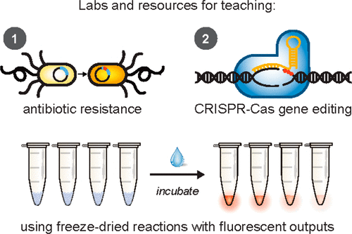
This is an image you’ll find in the abstract for the 2019 paper,

[downloaded from https://pubs.acs.org/doi/10.1021/acssynbio.8b00381]

Here are links and citations for the 2018 papers and the 2019 paper,

BioBits™ Explorer: A modular synthetic biology education kit by Ally Huang, Peter Q. Nguyen, Jessica C. Stark, Melissa K. Takahashi, Nina Donghia, Tom Ferrante, Aaron J. Dy, Karen J. Hsu, Rachel S. Dubner, Keith Pardee, Michael C. Jewett, and James J. Collins. Science Advances 01 Aug 2018: Vol. 4, no. 8, eaat5105 DOI: 10.1126/sciadv.aat5105

BioBits™ Bright: A fluorescent synthetic biology education kit by Jessica C. Stark, Ally Huang, Peter Q. Nguyen, Rachel S. Dubner, Karen J. Hsu, Thomas C. Ferrante, Mary Anderson, Ada Kanapskyte, Quinn Mucha, Jessica S. Packett, Palak Patel, Richa Patel, Deema Qaq, Tyler Zondor, Julie Burke, Thomas Martinez, Ashlee Miller-Berry, Aparna Puppala, Kara Reichert, Miriam Schmid, Lance Brand, Lander R. Hill, Jemima F. Chellaswamy, Nuhie Faheem, Suzanne Fetherling, Elissa Gong, Eddie Marie Gonzalzles, Teresa Granito, Jenna Koritsaris, Binh Nguyen, Sujud Ottman, Christina Palffy, Angela Patel, Sheila Skweres, Adriane Slaton, TaRhonda Woods, Nina Donghia, Keith Pardee, James J. Collins, and Michael C. Jewett. Science Advances 01 Aug 2018: Vol. 4, no. 8, eaat5107 DOI: 10.1126/sciadv.aat5107

BioBits Health: Classroom Activities Exploring Engineering, Biology, and Human Health with Fluorescent Readouts by Jessica C. Stark, Ally Huang, Karen J. Hsu, Rachel S. Dubner, Jason Forbrook, Suzanne Marshalla, Faith Rodriguez, Mechelle Washington, Grant A. Rybnicky, Peter Q. Nguyen, Brenna Hasselbacher, Ramah Jabri, Rijha Kamran, Veronica Koralewski, Will Wightkin, Thomas Martinez, and Michael C. Jewett. ACS Synth. Biol., Article ASAP
DOI: 10.1021/acssynbio.8b00381 Publication Date (Web): March 29, 2019

Copyright © 2019 American Chemical Society

Both of the 2018 papers appear to be open access while the 2019 paper is behind a paywall.

Should you be interested in acquiring a BioBits kit, you can check out the BioBits website. As for ‘conguering’ CRISPR, do we really need to look at it that way? Maybe a more humble appraoch could work just as well or even better, eh?

*’is’ removed from sentence on May 9, 2019.

Xenotransplantation—organs for transplantation in human patients—it’s a business and a science

The last time (June 18, 2018 post) I mentioned xenotransplantation (transplanting organs from one species into another species; see more here), it was in the context of an art/sci (or sciart) event coming to Vancouver (Canada).,

Patricia Piccinini’s Curious Imaginings Courtesy: Vancouver Biennale [downloaded from http://dailyhive.com/vancouver/vancouver-biennale-unsual-public-art-2018/]

The latest edition of the Vancouver Biennale was featured in a June 6, 2018 news item on the Daily Hive (Vancouver),

Melbourne artist Patricia Piccinini’s Curious Imaginings is expected to be one of the most talked about installations of the exhibit. Her style of “oddly captivating, somewhat grotesque, human-animal hybrid creature” is meant to be shocking and thought-provoking.

Piccinini’s interactive [emphasis mine] experience will “challenge us to explore the social impacts of emerging biotechnology and our ethical limits in an age where genetic engineering and digital technologies are already pushing the boundaries of humanity.”

Piccinini’s work will be displayed in the 105-year-old Patricia Hotel in Vancouver’s Strathcona neighbourhood. The 90-day ticketed exhibition [emphasis mine] is scheduled to open this September [2018].

(The show opens on Sept. 14, 2018.)

At the time, I had yet to stumble across Ingfei Chen’s thoughtful dive into the topic in her May 9, 2018 article for Slate.com,

In the United States, the clock is ticking for more than 114,700 adults and children waiting for a donated kidney or other lifesaving organ, and each day, nearly 20 of them die. Researchers are devising a new way to grow human organs inside other animals, but the method raises potentially thorny ethical issues. Other conceivable futuristic techniques sound like dystopian science fiction. As we envision an era of regenerative medicine decades from now, how far is society willing to go to solve the organ shortage crisis?

I found myself pondering this question after a discussion about the promises of stem cell technologies veered from the intriguing into the bizarre. I was interviewing bioengineer Zev Gartner, co-director and research coordinator of the Center for Cellular Construction at the University of California, San Francisco, about so-called organoids, tiny clumps of organlike tissue that can self-assemble from human stem cells in a Petri dish. These tissue bits are lending new insights into how our organs form and diseases take root. Some researchers even hope they can nurture organoids into full-size human kidneys, pancreases, and other organs for transplantation.

Certain organoid experiments have recently set off alarm bells, but when I asked Gartner about it, his radar for moral concerns was focused elsewhere. For him, the “really, really thought-provoking” scenarios involve other emerging stem cell–based techniques for engineering replacement organs for people, he told me. “Like blastocyst complementation,” he said.

Never heard of it? Neither had I. Turns out it’s a powerful new genetic engineering trick that researchers hope to use for growing human organs inside pigs or sheep—organs that could be genetically personalized for transplant patients, in theory avoiding immune-system rejection problems. The science still has many years to go, but if it pans out, it could be one solution to the organ shortage crisis. However, the prospect of creating hybrid animals with human parts and killing them to harvest organs has already raised a slew of ethical questions. In 2015, the National Institutes of Health placed a moratorium on federal funding of this nascent research area while it evaluated and discussed the issues.

As Gartner sees it, the debate over blastocyst complementation research—work that he finds promising—is just one of many conversations that society needs to have about the ethical and social costs and benefits of future technologies for making lifesaving transplant organs. “There’s all these weird ways that we could go about doing this,” he said, with a spectrum of imaginable approaches that includes organoids, interspecies organ farming, and building organs from scratch using 3D bioprinters. But even if it turns out we can produce human organs in these novel ways, the bigger issue, in each technological instance, may be whether we should.

Gartner crystallized things with a downright creepy example: “We know that the best bioreactor for tissues and organs for humans are human beings,” he said. Hypothetically, “the best way to get you a new heart would be to clone you, grow up a copy of yourself, and take the heart out.” [emphasis mine] Scientists could probably produce a cloned person with the technologies we already have, if money and ethics were of no concern. “But we don’t want to go there, right?” he added in the next breath. “The ethics involved in doing it are not compatible with who we want to be as a society.”

This sounds like Gartner may have been reading some science fiction, specifically, Lois McMaster Bujold and her Barrayar series where she often explored the ethics and possibilities of bioengineering. At this point, some of her work seems eerily prescient.

As for Chen’s article, I strongly encourage you to read it in its entirety if you have the time.

Medicine, healing, and big money

At about the same time, there was a May 31, 2018 news item on phys.org offering a perspective from some of the leaders in the science and the business (Note: Links have been removed),

Over the past few years, researchers led by George Church have made important strides toward engineering the genomes of pigs to make their cells compatible with the human body. So many think that it’s possible that, with the help of CRISPR technology, a healthy heart for a patient in desperate need might one day come from a pig.

“It’s relatively feasible to change one gene in a pig, but to change many dozens—which is quite clear is the minimum here—benefits from CRISPR,” an acronym for clustered regularly interspaced short palindromic repeats, said Church, the Robert Winthrop Professor of Genetics at Harvard Medical School (HMS) and a core faculty member of Harvard’s Wyss Institute for Biologically Inspired Engineering. Xenotransplantation is “one of few” big challenges (along with gene drives and de-extinction, he said) “that really requires the ‘oomph’ of CRISPR.”

To facilitate the development of safe and effective cells, tissues, and organs for future medical transplantation into human patients, Harvard’s Office of Technology Development has granted a technology license to the Cambridge biotech startup eGenesis.

Co-founded by Church and former HMS doctoral student Luhan Yang in 2015, eGenesis announced last year that it had raised $38 million to advance its research and development work. At least eight former members of the Church lab—interns, doctoral students, postdocs, and visiting researchers—have continued their scientific careers as employees there.

“The Church Lab is well known for its relentless pursuit of scientific achievements so ambitious they seem improbable—and, indeed, [for] its track record of success,” said Isaac Kohlberg, Harvard’s chief technology development officer and senior associate provost. “George deserves recognition too for his ability to inspire passion and cultivate a strong entrepreneurial drive among his talented research team.”

The license from Harvard OTD covers a powerful set of genome-engineering technologies developed at HMS and the Wyss Institute, including access to foundational intellectual property relating to the Church Lab’s 2012 breakthrough use of CRISPR, led by Yang and Prashant Mali, to edit the genome of human cells. Subsequent innovations that enabled efficient and accurate editing of numerous genes simultaneously are also included. The license is exclusive to eGenesis but limited to the field of xenotransplantation.

A May 30, 2018 Harvard University news release by Caroline Petty, which originated the news item, explores some of the issues associated with incubating humans organs in other species,

The prospect of using living, nonhuman organs, and concerns over the infectiousness of pathogens either present in the tissues or possibly formed in combination with human genetic material, have prompted the Food and Drug Administration to issue detailed guidance on xenotransplantation research and development since the mid-1990s. In pigs, a primary concern has been that porcine endogenous retroviruses (PERVs), strands of potentially pathogenic DNA in the animals’ genomes, might infect human patients and eventually cause disease. [emphases mine]

That’s where the Church lab’s CRISPR expertise has enabled significant advances. In 2015, the lab published important results in the journal Science, successfully demonstrating the use of genome engineering to eliminate all 62 PERVs in porcine cells. Science later called it “the most widespread CRISPR editing feat to date.”

In 2017, with collaborators at Harvard, other universities, and eGenesis, Church and Yang went further. Publishing again in Science, they first confirmed earlier researchers’ fears: Porcine cells can, in fact, transmit PERVs into human cells, and those human cells can pass them on to other, unexposed human cells. (It is still unknown under what circumstances those PERVs might cause disease.) In the same paper, they corrected the problem, announcing the embryogenesis and birth of 37 PERV-free pigs. [Note: My July 17, 2018 post features research which suggests CRISPR-Cas9 gene editing may cause greater genetic damage than had been thought.]

“Taken together, those innovations were stunning,” said Vivian Berlin, director of business development in OTD, who manages the commercialization strategy for much of Harvard’s intellectual property in the life sciences. “That was the foundation they needed, to convince both the scientific community and the investment community that xenotransplantation might become a reality.”

“After hundreds of tests, this was a critical milestone for eGenesis — and the entire field — and represented a key step toward safe organ transplantation from pigs,” said Julie Sunderland, interim CEO of eGenesis. “Building on this study, we hope to continue to advance the science and potential of making xenotransplantation a safe and routine medical procedure.”

Genetic engineering may undercut human diseases, but also could help restore extinct species, researcher says. [Shades of the Jurassic Park movies!]

It’s not, however, the end of the story: An immunological challenge remains, which eGenesis will need to address. The potential for a patient’s body to outright reject transplanted tissue has stymied many previous attempts at xenotransplantation. Church said numerous genetic changes must be achieved to make porcine organs fully compatible with human patients. Among these are edits to several immune functions, coagulation functions, complements, and sugars, as well as the PERVs.

“Trying the straight transplant failed almost immediately, within hours, because there’s a huge mismatch in the carbohydrates on the surface of the cells, in particular alpha-1-3-galactose, and so that was a showstopper,” Church explained. “When you delete that gene, which you can do with conventional methods, you still get pretty fast rejection, because there are a lot of other aspects that are incompatible. You have to take care of each of them, and not all of them are just about removing things — some of them you have to humanize. There’s a great deal of subtlety involved so that you get normal pig embryogenesis but not rejection.

“Putting it all together into one package is challenging,” he concluded.

In short, it’s the next big challenge for CRISPR.

Not unexpectedly, there is no mention of the CRISPR patent fight between Harvard/MIT’s (Massachusetts Institute of Technology) Broad Institute and the University of California at Berkeley (UC Berkeley). My March 15, 2017 posting featured an outcome where the Broad Institute won the first round of the fight. As I recall, it was a decision based on the principles associated with King Solomon, i.e., the US Patent Office, divided the baby and UCBerkeley got the less important part of the baby. As you might expect the decision has been appealed. In an April 30, 2018 piece, Scientific American reprinted an article about the latest round in the fight written by Sharon Begley for STAT (Note: Links have been removed),

All You Need to Know for Round 2 of the CRISPR Patent Fight

It’s baaaaack, that reputation-shredding, stock-moving fight to the death over key CRISPR patents. On Monday morning in Washington, D.C., the U.S. Court of Appeals for the Federal Circuit will hear oral arguments in University of California v. Broad Institute. Questions?

How did we get here? The patent office ruled in February 2017 that the Broad’s 2014 CRISPR patent on using CRISPR-Cas9 to edit genomes, based on discoveries by Feng Zhang, did not “interfere” with a patent application by UC based on the work of UC Berkeley’s Jennifer Doudna. In plain English, that meant the Broad’s patent, on using CRISPR-Cas9 to edit genomes in eukaryotic cells (all animals and plants, but not bacteria), was different from UC’s, which described Doudna’s experiments using CRISPR-Cas9 to edit DNA in a test tube—and it was therefore valid. The Patent Trial and Appeal Board concluded that when Zhang got CRISPR-Cas9 to work in human and mouse cells in 2012, it was not an obvious extension of Doudna’s earlier research, and that he had no “reasonable expectation of success.” UC appealed, and here we are.

For anyone who may not realize what the stakes are for these institutions, Linda Williams in a March 16, 1999 article for the LA Times had this to say about universities, patents, and money,

The University of Florida made about $2 million last year in royalties on a patent for Gatorade Thirst Quencher, a sports drink that generates some $500 million to $600 million a year in revenue for Quaker Oats Co.

The payments place the university among the top five in the nation in income from patent royalties.

Oh, but if some people on the Gainesville, Fla., campus could just turn back the clock. “If we had done Gatorade right, we would be getting $5 or $6 million (a year),” laments Donald Price, director of the university’s office of corporate programs. “It is a classic example of how not to handle a patent idea,” he added.

Gatorade was developed in 1965 when many universities were ill equipped to judge the commercial potential of ideas emerging from their research labs. Officials blew the university’s chance to control the Gatorade royalties when they declined to develop a professor’s idea.

The Gatorade story does not stop there and, even though it’s almost 20 years old, this article stands the test of time. I strongly encourage you to read it if the business end of patents and academia interest you or if you would like to develop more insight into the Broad Institute/UC Berkeley situation.

Getting back to the science, there is that pesky matter of diseases crossing over from one species to another. While, Harvard and eGenesis claim a victory in this area, it seems more work needs to be done.

Infections from pigs

An August 29, 2018 University of Alabama at Birmingham news release (also on EurekAlert) by Jeff Hansen, describes the latest chapter in the quest to provide more organs for transplantion,

A shortage of organs for transplantation — including kidneys and hearts — means that many patients die while still on waiting lists. So, research at the University of Alabama at Birmingham and other sites has turned to pig organs as an alternative. [emphasis mine]

Using gene-editing, researchers have modified such organs to prevent rejection, and research with primates shows the modified pig organs are well-tolerated.

An added step is needed to ensure the safety of these inter-species transplants — sensitive, quantitative assays for viruses and other infectious microorganisms in donor pigs that potentially could gain access to humans during transplantation.

The U.S. Food and Drug Administration requires such testing, prior to implantation, of tissues used for xenotransplantation from animals to humans. It is possible — though very unlikely — that an infectious agent in transplanted tissues could become an emerging infectious disease in humans.

In a paper published in Xenotransplantation, Mark Prichard, Ph.D., and colleagues at UAB have described the development and testing of 30 quantitative assays for pig infectious agents. These assays had sensitivities similar to clinical lab assays for viral loads in human patients. After validation, the UAB team also used the assays on nine sows and 22 piglets delivered from the sows through caesarian section.

“Going forward, ensuring the safety of these organs is of paramount importance,” Prichard said. “The use of highly sensitive techniques to detect potential pathogens will help to minimize adverse events in xenotransplantation.”

“The assays hold promise as part of the screening program to identify suitable donor animals, validate and release transplantable organs for research purposes, and monitor transplant recipients,” said Prichard, a professor in the UAB Department of Pediatrics and director of the Department of Pediatrics Molecular Diagnostics Laboratory.

The UAB researchers developed quantitative polymerase chain reaction, or qPCR, assays for 28 viruses sometimes found in pigs and two groups of mycoplasmas. They established reproducibility, sensitivity, specificity and lower limit of detection for each assay. All but three showed features of good quantitative assays, and the lower limit of detection values ranged between one and 16 copies of the viral or bacterial genetic material.

Also, the pig virus assays did not give false positives for some closely related human viruses.

As a start to understanding the infectious disease load in normal healthy animals and ensuring the safety of pig tissues used in xenotransplantation research, the researchers then screened blood, nasal swab and stool specimens from nine adult sows and 22 of their piglets delivered by caesarian section.

Mycoplasma species and two distinct herpesviruses were the most commonly detected microorganisms. Yet 14 piglets that were delivered from three sows infected with either or both herpesviruses were not infected with the herpesviruses, showing that transmission of these viruses from sow to the caesarian-delivery piglet was inefficient.

Prichard says the assays promise to enhance the safety of pig tissues for xenotransplantation, and they will also aid evaluation of human specimens after xenotransplantation.

The UAB researchers say they subsequently have evaluated more than 300 additional specimens, and that resulted in the detection of most of the targets. “The detection of these targets in pig specimens provides reassurance that the analytical methods are functioning as designed,” said Prichard, “and there is no a priori reason some targets might be more difficult to detect than others with the methods described here.”

As is my custom, here’s a link to and a citation for the paper,

Xenotransplantation panel for the detection of infectious agents in pigs by Caroll B. Hartline, Ra’Shun L. Conner, Scott H. James, Jennifer Potter, Edward Gray, Jose Estrada, Mathew Tector, A. Joseph Tector, Mark N. Prichard. Xenotransplantaion Volume 25, Issue 4 July/August 2018 e12427 DOI: https://doi.org/10.1111/xen.12427 First published: 18 August 2018

This paper is open access.

All this leads to questions about chimeras. If a pig is incubating organs with human cells it’s a chimera but then means the human receiving the organ becomes a chimera too. (For an example, see my Dec. 22, 2013 posting where there’s mention of a woman who received a trachea from a pig. Scroll down about 30% of the way.)

What is it to be human?

A question much beloved of philosophers and others, the question seems particularly timely with xenotransplantion and other developments such neuroprosthetics (cyborgs) and neuromorphic computing (brainlike computing).

As I’ve noted before, although not recently, popular culture offers a discourse on these issues. Take a look at the superhero movies and the way in which enhanced humans and aliens are presented. For example, X-Men comics and movies present mutants (humans with enhanced abilities) as despised and rejected. Video games (not really my thing but there is the Deus Ex series which has as its hero, a cyborg also offer insight into these issues.

Other than popular culture and in the ‘bleeding edge’ arts community, I can’t recall any public discussion on these matters arising from the extraordinary set of technologies which are being deployed or prepared for deployment in the foreseeable future.

(If you’re in Vancouver (Canada) from September 14 – December 15, 2018, you may want to check out Piccinini’s work. Also, there’s ” NCSU [North Carolina State University] Libraries, NC State’s Genetic Engineering and Society (GES) Center, and the Gregg Museum of Art & Design have issued a public call for art for the upcoming exhibition Art’s Work in the Age of Biotechnology: Shaping our Genetic Futures.” from my Sept. 6, 2018 posting. Deadline: Oct. 1, 2018.)

At a guess, there will be pushback from people who have no interest in debating what it is to be human as they already know, and will find these developments, when they learn about them, to be horrifying and unnatural.
New wound dressings with nanofibres for tissue regeneration

The Rotary Jet-Spinning manufacturing system was developed specifically as a therapeutic for the wounds of war. The dressings could be a good option for large wounds, such as burns, as well as smaller wounds on the face and hands, where preventing scarring is important. Illustration courtesy of Michael Rosnach/Harvard University

This image really gets the idea of regeneration across to the viewer while also informing you that this is medicine that comes from the military. A March 19,2018 news item on phys.org announces the work,

Researchers from the Harvard John A. Paulson School of Engineering and Applied Sciences (SEAS) and the Wyss Institute for Biologically Inspired Engineering have developed new wound dressings that dramatically accelerate healing and improve tissue regeneration. The two different types of nanofiber dressings, described in separate papers, use naturally-occurring proteins in plants and animals to promote healing and regrow tissue.

Our fiber manufacturing system was developed specifically for the purpose of developing therapeutics for the wounds of war,” said Kit Parker, the Tarr Family Professor of Bioengineering and Applied Physics at SEAS and senior author of the research. “As a soldier in Afghanistan, I witnessed horrible wounds and, at times, the healing process for those wounds was a horror unto itself. This research is a years-long effort by many people on my team to help with these problems.”

Parker is also a Core Faculty Member of the Wyss Institute.

The most recent paper, published in Biomaterials, describes a wound dressing inspired by fetal tissue.

A March 19, 2018 Harvard University John A. Paulson School of Engineering and Applied Science news release by Leah Burrows (also on EurekAlert), which originated the news item, provides some background information before launching into more detail about this latest work,

In the late 1970s, when scientists first started studying the wound-healing process early in development, they discovered something unexpected: Wounds incurred before the third trimester left no scars. This opened a range of possibilities for regenerative medicine. But for decades, researchers have struggled to replicate those unique properties of fetal skin.

Unlike adult skin, fetal skin has high levels of a protein called fibronectin, which assembles into the extracellular matrix and promotes cell binding and adhesion. Fibronectin has two structures: globular, which is found in blood, and fibrous, which is found in tissue. Even though fibrous fibronectin holds the most promise for wound healing, previous research focused on the globular structure, in part because manufacturing fibrous fibronectin was a major engineering challenge.

But Parker and his team are pioneers in the field of nanofiber engineering.

The researchers made fibrous fibronectin using a fiber-manufacturing platform called Rotary Jet-Spinning (RJS), developed by Parker’s Disease Biophysics Group. RJS works likes a cotton-candy machine — a liquid polymer solution, in this case globular fibronectin dissolved in a solvent, is loaded into a reservoir and pushed out through a tiny opening by centrifugal force as the device spins. As the solution leaves the reservoir, the solvent evaporates and the polymers solidify. The centrifugal force unfolds the globular protein into small, thin fibers. These fibers — less than one micrometer in diameter — can be collected to form a large-scale wound dressing or bandage.

“The dressing integrates into the wound and acts like an instructive scaffold, recruiting different stem cells that are relevant for regeneration and assisting in the healing process before being absorbed into the body,” said Christophe Chantre, a graduate student in the Disease Biophysics Group and first author of the paper.

In in vivo testing, the researchers found that wounds treated with the fibronectin dressing showed 84 percent tissue restoration within 20 days, compared with 55.6 percent restoration in wounds treated with a standard dressing.

The researchers also demonstrated that wounds treated with the fibronectin dressing had almost normal epidermal thickness and dermal architecture, and even regrew hair follicles — often considered one of the biggest challenges in the field of wound healing.

“This is an important step forward,” said Chantre. “Most work done on skin regeneration to date involves complex treatments combining scaffolds, cells, and even growth factors. Here we were able to demonstrate tissue repair and hair follicle regeneration using an entirely material approach. This has clear advantages for clinical translation.”

In another paper published in Advanced Healthcare Materials, the Disease Biophysics Group demonstrated a soy-based nanofiber that also enhances and promotes wound healing.

Soy protein contains both estrogen-like molecules — which have been shown to accelerate wound healing — and bioactive molecules similar to those that build and support human cells.

“Both the soy- and fibronectin-fiber technologies owe their success to keen observations in reproductive medicine,” said Parker. “During a woman’s cycle, when her estrogen levels go high, a cut will heal faster. If you do a surgery on a baby still in the womb, they have scar-less wound healing. Both of these new technologies are rooted in the most fascinating of all the topics in human biology — how we reproduce.”

In a similar way to fibronectin fibers, the research team used RJS to spin ultrathin soy fibers into wound dressings. In experiments, the soy- and cellulose-based dressing demonstrated a 72 percent increase in healing over wounds with no dressing and a 21 percent increase in healing over wounds dressed without soy protein.

“These findings show the great promise of soy-based nanofibers for wound healing,” said Seungkuk Ahn, a graduate student in the Disease Biophysics Group and first author of the paper. “These one-step, cost-effective scaffolds could be the next generation of regenerative dressings and push the envelope of nanofiber technology and the wound-care market.”

Both kinds of dressing, according to researchers, have advantages in the wound-healing space. The soy-based nanofibers — consisting of cellulose acetate and soy protein hydrolysate — are inexpensive, making them a good option for large-scale use, such as on burns. The fibronectin dressings, on the other hand, could be used for smaller wounds on the face and hands, where preventing scarring is important.

Here’s are links and citations for both papers mentioned in the news release,

Soy Protein/Cellulose Nanofiber Scaffolds Mimicking Skin Extracellular Matrix for Enhanced Wound Healing by Seungkuk Ahn, Christophe O. Chantre, Alanna R. Gannon, Johan U. Lind, Patrick H. Campbell, Thomas Grevesse, Blakely B. O’Connor, Kevin Kit Parker. Advanced Healthcare Materials https://doi.org/10.1002/adhm.201701175 First published: 23 January 2018

Production-scale fibronectin nanofibers promote wound closure and tissue repair in a dermal mouse model by Christophe O. Chantre, Patrick H. Campbell, Holly M. Golecki, Adrian T. Buganza, Andrew K. Capulli, Leila F. Deravi, Stephanie Dauth, Sean P. Sheehy, Jeffrey A.Paten. KarlGledhill, Yanne S. Doucet, Hasan E.Abaci, Seungkuk Ahn, Benjamin D.Pope, Jeffrey W.Ruberti, Simon P.Hoerstrup, Angela M.Christiano, Kevin Kit Parker. Biomaterials Volume 166, June 2018, Pages 96-108 https://doi.org/10.1016/j.biomaterials.2018.03.006 Available online 5 March 2018

Both papers are behind paywalls although you may want to check with ResearchGate where many researchers make their papers available for free.

One last comment, I noticed this at the end of Burrows’ news release,

The Harvard Office of Technology Development has protected the intellectual property relating to these projects and is exploring commercialization opportunities.

It reminded me of the patent battle between the Broad Institute (a Harvard University and Massachusetts Institute of Technology joint venture) and the University of California at Berkeley over CRISPR (clustered regularly interspaced short palindromic repeats) technology. (My March 15, 2017 posting describes the battle’s outcome.)

Lest we forget, there could be major financial rewards from this work.

A 3D printed eye cornea and a 3D printed copy of your brain (also: a Brad Pitt connection)

Sometimes it’s hard to keep up with 3D tissue printing news. I have two news bits, one concerning eyes and another concerning brains.

3D printed human corneas

A May 29, 2018 news item on ScienceDaily trumpets the news,

The first human corneas have been 3D printed by scientists at Newcastle University, UK.

It means the technique could be used in the future to ensure an unlimited supply of corneas.

As the outermost layer of the human eye, the cornea has an important role in focusing vision.

Yet there is a significant shortage of corneas available to transplant, with 10 million people worldwide requiring surgery to prevent corneal blindness as a result of diseases such as trachoma, an infectious eye disorder.

In addition, almost 5 million people suffer total blindness due to corneal scarring caused by burns, lacerations, abrasion or disease.

The proof-of-concept research, published today [May 29, 2018] in Experimental Eye Research, reports how stem cells (human corneal stromal cells) from a healthy donor cornea were mixed together with alginate and collagen to create a solution that could be printed, a ‘bio-ink’.

Here are the proud researchers with their cornea,

Caption: Dr. Steve Swioklo and Professor Che Connon with a dyed cornea. Credit: Newcastle University, UK

A May 30,2018 Newcastle University press release (also on EurekAlert but published on May 29, 2018), which originated the news item, adds more details,

Using a simple low-cost 3D bio-printer, the bio-ink was successfully extruded in concentric circles to form the shape of a human cornea. It took less than 10 minutes to print.

The stem cells were then shown to culture – or grow.

Che Connon, Professor of Tissue Engineering at Newcastle University, who led the work, said: “Many teams across the world have been chasing the ideal bio-ink to make this process feasible.

“Our unique gel – a combination of alginate and collagen – keeps the stem cells alive whilst producing a material which is stiff enough to hold its shape but soft enough to be squeezed out the nozzle of a 3D printer.

“This builds upon our previous work in which we kept cells alive for weeks at room temperature within a similar hydrogel. Now we have a ready to use bio-ink containing stem cells allowing users to start printing tissues without having to worry about growing the cells separately.”

The scientists, including first author and PhD student Ms Abigail Isaacson from the Institute of Genetic Medicine, Newcastle University, also demonstrated that they could build a cornea to match a patient’s unique specifications.

The dimensions of the printed tissue were originally taken from an actual cornea. By scanning a patient’s eye, they could use the data to rapidly print a cornea which matched the size and shape.

Professor Connon added: “Our 3D printed corneas will now have to undergo further testing and it will be several years before we could be in the position where we are using them for transplants.

“However, what we have shown is that it is feasible to print corneas using coordinates taken from a patient eye and that this approach has potential to combat the world-wide shortage.”

Here’s a link to and a citation for the paper,

3D bioprinting of a corneal stroma equivalent by Abigail Isaacson, Stephen Swioklo, Che J. Connon. Experimental Eye Research Volume 173, August 2018, Pages 188–193 and 2018 May 14 pii: S0014-4835(18)30212-4. doi: 10.1016/j.exer.2018.05.010. [Epub ahead of print]

This paper is behind a paywall.

A 3D printed copy of your brain

I love the title for this May 30, 2018 Wyss Institute for Biologically Inspired Engineering news release: Creating piece of mind by Lindsay Brownell (also on EurekAlert),

What if you could hold a physical model of your own brain in your hands, accurate down to its every unique fold? That’s just a normal part of life for Steven Keating, Ph.D., who had a baseball-sized tumor removed from his brain at age 26 while he was a graduate student in the MIT Media Lab’s Mediated Matter group. Curious to see what his brain actually looked like before the tumor was removed, and with the goal of better understanding his diagnosis and treatment options, Keating collected his medical data and began 3D printing his MRI [magnetic resonance imaging] and CT [computed tomography] scans, but was frustrated that existing methods were prohibitively time-intensive, cumbersome, and failed to accurately reveal important features of interest. Keating reached out to some of his group’s collaborators, including members of the Wyss Institute at Harvard University, who were exploring a new method for 3D printing biological samples.

“It never occurred to us to use this approach for human anatomy until Steve came to us and said, ‘Guys, here’s my data, what can we do?” says Ahmed Hosny, who was a Research Fellow with at the Wyss Institute at the time and is now a machine learning engineer at the Dana-Farber Cancer Institute. The result of that impromptu collaboration – which grew to involve James Weaver, Ph.D., Senior Research Scientist at the Wyss Institute; Neri Oxman, [emphasis mine] Ph.D., Director of the MIT Media Lab’s Mediated Matter group and Associate Professor of Media Arts and Sciences; and a team of researchers and physicians at several other academic and medical centers in the US and Germany – is a new technique that allows images from MRI, CT, and other medical scans to be easily and quickly converted into physical models with unprecedented detail. The research is reported in 3D Printing and Additive Manufacturing.

“I nearly jumped out of my chair when I saw what this technology is able to do,” says Beth Ripley, M.D. Ph.D., an Assistant Professor of Radiology at the University of Washington and clinical radiologist at the Seattle VA, and co-author of the paper. “It creates exquisitely detailed 3D-printed medical models with a fraction of the manual labor currently required, making 3D printing more accessible to the medical field as a tool for research and diagnosis.”

Imaging technologies like MRI and CT scans produce high-resolution images as a series of “slices” that reveal the details of structures inside the human body, making them an invaluable resource for evaluating and diagnosing medical conditions. Most 3D printers build physical models in a layer-by-layer process, so feeding them layers of medical images to create a solid structure is an obvious synergy between the two technologies.

However, there is a problem: MRI and CT scans produce images with so much detail that the object(s) of interest need to be isolated from surrounding tissue and converted into surface meshes in order to be printed. This is achieved via either a very time-intensive process called “segmentation” where a radiologist manually traces the desired object on every single image slice (sometimes hundreds of images for a single sample), or an automatic “thresholding” process in which a computer program quickly converts areas that contain grayscale pixels into either solid black or solid white pixels, based on a shade of gray that is chosen to be the threshold between black and white. However, medical imaging data sets often contain objects that are irregularly shaped and lack clear, well-defined borders; as a result, auto-thresholding (or even manual segmentation) often over- or under-exaggerates the size of a feature of interest and washes out critical detail.

The new method described by the paper’s authors gives medical professionals the best of both worlds, offering a fast and highly accurate method for converting complex images into a format that can be easily 3D printed. The key lies in printing with dithered bitmaps, a digital file format in which each pixel of a grayscale image is converted into a series of black and white pixels, and the density of the black pixels is what defines the different shades of gray rather than the pixels themselves varying in color.

Similar to the way images in black-and-white newsprint use varying sizes of black ink dots to convey shading, the more black pixels that are present in a given area, the darker it appears. By simplifying all pixels from various shades of gray into a mixture of black or white pixels, dithered bitmaps allow a 3D printer to print complex medical images using two different materials that preserve all the subtle variations of the original data with much greater accuracy and speed.

The team of researchers used bitmap-based 3D printing to create models of Keating’s brain and tumor that faithfully preserved all of the gradations of detail present in the raw MRI data down to a resolution that is on par with what the human eye can distinguish from about 9-10 inches away. Using this same approach, they were also able to print a variable stiffness model of a human heart valve using different materials for the valve tissue versus the mineral plaques that had formed within the valve, resulting in a model that exhibited mechanical property gradients and provided new insights into the actual effects of the plaques on valve function.

“Our approach not only allows for high levels of detail to be preserved and printed into medical models, but it also saves a tremendous amount of time and money,” says Weaver, who is the corresponding author of the paper. “Manually segmenting a CT scan of a healthy human foot, with all its internal bone structure, bone marrow, tendons, muscles, soft tissue, and skin, for example, can take more than 30 hours, even by a trained professional – we were able to do it in less than an hour.”

The researchers hope that their method will help make 3D printing a more viable tool for routine exams and diagnoses, patient education, and understanding the human body. “Right now, it’s just too expensive for hospitals to employ a team of specialists to go in and hand-segment image data sets for 3D printing, except in extremely high-risk or high-profile cases. We’re hoping to change that,” says Hosny.

In order for that to happen, some entrenched elements of the medical field need to change as well. Most patients’ data are compressed to save space on hospital servers, so it’s often difficult to get the raw MRI or CT scan files needed for high-resolution 3D printing. Additionally, the team’s research was facilitated through a joint collaboration with leading 3D printer manufacturer Stratasys, which allowed access to their 3D printer’s intrinsic bitmap printing capabilities. New software packages also still need to be developed to better leverage these capabilities and make them more accessible to medical professionals.

Despite these hurdles, the researchers are confident that their achievements present a significant value to the medical community. “I imagine that sometime within the next 5 years, the day could come when any patient that goes into a doctor’s office for a routine or non-routine CT or MRI scan will be able to get a 3D-printed model of their patient-specific data within a few days,” says Weaver.

Keating, who has become a passionate advocate of efforts to enable patients to access their own medical data, still 3D prints his MRI scans to see how his skull is healing post-surgery and check on his brain to make sure his tumor isn’t coming back. “The ability to understand what’s happening inside of you, to actually hold it in your hands and see the effects of treatment, is incredibly empowering,” he says.

“Curiosity is one of the biggest drivers of innovation and change for the greater good, especially when it involves exploring questions across disciplines and institutions. The Wyss Institute is proud to be a space where this kind of cross-field innovation can flourish,” says Wyss Institute Founding Director Donald Ingber, M.D., Ph.D., who is also the Judah Folkman Professor of Vascular Biology at Harvard Medical School (HMS) and the Vascular Biology Program at Boston Children’s Hospital, as well as Professor of Bioengineering at Harvard’s John A. Paulson School of Engineering and Applied Sciences (SEAS).

Here’s an image illustrating the work,

Caption: This 3D-printed model of Steven Keating’s skull and brain clearly shows his brain tumor and other fine details thanks to the new data processing method pioneered by the study’s authors. Credit: Wyss Institute at Harvard University

Here’s a link to and a citation for the paper,

From Improved Diagnostics to Presurgical Planning: High-Resolution Functionally Graded Multimaterial 3D Printing of Biomedical Tomographic Data Sets by Ahmed Hosny , Steven J. Keating, Joshua D. Dilley, Beth Ripley, Tatiana Kelil, Steve Pieper, Dominik Kolb, Christoph Bader, Anne-Marie Pobloth, Molly Griffin, Reza Nezafat, Georg Duda, Ennio A. Chiocca, James R.. Stone, James S. Michaelson, Mason N. Dean, Neri Oxman, and James C. Weaver. 3D Printing and Additive Manufacturing http://doi.org/10.1089/3dp.2017.0140 Online Ahead of Print:May 29, 2018

This paper appears to be open access.

A tangential Brad Pitt connection

It’s a bit of Hollywood gossip. There was some speculation in April 2018 that Brad Pitt was dating Dr. Neri Oxman highlighted in the Wyss Institute news release. Here’s a sample of an April 13, 2018 posting on Laineygossip (Note: A link has been removed),

It took him a long time to date, but he is now,” the insider tells PEOPLE. “He likes women who challenge him in every way, especially in the intellect department. Brad has seen how happy and different Amal has made his friend (George Clooney). It has given him something to think about.”

While a Pitt source has maintained he and Oxman are “just friends,” they’ve met up a few times since the fall and the insider notes Pitt has been flying frequently to the East Coast. He dropped by one of Oxman’s classes last fall and was spotted at MIT again a few weeks ago.

Pitt and Oxman got to know each other through an architecture project at MIT, where she works as a professor of media arts and sciences at the school’s Media Lab. Pitt has always been interested in architecture and founded the Make It Right Foundation, which builds affordable and environmentally friendly homes in New Orleans for people in need.

“One of the things Brad has said all along is that he wants to do more architecture and design work,” another source says. “He loves this, has found the furniture design and New Orleans developing work fulfilling, and knows he has a talent for it.”

It’s only been a week since Page Six first broke the news that Brad and Dr Oxman have been spending time together.

I’m fascinated by Oxman’s (and her colleagues’) furniture. Rose Brook writes about one particular Oxman piece in her March 27, 2014 posting for TCT magazine (Note: Links have been removed),

MIT Professor and 3D printing forerunner Neri Oxman has unveiled her striking acoustic chaise longue, which was made using Stratasys 3D printing technology.

Oxman collaborated with Professor W Craig Carter and Composer and fellow MIT Professor Tod Machover to explore material properties and their spatial arrangement to form the acoustic piece.

Christened Gemini, the two-part chaise was produced using a Stratasys Objet500 Connex3 multi-colour, multi-material 3D printer as well as traditional furniture-making techniques and it will be on display at the Vocal Vibrations exhibition at Le Laboratoire in Paris from March 28th 2014.

An Architect, Designer and Professor of Media, Arts and Science at MIT, Oxman’s creation aims to convey the relationship of twins in the womb through material properties and their arrangement. It was made using both subtractive and additive manufacturing and is part of Oxman’s ongoing exploration of what Stratasys’ ground-breaking multi-colour, multi-material 3D printer can do.

Brook goes on to explain how the chaise was made and the inspiration that led to it. Finally, it’s interesting to note that Oxman was working with Stratasys in 2014 and that this 2018 brain project is being developed in a joint collaboration with Statasys.

That’s it for 3D printing today.

Portable nanofibre fabrication device (point-of-use manufacturing)

A portable nanofiber fabrication device is quite an achievement although it seems it’s not quite ready for prime time yet. From a March 1, 2017 news item on Nanowerk (Note: A link has been removed),

Harvard researchers have developed a lightweight, portable nanofiber fabrication device that could one day be used to dress wounds on a battlefield or dress shoppers in customizable fabrics. The research was published recently in Macromolecular Materials and Engineering (“Design and Fabrication of Fibrous Nanomaterials Using Pull Spinning”)

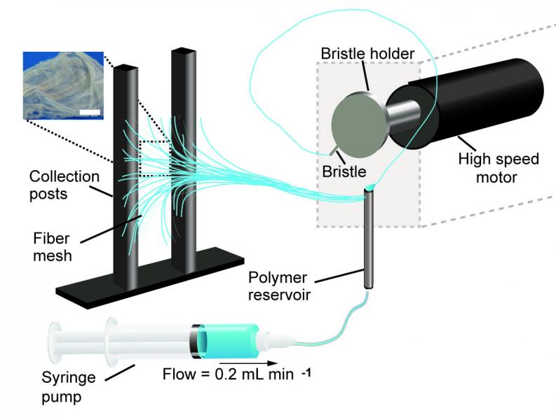
A schematic of the pull spinning apparatus with a side view illustration of a fiber being pulled from the polymer reservoir. The pull spinning system consists of a rotating bristle that dips and pulls a polymer jet in a spiral trajectory (Leila Deravi/Harvard University)

A March 1, 2017 Harvard University news release (also on EurekAlert) by Leah Burrow,, which originated the news item, describes the current process for nanofiber fabrication and explains how this technique is an improvement,

There are many ways to make nanofibers. These versatile materials — whose target applications include everything from tissue engineering to bullet proof vests — have been made using centrifugal force, capillary force, electric field, stretching, blowing, melting, and evaporation.

Each of these fabrication methods has pros and cons. For example, Rotary Jet-Spinning (RJS) and Immersion Rotary Jet-Spinning (iRJS) are novel manufacturing techniques developed in the Disease Biophysics Group at the Harvard John A. Paulson School of Engineering and Applied Sciences (SEAS) and the Wyss Institute for Biologically Inspired Engineering. Both RJS and iRJS dissolve polymers and proteins in a liquid solution and use centrifugal force or precipitation to elongate and solidify polymer jets into nanoscale fibers. These methods are great for producing large amounts of a range of materials – including DNA, nylon, and even Kevlar – but until now they haven’t been particularly portable.

The Disease Biophysics Group recently announced the development of a hand-held device that can quickly produce nanofibers with precise control over fiber orientation. Regulating fiber alignment and deposition is crucial when building nanofiber scaffolds that mimic highly aligned tissue in the body or designing point-of-use garments that fit a specific shape.

“Our main goal for this research was to make a portable machine that you could use to achieve controllable deposition of nanofibers,” said Nina Sinatra, a graduate student in the Disease Biophysics Group and co-first author of the paper. “In order to develop this kind of point-and-shoot device, we needed a technique that could produce highly aligned fibers with a reasonably high throughput.”

The new fabrication method, called pull spinning, uses a high-speed rotating bristle that dips into a polymer or protein reservoir and pulls a droplet from solution into a jet. The fiber travels in a spiral trajectory and solidifies before detaching from the bristle and moving toward a collector. Unlike other processes, which involve multiple manufacturing variables, pull spinning requires only one processing parameter — solution viscosity — to regulate nanofiber diameter. Minimal process parameters translate to ease of use and flexibility at the bench and, one day, in the field.

Pull spinning works with a range of different polymers and proteins. The researchers demonstrated proof-of-concept applications using polycaprolactone and gelatin fibers to direct muscle tissue growth and function on bioscaffolds, and nylon and polyurethane fibers for point-of-wear apparel.

“This simple, proof-of-concept study demonstrates the utility of this system for point-of-use manufacturing,” said Kit Parker, the Tarr Family Professor of Bioengineering and Applied Physics and director of the Disease Biophysics Group. “Future applications for directed production of customizable nanotextiles could extend to spray-on sportswear that gradually heats or cools an athlete’s body, sterile bandages deposited directly onto a wound, and fabrics with locally varying mechanical properties.”

Here’s a link to and a citation for the paper,

Design and Fabrication of Fibrous Nanomaterials Using Pull Spinning by Leila F. Deravi, Nina R. Sinatra, Christophe O. Chantre, Alexander P. Nesmith, Hongyan Yuan, Sahm K. Deravi, Josue A. Goss, Luke A. MacQueen, Mohammad R. Badrossamy, Grant M. Gonzalez, Michael D. Phillips, and Kevin Kit Parker. Macromolecular Materials and Engineering DOI: 10.1002/mame.201600404 Version of Record online: 17 JAN 2017

© 2017 WILEY-VCH Verlag GmbH & Co. KGaA, Weinheim

This paper is behind a paywall.

What is a multiregional brain-on-a-chip?

In response to having created a multiregional brain-on-a-chip, there’s an explanation from the team at Harvard University (which answers my question) in a Jan. 13, 2017 Harvard John A. Paulson School of Engineering and Applied Sciences news release (also on EurekAlert) by Leah Burrows,

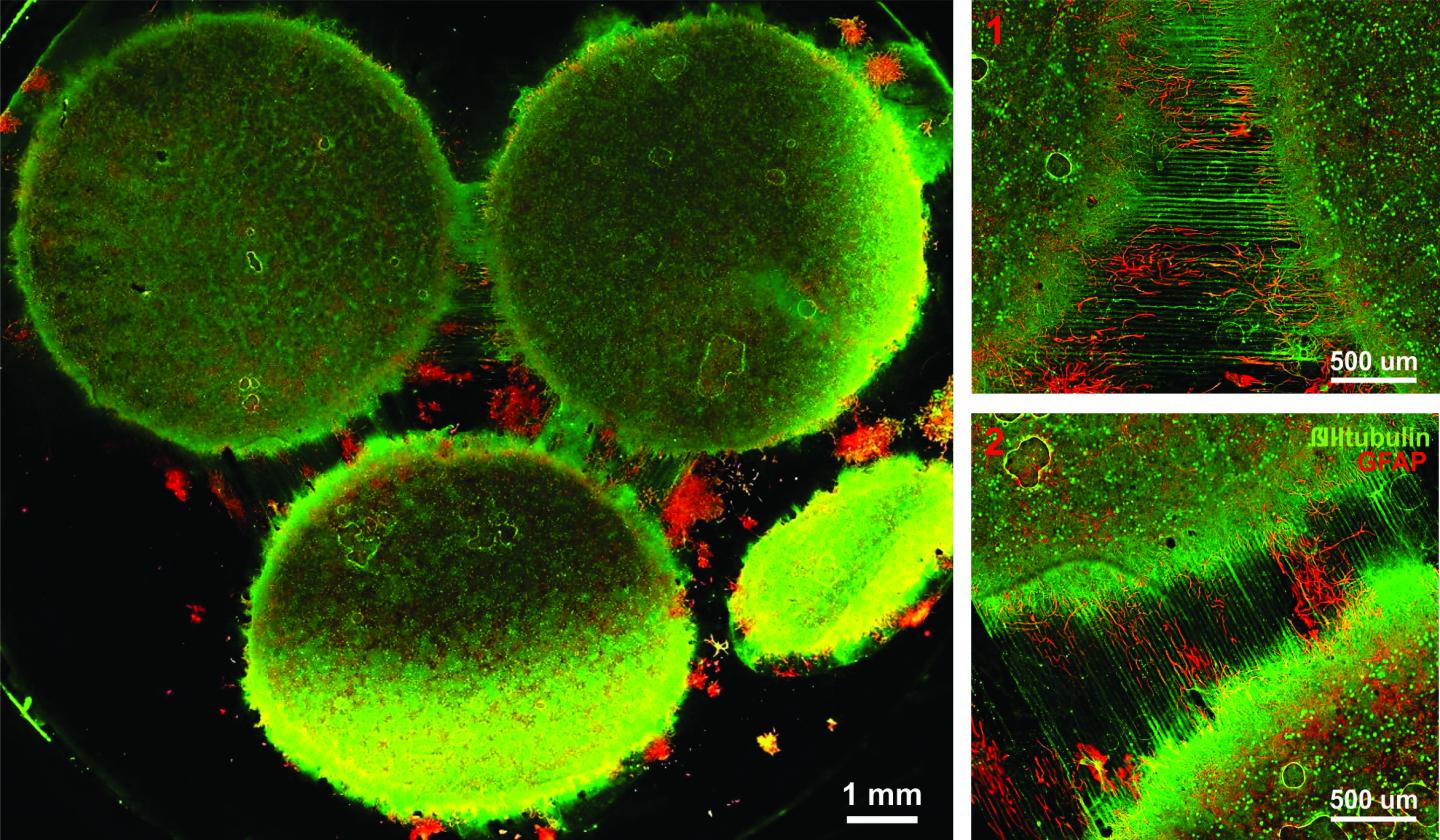
Harvard University researchers have developed a multiregional brain-on-a-chip that models the connectivity between three distinct regions of the brain. The in vitro model was used to extensively characterize the differences between neurons from different regions of the brain and to mimic the system’s connectivity.

“The brain is so much more than individual neurons,” said Ben Maoz, co-first author of the paper and postdoctoral fellow in the Disease Biophysics Group in the Harvard John A. Paulson School of Engineering and Applied Sciences (SEAS). “It’s about the different types of cells and the connectivity between different regions of the brain. When modeling the brain, you need to be able to recapitulate that connectivity because there are many different diseases that attack those connections.”

“Roughly twenty-six percent of the US healthcare budget is spent on neurological and psychiatric disorders,” said Kit Parker, the Tarr Family Professor of Bioengineering and Applied Physics Building at SEAS and Core Faculty Member of the Wyss Institute for Biologically Inspired Engineering at Harvard University. “Tools to support the development of therapeutics to alleviate the suffering of these patients is not only the human thing to do, it is the best means of reducing this cost.”

Researchers from the Disease Biophysics Group at SEAS and the Wyss Institute modeled three regions of the brain most affected by schizophrenia — the amygdala, hippocampus and prefrontal cortex.

They began by characterizing the cell composition, protein expression, metabolism, and electrical activity of neurons from each region in vitro.

“It’s no surprise that neurons in distinct regions of the brain are different but it is surprising just how different they are,” said Stephanie Dauth, co-first author of the paper and former postdoctoral fellow in the Disease Biophysics Group. “We found that the cell-type ratio, the metabolism, the protein expression and the electrical activity all differ between regions in vitro. This shows that it does make a difference which brain region’s neurons you’re working with.”

Next, the team looked at how these neurons change when they’re communicating with one another. To do that, they cultured cells from each region independently and then let the cells establish connections via guided pathways embedded in the chip.

The researchers then measured cell composition and electrical activity again and found that the cells dramatically changed when they were in contact with neurons from different regions.

“When the cells are communicating with other regions, the cellular composition of the culture changes, the electrophysiology changes, all these inherent properties of the neurons change,” said Maoz. “This shows how important it is to implement different brain regions into in vitro models, especially when studying how neurological diseases impact connected regions of the brain.”

To demonstrate the chip’s efficacy in modeling disease, the team doped different regions of the brain with the drug Phencyclidine hydrochloride — commonly known as PCP — which simulates schizophrenia. The brain-on-a-chip allowed the researchers for the first time to look at both the drug’s impact on the individual regions as well as its downstream effect on the interconnected regions in vitro.

The brain-on-a-chip could be useful for studying any number of neurological and psychiatric diseases, including drug addiction, post traumatic stress disorder, and traumatic brain injury.

“To date, the Connectome project has not recognized all of the networks in the brain,” said Parker. “In our studies, we are showing that the extracellular matrix network is an important part of distinguishing different brain regions and that, subsequently, physiological and pathophysiological processes in these brain regions are unique. This advance will not only enable the development of therapeutics, but fundamental insights as to how we think, feel, and survive.”

Here’s an image from the researchers,

Caption: Image of the in vitro model showing three distinct regions of the brain connected by axons. Credit: Disease Biophysics Group/Harvard University

Here’s a link to and a citation for the paper,

Neurons derived from different brain regions are inherently different in vitro: A novel multiregional brain-on-a-chip by Stephanie Dauth, Ben M Maoz, Sean P Sheehy, Matthew A Hemphill, Tara Murty, Mary Kate Macedonia, Angie M Greer, Bogdan Budnik, Kevin Kit Parker. Journal of Neurophysiology Published 28 December 2016 Vol. no. [?] , DOI: 10.1152/jn.00575.2016

This paper is behind a paywall and they haven’t included the vol. no. in the citation I’ve found.

Slip sliding away—making surfaces bacteria can’t grasp onto

Here’s another biomimicry story with a connection to Harvard University. From a Nov. 1, 2016 Beth Israel Deaconess Medical Center (Harvard Medical School Teaching Hospital) news release (also on EurekAlert),

Implanted medical devices like catheters, surgical mesh and dialysis systems are ideal surfaces on which bacteria can colonize and form hard-to-kill sheets called biofilms. Known as biofouling, this contamination of devices is responsible for more than half of the 1.7 million hospital-acquired infections in the United States each year.

In a report published in Biomaterials today, a team of scientists at Beth Israel Deaconess Medical Center (BIDMC), the Wyss Institute for Biologically Inspired Engineering and the John A. Paulson School of Engineering and Applied Sciences (SEAS) at Harvard University has demonstrated that an innovative, ultra-low adhesive coating prevented bacteria from attaching to surfaces treated with it, reducing bacterial adhesion by more than 98 percent in laboratory tests.

“Device related infections remain a significant problem in medicine, burdening society with millions of dollars in health care costs,” said Elliot Chaikof, MD, PhD, chair of the Roberta and Stephen R. Weiner Department of Surgery and Surgeon-in-Chief at BIDMC and an associate faculty member at the Wyss Institute. “Antibiotics alone will not solve this problem. We need to use new approaches to minimize the risk of infection, and this strategy is a very important step in that direction.”

The self-healing slippery surface coatings – known as ‘slippery liquid-infused porous surfaces’ (SLIPS) – were developed by Joanna Aizenberg, PhD, a Wyss Institute core faculty member, Professor of Chemistry and Chemical Biology and the Amy Smith Berylson Professor of Materials Science at SEAS at Harvard University. Inspired by the carnivorous Nepenthes pitcher plant that uses the slippery surface of its leaves to trap insects, Aizenberg engineered surface coatings that work to repel a variety of substances across a broad range of temperature, pressure and other environmental conditions. They are stable when exposed to UV light, and are low-cost and simple to manufacture. The current study is the first to demonstrate that SLIPS not only limit the ability of bacteria to adhere to surfaces, but also impede infection in an animal model.

SLIPS has been mentioned here before, most recently in a March 2, 2016 posting and before that in an Oct. 14, 2014 posting which appears to be precursor work for this latest research.

Getting back to the Nov. 1, 2016 news release, here’s more about plans for SLIPS and about recent trials,

“We are developing SLIPS recipes for a variety of medical applications by working with different medical-grade materials, ensuring the stability of the coating, and carefully pairing the non-fouling properties of the SLIPS materials to specific contaminates, environments and performance requirements,” said Aizenberg. “Here we have extended our repertoire and applied the SLIPS concept very convincingly to medical-grade lubricants, demonstrating its enormous potential in implanted devices prone to bacterial fouling and infection.”

In a series of trials, the researchers tested three SLIPS lubricants for their anti-adhesive qualities. First, they incubated disks of SLIPS-coated medical material ePTFE – a microporous form of Teflon – in a broth of Staphylococcus aureus (S. aureus), a generally harmless bacterium found in the nose and on skin, but one of the most common causes of hospital-acquired infections. After 48 hours, the three variations of SLIPS-treated disks demonstrated 98.3, 99.1 and 99.7 percent reductions in bacterial adhesion.

To test the material’s stability, the scientists performed the same experiment after soaking the SLIPS-coated samples for up to 21 days in a solution meant to simulate conditions inside a living mammal. After exposing these disks to S. aureus for 48 hours, the researchers found similar, nearly 100 percent reductions in bacterial adhesion.

Widely used clinically, medical mesh is particularly susceptible to bacterial infection. In another set of experiments to test the material’s biocompatibility, Chaikof and colleagues implanted small squares of SLIPS-treated mesh into murine models, injecting the site with S. aureus 24 hours later. Three days later, when the researchers removed the implanted mesh, they found little to no infection, compared with an infection rate of more than 90 percent among controls.

“Today, patients who receive implants often require antibiotics to keep the risk of bacterial infection at bay,” the authors wrote. “SLIPS coatings one day could obviate the widespread use of antibiotics and minimize the development of antibiotic resistant micro-organisms.”

“SLIPs have many promising medical applications that are in a very early stage of evaluation,” said Chaikof. “Clearly, there’s more work to be done before its introduction into the clinic, but this is one of a few studies that reinforces the exciting opportunities presented by this strategy to improve device performance and clinical outcomes.”

Here’s a link to and a citation for the paper,

An immobilized liquid interface prevents device associated bacterial infection in vivo by Jiaxuan Chen, Caitlin Howell, Carolyn A. Haller, Madhukar S. Patel, Perla Ayala, Katherine A. Moravec, Erbin Dai, Liying Liu, Irini Sotiri, Michael Aizenberg, Joanna Aizenberg, Elliot L. Chaikof. Biomaterials Volume 113, January 2017, Pages 80–92  http://dx.doi.org/10.1016/j.biomaterials.2016.09.028

This paper is behind a paywall.

Stronger more robust nanofibers for everything from bulletproof vests to cellular scaffolds (tissue engineering)

This work on a new technique for producing nanofibers comes from Harvard University’s School of Engineering and Applied Sciences and the Wyss Institute for Biologically Inspired Engineering (also at Harvard University). From an Oct. 10, 2016 news item on phys.org,

Fibrous materials—known for their toughness, durability and pliability—are used in everything from bulletproof vests to tires, filtration systems and cellular scaffolds for tissue engineering and regenerative medicine.

The properties of these materials are such that the smaller the fibers are, the stronger and tougher they become. But making certain fibers very small has been an engineering challenge.

Now, researchers from the Harvard John A. Paulson School of Engineering and Applied Sciences (SEAS) and the Wyss Institute for Biologically Inspired Engineering at Harvard have developed a new method to make and collect nanofibers and control their size and morphology. This could lead to stronger, more durable bulletproof vests and armor and more robust cellular scaffolding for tissue repair.

An Oct. 7, 2016 Harvard University press release by Leah Burrows, which originated the news item, describes the research in more detail (Note: A link has been removed),

Nanofibers are smaller than one micrometer in diameter.  Most nanofiber production platforms rely on dissolving polymers in a solution, which then evaporates as the fiber forms.

Rotary Jet-Spinning (RJS), the technique developed by Kit Parker’s Disease Biophysics Group, works likes a cotton candy machine. Parker is Tarr Family Professor of Bioengineering and Applied Physics at SEAS and a Core Member of the Wyss Institute. A liquid polymer solution is loaded into a reservoir and pushed out through a tiny opening by centrifugal force as the device spins. As the solution leaves the reservoir, the solvent evaporates and the polymers solidify and elongate into small, thin fibers.

“This advance is important because it allows us to manufacture ballistic protection that is much lighter, more flexible and more functional than what is available today,” said Parker, who in addition to his Harvard role is a lieutenant colonel in the United States Army Reserve and was motivated by his own combat experiences in Afghanistan. “Not only could it save lives but for the warfighter, it also could help reduce the repetitive injury motions that soldiers, sailors, marines and airmen have suffered over the last 15 years of the war on terror.”

“Rotary Jet-Spinning is great for most polymer fibers you want to make,” said Grant Gonzalez, a graduate student at SEAS and first author of the paper.  “However, some fibers require a solvent that doesn’t evaporate easily. Para-aramid, the polymer used in Kevlar® for example, is dissolved in sulfuric acid, which doesn’t evaporate off. The solution just splashes against the walls of the device without forming fibers.”

Nanofibers are smaller than one micrometer in diameter.  Most nanofiber production platforms rely on dissolving polymers in a solution, which then evaporates as the fiber forms.

Rotary Jet-Spinning (RJS), the technique developed by Kit Parker’s Disease Biophysics Group, works likes a cotton candy machine. Parker is Tarr Family Professor of Bioengineering and Applied Physics at SEAS and a Core Member of the Wyss Institute. A liquid polymer solution is loaded into a reservoir and pushed out through a tiny opening by centrifugal force as the device spins. As the solution leaves the reservoir, the solvent evaporates and the polymers solidify and elongate into small, thin fibers.

“This advance is important because it allows us to manufacture ballistic protection that is much lighter, more flexible and more functional than what is available today,” said Parker, who in addition to his Harvard role is a lieutenant colonel in the United States Army Reserve and was motivated by his own combat experiences in Afghanistan. “Not only could it save lives but for the warfighter, it also could help reduce the repetitive injury motions that soldiers, sailors, marines and airmen have suffered over the last 15 years of the war on terror.”

“Rotary Jet-Spinning is great for most polymer fibers you want to make,” said Grant Gonzalez, a graduate student at SEAS and first author of the paper.  “However, some fibers require a solvent that doesn’t evaporate easily. Para-aramid, the polymer used in Kevlar® for example, is dissolved in sulfuric acid, which doesn’t evaporate off. The solution just splashes against the walls of the device without forming fibers.”

Other methods, such as electrospinning, which uses an electric field to pull the polymer into a thin fiber, also have poor results with Kevlar and other polymers such as alginate used for tissue scaffolding and DNA.

The Harvard team overcame these challenges by developing a wet-spinning platform, which uses the same principles as the RJS system but relies on precipitation rather than evaporation to separate the solvent from the polymer.

In this system, called immersion Rotary Jet-Spinning (iRJS), when the polymer solution shoots out of the reservoir, it first passes through an area of open air, where the polymers elongate and the chains align. Then the solution hits a liquid bath that removes the solvent and precipitates the polymers to form solid fibers. Since the bath is also spinning — like water in a salad spinner — the nanofibers follow the stream of the vortex and wrap around a rotating collector at the base of the device.

Using this system, the team produced Nylon, DNA, alginate and ballistic resistant para-aramid nanofibers. The team could tune the fiber’s diameter by changing the solution concentration, the rotational speed and the distance the polymer traveled from the reservoir to the bath.

“By being able to modulate fiber strength, we can create a cellular scaffold that can mimic skeleton muscle and native tissues,” said Gonzalez.  “This platform could enable us to create a wound dressing out of alginate material or seed and mature cells on scaffolding for tissue engineering.”

Because the fibers were collected by a spinning vortex, the system also produced well-aligned sheets of nanofibers, which is important for scaffolding and ballistic resistant materials.

This is the ‘candy floss’ technique at work,

Rotary Jet-Spinning (RJS) works likes a cotton candy machine. A liquid polymer solution is loaded into a reservoir and pushed out through a tiny opening by centrifugal force as the device spins. As the solution leaves the reservoir, the solvent evaporates and the polymers solidify and elongate into small, thin fibers. Courtesy: Harvard University

Rotary Jet-Spinning (RJS) works likes a cotton candy machine. A liquid polymer solution is loaded into a reservoir and pushed out through a tiny opening by centrifugal force as the device spins. As the solution leaves the reservoir, the solvent evaporates and the polymers solidify and elongate into small, thin fibers. Courtesy: Harvard University

Here’s a link to and a citation for the paper,

Production of Synthetic, Para-Aramid and Biopolymer Nanofibers by Immersion Rotary Jet-Spinning by Grant M. Gonzalez, Luke A. MacQueen, Johan U. Lind, Stacey A. Fitzgibbons, Christophe O. Chantre, Isabelle Huggler, Holly M. Golecki, Josue A. Goss, Kevin Kit Parker. Macromolecular Materials and Engineering DOI: 10.1002/mame.201600365 Version of Record online: 7 OCT 2016

© 2016 WILEY-VCH Verlag GmbH & Co. KGaA, Weinheim

This paper is behind a paywall.

Sutures that can gather data wirelessly

Are sutures which gather data hackable? It’s a little early to start thinking about that issue as this seems to be brand new research. A July 18, 2016 news item on ScienceDaily tells more,

For the first time, researchers led by Tufts University engineers have integrated nano-scale sensors, electronics and microfluidics into threads — ranging from simple cotton to sophisticated synthetics — that can be sutured through multiple layers of tissue to gather diagnostic data wirelessly in real time, according to a paper published online July 18 [2016] in Microsystems & Nanoengineering. The research suggests that the thread-based diagnostic platform could be an effective substrate for a new generation of implantable diagnostic devices and smart wearable systems.

A July 18, 2016 Tufts University news release (also on EurekAlert), which originated the news item, provides more detail,

The researchers used a variety of conductive threads that were dipped in physical and chemical sensing compounds and connected to wireless electronic circuitry to create a flexible platform that they sutured into tissue in rats as well as in vitro. The threads collected data on tissue health (e.g. pressure, stress, strain and temperature), pH and glucose levels that can be used to determine such things as how a wound is healing, whether infection is emerging, or whether the body’s chemistry is out of balance. The results were transmitted wirelessly to a cell phone and computer.

The three-dimensional platform is able to conform to complex structures such as organs, wounds or orthopedic implants.

While more study is needed in a number of areas, including investigation of long-term biocompatibility, researchers said initial results raise the possibility of optimizing patient-specific treatments.

“The ability to suture a thread-based diagnostic device intimately in a tissue or organ environment in three dimensions adds a unique feature that is not available with other flexible diagnostic platforms,” said Sameer Sonkusale, Ph.D., corresponding author on the paper and director of the interdisciplinary Nano Lab in the Department of Electrical and Computer Engineering at Tufts School of Engineering. “We think thread-based devices could potentially be used as smart sutures for surgical implants, smart bandages to monitor wound healing, or integrated with textile or fabric as personalized health monitors and point-of-care diagnostics.”

Until now, the structure of substrates for implantable devices has essentially been two-dimensional, limiting their usefulness to flat tissue such as skin, according to the paper. Additionally, the materials in those substrates are expensive and require specialized processing.

Here’s a link to and a citation for the paper,

A toolkit of thread-based microfluidics, sensors, and electronics for 3D tissue embedding for medical diagnostics by Pooria Mostafalu, Mohsen Akbari, Kyle A. Alberti, Qiaobing Xu, Ali Khademhosseini, & Sameer R. Sonkusale. Microsystems & Nanoengineering 2, Article number: 16039 (2016) doi:10.1038/micronano.2016.39 Published online 18 July 2016

This paper is open access.