It’s usually silver nanoparticles (with a nod to titanium dioxide as another problem nanoparticle) which star in scenarios regarding environmental concerns, especially with water. According to an Aug. 28, 2018 news item on Nanowerk, gold nanoparticles under certain conditions could also pose problems,
It turns out gold isn’t always the shining example of a biologically stable material that it’s assumed to be, according to environmental engineers at Duke’s Center for the Environmental Implications of NanoTechnology (CEINT).
In a nanoparticle form, the normally very stable, inert, noble metal actually gets dismantled by a microbe found on a Brazilian aquatic weed.
While the findings don’t provide dire warnings about any unknown toxic effects of gold, they do provide a warning to researchers on how it is used in certain experiments.
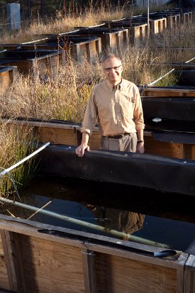
Here’s an image of one of the researchers standing in the test bed where they made their discovery (the caption will help to make sense of the reference to mesocosms in the news release, which follows,,
Mark Wiesner stands with rows of mesocosms—small, manmade structures containing different plants and microorganisms meant to represent a natural environment with experimental controls. Courtesy: Duke University
CEINT researchers from Duke, Carnegie Mellon and the University of Kentucky were running an experiment to investigate how nanoparticles used as a commercial pesticide affect wetland environments in the presence of added nutrients. Although real-world habitats often receive doses of both pesticides and fertilizers, most studies on the environmental effects of such compounds only look at a single contaminant at a time.
For nine months, the researchers released low doses of nitrogen, phosphorus and copper hydroxide nanoparticles into wetland mesocosms [emphasis mine]– small, manmade structures containing different plants and microorganisms meant to represent a natural environment with experimental controls. The goal was to see where the nanoparticle pesticides ended up and how they affected the plant and animal life within the mesocosm.
The researchers also released low doses of gold nanoparticles as tracers, assuming the biologically inert nanoparticles would remain stable while migrating through the ecosystem. This would help the researchers interpret data on the pesticide particles that partly dissolve by showing them how a solid metal particle acts within the system.
But when the researchers went to analyze their results, they found that many of the gold nanoparticles had been oxidized and dissolved.
“We were taken completely by surprise,” said Mark Wiesner, the James B. Duke Professor and chair of civil and environmental engineering at Duke. “The nanoparticles that were supposed to be the most stable turned out to be the least stable of all.”
After further inspection, the researchers found the culprit — the microbiome growing on a common Brazilian waterweed called Egeria densa. Many bacteria secrete chemicals to essentially mine metallic nutrients from their surroundings. With their metabolism spiked by the experiment’s added nutrients, the bacteria living on the E. densa were catalyzing the reaction to dissolve the gold nanoparticles.
This process wouldn’t pose any threat [emphasis mine] to humans or other animal species in the wild. But when researchers design experiments with the assumption that their gold nanoparticles will remain intact, the process can confound the interpretation of their results.
“The assumption that gold is inert did not hold in these experiments,” said Wiesner. “This is a good lesson that underscores how real, complex environments, that include for example the bacteria growing on leaves, can give very different results from experiments run in a laboratory setting that do not include these complexities.”
Here’s a link to and a citation for the paper,
Gold nanoparticle biodissolution by a freshwater macrophyte and its associated microbiome by Astrid Avellan, Marie Simonin, Eric McGivney, Nathan Bossa, Eleanor Spielman-Sun, Jennifer D. Rocca, Emily S. Bernhardt, Nicholas K. Geitner, Jason M. Unrine, Mark R. Wiesner, & Gregory V. Lowry. Nature Nanotechnology (2018) DOI: https://doi.org/10.1038/s41565-018-0231-y Published
I have heard of phytomining in soil remediation efforts (reclaiming nanoscale metals in plants near mining operations; you can find a more detailed definition here at Wiktionary) but, in this case, scientists have discovered plant tissues with nanoscale gold in an area which has no known deposits of gold. From a June 14, 2018 news item on Nanowwerk (Note: A link has been removed),
Plants containing the element gold are already widely known. The flowering perennial plant alfafa, for example, has been cultivated by scientists to contain pure gold in its plant tissue. Now researchers from the Sun Yat-sen University in China have identified and investigated the characteristics of gold nanoparticles in two plant species growing in their natural environments.
The study, led by Xiaoen Luo, is published in Environmental Chemistry Letters (“Discovery of nano-sized gold particles in natural plant tissues”) and has implications for the way gold nanoparticles are produced and absorbed from the environment.
Xiaoen Luo and her colleagues investigated the perennial shrub B. nivea and the annual or biennial weed Erigeron Canadensis. The researchers collected and prepared samples of both plants so that they could be examined using the specialist analytical tool called field-emission transmission electron microscope (TEM).
Gold-bearing nanoparticles – tiny gold particles fused with another element such as oxygen or copper – were found in both types of plant. In E. Canadensis these particles were around 20-50 nm in diameter and had an irregular form. The gold-bearing particles in B. nivea were circular, elliptical or bone-rod shaped with smooth edges and were 5-15 nm.
“The abundance of gold in the crust is very low and there was no metal deposit in the sampling area so we speculate that the source of these gold nanoparticles is a nearby electroplating plant that uses gold in its operations, “ explains Jianjin Cao who is a co-author of the study.
Most of the characteristics of the nanoparticles matched those of artificial particles rather than naturally occurring nanoparticles, which would support this theory. The researchers believe that the gold-bearing particles were absorbed through the pores of the plants directly, indicating that gold could be accumulated from the soil, water or air.
“Discovering gold-bearing nanoparticles in natural plant tissues is of great significance and allows new possibilities to clean up areas contaminated with nanoparticles, and also to enrich gold nanoparticles using plants,” says Xiaoen Luo.
The researchers plan to further study the migration mechanism, storage locations and growth patterns of gold nanoparticles in plants and also verify the absorbing capacity of different plants for gold nanoparticles in polluted areas.
For anyone who’d like to find out more about electroplating, there’s this January 25, 2018 article by Anne Marie Helmenstine for ThoughtCo.
The Rotary Jet-Spinning manufacturing system was developed specifically as a therapeutic for the wounds of war. The dressings could be a good option for large wounds, such as burns, as well as smaller wounds on the face and hands, where preventing scarring is important. Illustration courtesy of Michael Rosnach/Harvard University
This image really gets the idea of regeneration across to the viewer while also informing you that this is medicine that comes from the military. A March 19,2018 news item on phys.org announces the work,
Researchers from the Harvard John A. Paulson School of Engineering and Applied Sciences (SEAS) and the Wyss Institute for Biologically Inspired Engineering have developed new wound dressings that dramatically accelerate healing and improve tissue regeneration. The two different types of nanofiber dressings, described in separate papers, use naturally-occurring proteins in plants and animals to promote healing and regrow tissue.
Our fiber manufacturing system was developed specifically for the purpose of developing therapeutics for the wounds of war,” said Kit Parker, the Tarr Family Professor of Bioengineering and Applied Physics at SEAS and senior author of the research. “As a soldier in Afghanistan, I witnessed horrible wounds and, at times, the healing process for those wounds was a horror unto itself. This research is a years-long effort by many people on my team to help with these problems.”
Parker is also a Core Faculty Member of the Wyss Institute.
The most recent paper, published in Biomaterials, describes a wound dressing inspired by fetal tissue.
In the late 1970s, when scientists first started studying the wound-healing process early in development, they discovered something unexpected: Wounds incurred before the third trimester left no scars. This opened a range of possibilities for regenerative medicine. But for decades, researchers have struggled to replicate those unique properties of fetal skin.
Unlike adult skin, fetal skin has high levels of a protein called fibronectin, which assembles into the extracellular matrix and promotes cell binding and adhesion. Fibronectin has two structures: globular, which is found in blood, and fibrous, which is found in tissue. Even though fibrous fibronectin holds the most promise for wound healing, previous research focused on the globular structure, in part because manufacturing fibrous fibronectin was a major engineering challenge.
But Parker and his team are pioneers in the field of nanofiber engineering.
The researchers made fibrous fibronectin using a fiber-manufacturing platform called Rotary Jet-Spinning (RJS), developed by Parker’s Disease Biophysics Group. RJS works likes a cotton-candy machine — a liquid polymer solution, in this case globular fibronectin dissolved in a solvent, is loaded into a reservoir and pushed out through a tiny opening by centrifugal force as the device spins. As the solution leaves the reservoir, the solvent evaporates and the polymers solidify. The centrifugal force unfolds the globular protein into small, thin fibers. These fibers — less than one micrometer in diameter — can be collected to form a large-scale wound dressing or bandage.
“The dressing integrates into the wound and acts like an instructive scaffold, recruiting different stem cells that are relevant for regeneration and assisting in the healing process before being absorbed into the body,” said Christophe Chantre, a graduate student in the Disease Biophysics Group and first author of the paper.
In in vivo testing, the researchers found that wounds treated with the fibronectin dressing showed 84 percent tissue restoration within 20 days, compared with 55.6 percent restoration in wounds treated with a standard dressing.
The researchers also demonstrated that wounds treated with the fibronectin dressing had almost normal epidermal thickness and dermal architecture, and even regrew hair follicles — often considered one of the biggest challenges in the field of wound healing.
“This is an important step forward,” said Chantre. “Most work done on skin regeneration to date involves complex treatments combining scaffolds, cells, and even growth factors. Here we were able to demonstrate tissue repair and hair follicle regeneration using an entirely material approach. This has clear advantages for clinical translation.”
In another paper published in Advanced Healthcare Materials, the Disease Biophysics Group demonstrated a soy-based nanofiber that also enhances and promotes wound healing.
Soy protein contains both estrogen-like molecules — which have been shown to accelerate wound healing — and bioactive molecules similar to those that build and support human cells.
“Both the soy- and fibronectin-fiber technologies owe their success to keen observations in reproductive medicine,” said Parker. “During a woman’s cycle, when her estrogen levels go high, a cut will heal faster. If you do a surgery on a baby still in the womb, they have scar-less wound healing. Both of these new technologies are rooted in the most fascinating of all the topics in human biology — how we reproduce.”
In a similar way to fibronectin fibers, the research team used RJS to spin ultrathin soy fibers into wound dressings. In experiments, the soy- and cellulose-based dressing demonstrated a 72 percent increase in healing over wounds with no dressing and a 21 percent increase in healing over wounds dressed without soy protein.
“These findings show the great promise of soy-based nanofibers for wound healing,” said Seungkuk Ahn, a graduate student in the Disease Biophysics Group and first author of the paper. “These one-step, cost-effective scaffolds could be the next generation of regenerative dressings and push the envelope of nanofiber technology and the wound-care market.”
Both kinds of dressing, according to researchers, have advantages in the wound-healing space. The soy-based nanofibers — consisting of cellulose acetate and soy protein hydrolysate — are inexpensive, making them a good option for large-scale use, such as on burns. The fibronectin dressings, on the other hand, could be used for smaller wounds on the face and hands, where preventing scarring is important.
Here’s are links and citations for both papers mentioned in the news release,
Production-scale fibronectin nanofibers promote wound closure and tissue repair in a dermal mouse model by Christophe O. Chantre, Patrick H. Campbell, Holly M. Golecki, Adrian T. Buganza, Andrew K. Capulli, Leila F. Deravi, Stephanie Dauth, Sean P. Sheehy, Jeffrey A.Paten. KarlGledhill, Yanne S. Doucet, Hasan E.Abaci, Seungkuk Ahn, Benjamin D.Pope, Jeffrey W.Ruberti, Simon P.Hoerstrup, Angela M.Christiano, Kevin Kit Parker. Biomaterials Volume 166, June 2018, Pages 96-108 https://doi.org/10.1016/j.biomaterials.2018.03.006 Available online 5 March 2018
Both papers are behind paywalls although you may want to check with ResearchGate where many researchers make their papers available for free.
One last comment, I noticed this at the end of Burrows’ news release,
The Harvard Office of Technology Development has protected the intellectual property relating to these projects and is exploring commercialization opportunities.
It reminded me of the patent battle between the Broad Institute (a Harvard University and Massachusetts Institute of Technology joint venture) and the University of California at Berkeley over CRISPR (clustered regularly interspaced short palindromic repeats) technology. (My March 15, 2017 posting describes the battle’s outcome.)
Lest we forget, there could be major financial rewards from this work.
ARPICO (Society of Italian Researchers and Professionals in Western Canada) is hosting a talk on the topic of genetically modified food. Here’s more from their May 20, 2018 announcement (received via email),
Our third speaking event of the year has been scheduled for Monday, June 4th, 2018 at the Italian Cultural Centre – Museum & Art Gallery. Marie-Claude Fortin’s talk will discuss food systems derived from biotechnology (often referred to as GMO) and their comparison with traditional farming processes, both technical and ethical. You can read a summary of Marie-Claude Fortin’s lecture as well as her short professional biography at the bottom of this message.
Ahead of the speaking event, ARPICO will be holding its 2018 Annual General Meeting in the same location. We encourage everyone to participate in the AGM, have their say on ARPICO’s matters and possibly volunteer for the Board of Directors.
Genetically-Engineered Food: Facts, Ethical Considerations and World Hunger
In this lecture we will explore a part of our food system, which has received much press, but which consumers still misunderstand: food derived from biotechnology often referred to as genetically modified organisms. We will be learning about the types of plants and animals which are genetically engineered and part of our everyday food system and the reasons for which they have been transformed genetically. We will be looking at the issue from several different angles. You are encouraged to approach the topic with an open mind, and learn how the technology is being used. We will start by understanding the differences between traditional plant breeding, conventional plant breeding, transgenic technology and genome editing. The latter two processes are considered genetic engineering technologies but all of them constitute a continuum of techniques employed to improve domestic plants and animals. We will then go over the ethical paradigms related to genetically engineered food represented by the European and North American points of view. Finally, we will discuss the strengths and weaknesses associated with genetic engineering as a tool to solve world hunger.
Marie-Claude Fortin is a former Research Scientist with Agriculture and Agri-Food Canada, Associate Editor with Crop Science Society of America, Board Member of the Soil and Water Conservation Society and Adjunct Professor at the University of British Columbia (UBC) and currently responsible for the shared research infrastructure portfolio at the UBC Vice-President Research & Innovation Office. Her main areas of research expertise are crop and soil sciences with special interests in measuring and modeling crop development and various processes on agricultural land: water and nitrogen fertilizer flow through the soil profile, emissions of greenhouse gases and soil physical properties. Her research shows that sustainable crop management practices result in soil environments, which are conducive to resilient crop production and organic matter buildup, which is the process of storing carbon in soils, a most important process in this era of climate change. For the past 18 years, Marie-Claude has been teaching food systems courses at UBC [University of British Columbia], emphasizing impacts of decisions made at the corporate, national and local levels on the economic, environmental and social sustainability of the food system, including impacts of organic and industrial agriculture and adoption of genetically engineered crops and animals, on farmers and consumers.
WHEN (AGM): Monday, June 4th, 2018 at 6:00pm (doors open at 5:50pm)
WHEN (EVENT): Monday, June 4th, 2018 at 7:00pm (doors open at 6:45pm)
WHERE: Italian Cultural Centre – Museum & Art Gallery – 3075 Slocan St, Vancouver, BC, V5M 3E4
Tickets are FREE, but all individuals are requested to obtain “free-admission” tickets on EventBrite site due to limited seating at the venue. Organizers need accurate registration numbers to manage wait lists and prepare name tags.
All ARPICO events are 100% staffed by volunteer organizers and helpers, however, room rental, stationery, and guest refreshments are costs incurred and underwritten by members of ARPICO. Therefore to be fair, all audience participants are asked to donate to the best of their ability at the door or via EventBrite to “help” defray costs of the event.
FAQs
Where can I contact the organizer with any questions? info@arpico.ca
Do I have to bring my printed ticket to the event? No, you do not. Your name will be on our Registration List at the Check-in Desk.
Is my registration/ticket transferrable? If you are unable to attend, another person may use your ticket. Please send us an email at info@arpico.ca of this substitution to correct our audience Registration List and to prepare guest name tags.
Can I update my registration information? Yes. If you have any questions, contact us at info@arpico.ca
I am having trouble using EventBrite and cannot reserve my ticket(s). Can someone at ARPICO help me with my ticket reservation? Of course, simply send your ticket request to us at info@arpico.ca so we help you.
Since the 2016 approval, AquAdvantage salmon, 4.5M tonnes has been sold in Canada according to an Aug. 8, 2017 article by Sima Shakeri for Huffington Post (Note: Links have been removed),
After decades of trying to get approval by in North America, genetically modified Atlantic salmon has been sold to consumers in Canada.
AquaBounty Technologies, an American company that produces the Atlantic salmon, confirmed it had sold 4.5 tonnes of the modified fish on August 4 [2017], the Scientific American reported.
The fish have been engineered with a growth hormone gene from Chinook salmon to grow faster than regular salmon and require less food. They take about 18 months to reach market size, which is much quicker than the 30 months or so for conventional salmon.
The Washington Post wrote AquaBounty’s salmon also contains a gene from the ocean pout that makes the salmon produce the growth hormone gene all-year-round.
The company produces the eggs in a facility in P.E.I., which is currently being expanded, and then they’re shipped to Panama where the fish are raised.
Health Canada assessed the AquAdvantage salmon and concluded it “did not pose a greater risk to human health than salmon currently available on the Canadian market,” and that it would have no impact on allergies nor a difference in nutritional value compared to other farmed salmon.
Because of that, the AquAdvantage product is not required to be specially labelled as genetically modified, and is up to the discretion of retailers.
…
As for gene editing, I don’t follow everything in that area of endeavour but I have (more or less) kept track of CRISPR ((clustered regularly interspaced short palindromic repeat). Just use CRISPR as the search term for the blog search function to find what’s here.
This looks to be a very interesting talk and good for ARPICO for tackling a ‘difficult’ topic. I hope they have a lively, convivial, and open discussion.
Researcher Bor-Kai Hsiung’s work has graced this blog before but the topic was tarantulas and their structural colour. This time, it’s all about Australian peacock spiders and their structural colour according to a December 22, 2017 news item on ScienceDaily,
Even if you are arachnophobic, you probably have seen pictures or videos of Australian peacock spiders (Maratus spp.). These tiny spiders are only 1-5 mm long but are famous for their flamboyant courtship displays featuring diverse and intricate body colorations, patterns, and movements.
The spiders extremely large anterior median eyes have excellent color vision and combine with their bright colors to make peacock spiders cute enough to cure most people of their arachnophobia. But these displays aren’t just pretty to look at, they also inspire new ways for humans to produce color in technology.
One species of peacock spider — the rainbow peacock spider (Maratus robinsoni) is particularly neat, because it showcases an intense rainbow iridescent signal in males’ courtship displays to the females. This is the first known instance in nature of males using an entire rainbow of colors to entice females. Dr. Bor-Kai Hsiung led an international team of researchers from the US (UAkron, Cal Tech, UC San Diego, UNL [University of Nebraska-Lincoln]), Belgium (Ghent University), Netherlands (UGroningen), and Australia to discover how rainbow peacock spiders produce this unique multi-color iridescent signal.
Using a diverse array of research techniques, including light and electron microscopy, hyperspectral imaging, imaging scatterometry, nano 3D printing and optical modeling, the team found the origin of this intense rainbow iridescence emerged from specialized abdominal scales of the spiders. These scales have an airfoil-like microscopic 3D contour with nanoscale diffraction grating structures on the surface.
The interaction between the surface nano-diffraction grating and the microscopic curvature of the scales enables separation and isolation of light into its component wavelengths at finer angles and smaller distances than are possible with current manmade engineering technologies.
Inspiration from these super iridescent scales can be used to overcome current limitations in spectral manipulation, and to further reduce the size of optical spectrometers for applications where fine-scale spectral resolution is required in a very small package, notably instruments on space missions, or wearable chemical detection systems. And it could have a wide array of implications to fields ranging from life sciences and biotechnologies to material sciences and engineering.
Here’s a video of an Australian rainbow peacock spider,
Here’s more from the YouTube description published on April 13, 2017 by Peacockspiderman,
Scenes of Maratus robinsoni, a spider Peter Robinson discovered and David Hill and I named it after him in 2012. You can read our description on pages 36-41 in Peckhamia 103.2, which can be downloaded from the Peckhamia website http://peckhamia.com/peckhamia_number…. This is one of the two smallest species of peacock spider (2.5 mm long) and the only spider we know of in which colour changes occur every time it moves, this video was created to document this. Music: ‘Be Still’ by Johannes Bornlöf licensed through my MCN ‘Brave Bison’ from ‘Epidemic Sound’ For licensing inquiries please contact Brave Bison licensing@bravebison.io
The University of California at San Diego also published a December 22, 2017 news release about this work, which covers some of the same ground while providing a few new tidbits of information,
Brightly colored Australian peacock spiders (Maratus spp.) captivate even the most arachnophobic viewers with their flamboyant courtship displays featuring diverse and intricate body colorations, patterns, and movements – all packed into miniature bodies measuring less than five millimeters in size for many species. However, these displays are not just pretty to look at. They also inspire new ways for humans to produce color in technology.
One species of peacock spider – the rainbow peacock spider (Maratus robinsoni) – is particularly impressive, because it showcases an intense rainbow iridescent signal in males’ courtship displays to females. This is the first known instance in nature of males using an entire rainbow of colors to entice females to mate. But how do males make their rainbows? A new study published in Nature Communications looked to answer that question.
Figuring out the answers was inherently interdisciplinary so Bor-Kai Hsiung, a postdoctoral scholar at Scripps Institution of Oceanography at the University of California San Diego, assembled an international team that included biologists, physicists and engineers. Starting while he was a Ph.D. student at The University of Akron under the mentorship of Todd Blackledge and Matthew Shawkey, the team included researchers from UA, Scripps Oceanography, California Institute of Technology, and University of Nebraska-Lincoln, the University of Ghent in Belgium, University of Groningen in Netherlands, and Australia to discover how rainbow peacock spiders produce this unique iridescent signal.
The team investigated the spider’s photonic structures using techniques that included light and electron microscopy, hyperspectral imaging, imaging scatterometry and optical modeling to generate hypotheses about how the spider’s scale generate such intense rainbows. The team then used cutting-edge nano 3D printing to fabricate different prototypes to test and validate their hypotheses. In the end, they found that the intense rainbow iridescence emerged from specialized abdominal scales on the spiders. These scales combine an airfoil-like microscopic 3D contour with nanoscale diffraction grating structures on the surface. It is the interaction between the surface nano-diffraction grating and the microscopic curvature of the scales that enables separation and isolation of light into its component wavelengths at finer angles and smaller distances than are possible with current engineering technologies.
“Who knew that such a small critter would create such an intense iridescence using extremely sophisticated mechanisms that will inspire optical engineers,” said Dimitri Deheyn, Hsuing’s advisor at Scripps Oceanography and a coauthor of the study.
For Hsiung, the finding wasn’t quite so unexpected.
“One of the main questions that I wanted to address in my Ph.D. dissertation was ‘how does nature modulate iridescence?’ From a biomimicry perspective, to fully understand and address a question, one has to take extremes from both ends into consideration. I purposefully chose to study these tiny spiders with intense iridescence after having investigated the non-iridescent blue tarantulas,” said Hsiung.
The mechanism behind these tiny rainbows may inspire new color technology, but would not have been discovered without research combining basic natural history with physics and engineering, the researchers said.
“Nanoscale 3D printing allowed us to experimentally validate our models, which was really exciting,” said Shawkey. “We hope that these techniques will become common in the future.”
“As an engineer, what I found fascinating about these spider structural colors is how these long evolved complex structures can still outperform human engineering,” said Radwanul Hasan Siddique, a postdoctoral scholar at Caltech and study coauthor. “Even with high-end fabrication techniques, we could not replicate the exact structures. I wonder how the spiders assemble these fancy structural patterns in the first place!”
Inspiration from these super iridescent spider scales can be used to overcome current limitations in spectral manipulation, and to reduce the size of optical spectrometers for applications where fine-scale spectral resolution is required in a very small package, notably instruments on space missions, or wearable chemical detection systems.
In the end, peacock spiders don’t just produce nature’s smallest rainbows.They could also have implications for a wide array of fields ranging from life sciences and biotechnologies to material sciences and engineering.
Before citing the paper and providing a link, here’s a story by Robert F. Service for Science magazine about attempts to capitalize on ‘spider technology’, in this case spider silk,
The hype over spider silk has been building since 1710. That was the year François Xavier Bon de Saint Hilaire, president of the Royal Society of Sciences in Montpellier, France, wrote to his colleagues, “You will be surpriz’d to hear, that Spiders make a Silk, as beautiful, strong and glossy, as common Silk.” Modern pitches boast that spider silk is five times stronger than steel yet more flexible than rubber. If it could be made into ropes, a macroscale web would be able to snare a jetliner.
The key word is “if.” Researchers first cloned a spider silk gene in 1990, in hopes of incorporating it into other organisms to produce the silk. (Spiders can’t be farmed like silkworms because they are territorial and cannibalistic.) Today, Escherichia coli bacteria, yeasts, plants, silkworms, and even goats have been genetically engineered to churn out spider silk proteins, though the proteins are often shorter and simpler than the spiders’ own. Companies have managed to spin those proteins into enough high-strength thread to produce a few prototype garments, including a running shoe by Adidas and a lightweight parka by The North Face. But so far, companies have struggled to mass produce these supersilks.
Some executives say that may finally be about to change. One Emeryville, California-based startup, Bolt Threads, says it has perfected growing spider silk proteins in yeast and is poised to turn out tons of spider silk thread per year. In Lansing, Michigan, Kraig Biocraft Laboratories says it needs only to finalize negotiations with silkworm farms in Vietnam to produce mass quantities of a combination spider/silkworm silk, which the U.S. Army is now testing for ballistics protection. …
I encourage you to read Service’s article in its entirety if the commercialization prospects for spider silk interest you as it includes gems such as this,
Spider silk proteins are already making their retail debut—but in cosmetics and medical devices, not high-strength fibers. AMSilk grows spider silk proteins in E. coli and dries the purified protein into powders or mixes it into gels, for use as additives for personal care products, such as moisture-retaining skin lotions. The silk proteins supposedly help the lotions form a very smooth, but breathable, layer over the skin. Römer says the company now sells tons of its purified silk protein ingredients every year.
…
Finally, here’s a citation for and a link to the paper about Australian peacock spiders and nanophotonics,
Rainbow peacock spiders inspire miniature super-iridescent optics by Bor-Kai Hsiung, Radwanul Hasan Siddique, Doekele G. Stavenga, Jürgen C. Otto, Michael C. Allen, Ying Liu, Yong-Feng Lu, Dimitri D. Deheyn, Matthew D. Shawkey, & Todd A. Blackledge. Nature Communications 8, Article number: 2278 (2017) doi:10.1038/s41467-017-02451-x Published online: 22 December 2017
The American Chemical Society (ACS) and the Massachusetts Institute of Technology (MIT) have both issued news releases about the latest in bioluminescence.The researchers tested their work on watercress, a vegetable that was viewed in almost sacred terms in my family; it was not easily available in Vancouver (Canada) when I was child.
My father would hunt down fresh watercress by checking out the Chinese grocery stores. He could spot the fresh stuff from across the street while driving at 30 miles or more per hour. Spotting it entailed an immediate hunt for parking (my father hated to pay so we might have go around the block a few times or more) and a dash out of the car to ensure that he got his watercress before anyone else spotted it. These days it’s much more easily available and, thankfully, my father has passed on so he won’t have to think about glowing watercress.
The 2009 film “Avatar” created a lush imaginary world, illuminated by magical, glowing plants. Now researchers are starting to bring this spellbinding vision to life to help reduce our dependence on artificial lighting. They report in ACS’ journal Nano Letters a way to infuse plants with the luminescence of fireflies.
Nature has produced many bioluminescent organisms, however, plants are not among them. Most attempts so far to create glowing greenery — decorative tobacco plants in particular — have relied on introducing the genes of luminescent bacteria or fireflies through genetic engineering. But getting all the right components to the right locations within the plants has been a challenge. To gain better control over where light-generating ingredients end up, Michael S. Strano and colleagues recently created nanoparticles that travel to specific destinations within plants. Building on this work, the researchers wanted to take the next step and develop a “nanobionic,” glowing plant.
The team infused watercress and other plants with three different nanoparticles in a pressurized bath. The nanoparticles were loaded with light-emitting luciferin; luciferase, which modifies luciferin and makes it glow; and coenzyme A, which boosts luciferase activity. Using size and surface charge to control where the sets of nanoparticles could go within the plant tissues, the researchers could optimize how much light was emitted. Their watercress was half as bright as a commercial 1 microwatt LED and 100,000 times brighter than genetically engineered tobacco plants. Also, the plant could be turned off by adding a compound that blocks luciferase from activating luciferin’s glow.
Imagine that instead of switching on a lamp when it gets dark, you could read by the light of a glowing plant on your desk.
MIT engineers have taken a critical first step toward making that vision a reality. By embedding specialized nanoparticles into the leaves of a watercress plant, they induced the plants to give off dim light for nearly four hours. They believe that, with further optimization, such plants will one day be bright enough to illuminate a workspace.
“The vision is to make a plant that will function as a desk lamp — a lamp that you don’t have to plug in. The light is ultimately powered by the energy metabolism of the plant itself,” says Michael Strano, the Carbon P. Dubbs Professor of Chemical Engineering at MIT and the senior author of the study
This technology could also be used to provide low-intensity indoor lighting, or to transform trees into self-powered streetlights, the researchers say.
MIT postdoc Seon-Yeong Kwak is the lead author of the study, which appears in the journal Nano Letters.
Nanobionic plants
Plant nanobionics, a new research area pioneered by Strano’s lab, aims to give plants novel features by embedding them with different types of nanoparticles. The group’s goal is to engineer plants to take over many of the functions now performed by electrical devices. The researchers have previously designed plants that can detect explosives and communicate that information to a smartphone, as well as plants that can monitor drought conditions.
Lighting, which accounts for about 20 percent of worldwide energy consumption, seemed like a logical next target. “Plants can self-repair, they have their own energy, and they are already adapted to the outdoor environment,” Strano says. “We think this is an idea whose time has come. It’s a perfect problem for plant nanobionics.”
To create their glowing plants, the MIT team turned to luciferase, the enzyme that gives fireflies their glow. Luciferase acts on a molecule called luciferin, causing it to emit light. Another molecule called co-enzyme A helps the process along by removing a reaction byproduct that can inhibit luciferase activity.
The MIT team packaged each of these three components into a different type of nanoparticle carrier. The nanoparticles, which are all made of materials that the U.S. Food and Drug Administration classifies as “generally regarded as safe,” help each component get to the right part of the plant. They also prevent the components from reaching concentrations that could be toxic to the plants.
The researchers used silica nanoparticles about 10 nanometers in diameter to carry luciferase, and they used slightly larger particles of the polymers PLGA and chitosan to carry luciferin and coenzyme A, respectively. To get the particles into plant leaves, the researchers first suspended the particles in a solution. Plants were immersed in the solution and then exposed to high pressure, allowing the particles to enter the leaves through tiny pores called stomata.
Particles releasing luciferin and coenzyme A were designed to accumulate in the extracellular space of the mesophyll, an inner layer of the leaf, while the smaller particles carrying luciferase enter the cells that make up the mesophyll. The PLGA particles gradually release luciferin, which then enters the plant cells, where luciferase performs the chemical reaction that makes luciferin glow.
The researchers’ early efforts at the start of the project yielded plants that could glow for about 45 minutes, which they have since improved to 3.5 hours. The light generated by one 10-centimeter watercress seedling is currently about one-thousandth of the amount needed to read by, but the researchers believe they can boost the light emitted, as well as the duration of light, by further optimizing the concentration and release rates of the components.
Plant transformation
Previous efforts to create light-emitting plants have relied on genetically engineering plants to express the gene for luciferase, but this is a laborious process that yields extremely dim light. Those studies were performed on tobacco plants and Arabidopsis thaliana, which are commonly used for plant genetic studies. However, the method developed by Strano’s lab could be used on any type of plant. So far, they have demonstrated it with arugula, kale, and spinach, in addition to watercress.
For future versions of this technology, the researchers hope to develop a way to paint or spray the nanoparticles onto plant leaves, which could make it possible to transform trees and other large plants into light sources.
“Our target is to perform one treatment when the plant is a seedling or a mature plant, and have it last for the lifetime of the plant,” Strano says. “Our work very seriously opens up the doorway to streetlamps that are nothing but treated trees, and to indirect lighting around homes.”
The researchers have also demonstrated that they can turn the light off by adding nanoparticles carrying a luciferase inhibitor. This could enable them to eventually create plants that shut off their light emission in response to environmental conditions such as sunlight, the researchers say.
Here’s a link to and a citation for the paper,
A Nanobionic Light-Emitting Plant by Seon-Yeong Kwak, Juan Pablo Giraldo, Min Hao Wong, Volodymyr B. Koman, Tedrick Thomas Salim Lew, Jon Ell, Mark C. Weidman, Rosalie M. Sinclair, Markita P. Landry, William A. Tisdale, and Michael S. Strano. Nano Lett., 2017, 17 (12), pp 7951–7961 DOI: 10.1021/acs.nanolett.7b04369 Publication Date (Web): November 17, 2017
I’ve written a couple times about Greg Gage and his Backyard Brains, first, in a March 28, 2012 posting (scroll down about 40% of the way for the mention of the first [?] ‘SpikerBox’) and, most recently, in a June 26, 2013 posting (scroll down about 25% of the way for the mention of a RoboRoach Kickstater project from Backyard Brains) which also featured the launch of a new educational product and a TED [technology education design] talk.
Here’s the latest from an Oct. 10, 2017 news release (received via email),
Backyard Brains Releases Plant SpikerBox, unlocking the Secret Electrical Language used in Plants
The first consumer device to investigate how plants create behaviors through electrophysiology and to enable interspecies plant to plant communication.
ANN ARBOR, MI, OCTOBER 10, 2017–Today Backyard Brains launched the Plant SpikerBox, the first ever science kit designed to reveal the wonderful nature behind plant behavior through electrophysiology experiments done at home or in the classroom. The new SpikerBox launched alongside three new experiments, enabling users to explore Venus Flytrap and Sensitive Mimosa signals and to perform a jaw-dropping Interspecies Plant-Plant-Communicator experiment. The Plant SpikerBox and all three experiments are featured in a live talk from TED2017 given by Backyard Brains CEO and cofounder Dr. Greg Gage which was released today on https://ted.com.
Backyard Brains received viral attention for their previous videos, TED talks, and for their mission to create hands-on neuroscience experiments for everyone. The company (run by professional neuroscientists) produces consumer-friendly versions of expensive graduate lab equipment used at top research universities around the world. The new plant experiments and device facilitate the growing movement of DIY [do it yourself] scientists, made up of passionate amateurs, students, parents, and teachers.
Like previous inventions, the Plant SpikerBox is extremely easy to use, making it accessible for students as young as middle school. The device works by recording the electrical activity responsible for different plant behaviors. For example, the Venus Flytrap uses an electrical signal to determine if prey has landed in its trap; the SpikerBox reveals these invisible messages and allows you to visualize them on your mobile device. For the first time ever, you can peer into the fascinating world of plant signaling and plant behaviors.
The new SpikerBox features an “Interspecies Plant-Plant-Communicator” which demonstrates the ubiquitous nature of electrical signaling seen in humans, insects, and plants. With this device, one can capture the electrical message (called an action potential) from one plant’s behavior, and send it to a different plant to activate another behavior.
Co-founder and CEO Greg Gage explains, “Itis surprising to many people that plants use electrical messages similar to those used by the neurons in our brains. I was shocked to hear that. Many neuroscientists are. But if you think about it, it [sic] does make sense. Our nervous system evolved to react quickly. Electricity is fast. The plants we are studying also need to react quickly, so it makes sense they would develop a similar system. To be clear: No, plants don’t have brains, but they do exhibit behaviors and they do use electric messages called ‘Action Potentials’ like we do to send information. The benefit of these plant experiments then is twofold: First, we can simply demonstrate fundamental neuroscience principles, and second, we can spread the wonder of understanding how living creatures work and hopefully encourage others to make a career in life sciences!”
The Plant SpikerBox is a trailblazer, bringing plant electrophysiology to the public for the first time ever. It is designed to work with the Backyard Brains SpikeRecorder software which is available to download for free on their website or in mobile app stores. The three plant experiments are just a few of the dozens of free experiments available on the Backyard Brains website. The Plant SpikerBox is available now for $149.99.
About Backyard Brains
A staggering 1 in 5 people will develop a neurological disorder in their lifetime, making the need for neuroscience studies urgent. Backyard Brains passionately responds with their motto “Neuroscience for Everyone,” providing exposure, education, and experiment kits to students of all ages. Founded in 2010 in Ann Arbor, MI by University of Michigan Neuroscience graduate students Greg Gage and Tim Marzullo, Backyard Brains have been dubbed Champions of Change at an Obama White House ceremony and have won prestigious awards from the National Institutes of Health and the Society for Neuroscience. To learn more, visit BackyardBrains.com
You can find an embedded video of Greg Gage’s TED talk and Plant SpikerBox launch along with links to experiments you could run with it on Backyard Brains’ Plant SpikerBox product page.
Your nervous system allows you to sense and respond quickly to the environment around you. You have a nervous system, animals have nervous systems, but plants do not. But not having a nervous system does not mean you cannot sense and respond to the world. Plants can certainly sense the environment around them and move. You have seen your plants slowly turn their leaves towards sunlight by the window over a week, open their flowers in the day, and close their flowers during the night. Some plants can move in much more dramatic fashion, such as the Venus Flytrap and the Sensitive Mimosa.
The Venus Flytrap comes from the swamps of North Carolina, USA, and lives in very nutrient-poor, water-logged soil. It photosynthesizes like other plants, but it can’t always rely on the sunlight for food. To supplement its food supply it traps and eats insects, extracting from them the nitrogen and phosphorous needed to form plant food (amino acids, nucleic acids, and other molecules).
If you look closely at the Venus Flytrap, you will notice it has very tiny “Trigger Hairs” inside its trap leaves.
If a wayward, unsuspecting insect touches a trigger hair, an Action Potential occurs in the leaves. This is a different Action Potential than what we are used to seeing in neurons, as it’s based on the movement of calcium, potassium, and chloride ions (vs. movement of potassium and sodium as in the Action Potentials of neurons and muscles), and it is muuuuuuuuucccchhhhhh longer than anything we’ve seen before.
If the trigger hair is touched twice within 20 seconds (firing two Action Potentials within 20 seconds), the trap closes. The trap is not closing due to muscular action (plants do not have muscles), but rather due to an osmotic, rapid change in the shape of curvature of the trap leaves. Interestingly, the firing of Action Potentials is not always reliable, depending on time of year, temperature, health of plant, and/or other factors. Quite different from we humans, Action Potential failure is not devastating to a Venus Flytrap.
We can observe this plant Action Potential using our Plant SpikerBox. Welcome to the Brave New World of Plant Electrophysiology.
Downloads
Before you begin, make sure you have the Backyard Brains SpikeRecorder. The Backyard Brains SpikeRecorder program allows you to visualize and save data on your computer when doing experiments.
….
I did feel a bit sorry for the Venus Flytrap in Greg Gage’s TED talk which was fooled into closing its trap. According to Gage, the Venus Flytrap has limited number of times it can close its trap and after the last time, it dies. On the other hand, I eat meat and use leather goods so there is not pedestal for me to perch on.
For anyone who caught the Brittany Spears reference in the headline in this posting,
From exploring outer space with Brittany Spears to exploring plant communication and neuroscience in your back yard, science can be found in many different places.
The answer to the question about whether brains and plants are alike is the standard ‘yes and no’. That said, there are some startling similarities from a statistical perspective (from a July 6, 2017 Salk Institute news release (also received via email; Note: Links have been removed),
Plants and brains are more alike than you might think: Salk scientists discovered that the mathematical rules governing how plants grow are similar to how brain cells sprout connections. The new work, published in Current Biology on July 6, 2017, and based on data from 3D laser scanning of plants, suggests there may be universal rules of logic governing branching growth across many biological systems.
“Our project was motivated by the question of whether, despite all the diversity we see in plant forms, there is some form or structure they all share,” says Saket Navlakha, assistant professor in Salk’s Center for Integrative Biology and senior author of the paper. “We discovered that there is—and, surprisingly, the variation in how branches are distributed in space can be described mathematically by something called a Gaussian function, which is also known as a bell curve.”
Being immobile, plants have to find creative strategies for adjusting their architecture to address environmental challenges, like being shaded by a neighbor. The diversity in plant forms, from towering redwoods to creeping thyme, is a visible sign of these strategies, but Navlakha wondered if there was some unseen organizing principle at work. To find out, his team used high-precision 3D scanning technology to measure the architecture of young plants over time and quantify their growth in ways that could be analyzed mathematically.
“This collaboration arose from a conversation that Saket and I had shortly after his arrival at Salk,” says Professor and Director of the Plant Molecular and Cellular Biology Laboratory Joanne Chory, who, along with being the Howard H. and Maryam R. Newman Chair in Plant Biology, is also a Howard Hughes Medical Investigator and one of the paper’s coauthors. “We were able to fund our studies thanks to Salk’s innovation grant program and the Howard Hughes Medical Institute.”
The team began with three agriculturally valuable crops: sorghum, tomato and tobacco. The researchers grew the plants from seeds under conditions the plants might experience naturally (shade, ambient light, high light, high heat and drought). Every few days for a month, first author Adam Conn scanned each plant to digitally capture its growth. In all, Conn scanned almost 600 plants.
“We basically scanned the plants like you would scan a piece of paper,” says Conn, a Salk research assistant. “But in this case the technology is 3D and allows us to capture a complete form—the full architecture of how the plant grows and distributes branches in space.”
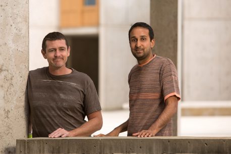
From left: Adam Conn and Saket Navlakha Credit: Salk Institute
Each plant’s digital representation is called a point cloud, a set of 3D coordinates in space that can be analyzed computationally. With the new data, the team built a statistical description of theoretically possible plant shapes by studying the plant’s branch density function. The branch density function depicts the likelihood of finding a branch at any point in the space surrounding a plant.
This model revealed three properties of growth: separability, self-similarity and a Gaussian branch density function. Separability means that growth in one spatial direction is independent of growth in other directions. According to Navlakha, this property means that growth is very simple and modular, which may let plants be more resilient to changes in their environment. Self-similarity means that all the plants have the same underlying shape, even though some plants may be stretched a little more in one direction, or squeezed in another direction. In other words, plants don’t use different statistical rules to grow in shade than they do to grow in bright light. Lastly, the team found that, regardless of plant species or growth conditions, branch density data followed a Gaussian distribution that is truncated at the boundary of the plant. Basically, this says that branch growth is densest near the plant’s center and gets less dense farther out following a bell curve.
The high level of evolutionary efficiency suggested by these properties is surprising. Even though it would be inefficient for plants to evolve different growth rules for every type of environmental condition, the researchers did not expect to find that plants would be so efficient as to develop only a single functional form. The properties they identified in this work may help researchers evaluate new strategies for genetically engineering crops.
Previous work by one of the paper’s authors, Charles Stevens, a professor in Salk’s Molecular Neurobiology Laboratory, found the same three mathematical properties at work in brain neurons. “The similarity between neuronal arbors and plant shoots is quite striking, and it seems like there must be an underlying reason,” says Stevens. “Probably, they both need to cover a territory as completely as possible but in a very sparse way so they don’t interfere with each other.”
The next challenge for the team is to identify what might be some of the mechanisms at the molecular level driving these changes. Navlakha adds, “We could see whether these principles deviate in other agricultural species and maybe use that knowledge in selecting plants to improve crop yields.”
Should you not be able to access the news release, you can find the information in a July 6, 2017 news item on ScienceDaily.
For the paper, here’s a link and a citation,
A Statistical Description of Plant Shoot Architecture by Adam Conn, Ullas V. Pedmale4, Joanne Chory, Charles F. Stevens, Saket Navlakha. Current Biology DOI: http://dx.doi.org/10.1016/j.cub.2017.06.009 Publication stage: In Press Corrected Proof July 2017
This paper is behind a paywall.
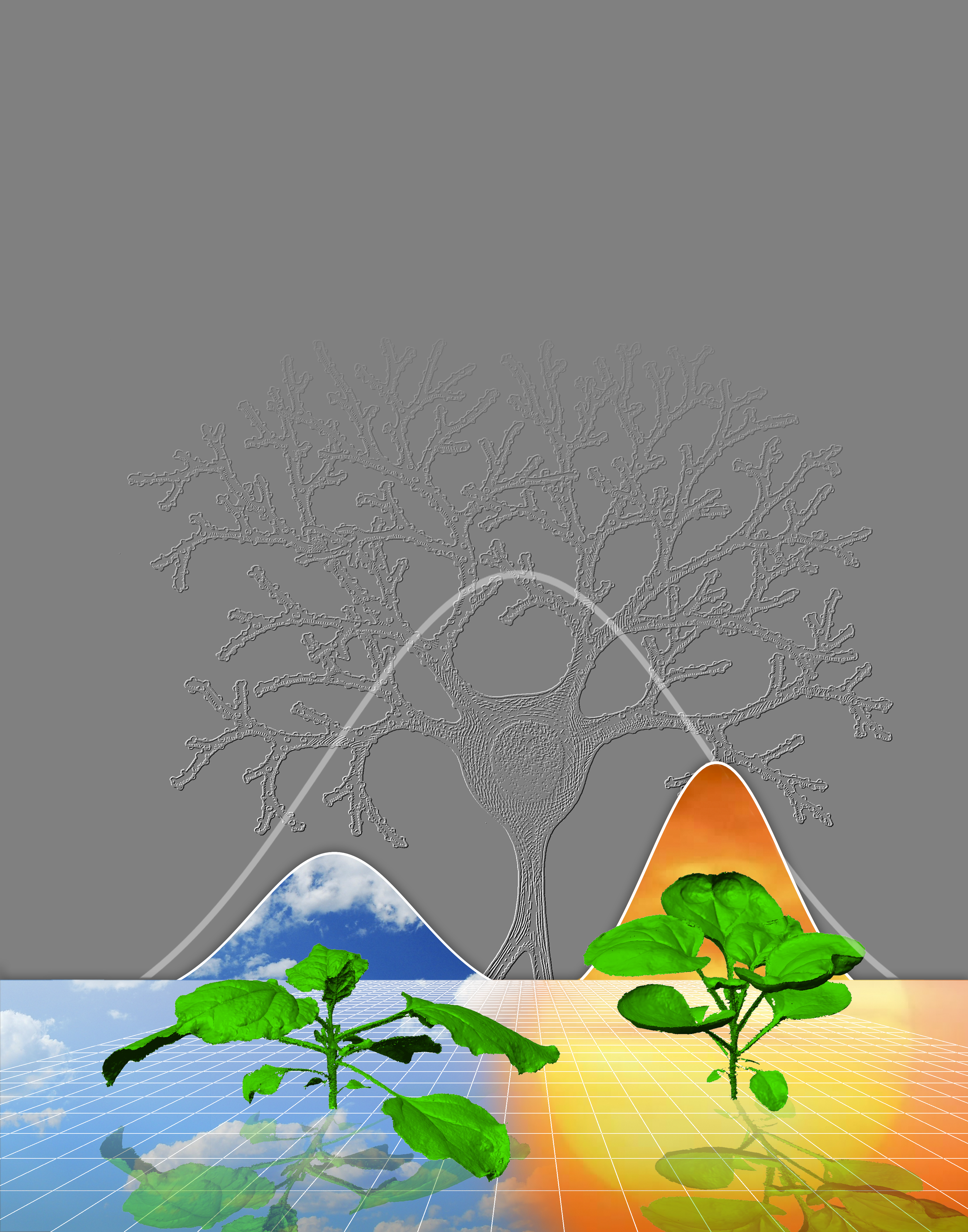
Here’s an image that illustrates the principles the researchers are attempting to establish,
This illustration represents how plants use the same rules to grow under widely different conditions (for example, cloudy versus sunny), and that the density of branches in space follows a Gaussian (“bell curve”) distribution, which is also true of neuronal branches in the brain. Credit: Salk Institute
It’s usually organ-on-a-chip or lab-on-a-chip or human-on-a-chip; this is my first tree-on-a-chip.
Engineers have designed a microfluidic device they call a “tree-on-a-chip,” which mimics the pumping mechanism of trees and other plants. Courtesy: MIT
Trees and other plants, from towering redwoods to diminutive daisies, are nature’s hydraulic pumps. They are constantly pulling water up from their roots to the topmost leaves, and pumping sugars produced by their leaves back down to the roots. This constant stream of nutrients is shuttled through a system of tissues called xylem and phloem, which are packed together in woody, parallel conduits.
Now engineers at MIT [Massachusetts Institute of Technology] and their collaborators have designed a microfluidic device they call a “tree-on-a-chip,” which mimics the pumping mechanism of trees and plants. Like its natural counterparts, the chip operates passively, requiring no moving parts or external pumps. It is able to pump water and sugars through the chip at a steady flow rate for several days. The results are published this week in Nature Plants.
Anette “Peko” Hosoi, professor and associate department head for operations in MIT’s Department of Mechanical Engineering, says the chip’s passive pumping may be leveraged as a simple hydraulic actuator for small robots. Engineers have found it difficult and expensive to make tiny, movable parts and pumps to power complex movements in small robots. The team’s new pumping mechanism may enable robots whose motions are propelled by inexpensive, sugar-powered pumps.
“The goal of this work is cheap complexity, like one sees in nature,” Hosoi says. “It’s easy to add another leaf or xylem channel in a tree. In small robotics, everything is hard, from manufacturing, to integration, to actuation. If we could make the building blocks that enable cheap complexity, that would be super exciting. I think these [microfluidic pumps] are a step in that direction.”
Hosoi’s co-authors on the paper are lead author Jean Comtet, a former graduate student in MIT’s Department of Mechanical Engineering; Kaare Jensen of the Technical University of Denmark; and Robert Turgeon and Abraham Stroock, both of Cornell University.
A hydraulic lift
The group’s tree-inspired work grew out of a project on hydraulic robots powered by pumping fluids. Hosoi was interested in designing hydraulic robots at the small scale, that could perform actions similar to much bigger robots like Boston Dynamic’s Big Dog, a four-legged, Saint Bernard-sized robot that runs and jumps over rough terrain, powered by hydraulic actuators.
“For small systems, it’s often expensive to manufacture tiny moving pieces,” Hosoi says. “So we thought, ‘What if we could make a small-scale hydraulic system that could generate large pressures, with no moving parts?’ And then we asked, ‘Does anything do this in nature?’ It turns out that trees do.”
The general understanding among biologists has been that water, propelled by surface tension, travels up a tree’s channels of xylem, then diffuses through a semipermeable membrane and down into channels of phloem that contain sugar and other nutrients.
The more sugar there is in the phloem, the more water flows from xylem to phloem to balance out the sugar-to-water gradient, in a passive process known as osmosis. The resulting water flow flushes nutrients down to the roots. Trees and plants are thought to maintain this pumping process as more water is drawn up from their roots.
“This simple model of xylem and phloem has been well-known for decades,” Hosoi says. “From a qualitative point of view, this makes sense. But when you actually run the numbers, you realize this simple model does not allow for steady flow.”
In fact, engineers have previously attempted to design tree-inspired microfluidic pumps, fabricating parts that mimic xylem and phloem. But they found that these designs quickly stopped pumping within minutes.
It was Hosoi’s student Comtet who identified a third essential part to a tree’s pumping system: its leaves, which produce sugars through photosynthesis. Comtet’s model includes this additional source of sugars that diffuse from the leaves into a plant’s phloem, increasing the sugar-to-water gradient, which in turn maintains a constant osmotic pressure, circulating water and nutrients continuously throughout a tree.
Running on sugar
With Comtet’s hypothesis in mind, Hosoi and her team designed their tree-on-a-chip, a microfluidic pump that mimics a tree’s xylem, phloem, and most importantly, its sugar-producing leaves.
To make the chip, the researchers sandwiched together two plastic slides, through which they drilled small channels to represent xylem and phloem. They filled the xylem channel with water, and the phloem channel with water and sugar, then separated the two slides with a semipermeable material to mimic the membrane between xylem and phloem. They placed another membrane over the slide containing the phloem channel, and set a sugar cube on top to represent the additional source of sugar diffusing from a tree’s leaves into the phloem. They hooked the chip up to a tube, which fed water from a tank into the chip.
With this simple setup, the chip was able to passively pump water from the tank through the chip and out into a beaker, at a constant flow rate for several days, as opposed to previous designs that only pumped for several minutes.
“As soon as we put this sugar source in, we had it running for days at a steady state,” Hosoi says. “That’s exactly what we need. We want a device we can actually put in a robot.”
Hosoi envisions that the tree-on-a-chip pump may be built into a small robot to produce hydraulically powered motions, without requiring active pumps or parts.
“If you design your robot in a smart way, you could absolutely stick a sugar cube on it and let it go,” Hosoi says.
This research was supported, in part, by the Defense Advance Research Projects Agency [DARPA].
A Feb. 27, 2017 article on Nanowerk describes research which could turn living plants into solar cells and panels (Note: Links have been removed),
Plants power life on Earth. They are the original food source supplying energy to almost all living organisms and the basis of the fossil fuels that feed the power demands of the modern world. But burning the remnants of long-dead forests is changing the world in dangerous ways. Can we better harness the power of living plants today?
One way might be to turn plants into natural solar power stations that could convert sunlight into energy far more efficiently. To do this, we’d need a way of getting the energy out in the form of electricity. One company has found a way to harvest electrons deposited by plants into the soil beneath them. But new research (PNAS, “In vivo polymerization and manufacturing of wires and supercapacitors in plants”) from Finland looks at tapping plants’ energy directly by turning their internal structures into electric circuits.
A Feb. 27, 2017 essay by Stuart Thompson for The Conversation (which originated the article) explains the principles underlying the research (Note: A link has been removed),
Plants contain water-filled tubes called “xylem elements” that carry water from their roots to their leaves. The water flow also carries and distributes dissolved nutrients and other things such as chemical signals. The Finnish researchers, whose work is published in PNAS, developed a chemical that was fed into a rose cutting to form a solid material that could carry and store electricity.
Previous experiments have used a chemical called PEDOT to form conducting wires in the xylem, but it didn’t penetrate further into the plant. For the new research, they designed a molecule called ETE-S that forms similar electrical conductors but can also be carried wherever the stream of water travelling though the xylem goes.
This flow is driven by the attraction between water molecules. When water in a leaf evaporates, it pulls on the chain of molecules left behind, dragging water up through the plant all the way from the roots. You can see this for yourself by placing a plant cutting in food colouring and watching the colour move up through the xylem. The researchers’ method was so similar to the food colouring experiment that they could see where in the plant their electrical conductor had travelled to from its colour.
The result was a complex electronic network permeating the leaves and petals, surrounding their cells and replicating their pattern. The wires that formed conducted electricity up to a hundred times better than those made from PEDOT and could also store electrical energy in the same way as an electronic component called a capacitor.
I recommend reading Thompson’s piece in its entirety.