
Tag Archives: Leila F. Deravi

Enhance sunscreen without harming the environment by using octopus and squid pigments

These days it seems experts are encouraging people wear sunscreen all year round. Anyway, that’s my excuse for claiming that this is a timely announcement, from a July 22, 2024 news item on phys.org,

When Northeastern [Northeastern University; Boston, Massachusetts] graduate Camille Martin and associate professor Leila Deravi co-founded Seaspire, a skincare ingredients company inspired by pigment in octopus and squid, their goal was to create a product that is good for your skin and the environment.

New research shows that they are on the right track.

A July 19, 2024 Northeastern University news release by Cynthia McCormick Hibbert, which originated the news item, reveals more about the research, Note: Links have been removed,

A paper published in the International Journal of Cosmetic Science says that Xanthochrome, a synthesized version of a molecule found in cephalopods such as squid, octopus and cuttlefish, boosts levels of sunscreen protection in combination with zinc oxide while having no adverse effects on coral cuttings.

The marine safety findings are important because “there’s a lot of toxicities involved with (traditional) UV filters in sunscreens,” says Deravi, who is Seaspire’s scientific adviser and an associate professor of chemistry and chemical biology.

“Some of the chemical UV-filters in particular are known to create reactive oxygen species that are not only bad for the environment but can also seep into our skin and cause systemic toxicities,” she says.

The result is a pressing need for environmentally friendly ingredients, says Martin, who got her Ph.D. in chemistry from Northeastern in 2019 and has served as Seaspire’s CEO since its founding that year.

“The industry is really excited about new materials innovations,” she says. “Everything we do as a biotechnology company is centered around leveraging marine animals as a source of inspiration for the next generation of skin care ingredients.”

From lab to market

The goal of Seaspire, Martin says, is to make Xanthochrome available to skin care product manufacturers and distributors up and down the supply chain so that it ends up in a wide range of ski care and personal care products including sunscreens, anti-aging applications and functional color cosmetics.

“We are just wrapping up the research and development on it now and actively looking for partnerships to bring this to market,” Deravi says.

Produced as a brown, textured powder, Xanthochrome has potent antioxidant and skin restorative properties as well as having light scattering qualities that provide protection against photoaging, Martin and Deravi say.

Martin says Xanthochrome is the trade name for a chemically synthesized version of xanthommatin, which is found in the skin of cuttlefish, octopus and squid and in insects as well.

“The secret to the cephalopods’ unique coloration is derived from its multifunctional chemical compounds, which we identified in our lab at Northeastern,” Deravi says.

“Camille’s Ph.D. work was the first to show that these small molecules inside cephalopod skin that contribute to camouflage in the animal also have really interesting antioxidant properties,” Deravi says.

“They’re free radical scavengers, which are very important for skin health and skin barrier function,” she says.

“And then they also have pretty important optical properties protecting against exposure to sunlight, which is the main function of some UV filters and sunscreens,” Deravi says.

“We didn’t create a new molecule,” Martin says. “We were able to isolate and characterize the properties of the biomolecules found within cephalopods, engineer a bio-identical version of the naturally occurring material and position Xanthochrome as a new active ingredient that provides a wide range of skin care benefits.”

“It’s a really interesting space where you have a single molecule that can have so many functions,” she says.

Previous research showed Xanthochrome, unlike the parabens that often go into sunscreens, is not an endocrine disruptor.

The most recent study shows that it boosts the ultraviolet protection of zinc oxide, which the U.S. Food and Drug Administration considers a safe and effective ingredient in sunscreen, by 28% and the blocking potential of visible light by 45%.

It also showed Xanthochrome did not have an adverse effect on coral cuttings even at concentrations five times higher than what are used in typical formulations.

Martin and Deravi hope that skincare product manufacturers see Xanthochrome as a next-generation ingredient on the heels of retinoids and vitamin C and hyaluronic acid.

“We’re creating products that can really be applied and adopted across a wide range of users,” Martin says. “We are creating something that is not only safe for all people, but also the environment.”

“You have to prove the new raw materials are safe for humans and also for the ocean, where ultimately every product is going to get washed into,” Deravi says.

Here’s a link to and a citation for the paper,

Using cephalopod-inspired chemistry to extend long-wavelength ultraviolet and visible light protection of mineral sunscreens by Leila F. Deravi, Isabel Cui, Camille A. Martin. International Journal of Cosmetic Science (2024) DOI: https://doi.org/10.1111/ics.12993 First published: 19 July 2024

This paper is behind a paywall.

The Seaspire Skincare website does not have any information about where you might access products with Xanthochrome. I’ll be keeping watch hoping to see some products in the not too distant future.

New wound dressings with nanofibres for tissue regeneration

The Rotary Jet-Spinning manufacturing system was developed specifically as a therapeutic for the wounds of war. The dressings could be a good option for large wounds, such as burns, as well as smaller wounds on the face and hands, where preventing scarring is important. Illustration courtesy of Michael Rosnach/Harvard University

This image really gets the idea of regeneration across to the viewer while also informing you that this is medicine that comes from the military. A March 19,2018 news item on phys.org announces the work,

Researchers from the Harvard John A. Paulson School of Engineering and Applied Sciences (SEAS) and the Wyss Institute for Biologically Inspired Engineering have developed new wound dressings that dramatically accelerate healing and improve tissue regeneration. The two different types of nanofiber dressings, described in separate papers, use naturally-occurring proteins in plants and animals to promote healing and regrow tissue.

Our fiber manufacturing system was developed specifically for the purpose of developing therapeutics for the wounds of war,” said Kit Parker, the Tarr Family Professor of Bioengineering and Applied Physics at SEAS and senior author of the research. “As a soldier in Afghanistan, I witnessed horrible wounds and, at times, the healing process for those wounds was a horror unto itself. This research is a years-long effort by many people on my team to help with these problems.”

Parker is also a Core Faculty Member of the Wyss Institute.

The most recent paper, published in Biomaterials, describes a wound dressing inspired by fetal tissue.

A March 19, 2018 Harvard University John A. Paulson School of Engineering and Applied Science news release by Leah Burrows (also on EurekAlert), which originated the news item, provides some background information before launching into more detail about this latest work,

In the late 1970s, when scientists first started studying the wound-healing process early in development, they discovered something unexpected: Wounds incurred before the third trimester left no scars. This opened a range of possibilities for regenerative medicine. But for decades, researchers have struggled to replicate those unique properties of fetal skin.

Unlike adult skin, fetal skin has high levels of a protein called fibronectin, which assembles into the extracellular matrix and promotes cell binding and adhesion. Fibronectin has two structures: globular, which is found in blood, and fibrous, which is found in tissue. Even though fibrous fibronectin holds the most promise for wound healing, previous research focused on the globular structure, in part because manufacturing fibrous fibronectin was a major engineering challenge.

But Parker and his team are pioneers in the field of nanofiber engineering.

The researchers made fibrous fibronectin using a fiber-manufacturing platform called Rotary Jet-Spinning (RJS), developed by Parker’s Disease Biophysics Group. RJS works likes a cotton-candy machine — a liquid polymer solution, in this case globular fibronectin dissolved in a solvent, is loaded into a reservoir and pushed out through a tiny opening by centrifugal force as the device spins. As the solution leaves the reservoir, the solvent evaporates and the polymers solidify. The centrifugal force unfolds the globular protein into small, thin fibers. These fibers — less than one micrometer in diameter — can be collected to form a large-scale wound dressing or bandage.

“The dressing integrates into the wound and acts like an instructive scaffold, recruiting different stem cells that are relevant for regeneration and assisting in the healing process before being absorbed into the body,” said Christophe Chantre, a graduate student in the Disease Biophysics Group and first author of the paper.

In in vivo testing, the researchers found that wounds treated with the fibronectin dressing showed 84 percent tissue restoration within 20 days, compared with 55.6 percent restoration in wounds treated with a standard dressing.

The researchers also demonstrated that wounds treated with the fibronectin dressing had almost normal epidermal thickness and dermal architecture, and even regrew hair follicles — often considered one of the biggest challenges in the field of wound healing.

“This is an important step forward,” said Chantre. “Most work done on skin regeneration to date involves complex treatments combining scaffolds, cells, and even growth factors. Here we were able to demonstrate tissue repair and hair follicle regeneration using an entirely material approach. This has clear advantages for clinical translation.”

In another paper published in Advanced Healthcare Materials, the Disease Biophysics Group demonstrated a soy-based nanofiber that also enhances and promotes wound healing.

Soy protein contains both estrogen-like molecules — which have been shown to accelerate wound healing — and bioactive molecules similar to those that build and support human cells.

“Both the soy- and fibronectin-fiber technologies owe their success to keen observations in reproductive medicine,” said Parker. “During a woman’s cycle, when her estrogen levels go high, a cut will heal faster. If you do a surgery on a baby still in the womb, they have scar-less wound healing. Both of these new technologies are rooted in the most fascinating of all the topics in human biology — how we reproduce.”

In a similar way to fibronectin fibers, the research team used RJS to spin ultrathin soy fibers into wound dressings. In experiments, the soy- and cellulose-based dressing demonstrated a 72 percent increase in healing over wounds with no dressing and a 21 percent increase in healing over wounds dressed without soy protein.

“These findings show the great promise of soy-based nanofibers for wound healing,” said Seungkuk Ahn, a graduate student in the Disease Biophysics Group and first author of the paper. “These one-step, cost-effective scaffolds could be the next generation of regenerative dressings and push the envelope of nanofiber technology and the wound-care market.”

Both kinds of dressing, according to researchers, have advantages in the wound-healing space. The soy-based nanofibers — consisting of cellulose acetate and soy protein hydrolysate — are inexpensive, making them a good option for large-scale use, such as on burns. The fibronectin dressings, on the other hand, could be used for smaller wounds on the face and hands, where preventing scarring is important.

Here’s are links and citations for both papers mentioned in the news release,

Soy Protein/Cellulose Nanofiber Scaffolds Mimicking Skin Extracellular Matrix for Enhanced Wound Healing by Seungkuk Ahn, Christophe O. Chantre, Alanna R. Gannon, Johan U. Lind, Patrick H. Campbell, Thomas Grevesse, Blakely B. O’Connor, Kevin Kit Parker. Advanced Healthcare Materials https://doi.org/10.1002/adhm.201701175 First published: 23 January 2018

Production-scale fibronectin nanofibers promote wound closure and tissue repair in a dermal mouse model by Christophe O. Chantre, Patrick H. Campbell, Holly M. Golecki, Adrian T. Buganza, Andrew K. Capulli, Leila F. Deravi, Stephanie Dauth, Sean P. Sheehy, Jeffrey A.Paten. KarlGledhill, Yanne S. Doucet, Hasan E.Abaci, Seungkuk Ahn, Benjamin D.Pope, Jeffrey W.Ruberti, Simon P.Hoerstrup, Angela M.Christiano, Kevin Kit Parker. Biomaterials Volume 166, June 2018, Pages 96-108 https://doi.org/10.1016/j.biomaterials.2018.03.006 Available online 5 March 2018

Both papers are behind paywalls although you may want to check with ResearchGate where many researchers make their papers available for free.

One last comment, I noticed this at the end of Burrows’ news release,

The Harvard Office of Technology Development has protected the intellectual property relating to these projects and is exploring commercialization opportunities.

It reminded me of the patent battle between the Broad Institute (a Harvard University and Massachusetts Institute of Technology joint venture) and the University of California at Berkeley over CRISPR (clustered regularly interspaced short palindromic repeats) technology. (My March 15, 2017 posting describes the battle’s outcome.)

Lest we forget, there could be major financial rewards from this work.
Portable nanofibre fabrication device (point-of-use manufacturing)

A portable nanofiber fabrication device is quite an achievement although it seems it’s not quite ready for prime time yet. From a March 1, 2017 news item on Nanowerk (Note: A link has been removed),

Harvard researchers have developed a lightweight, portable nanofiber fabrication device that could one day be used to dress wounds on a battlefield or dress shoppers in customizable fabrics. The research was published recently in Macromolecular Materials and Engineering (“Design and Fabrication of Fibrous Nanomaterials Using Pull Spinning”)

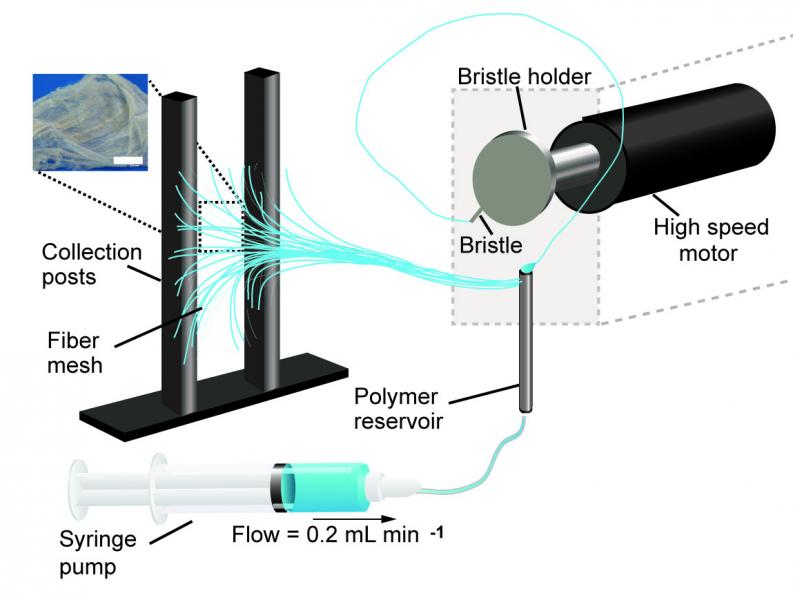
A schematic of the pull spinning apparatus with a side view illustration of a fiber being pulled from the polymer reservoir. The pull spinning system consists of a rotating bristle that dips and pulls a polymer jet in a spiral trajectory (Leila Deravi/Harvard University)

A March 1, 2017 Harvard University news release (also on EurekAlert) by Leah Burrow,, which originated the news item, describes the current process for nanofiber fabrication and explains how this technique is an improvement,

There are many ways to make nanofibers. These versatile materials — whose target applications include everything from tissue engineering to bullet proof vests — have been made using centrifugal force, capillary force, electric field, stretching, blowing, melting, and evaporation.

Each of these fabrication methods has pros and cons. For example, Rotary Jet-Spinning (RJS) and Immersion Rotary Jet-Spinning (iRJS) are novel manufacturing techniques developed in the Disease Biophysics Group at the Harvard John A. Paulson School of Engineering and Applied Sciences (SEAS) and the Wyss Institute for Biologically Inspired Engineering. Both RJS and iRJS dissolve polymers and proteins in a liquid solution and use centrifugal force or precipitation to elongate and solidify polymer jets into nanoscale fibers. These methods are great for producing large amounts of a range of materials – including DNA, nylon, and even Kevlar – but until now they haven’t been particularly portable.

The Disease Biophysics Group recently announced the development of a hand-held device that can quickly produce nanofibers with precise control over fiber orientation. Regulating fiber alignment and deposition is crucial when building nanofiber scaffolds that mimic highly aligned tissue in the body or designing point-of-use garments that fit a specific shape.

“Our main goal for this research was to make a portable machine that you could use to achieve controllable deposition of nanofibers,” said Nina Sinatra, a graduate student in the Disease Biophysics Group and co-first author of the paper. “In order to develop this kind of point-and-shoot device, we needed a technique that could produce highly aligned fibers with a reasonably high throughput.”

The new fabrication method, called pull spinning, uses a high-speed rotating bristle that dips into a polymer or protein reservoir and pulls a droplet from solution into a jet. The fiber travels in a spiral trajectory and solidifies before detaching from the bristle and moving toward a collector. Unlike other processes, which involve multiple manufacturing variables, pull spinning requires only one processing parameter — solution viscosity — to regulate nanofiber diameter. Minimal process parameters translate to ease of use and flexibility at the bench and, one day, in the field.

Pull spinning works with a range of different polymers and proteins. The researchers demonstrated proof-of-concept applications using polycaprolactone and gelatin fibers to direct muscle tissue growth and function on bioscaffolds, and nylon and polyurethane fibers for point-of-wear apparel.

“This simple, proof-of-concept study demonstrates the utility of this system for point-of-use manufacturing,” said Kit Parker, the Tarr Family Professor of Bioengineering and Applied Physics and director of the Disease Biophysics Group. “Future applications for directed production of customizable nanotextiles could extend to spray-on sportswear that gradually heats or cools an athlete’s body, sterile bandages deposited directly onto a wound, and fabrics with locally varying mechanical properties.”

Here’s a link to and a citation for the paper,

Design and Fabrication of Fibrous Nanomaterials Using Pull Spinning by Leila F. Deravi, Nina R. Sinatra, Christophe O. Chantre, Alexander P. Nesmith, Hongyan Yuan, Sahm K. Deravi, Josue A. Goss, Luke A. MacQueen, Mohammad R. Badrossamy, Grant M. Gonzalez, Michael D. Phillips, and Kevin Kit Parker. Macromolecular Materials and Engineering DOI: 10.1002/mame.201600404 Version of Record online: 17 JAN 2017

© 2017 WILEY-VCH Verlag GmbH & Co. KGaA, Weinheim

This paper is behind a paywall.