Tag Archives: nanofibers

Suit up with nanofiber for protection against explosions and high temperatures

Where explosions are concerned you might expect to see some army research and you would be right. A June 29, 2020 news item on ScienceDaily breaks the news,

Since World War I, the vast majority of American combat casualties has come not from gunshot wounds but from explosions. Today, most soldiers wear a heavy, bullet-proof vest to protect their torso but much of their body remains exposed to the indiscriminate aim of explosive fragments and shrapnel.

Designing equipment to protect extremities against the extreme temperatures and deadly projectiles that accompany an explosion has been difficult because of a fundamental property of materials. Materials that are strong enough to protect against ballistic threats can’t protect against extreme temperatures and vice versa. As a result, much of today’s protective equipment is composed of multiple layers of different materials, leading to bulky, heavy gear that, if worn on the arms and legs, would severely limit a soldier’s mobility.

Now, Harvard University researchers, in collaboration with the U.S. Army Combat Capabilities Development Command Soldier Center (CCDC SC) and West Point, have developed a lightweight, multifunctional nanofiber material that can protect wearers from both extreme temperatures and ballistic threats.

A June 29, 2020 Harvard University news release (also on EurekAlert) by Leah Burrows, which originated the news item, expands on the theme,

“When I was in combat in Afghanistan, I saw firsthand how body armor could save lives,” said senior author Kit Parker, the Tarr Family Professor of Bioengineering and Applied Physics at the Harvard John A. Paulson School of Engineering and Applied Sciences (SEAS) and a lieutenant colonel in the United States Army Reserve. “I also saw how heavy body armor could limit mobility. As soldiers on the battlefield, the three primary tasks are to move, shoot, and communicate. If you limit one of those, you decrease survivability and you endanger mission success.”

“Our goal was to design a multifunctional material that could protect someone working in an extreme environment, such as an astronaut, firefighter or soldier, from the many different threats they face,” said Grant M. Gonzalez, a postdoctoral fellow at SEAS and first author of the paper.

In order to achieve this practical goal, the researchers needed to explore the tradeoff between mechanical protection and thermal insulation, properties rooted in a material’s molecular structure and orientation.

Materials with strong mechanical protection, such as metals and ceramics, have a highly ordered and aligned molecular structure. This structure allows them to withstand and distribute the energy of a direct blow. Insulating materials, on the other hand, have a much less ordered structure, which prevents the transmission of heat through the material.

Kevlar and Twaron are commercial products used extensively in protective equipment and can provide either ballistic or thermal protection, depending on how they are manufactured. Woven Kevlar, for example, has a highly aligned crystalline structure and is used in protective bulletproof vests. Porous Kevlar aerogels, on the other hand, have been shown to have high thermal insulation.

“Our idea was to use this Kevlar polymer to combine the woven, ordered structure of fibers with the porosity of aerogels to make long, continuous fibers with porous spacing in between,” said Gonzalez. “In this system, the long fibers could resist a mechanical impact while the pores would limit heat diffusion.”

The research team used immersion Rotary Jet-Spinning (iRJS), a technique developed by Parker’s Disease Biophysics Group, to manufacture the fibers. In this technique, a liquid polymer solution is loaded into a reservoir and pushed out through a tiny opening by centrifugal force as the device spins. When the polymer solution shoots out of the reservoir, it first passes through an area of open air, where the polymers elongate and the chains align. Then the solution hits a liquid bath that removes the solvent and precipitates the polymers to form solid fibers. Since the bath is also spinning — like water in a salad spinner — the nanofibers follow the stream of the vortex and wrap around a rotating collector at the base of the device.

By tuning the viscosity of the liquid polymer solution, the researchers were able to spin long, aligned nanofibers into porous sheets — providing enough order to protect against projectiles but enough disorder to protect against heat. In about 10 minutes, the team could spin sheets about 10 by 30 centimeters in size.

To test the sheets, the Harvard team turned to their collaborators to perform ballistic tests. Researchers at CCDC SC in Natick, Massachusetts simulated shrapnel impact by shooting large, BB-like projectiles at the sample. The team performed tests by sandwiching the nanofiber sheets between sheets of woven Twaron. They observed little difference in protection between a stack of all woven Twaron sheets and a combined stack of woven Twaron and spun nanofibers.

“The capabilities of the CCDC SC allow us to quantify the successes of our fibers from the perspective of protective equipment for warfighters, specifically,” said Gonzalez.

“Academic collaborations, especially those with distinguished local universities such as Harvard, provide CCDC SC the opportunity to leverage cutting-edge expertise and facilities to augment our own R&D capabilities,” said Kathleen Swana, a researcher at CCDC SC and one of the paper’s authors. “CCDC SC, in return, provides valuable scientific and soldier-centric expertise and testing capabilities to help drive the research forward.”

In testing for thermal protection, the researchers found that the nanofibers provided 20 times the heat insulation capability of commercial Twaron and Kevlar.

“While there are improvements that could be made, we have pushed the boundaries of what’s possible and started moving the field towards this kind of multifunctional material,” said Gonzalez.

“We’ve shown that you can develop highly protective textiles for people that work in harm’s way,” said Parker. “Our challenge now is to evolve the scientific advances to innovative products for my brothers and sisters in arms.”

Harvard’s Office of Technology Development has filed a patent application for the technology and is actively seeking commercialization opportunities.

Here’s a link to and a citation for the paper,

para-Aramid Fiber Sheets for Simultaneous Mechanical and Thermal Protection in Extreme Environments by Grant M. Gonzalez, Janet Ward, John Song, Kathleen Swana, Stephen A. Fossey, Jesse L. Palmer, Felita W. Zhang, Veronica M. Lucian, Luca Cera, John F. Zimmerman, F. John Burpo, Kevin Kit Parker. Matter DOI: https://doi.org/10.1016/j.matt.2020.06.001 Published:June 29, 2020

This paper is behind a paywall.

While this is the first time I’ve featured clothing/armour that’s protective against explosions I have on at least two occasions featured bulletproof clothing in a Canadian context. A November 4, 2013 posting had a story about a Toronto-based tailoring establishment, Garrison Bespoke, that was going to publicly test a bulletproof business suit. Should you be interested, it is possible to order the suit here. There’s also a February 11, 2020 posting announcing research into “Comfortable, bulletproof clothing for Canada’s Department of National Defence.”
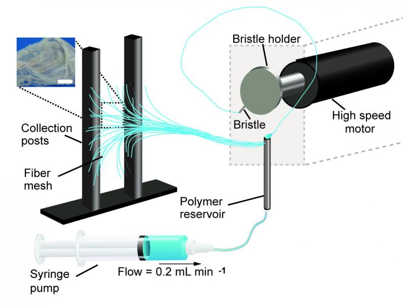
A new platform for culturing stem cells: a Multiplexed Artificial Cellular Microenvironment array

Japanese scientists have developed a more precise method for culturing stem cells according to a March 14, 2017 news item on Nanowerk,

A team of researchers in Japan has developed a new platform for culturing human pluripotent stem cells that provides far more control of culture conditions than previous tools by using micro and nanotechnologies.

The Multiplexed Artificial Cellular Microenvironment (MACME) array places nanofibres, mimicking cellular matrices, into fluid-filled micro-chambers of precise sizes, which mimic extracellular environments.

Caption: The Multiplexed Artificial Cellular Microenvironment (MACME) array, consisted with a microfluidic structure and nanofibre array for mimicking cellular microenvironments. Credit: Kyoto University iCeMS

A March 17, 2017 Kyoto University press release (also on EurekAlert), which originated the news item, explains the research in more detail,

Human pluripotent stems cells (hPSCs) hold great promise for tissue engineering, regenerative medicine and cell-based therapies because they can become any type of cell. The environment surrounding the cells plays a major role in determining what tissues they become, if they replicate into more cells, or die. However, understanding these interactions has been difficult because researchers have lacked tools that work on the appropriate scale.

Often, stem cells are cultured in a cell culture medium in small petri dishes. While factors such as medium pH levels and nutrients can be controlled, the artificial set up is on the macroscopic scale and does not allow for precise control of the physical environment surrounding the cells.

The MACME array miniaturizes this set up, culturing stem cells in rows of micro-chambers of cell culture medium. It also takes it a step further by placing nanofibers in these chambers to mimic the structures found around cells.

Led by Ken-ichiro Kamei of Kyoto University’s Institute for Integrated Cell-Material Sciences (iCeMS), the team tested a variety of nanofiber materials and densities, micro-chamber heights and initial stem cell densities to determine the best combination that encourages human pluripotent stem cells to replicate.

They stained the cells with several fluorescent markers and used a microscope to see if the cells died, replicated or differentiated into tissues.

Their analysis revealed that gelatin nanofibers and medium-sized chambers that create medium seed cell density provided the best environment for the stem cells to continue to multiply. The quantity and density of neighboring cells strongly influences cell survival.

The array is an “optimal and powerful approach for understanding how environmental cues regulate cellular functions,” the researchers conclude in a recently published paper in the journal Small.

This array appears to be the first time multiple kinds of extracellular environments can be mounted onto a single device, making it much easier to compare how different environments influence cells.

The MACME array could substantially reduce experiment costs compared to conventional tools, in part because it is low volume and requires less cell culture medium. The array does not require any special equipment and is compatible with both commonly used laboratory pipettes and automated pipette systems for performing high-throughput screening.

Here’s a link to and a citation for the paper,

Microfluidic-Nanofiber Hybrid Array for Screening of Cellular Microenvironments by Ken-ichiro Kamei, Yasumasa Mashimo, Momoko Yoshioka, Yumie Tokunaga, Christopher Fockenberg, Shiho Terada, Yoshie Koyama, Minako Nakajima, Teiko Shibata-Seki, Li Liu, Toshihiro Akaike, Eiry Kobatake, Siew-Eng How, Motonari Uesugi, and Yong Chen. Small DOI: 10.1002/smll.201603104 Version of Record online: 8 MAR 2017

© 2017 WILEY-VCH Verlag GmbH & Co. KGaA, Weinheim

This paper is behind a paywall.

Portable nanofibre fabrication device (point-of-use manufacturing)

A portable nanofiber fabrication device is quite an achievement although it seems it’s not quite ready for prime time yet. From a March 1, 2017 news item on Nanowerk (Note: A link has been removed),

Harvard researchers have developed a lightweight, portable nanofiber fabrication device that could one day be used to dress wounds on a battlefield or dress shoppers in customizable fabrics. The research was published recently in Macromolecular Materials and Engineering (“Design and Fabrication of Fibrous Nanomaterials Using Pull Spinning”)

A schematic of the pull spinning apparatus with a side view illustration of a fiber being pulled from the polymer reservoir. The pull spinning system consists of a rotating bristle that dips and pulls a polymer jet in a spiral trajectory (Leila Deravi/Harvard University)

A March 1, 2017 Harvard University news release (also on EurekAlert) by Leah Burrow,, which originated the news item, describes the current process for nanofiber fabrication and explains how this technique is an improvement,

There are many ways to make nanofibers. These versatile materials — whose target applications include everything from tissue engineering to bullet proof vests — have been made using centrifugal force, capillary force, electric field, stretching, blowing, melting, and evaporation.

Each of these fabrication methods has pros and cons. For example, Rotary Jet-Spinning (RJS) and Immersion Rotary Jet-Spinning (iRJS) are novel manufacturing techniques developed in the Disease Biophysics Group at the Harvard John A. Paulson School of Engineering and Applied Sciences (SEAS) and the Wyss Institute for Biologically Inspired Engineering. Both RJS and iRJS dissolve polymers and proteins in a liquid solution and use centrifugal force or precipitation to elongate and solidify polymer jets into nanoscale fibers. These methods are great for producing large amounts of a range of materials – including DNA, nylon, and even Kevlar – but until now they haven’t been particularly portable.

The Disease Biophysics Group recently announced the development of a hand-held device that can quickly produce nanofibers with precise control over fiber orientation. Regulating fiber alignment and deposition is crucial when building nanofiber scaffolds that mimic highly aligned tissue in the body or designing point-of-use garments that fit a specific shape.

“Our main goal for this research was to make a portable machine that you could use to achieve controllable deposition of nanofibers,” said Nina Sinatra, a graduate student in the Disease Biophysics Group and co-first author of the paper. “In order to develop this kind of point-and-shoot device, we needed a technique that could produce highly aligned fibers with a reasonably high throughput.”

The new fabrication method, called pull spinning, uses a high-speed rotating bristle that dips into a polymer or protein reservoir and pulls a droplet from solution into a jet. The fiber travels in a spiral trajectory and solidifies before detaching from the bristle and moving toward a collector. Unlike other processes, which involve multiple manufacturing variables, pull spinning requires only one processing parameter — solution viscosity — to regulate nanofiber diameter. Minimal process parameters translate to ease of use and flexibility at the bench and, one day, in the field.

Pull spinning works with a range of different polymers and proteins. The researchers demonstrated proof-of-concept applications using polycaprolactone and gelatin fibers to direct muscle tissue growth and function on bioscaffolds, and nylon and polyurethane fibers for point-of-wear apparel.

“This simple, proof-of-concept study demonstrates the utility of this system for point-of-use manufacturing,” said Kit Parker, the Tarr Family Professor of Bioengineering and Applied Physics and director of the Disease Biophysics Group. “Future applications for directed production of customizable nanotextiles could extend to spray-on sportswear that gradually heats or cools an athlete’s body, sterile bandages deposited directly onto a wound, and fabrics with locally varying mechanical properties.”

Here’s a link to and a citation for the paper,

Design and Fabrication of Fibrous Nanomaterials Using Pull Spinning by Leila F. Deravi, Nina R. Sinatra, Christophe O. Chantre, Alexander P. Nesmith, Hongyan Yuan, Sahm K. Deravi, Josue A. Goss, Luke A. MacQueen, Mohammad R. Badrossamy, Grant M. Gonzalez, Michael D. Phillips, and Kevin Kit Parker. Macromolecular Materials and Engineering DOI: 10.1002/mame.201600404 Version of Record online: 17 JAN 2017

© 2017 WILEY-VCH Verlag GmbH & Co. KGaA, Weinheim

This paper is behind a paywall.

Stronger more robust nanofibers for everything from bulletproof vests to cellular scaffolds (tissue engineering)

This work on a new technique for producing nanofibers comes from Harvard University’s School of Engineering and Applied Sciences and the Wyss Institute for Biologically Inspired Engineering (also at Harvard University). From an Oct. 10, 2016 news item on phys.org,

Fibrous materials—known for their toughness, durability and pliability—are used in everything from bulletproof vests to tires, filtration systems and cellular scaffolds for tissue engineering and regenerative medicine.

The properties of these materials are such that the smaller the fibers are, the stronger and tougher they become. But making certain fibers very small has been an engineering challenge.

Now, researchers from the Harvard John A. Paulson School of Engineering and Applied Sciences (SEAS) and the Wyss Institute for Biologically Inspired Engineering at Harvard have developed a new method to make and collect nanofibers and control their size and morphology. This could lead to stronger, more durable bulletproof vests and armor and more robust cellular scaffolding for tissue repair.

An Oct. 7, 2016 Harvard University press release by Leah Burrows, which originated the news item, describes the research in more detail (Note: A link has been removed),

Nanofibers are smaller than one micrometer in diameter.  Most nanofiber production platforms rely on dissolving polymers in a solution, which then evaporates as the fiber forms.

Rotary Jet-Spinning (RJS), the technique developed by Kit Parker’s Disease Biophysics Group, works likes a cotton candy machine. Parker is Tarr Family Professor of Bioengineering and Applied Physics at SEAS and a Core Member of the Wyss Institute. A liquid polymer solution is loaded into a reservoir and pushed out through a tiny opening by centrifugal force as the device spins. As the solution leaves the reservoir, the solvent evaporates and the polymers solidify and elongate into small, thin fibers.

“This advance is important because it allows us to manufacture ballistic protection that is much lighter, more flexible and more functional than what is available today,” said Parker, who in addition to his Harvard role is a lieutenant colonel in the United States Army Reserve and was motivated by his own combat experiences in Afghanistan. “Not only could it save lives but for the warfighter, it also could help reduce the repetitive injury motions that soldiers, sailors, marines and airmen have suffered over the last 15 years of the war on terror.”

“Rotary Jet-Spinning is great for most polymer fibers you want to make,” said Grant Gonzalez, a graduate student at SEAS and first author of the paper.  “However, some fibers require a solvent that doesn’t evaporate easily. Para-aramid, the polymer used in Kevlar® for example, is dissolved in sulfuric acid, which doesn’t evaporate off. The solution just splashes against the walls of the device without forming fibers.”

Nanofibers are smaller than one micrometer in diameter.  Most nanofiber production platforms rely on dissolving polymers in a solution, which then evaporates as the fiber forms.

Rotary Jet-Spinning (RJS), the technique developed by Kit Parker’s Disease Biophysics Group, works likes a cotton candy machine. Parker is Tarr Family Professor of Bioengineering and Applied Physics at SEAS and a Core Member of the Wyss Institute. A liquid polymer solution is loaded into a reservoir and pushed out through a tiny opening by centrifugal force as the device spins. As the solution leaves the reservoir, the solvent evaporates and the polymers solidify and elongate into small, thin fibers.

“This advance is important because it allows us to manufacture ballistic protection that is much lighter, more flexible and more functional than what is available today,” said Parker, who in addition to his Harvard role is a lieutenant colonel in the United States Army Reserve and was motivated by his own combat experiences in Afghanistan. “Not only could it save lives but for the warfighter, it also could help reduce the repetitive injury motions that soldiers, sailors, marines and airmen have suffered over the last 15 years of the war on terror.”

“Rotary Jet-Spinning is great for most polymer fibers you want to make,” said Grant Gonzalez, a graduate student at SEAS and first author of the paper.  “However, some fibers require a solvent that doesn’t evaporate easily. Para-aramid, the polymer used in Kevlar® for example, is dissolved in sulfuric acid, which doesn’t evaporate off. The solution just splashes against the walls of the device without forming fibers.”

Other methods, such as electrospinning, which uses an electric field to pull the polymer into a thin fiber, also have poor results with Kevlar and other polymers such as alginate used for tissue scaffolding and DNA.

The Harvard team overcame these challenges by developing a wet-spinning platform, which uses the same principles as the RJS system but relies on precipitation rather than evaporation to separate the solvent from the polymer.

In this system, called immersion Rotary Jet-Spinning (iRJS), when the polymer solution shoots out of the reservoir, it first passes through an area of open air, where the polymers elongate and the chains align. Then the solution hits a liquid bath that removes the solvent and precipitates the polymers to form solid fibers. Since the bath is also spinning — like water in a salad spinner — the nanofibers follow the stream of the vortex and wrap around a rotating collector at the base of the device.

Using this system, the team produced Nylon, DNA, alginate and ballistic resistant para-aramid nanofibers. The team could tune the fiber’s diameter by changing the solution concentration, the rotational speed and the distance the polymer traveled from the reservoir to the bath.

“By being able to modulate fiber strength, we can create a cellular scaffold that can mimic skeleton muscle and native tissues,” said Gonzalez.  “This platform could enable us to create a wound dressing out of alginate material or seed and mature cells on scaffolding for tissue engineering.”

Because the fibers were collected by a spinning vortex, the system also produced well-aligned sheets of nanofibers, which is important for scaffolding and ballistic resistant materials.

This is the ‘candy floss’ technique at work,

Rotary Jet-Spinning (RJS) works likes a cotton candy machine. A liquid polymer solution is loaded into a reservoir and pushed out through a tiny opening by centrifugal force as the device spins. As the solution leaves the reservoir, the solvent evaporates and the polymers solidify and elongate into small, thin fibers. Courtesy: Harvard University

Rotary Jet-Spinning (RJS) works likes a cotton candy machine. A liquid polymer solution is loaded into a reservoir and pushed out through a tiny opening by centrifugal force as the device spins. As the solution leaves the reservoir, the solvent evaporates and the polymers solidify and elongate into small, thin fibers. Courtesy: Harvard University

Here’s a link to and a citation for the paper,

Production of Synthetic, Para-Aramid and Biopolymer Nanofibers by Immersion Rotary Jet-Spinning by Grant M. Gonzalez, Luke A. MacQueen, Johan U. Lind, Stacey A. Fitzgibbons, Christophe O. Chantre, Isabelle Huggler, Holly M. Golecki, Josue A. Goss, Kevin Kit Parker. Macromolecular Materials and Engineering DOI: 10.1002/mame.201600365 Version of Record online: 7 OCT 2016

© 2016 WILEY-VCH Verlag GmbH & Co. KGaA, Weinheim

This paper is behind a paywall.

Oil spill cleanups with supergelators

Researchers in Singapore have proposed a new technology for cleaning up oil spills, according to a June 17, 2016 news item on Nanowerk,

Large-scale oil spills, where hundreds of tons of petroleum products are accidentally released into the oceans, not only have devastating effects on the environment, but have significant socio-economic impact as well [1].

Current techniques of cleaning up oil spills are not very efficient and may even cause further pollution or damage to the environment. These methods, which include the use of toxic detergent-like compounds called dispersants or burning of the oil slick, result in incomplete removal of the oil. The oil molecules remain in the water over long periods and may even be spread over a larger area as they are carried by wind and waves. Further, burning can only be applied to fresh oil slicks of at least 3 millimeters thick, and this process would also cause secondary environmental pollution.

In a bid to improve the technology utilized by cleanup crews to manage and contain such large spills, researchers from the Institute of Bioengineering and Nanotechnology (IBN) of A*STAR [located in Singapore] have invented a smart oil-scavenging material or supergelators that could help clean up oil spills efficiently and rapidly to prevent secondary pollution.

These supergelators are derived from highly soluble small organic molecules, which instantly self-assemble into nanofibers to form a 3D net that traps the oil molecules so that they can be removed easily from the surface of the water.

A June 17, 2016 IBN A*STAR media release, which originated the news item, provides more detail,

“Marine oil spills have a disastrous impact on the environment and marine life, and result in an enormous economic burden on society. Our rapid-acting supergelators offer an effective cleanup solution that can help to contain the severe environmental damage and impact of such incidents in the future,” said IBN Executive Director Professor Jackie Y. Ying.

Motivated by the urgent need for a more effective oil spill control solution, the IBN researchers developed new compounds that dissolve easily in environmentally friendly solvents and gel rapidly upon contact with oil. The supergelator molecules arrange themselves into a 3D network, entangling the oil molecules into clumps that can then be easily skimmed off the water’s surface.

“The most interesting and useful characteristic of our molecules is their ability to stack themselves on top of each other. These stacked columns allow our researchers to create and test different molecular constructions, while finding the best structure that will yield the desired properties,” said IBN Team Leader and Principal Research Scientist Dr Huaqiang Zeng. (Animation: Click to see how the supergelators stack themselves into columns.)

IBN’s supergelators have been tested on various types of weathered and unweathered crude oil in seawater, and have been found to be effective in solidifying all of them. The supergelators take only minutes to solidify the oil at room temperature for easy removal from water. In addition, tests carried out by the research team showed that the supergelator was not toxic to human cells, as well as zebrafish embryos and larvae. The researchers believe that these qualities would make the supergelators suitable for use in large oil spill areas.

The Institute is looking for industrial partners to further develop its technology for commercial use. [emphasis mine]

Video: Click to watch the supergelators in action

  1. The well documented BP Gulf of Mexico oil well accident in 2010 was a catastrophe on an unprecedented scale, with damages amounting to hundreds of billions of dollars. Its wide-ranging effects on the marine ecosystem, as well as the fishing and tourism industries, can still be felt six years on.

Here’s a link to and a citation for the paper,

Instant Room-Temperature Gelation of Crude Oil by Chiral Organogelators by Changliang Ren, Grace Hwee Boon Ng, Hong Wu, Kiat-Hwa Chan, Jie Shen, Cathleen Teh, Jackie Y. Ying, and Huaqiang Zeng. Chem. Mater., 2016, 28 (11), pp 4001–4008 DOI: 10.1021/acs.chemmater.6b01367 Publication Date (Web): May 10, 2016

Copyright © 2016 American Chemical Society

This paper is behind a paywall.

I have featured other nanotechnology-enabled oil spill cleanup solutions here. One of the more recent pieces is my Dec. 7, 2015 post about boron nitride sponges. The search terms: ‘oil spill’ and ‘oil spill cleanup’ will help you unearth more.

There have been some promising possibilities and I hope one day these clean up technologies will be brought to market.

Watching artificial nanofibres self-sort in real-time

A May 31, 2016 news item on phys.org describes research on self-assembling fibres at Kyoto University (Japan) by referencing the ancient Greek mythological figure, Psyche,

The Greek goddess Psyche borrowed help from ants to sort a room full of different grains. Cells, on the other hand, do something similar without Olympian assistance, as they organize molecules into robust, functional fibers. Now scientists are able to see self-sorting phenomena happen in real time with artificial molecules.

The achievement, reported in Nature Chemistry, elucidates how two different types of nanofibers sort themselves into organized structures under artificial conditions.

“Basic cellular structures, such as actin filaments, come into being through the autonomous self-sorting of individual molecules, even though a tremendous variety of proteins and small molecules are present inside the cell,” says lead author Hajime Shigemitsu, a researcher in Itaru Hamachi’s lab at Kyoto University.

A May 30, 2016 Kyoto University news release (also on EurekAlert), which originated the news item, expands on the theme,

“Imagine a box filled with an assortment of building blocks — it’s as if the same type of blocks started sorting themselves into neat bundles all on their own. In living cells, such phenomena always happen, enabling accurate self-assembling of proteins, which is essential for cell functions.”

“If we are able to control self-sorting with artificial molecules, we can work toward developing intelligent, next-generation biomimics that possess the flexibility and diversity of functions that exist in a living cell.”

Study co-author Ryou Kubota explains that previous studies have already made artificial molecules build themselves into fibers — but only when there was one type of molecule around. Having a jumble of types, on the other hand, made the molecules confused.

“The difficulty in inducing self-assembly with artificial molecules is that they don’t recognize the same type of molecule, unlike molecules in the natural world. Different types of artificial molecules interact with each other and make an unsorted cluster.”

From a database of structural analyses, Hamachi and colleagues discovered a combination of nanofibers — namely a peptide-based and lipid-based hydrogelator — that would make sorted fibers without mixing with the other. They then tethered the fibers with fluorescent probes; with a type of microscope typically used in cell imaging, the team was able to observe directly and in real-time how the artificial molecules sorted themselves.

“Ultimately, this finding could help develop new materials that respond dynamically to different environments and stimuli,” elaborates Hamachi. “This insight is not only useful for materials science, but may also provide useful clues for understanding self-organization in cells.”

Here’s a link to and a citation for the paper,

In situ real-time imaging of self-sorted supramolecular nanofibres by Shoji Onogi, Hajime Shigemitsu, Tatsuyuki Yoshii, Tatsuya Tanida, Masato Ikeda, Ryou Kubota, & Itaru Hamachi. Nature Chemistry (2016) doi:10.1038/nchem.2526 Published online 30 May 2016

This paper is behind a paywall bu the researchers have made a video of the self-sorting proteins freely available,

Brushing your way to nanofibres

The scientists are using what looks like a hairbrush to create nanofibres ,

Figure 2: Brush-spinning of nanofibers. (Reprinted with permission by Wiley-VCH Verlag)) [downloaded from http://www.nanowerk.com/spotlight/spotid=41398.php]

Figure 2: Brush-spinning of nanofibers. (Reprinted with permission by Wiley-VCH Verlag)) [downloaded from http://www.nanowerk.com/spotlight/spotid=41398.php]

A Sept. 23, 2015 Nanowerk Spotlight article by Michael Berger provides an in depth look at this technique (developed by a joint research team of scientists from the University of Georgia, Princeton University, and Oxford University) which could make producing nanofibers for use in scaffolds (tissue engineering and other applications) more easily and cheaply,

Polymer nanofibers are used in a wide range of applications such as the design of new composite materials, the fabrication of nanostructured biomimetic scaffolds for artificial bones and organs, biosensors, fuel cells or water purification systems.

“The simplest method of nanofiber fabrication is direct drawing from a polymer solution using a glass micropipette,” Alexander Tokarev, Ph.D., a Research Associate in the Nanostructured Materials Laboratory at the University of Georgia, tells Nanowerk. “This method however does not scale up and thus did not find practical applications. In our new work, we introduce a scalable method of nanofiber spinning named touch-spinning.”

James Cook in a Sept. 23, 2015 article for Materials Views provides a description of the technology,

A glass rod is glued to a rotating stage, whose diameter can be chosen over a wide range of a few centimeters to more than 1 m. A polymer solution is supplied, for example, from a needle of a syringe pump that faces the glass rod. The distance between the droplet of polymer solution and the tip of the glass rod is adjusted so that the glass rod contacts the polymer droplet as it rotates.

Following the initial “touch”, the polymer droplet forms a liquid bridge. As the stage rotates the bridge stretches and fiber length increases, with the diameter decreasing due to mass conservation. It was shown that the diameter of the fiber can be precisely controlled down to 40 nm by the speed of the stage rotation.

The method can be easily scaled-up by using a round hairbrush composed of 600 filaments.

When the rotating brush touches the surface of a polymer solution, the brush filaments draw many fibers simultaneously producing hundred kilometers of fibers in minutes.

The drawn fibers are uniform since the fiber diameter depends on only two parameters: polymer concentration and speed of drawing.

Returning to Berger’s Spotlight article, there is an important benefit with this technique,

As the team points out, one important aspect of the method is the drawing of single filament fibers.

These single filament fibers can be easily wound onto spools of different shapes and dimensions so that well aligned one-directional, orthogonal or randomly oriented fiber meshes with a well-controlled average mesh size can be fabricated using this very simple method.

“Owing to simplicity of the method, our set-up could be used in any biomedical lab and facility,” notes Tokarev. “For example, a customized scaffold by size, dimensions and othermorphologic characteristics can be fabricated using donor biomaterials.”

Berger’s and Cook’s articles offer more illustrations and details.

Here’s a link to and a citation for the paper,

Touch- and Brush-Spinning of Nanofibers by Alexander Tokarev, Darya Asheghal, Ian M. Griffiths, Oleksandr Trotsenko, Alexey Gruzd, Xin Lin, Howard A. Stone, and Sergiy Minko. Advanced Materials DOI: 10.1002/adma.201502768ViewFirst published: 23 September 2015

This paper is behind a paywall.

Using music to align your nanofibers

It’s always nice to feature a ‘nano and music’ research story, my Nov. 6, 2013 posting being, until now, the most recent. A Jan. 8, 2014 news item on Nanowerk describes Japanese researchers’ efforts with nanofibers (Note: A link has been removed),

Humans create and perform music for a variety of purposes, such as aesthetic pleasure, healing, religion, and ceremony. Accordingly, a scientific question arises: Can molecules or molecular assemblies interact physically with the sound vibrations of music? In the journal ChemPlusChem (“Acoustic Alignment of a Supramolecular Nanofiber in Harmony with the Sound of Music”), Japanese researchers have now revealed their physical interaction. When classical music was playing, a designed supramolecular nanofiber in a solution dynamically aligned in harmony with the sound of music.

Sound is vibration of matter, having a frequency, in which certain physical interactions occur between the acoustically vibrating media and solute molecules or molecular assemblies. Music is an art form consisting of the sound and silence expressed through time, and characterized by rhythm, harmony, and melody. The question of whether music can cause any kind of molecular or macromolecular event is controversial, and the physical interaction between the molecules and the sound of music has never been reported.

The Jan. 8, 2014 Chemistry Views article, which originated the news item, provides more detail,

Scientists working at Kobe University and Kobe City College of Technology, Japan, have now developed a supramolecular nanofiber, composed of an anthracene derivative, which can dynamically align by sensing acoustic streaming flows generated by the sound of music. Time course linear dichroism (LD) spectroscopy could visualize spectroscopically the dynamic acoustic alignments of the nanofiber in the solution. The nanofiber aligns upon exposure to the audible sound wave, with frequencies up to 1000 Hz, with quick responses to the sound and silence, and amplitude and frequency changes of the sound wave. The sheared flows generated around glass-surface boundary layer and the crossing area of the downward and upward flows allow shear-induced alignments of the nanofiber.
Music is composed of the multi complex sounds and silence, which characteristically change in the course of its playtime. The team, led by A. Tsuda, uses “Symphony No. 5 in C minor, First movement: Allegro con brio” written by Beethoven, and “Symphony No. 40 in G minor, K. 550, First movement”, written by Mozart in the experiments. When the classical music was playing, the sample solution gave the characteristic LD profile of the music, where the nanofiber dynamically aligned in harmony with the sound of music.

Here’s an imagie illustrating the scientists’ work with music,

[downloaded from http://www.chemistryviews.org/details/ezine/5712621/Musical_Molecules.html]

[downloaded from http://www.chemistryviews.org/details/ezine/5712621/Musical_Molecules.html]

Here’s a link to and a citation for the paper,

Acoustic Alignment of a Supramolecular Nanofiber in Harmony with the Sound of Music by Ryosuke Miura, Yasunari Ando, Yasuhisa Hotta, Yoshiki Nagatani, Akihiko Tsuda, ChemPlusChem 2014.  DOI: 10.1002/cplu.201300400

This is an open access paper as of Jan. 8, 2014. If the above link does not work, try this .