Tag Archives: regenerative medicine

Treating traumatic muscle loss with tissue nanotransfection

A November 9, 2022 news item on ScienceDaily announces some work from Indiana University (US),

Technology developed by researchers at the Indiana University School of Medicine that can change skin tissue into blood vessels and nerve cells has also shown promise as a treatment for traumatic muscle loss.

Tissue nanotransfection is a minimally invasive nanochip device that can reprogram tissue function by applying a harmless electric spark to deliver specific genes in a fraction of a second.

A November 9, 2022 Indiana University news release (also on EurekAlert), which originated the news item, provides additional technical details, Note: Links have been removed,

A new study, published in Nature Partner Journals Regenerative Medicine, tested tissue nanotransfection-based gene therapy as a treatment, with the goal of delivering a gene known to be a major driver of muscle repair and regeneration. They found that muscle function improved when tissue nanotransfection was used as a therapy for seven days following volumetric muscle loss in rats. It is the first study to report that tissue nanotransfection technology can be used to generate muscle tissue and demonstrates its benefit in addressing volumetric muscle loss.

Volumetric muscle loss is the traumatic or surgical loss of skeletal muscle that results in compromised muscle strength and mobility. Incapable of regenerating the amount of lost tissue, the affected muscle undergoes substantial loss of function, thus compromising quality of life. A 20 percent loss in mass can result in an up to 90 percent loss in muscle function.

Current clinical treatments for volumetric muscle loss are physical therapy or autologous tissue transfer (using a person’s own tissue), the outcomes of which are promising but call for improved treatment regimens.

“We are encouraged that tissue nanotransfection is emerging as a versatile platform technology for gene delivery, gene editing and in vivo tissue reprogramming,” said Chandan Sen, director of the Indiana Center for Regenerative Medicine and Engineering, associate vice president for research and Distinguished Professor at the IU School of Medicine. “This work proves the potential of tissue nanotransfection in muscle tissue, opening up a new avenue of investigational pursuit that should help in addressing traumatic muscle loss. Importantly, it demonstrates the versatility of the tissue nanotransfection technology platform in regenerative medicine.”

Sen also leads the regenerative medicine and engineering scientific pillar of the IU Precision Health Initiative and is lead author on the new publication.

The Indiana Center for Regenerative Medicine and Engineering is home to the tissue nanotransfection technology for in vivo tissue reprogramming, gene delivery and gene editing. So far, tissue nanotransfection has also been achieved in blood vessel and nerve tissue. In addition, recent work has shown that topical tissue nanotransfection can achieve cell-specific gene editing of skin wound tissue to improve wound closure.

Here’s a link to and a citation for the paper,

Myogenic tissue nanotransfection improves muscle torque recovery following volumetric muscle loss by Andrew Clark, Subhadip Ghatak, Poornachander Reddy Guda, Mohamed S. El Masry, Yi Xuan, Amy Y. Sato, Teresita Bellido & Chandan K. Sen. npj Regenerative Medicine volume 7, Article number: 63 (2022) DOI: https://doi.org/10.1038/s41536-022-00259-y Published: 20 October 2022

This paper is open access.

This is a very nice image of a delighted Dr. Sen,

Caption Chandan Sen Credit: Photo by Liz Kaye, Indiana University

Tissue nanotransfection

I’m wondering how I missed the research from last year (2021) which foregrounds this latest work. Ah well. It happens. Making up for lost time, here’s a July 18, 2022 news item on phys.org about tissue nanotransfection, Note: Links have been removed,

The Indiana Center for Regenerative Medicine and Engineering (ICRME) at Indiana University School of Medicine is home to tissue nanotransfection (TNT) regenerative medicine technology that achieves functional tissue reprogramming in the live body. Last year, ICRME researchers published on how to manufacture the TNT 2.0 silicon chip hardware in Nature Protocol. Now, their research demonstrates for the first time that TNT can serve as a non-viral, topical gene-editing delivery device.

TNT is a minimally invasive device that can reprogram tissue function in the live body by applying pulses of harmless, electric sparks to deliver specific genes of interest to the skin.

“TNT-based delivery can achieve cell-specific gene editing,” said corresponding author Chandan K. Sen, Ph.D., the J. Stanley Battersby Chair and distinguished professor of surgery, director of the ICRME at IU School of Medicine and executive director of the Indiana University Health Comprehensive Wound Care Center. “Your skin has thousands of genes and in chronic wounds many key genes are silenced by DNA methylation. TNT-based gene editing technology can remove that barrier.”

A July 18, 2022 Indiana University School of Medicine news release (also on EurekAlert), which originated the news item, updates the information with some of the latest research, Note: Links have been removed,

In this study, genome-wide methylation was observed in the chronic wound tissue of patients. This was reproduced in an experimental murine model. TNT-based, cell-specific gene editing rescued wound healing. Results were published recently [July 12, 2022] in the Journal of Clinical Investigation.

Previous TNT application studies reported on the rescue of injured legs, diabetic neuropathy, crushed nerve and the stroke-affected brain. This is the first time promoter methylation of genes is recognized as a critical barrier to wound healing. In this study, ICRME investigators found that P53 methylation and gene silencing as a critical barrier to cutaneous wound epithelial-to-mesenchymal transition (EMT), a mechanism that is necessary to close skin wounds. TNT based non-viral keratinocyte-specific demethylation of P53 gene rescued EMT and achieved wound closure.

Chronic wounds can result in serious and sometimes life-threatening complications from an abundance of dying and necrotic tissue, such as cellulitis, lower-extremity amputation and sepsis. Treating chronic wounds is estimated to cost the United States health care system $28 billion annually, which amplifies the need to test novel treatments to prevent amputation, save lives and lower health care costs.

“Inspired by observations in chronic wound patients, this work has achieved an important milestone highlighting the need to de-silence genes at the wound-site,” said first author Kanhaiya Singh, PhD, assistant professor of surgery and an investigator at the ICRME.

Here are two links and citations. First, the earlier work,

Fabrication and use of silicon hollow-needle arrays to achieve tissue nanotransfection in mouse tissue in vivo by Yi Xuan, Subhadip Ghatak, Andrew Clark, Zhigang Li, Savita Khanna, Dongmin Pak, Mangilal Agarwal, Sashwati Roy, Peter Duda & Chandan K. Sen. Nature Protocols volume 16, pages 5707–5738 (2021) DOI: https://doi.org/10.1038/s41596-021-00631-0 Published: 26 November 2021 Issue Date: December 2021

This paper is behind a paywall.

Now, the latest work

Genome-wide DNA hypermethylation opposes healing in chronic wound patients by impairing epithelial-to-mesenchymal transition by Kanhaiya Singh, Yashika Rustagi, Ahmed S. Abouhashem, Saba Tabasum, Priyanka Verma, Edward Hernandez, Durba Pal, Dolly K. Khona, Sujit K. Mohanty, Manishekhar Kumar, Rajneesh Srivastava, Poornachander R Guda, Sumit S. Verma, Sanskruti Mahajan, Jackson A. Killian, Logan A. Walker, Subhadip Ghatak, Shomita S. Mathew-Steiner, Kristen Wanczyk, Sheng Liu, Jun Wan, Pearlly Yan, Ralf Bundschuh, Savita Khanna, Gayle M. Gordillo, Michael P. Murphy, Sashwati Roy, and Chandan K. Sen. J Clin Invest. DOI: https://doi.org/10.1172/JCI157279 Published: July 12, 2022 Version 1 (In-Press Preview) Version 2: J Clin Invest. 2022;132(17):e157279. https://doi.org/10.1172/JCI157279. Volume 132, Issue 17 Published September 1, 2022

This paper is open access.

Asparagus spinal cord?

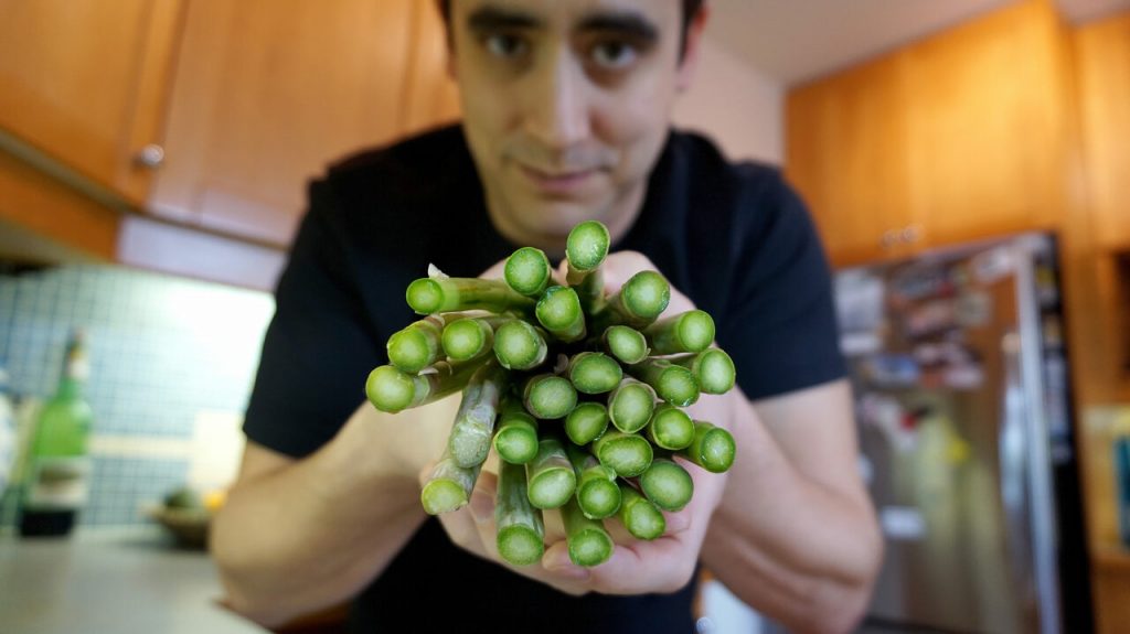
I love this picture,

Pelling in the kitchen with asparagus, the veggie that inspired his work on spinal cord injuries. Credit: Andrew Pelling?

The image accompanies Cari Shane’s August 4, 2021 article for Atlas Obscura’s Gastro Obscura about Andrew Pelling and his asparagus-based scaffolds for spinal cord stem cells (Note: A link has been removed),

Around 10 years ago, Pelling [Dr. Andrew Pelling at the University of Ottawa], a biophysicist, started thinking with his team about materials that could be used to reconstruct damaged or diseased human tissues. Surrounded by a rainbow of fresh fruits and vegetables at his University of Ottawa lab, Pelling and his team dismantle biological systems, mixing and matching parts, and put them back together in new and creative ways. It’s a little bit like a hacker who takes parts from a phone, a computer, and a car to build a robotic arm. Or like Mary Shelley’s Dr. Frankenstein, who built a monster out of cadavers. Except Pelling’s team has turned an apple into an ear and, most recently, a piece of asparagus into a scaffold for spinal-cord implants.

Pelling believes the future of regenerative medicine—which uses external therapies to help the body heal, the same way a cut heals by itself or a broken bone can mend without surgery—is in the supermarket produce aisle. He calls it “augmented biology,” and it’s a lot less expensive—by thousands and thousands of dollars—than implanting organs donated by humans, taken from animals, or manmade or bioengineered from animal tissue.

Decellularization as a process for implantation is fairly new, developed in the mid 1990s primarily by Doris Taylor. By washing out the genetic materials that make an apple an apple, for example, you are left with plant tissue, or a “cellulose mesh,” explains Pelling. “What we’re doing is washing out all the plant DNA, RNA proteins, all that sort of stuff that can cause immune responses, and rejection. And we’re just leaving behind the fiber in a plant—like literally the stuff that gets stuck in your teeth.”

When Pelling noticed the resemblance between a decellularized apple slice and an ear, he saw the true potential of his lab games. If he implanted the apple scaffolding into a living animal, he wondered, would it “be accepted” and vascularize? That is, would the test animal’s body glom onto the plant cells as if they weren’t a dangerous, foreign body and instead send out signals to create a blood supply, allowing the plant tissue to become a living part of the animal’s body? The answer was yes. “Suddenly, and by accident, we developed a material that has huge therapeutic and regenerative potential,” says Pelling. The apple ear does not enable hearing, and it remains in the animal-testing phase, but it may have applications for aesthetic implantation.

Soon after his breakthrough apple experiment, which was published in 2016 and earned him the moniker of “mad scientist,” Pelling shifted his focus to asparagus. The idea hit him when he was cooking. Looking at the end of a spear, he thought, “Hey, it looks like a spinal cord. What the hell? Maybe we can do something,” he says.

… Pelling implanted decellularized asparagus tissue under the skin of a lab rat. In just a few weeks, blood vessels flowed through the asparagus scaffolding; healthy cells from the animal moved into the tissue and turned the scaffold into living tissue. “The surprise here was that the body, instead of rejecting this material, it actually integrated into the material,” says Pelling. In the bioengineering world, getting that to happen has typically been a major challenge.

And then came the biggest surprise of all. Rats with severed spinal cords that had been implanted with the asparagus tissue were able to walk again, just a few weeks after implantation. …

While using asparagus tissue as scaffolding to repair spinal cords is not a “miracle cure,” says Pelling, it’s unlike the kinds of implants that have come before. Donated or manufactured organs are historically both more complicated and more expensive. Pelling’s implants were “done without stem cells or electrical stimulation or exoskeletons, or any of the usual approaches, but rather using very low cost, accessible materials that we honestly just bought at the grocery store,” he says, “and, we achieved the same level of recovery.” (At least in animal tests.) Plus, whereas patients usually need lifelong immunosuppressants, which can have negative side effects, to prevent their body from rejecting an implant, that doesn’t seem necessary with Pelling’s plant-based implants. And, so far, the plant-based implants don’t seem to break down over time like traditional spinal-cord implants. “The inertness of plant tissue is exactly why it’s so biocompatible,” says Pelling.

In October 2020, the asparagus implant was designated as a “breakthrough device” by the FDA [US Food and Drug Administration]. The designation means human trials will be fast-tracked and likely begin in a few years. …

Shane’s August 4, 2021 article is fascinating and well illustrated with a number of embedded images. If you have the time and the inclination, do read it.

More of Pelling’s work can be found here at the Pelling Lab website. He was mentioned (by name only as a participant in the second Canadian DIY Biology Summit organized by the Public Health Agency of Canada [PHAC]) here in an April 21, 2020 posting (my 10 year review of science culture in Canada). You’ll find the Pelling mention under the DIY Biology subhead about 20% of the way down the screen.

A new generation of xenobots made with frog cells

I meant to feature this work last year when it was first announced so I’m delighted a second chance has come around so soon after. From a March 31, 2021 news item on ScienceDaily,

Last year, a team of biologists and computer scientists from Tufts University and the University of Vermont (UVM) created novel, tiny self-healing biological machines from frog cells called “Xenobots” that could move around, push a payload, and even exhibit collective behavior in the presence of a swarm of other Xenobots.

Get ready for Xenobots 2.0.

Here’s a video of the Xenobot 2.0. It’s amazing but, for anyone who has problems with animal experimentation, this may be disturbing,


The next version of Xenobots have been created – they’re faster, live longer, and can now record information. (Source: Doug Blackiston & Emma Lederer)

A March 31, 2021 Tufts University news release by Mike Silver (also on EurekAlert and adapted and published as Scientists Create the Next Generation of Living Robots on the University of Vermont website as a UVM Today story),

The same team has now created life forms that self-assemble a body from single cells, do not require muscle cells to move, and even demonstrate the capability of recordable memory. The new generation Xenobots also move faster, navigate different environments, and have longer lifespans than the first edition, and they still have the ability to work together in groups and heal themselves if damaged. The results of the new research were published today [March 31, 2021] in Science Robotics.

Compared to Xenobots 1.0, in which the millimeter-sized automatons were constructed in a “top down” approach by manual placement of tissue and surgical shaping of frog skin and cardiac cells to produce motion, the next version of Xenobots takes a “bottom up” approach. The biologists at Tufts took stem cells from embryos of the African frog Xenopus laevis (hence the name “Xenobots”) and allowed them to self-assemble and grow into spheroids, where some of the cells after a few days differentiated to produce cilia – tiny hair-like projections that move back and forth or rotate in a specific way. Instead of using manually sculpted cardiac cells whose natural rhythmic contractions allowed the original Xenobots to scuttle around, cilia give the new spheroidal bots “legs” to move them rapidly across a surface. In a frog, or human for that matter, cilia would normally be found on mucous surfaces, like in the lungs, to help push out pathogens and other foreign material. On the Xenobots, they are repurposed to provide rapid locomotion. 

“We are witnessing the remarkable plasticity of cellular collectives, which build a rudimentary new ‘body’ that is quite distinct from their default – in this case, a frog – despite having a completely normal genome,” said Michael Levin, Distinguished Professor of Biology and director of the Allen Discovery Center at Tufts University, and corresponding author of the study. “In a frog embryo, cells cooperate to create a tadpole. Here, removed from that context, we see that cells can re-purpose their genetically encoded hardware, like cilia, for new functions such as locomotion. It is amazing that cells can spontaneously take on new roles and create new body plans and behaviors without long periods of evolutionary selection for those features.”

“In a way, the Xenobots are constructed much like a traditional robot.  Only we use cells and tissues rather than artificial components to build the shape and create predictable behavior.” said senior scientist Doug Blackiston, who co-first authored the study with research technician Emma Lederer. “On the biology end, this approach is helping us understand how cells communicate as they interact with one another during development, and how we might better control those interactions.”

While the Tufts scientists created the physical organisms, scientists at UVM were busy running computer simulations that modeled different shapes of the Xenobots to see if they might exhibit different behaviors, both individually and in groups. Using the Deep Green supercomputer cluster at UVM’s Vermont Advanced Computing Core, the team, led by computer scientists and robotics experts Josh Bongard and Sam Kriegman, simulated the Xenbots under hundreds of thousands of random environmental conditions using an evolutionary algorithm.  These simulations were used to identify Xenobots most able to work together in swarms to gather large piles of debris in a field of particles

“We know the task, but it’s not at all obvious — for people — what a successful design should look like. That’s where the supercomputer comes in and searches over the space of all possible Xenobot swarms to find the swarm that does the job best,” says Bongard. “We want Xenobots to do useful work. Right now we’re giving them simple tasks, but ultimately we’re aiming for a new kind of living tool that could, for example, clean up microplastics in the ocean or contaminants in soil.” 

It turns out, the new Xenobots are much faster and better at tasks such as garbage collection than last year’s model, working together in a swarm to sweep through a petri dish and gather larger piles of iron oxide particles. They can also cover large flat surfaces, or travel through narrow capillary tubes.

These studies also suggest that the in silico [computer] simulations could in the future optimize additional features of biological bots for more complex behaviors. One important feature added in the Xenobot upgrade is the ability to record information.

Now with memory

A central feature of robotics is the ability to record memory and use that information to modify the robot’s actions and behavior. With that in mind, the Tufts scientists engineered the Xenobots with a read/write capability to record one bit of information, using a fluorescent reporter protein called EosFP, which normally glows green. However, when exposed to light at 390nm wavelength, the protein emits red light instead. 

The cells of the frog embryos were injected with messenger RNA coding for the EosFP protein before stem cells were excised to create the Xenobots. The mature Xenobots now have a built-in fluorescent switch which can record exposure to blue light around 390nm.
The researchers tested the memory function by allowing 10 Xenobots to swim around a surface on which one spot is illuminated with a beam of 390nm light. After two hours, they found that three bots emitted red light. The rest remained their original green, effectively recording the “travel experience” of the bots.

This proof of principle of molecular memory could be extended in the future to detect and record not only light but also the presence of radioactive contamination, chemical pollutants, drugs, or a disease condition. Further engineering of the memory function could enable the recording of multiple stimuli (more bits of information) or allow the bots to release compounds or change behavior upon sensation of stimuli. 

“When we bring in more capabilities to the bots, we can use the computer simulations to design them with more complex behaviors and the ability to carry out more elaborate tasks,” said Bongard. “We could potentially design them not only to report conditions in their environment but also to modify and repair conditions in their environment.”

Xenobot, heal thyself

“The biological materials we are using have many features we would like to someday implement in the bots – cells can act like sensors, motors for movement, communication and computation networks, and recording devices to store information,” said Levin. “One thing the Xenobots and future versions of biological bots can do that their metal and plastic counterparts have difficulty doing is constructing their own body plan as the cells grow and mature, and then repairing and restoring themselves if they become damaged. Healing is a natural feature of living organisms, and it is preserved in Xenobot biology.” 

The new Xenobots were remarkably adept at healing and would close the majority of a severe full-length laceration half their thickness within 5 minutes of the injury. All injured bots were able to ultimately heal the wound, restore their shape and continue their work as before. 

Another advantage of a biological robot, Levin adds, is metabolism. Unlike metal and plastic robots, the cells in a biological robot can absorb and break down chemicals and work like tiny factories synthesizing and excreting chemicals and proteins. The whole field of synthetic biology – which has largely focused on reprogramming single celled organisms to produce useful molecules – can now be exploited in these multicellular creatures

Like the original Xenobots, the upgraded bots can survive up to ten days on their embryonic energy stores and run their tasks without additional energy sources, but they can also carry on at full speed for many months if kept in a “soup” of nutrients. 

What the scientists are really after

An engaging description of the biological bots and what we can learn from them is presented in a TED talk by Michael Levin. In his TED Talk, professor Levin describes not only the remarkable potential for tiny biological robots to carry out useful tasks in the environment or potentially in therapeutic applications, but he also points out what may be the most valuable benefit of this research – using the bots to understand how individual cells come together, communicate, and specialize to create a larger organism, as they do in nature to create a frog or human. It’s a new model system that can provide a foundation for regenerative medicine.

Xenobots and their successors may also provide insight into how multicellular organisms arose from ancient single celled organisms, and the origins of information processing, decision making and cognition in biological organisms. 

Recognizing the tremendous future for this technology, Tufts University and the University of Vermont have established the Institute for Computer Designed Organisms (ICDO), to be formally launched in the coming months, which will pull together resources from each university and outside sources to create living robots with increasingly sophisticated capabilities.

The ultimate goal for the Tufts and UVM researchers is not only to explore the full scope of biological robots they can make; it is also to understand the relationship between the ‘hardware’ of the genome and the ‘software’ of cellular communications that go into creating organized tissues, organs and limbs. Then we can gain greater control of that morphogenesis for regenerative medicine, and the treatment of cancer and diseases of aging.

Here’s a link to and a citation for the paper,

A cellular platform for the development of synthetic living machines by Douglas Blackiston, Emma Lederer, Sam Kriegman, Simon Garnier, Joshua Bongard, and Michael Levin. Science Robotics 31 Mar 2021: Vol. 6, Issue 52, eabf1571 DOI: 10.1126/scirobotics.abf1571

This paper is behind a paywall.

Cortical spheroids (like mini-brains) could unlock (larger) brain’s mysteries

A March 19, 2021 Northwestern University news release on EurekAlert announces the creation of a device designed to monitor brain organoids (for anyone unfamiliar with brain organoids there’s more information after the news),

A team of scientists, led by researchers at Northwestern University, Shirley Ryan AbilityLab and the University of Illinois at Chicago (UIC), has developed novel technology promising to increase understanding of how brains develop, and offer answers on repairing brains in the wake of neurotrauma and neurodegenerative diseases.

Their research is the first to combine the most sophisticated 3-D bioelectronic systems with highly advanced 3-D human neural cultures. The goal is to enable precise studies of how human brain circuits develop and repair themselves in vitro. The study is the cover story for the March 19 [March 17, 2021 according to the citation] issue of Science Advances.

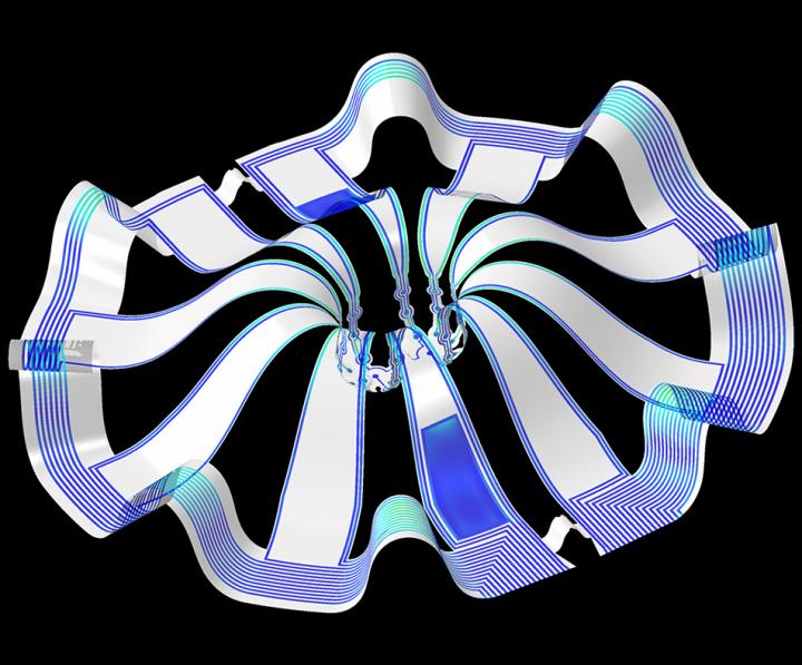
The cortical spheroids used in the study, akin to “mini-brains,” were derived from human-induced pluripotent stem cells. Leveraging a 3-D neural interface system that the team developed, scientists were able to create a “mini laboratory in a dish” specifically tailored to study the mini-brains and collect different types of data simultaneously. Scientists incorporated electrodes to record electrical activity. They added tiny heating elements to either keep the brain cultures warm or, in some cases, intentionally overheated the cultures to stress them. They also incorporated tiny probes — such as oxygen sensors and small LED lights — to perform optogenetic experiments. For instance, they introduced genes into the cells that allowed them to control the neural activity using different-colored light pulses.

This platform then enabled scientists to perform complex studies of human tissue without directly involving humans or performing invasive testing. In theory, any person could donate a limited number of their cells (e.g., blood sample, skin biopsy). Scientists can then reprogram these cells to produce a tiny brain spheroid that shares the person’s genetic identity. The authors believe that, by combining this technology with a personalized medicine approach using human stem cell-derived brain cultures, they will be able to glean insights faster and generate better, novel interventions.

“The advances spurred by this research will offer a new frontier in the way we study and understand the brain,” said Shirley Ryan AbilityLab’s Dr. Colin Franz, co-lead author on the paper who led the testing of the cortical spheroids. “Now that the 3-D platform has been developed and validated, we will be able to perform more targeted studies on our patients recovering from neurological injury or battling a neurodegenerative disease.”

Yoonseok Park, postdoctoral fellow at Northwestern University and co-lead author, added, “This is just the beginning of an entirely new class of miniaturized, 3-D bioelectronic systems that we can construct to expand the capacity of the regenerative medicine field. For example, our next generation of device will support the formation of even more complex neural circuits from brain to muscle, and increasingly dynamic tissues like a beating heart.”

Current electrode arrays for tissue cultures are 2-D, flat and unable to match the complex structural designs found throughout nature, such as those found in the human brain. Moreover, even when a system is 3-D, it is extremely challenging to incorporate more than one type of material into a small 3-D structure. With this advance, however, an entire class of 3-D bioelectronics devices has been tailored for the field of regenerative medicine.

“Now, with our small, soft 3-D electronics, the capacity to build devices that mimic the complex biological shapes found in the human body is finally possible, providing a much more holistic understanding of a culture,” said Northwestern’s John Rogers, who led the technology development using technology similar to that found in phones and computers. “We no longer have to compromise function to achieve the optimal form for interfacing with our biology.”

As a next step, scientists will use the devices to better understand neurological disease, test drugs and therapies that have clinical potential, and compare different patient-derived cell models. This understanding will then enable a better grasp of individual differences that may account for the wide variation of outcomes seen in neurological rehabilitation.

“As scientists, our goal is to make laboratory research as clinically relevant as possible,” said Kristen Cotton, research assistant in Dr. Franz’s lab. “This 3-D platform opens the door to new experiments, discovery and scientific advances in regenerative neurorehabilitation medicine that have never been possible.”

Caption: Three dimensional multifunctional neural interfaces for cortical spheroids and engineered assembloids Credit: Northwestern University

As for what brain ogranoids might be, Carl Zimmer in an Aug. 29, 2019 article for the New York Times provides an explanation,

Organoids Are Not Brains. How Are They Making Brain Waves?

Two hundred and fifty miles over Alysson Muotri’s head, a thousand tiny spheres of brain cells were sailing through space.

The clusters, called brain organoids, had been grown a few weeks earlier in the biologist’s lab here at the University of California, San Diego. He and his colleagues altered human skin cells into stem cells, then coaxed them to develop as brain cells do in an embryo.

The organoids grew into balls about the size of a pinhead, each containing hundreds of thousands of cells in a variety of types, each type producing the same chemicals and electrical signals as those cells do in our own brains.

In July, NASA packed the organoids aboard a rocket and sent them to the International Space Station to see how they develop in zero gravity.

Now the organoids were stowed inside a metal box, fed by bags of nutritious broth. “I think they are replicating like crazy at this stage, and so we’re going to have bigger organoids,” Dr. Muotri said in a recent interview in his office overlooking the Pacific.

What, exactly, are they growing into? That’s a question that has scientists and philosophers alike scratching their heads.

On Thursday, Dr. Muotri and his colleagues reported that they  have recorded simple brain waves in these organoids. In mature human brains, such waves are produced by widespread networks of neurons firing in synchrony. Particular wave patterns are linked to particular forms of brain activity, like retrieving memories and dreaming.

As the organoids mature, the researchers also found, the waves change in ways that resemble the changes in the developing brains of premature babies.

“It’s pretty amazing,” said Giorgia Quadrato, a neurobiologist at the University of Southern California who was not involved in the new study. “No one really knew if that was possible.”

But Dr. Quadrato stressed it was important not to read too much into the parallels. What she, Dr. Muotri and other brain organoid experts build are clusters of replicating brain cells, not actual brains.

If you have the time, I recommend reading Zimmer’s article in its entirety. Perhaps not coincidentally, Zimmer has an excerpt titled “Lab-Grown Brain Organoids Aren’t Alive. But They’re Not Not Alive, Either.” published in Slate.com,

From Life’s Edge: The Search For What It Means To Be Alive by Carl Zimmer, published by Dutton, an imprint of Penguin Publishing Group, a division of Penguin Random House, LLC. Copyright © 2021 by Carl Zimmer.

Cleber Trujillo led me to a windowless room banked with refrigerators, incubators, and microscopes. He extended his blue-gloved hands to either side and nearly touched the walls. “This is where we spend half our day,” he said.

In that room Trujillo and a team of graduate students raised a special kind of life. He opened an incubator and picked out a clear plastic box. Raising it above his head, he had me look up at it through its base. Inside the box were six circular wells, each the width of a cookie and filled with what looked like watered-down grape juice. In each well 100 pale globes floated, each the size of a housefly head.

Getting back to the research about monitoring brain organoids, here’s a link to and a citation for the paper about cortical spheroids,

Three-dimensional, multifunctional neural interfaces for cortical spheroids and engineered assembloids by Yoonseok Park, Colin K. Franz, Hanjun Ryu, Haiwen Luan, Kristen Y. Cotton, Jong Uk Kim, Ted S. Chung, Shiwei Zhao, Abraham Vazquez-Guardado, Da Som Yang, Kan Li, Raudel Avila, Jack K. Phillips, Maria J. Quezada, Hokyung Jang, Sung Soo Kwak, Sang Min Won, Kyeongha Kwon, Hyoyoung Jeong, Amay J. Bandodkar, Mengdi Han, Hangbo Zhao, Gabrielle R. Osher, Heling Wang, KunHyuck Lee, Yihui Zhang, Yonggang Huang, John D. Finan and John A. Rogers. Science Advances 17 Mar 2021: Vol. 7, no. 12, eabf9153 DOI: 10.1126/sciadv.abf9153

This paper appears to be open access.

According to a March 22, 2021 posting on the Shirley Riley AbilityLab website, the paper is featured on the front cover of Science Advances (vol. 7 no. 12).

Transplanting healthy neurons could be possible with walking molecules and 3D printing

A February 23, 2021 news item on ScienceDaily announces work which may lead to healing brain injuries and diseases,

Imagine if surgeons could transplant healthy neurons into patients living with neurodegenerative diseases or brain and spinal cord injuries. And imagine if they could “grow” these neurons in the laboratory from a patient’s own cells using a synthetic, highly bioactive material that is suitable for 3D printing.

By discovering a new printable biomaterial that can mimic properties of brain tissue, Northwestern University researchers are now closer to developing a platform capable of treating these conditions using regenerative medicine.

A February 22, 2021 Northwestern University news release (also received by email and available on EurekAlert) by Lila Reynolds, which originated the news item, delves further into self-assembling ‘walking’ molecules and the nanofibers resulting in a new material designed to promote the growth of healthy neurons,

A key ingredient to the discovery is the ability to control the self-assembly processes of molecules within the material, enabling the researchers to modify the structure and functions of the systems from the nanoscale to the scale of visible features. The laboratory of Samuel I. Stupp published a 2018 paper in the journal Science which showed that materials can be designed with highly dynamic molecules programmed to migrate over long distances and self-organize to form larger, “superstructured” bundles of nanofibers.

Now, a research group led by Stupp has demonstrated that these superstructures can enhance neuron growth, an important finding that could have implications for cell transplantation strategies for neurodegenerative diseases such as Parkinson’s and Alzheimer’s disease, as well as spinal cord injury.

“This is the first example where we’ve been able to take the phenomenon of molecular reshuffling we reported in 2018 and harness it for an application in regenerative medicine,” said Stupp, the lead author on the study and the director of Northwestern’s Simpson Querrey Institute. “We can also use constructs of the new biomaterial to help discover therapies and understand pathologies.

Walking molecules and 3D printing

The new material is created by mixing two liquids that quickly become rigid as a result of interactions known in chemistry as host-guest complexes that mimic key-lock interactions among proteins, and also as the result of the concentration of these interactions in micron-scale regions through a long scale migration of “walking molecules.”

The agile molecules cover a distance thousands of times larger than themselves in order to band together into large superstructures. At the microscopic scale, this migration causes a transformation in structure from what looks like an uncooked chunk of ramen noodles into ropelike bundles.

“Typical biomaterials used in medicine like polymer hydrogels don’t have the capabilities to allow molecules to self-assemble and move around within these assemblies,” said Tristan Clemons, a research associate in the Stupp lab and co-first author of the paper with Alexandra Edelbrock, a former graduate student in the group. “This phenomenon is unique to the systems we have developed here.”

Furthermore, as the dynamic molecules move to form superstructures, large pores open that allow cells to penetrate and interact with bioactive signals that can be integrated into the biomaterials.

Interestingly, the mechanical forces of 3D printing disrupt the host-guest interactions in the superstructures and cause the material to flow, but it can rapidly solidify into any macroscopic shape because the interactions are restored spontaneously by self-assembly. This also enables the 3D printing of structures with distinct layers that harbor different types of neural cells in order to study their interactions.

Signaling neuronal growth

The superstructure and bioactive properties of the material could have vast implications for tissue regeneration. Neurons are stimulated by a protein in the central nervous system known as brain-derived neurotrophic factor (BDNF), which helps neurons survive by promoting synaptic connections and allowing neurons to be more plastic. BDNF could be a valuable therapy for patients with neurodegenerative diseases and injuries in the spinal cord but these proteins degrade quickly in the body and are expensive to produce.

One of the molecules in the new material integrates a mimic of this protein that activates its receptor known as Trkb, and the team found that neurons actively penetrate the large pores and populate the new biomaterial when the mimetic signal is present. This could also create an environment in which neurons differentiated from patient-derived stem cells mature before transplantation.

Now that the team has applied a proof of concept to neurons, Stupp believes he could now break into other areas of regenerative medicine by applying different chemical sequences to the material. Simple chemical changes in the biomaterials would allow them to provide signals for a wide range of tissues.

“Cartilage and heart tissue are very difficult to regenerate after injury or heart attacks, and the platform could be used to prepare these tissues in vitro from patient-derived cells,” Stupp said. “These tissues could then be transplanted to help restore lost functions. Beyond these interventions, the materials could be used to build organoids to discover therapies or even directly implanted into tissues for regeneration since they are biodegradable.”

Here’s a link to and a citation for the paper,

Superstructured Biomaterials Formed by Exchange Dynamics and Host–Guest Interactions in Supramolecular Polymers by Alexandra N. Edelbrock, Tristan D. Clemons, Stacey M. Chin, Joshua J. W. Roan, Eric P. Bruckner, Zaida Álvarez, Jack F. Edelbrock, Kristen S. Wek, Samuel I. Stupp. Advanced Science DOI: https://doi.org/10.1002/advs.202004042 First published: 22 February 2021

This paper is open access.

Congratulations to Molly Shoichet (her hydrogels are used in regenerative medicine and more) for winning the $1 million Gerhard Herzberg Canada Gold Medal

I imagine that most anyone who’s been in contact with Ms. Shoichet is experiencing a thrill on hearing this morning’s (November 10, 2020) news about winning Canada’s highest honour for science and engineering research. (Confession: she, very kindly, once gave me a brief interview for a posting on this blog, more about that later).

Why Molly Shoichet won the Gerhard Herzberg Canada Gold Medal

Emily Chung’s Nov. 10, 2020 news item on the Canadian Broadcasting Corporation (CBC) website announces the exciting news (Note: Links have been removed),

A Toronto chemical engineering professor has won the $1 million Gerhard Herzberg Canada Gold Medal, the country’s top science prize, for her work designing gels that mimic human tissues.

The Natural Sciences and Engineering Research Council of Canada (NSERC) announced Tuesday [Nov. 10, 2020] that Molly Shoichet, professor of chemical engineering and applied chemistry and Canada Research Chair in Tissue Engineering at the University of Toronto is this year’s recipient of the award, which recognizes “sustained excellence” and “overall influence” of research conducted in Canada in the natural sciences or engineering.

Shoichet’s hydrogels are used for drug development and  delivery and regenerative medicine to heal injuries and treat diseases.

NSERC said Shoichet’s work has led to the development of several “game-changing” applications of such materials. They “delivered a crucial breakthrough” by allowing cells to be grown in three dimensions as they do in the body, rather than the two dimensions they typically do in a petri dish.

Hydrogels are polymer materials — materials such as plastics, made of repeating units — that become swollen with water.

“If you’ve ever eaten Jell-o, that’s a hydrogel,” Shoichet said. Slime and the absorbent material inside disposable diapers are also hydrogels.

Shoichet was born in Toronto, and studied science and engineering at the Massachusetts Institute of Technology and the University of Massachusetts Amherst. After graduating, she worked in the biotech industry alongside “brilliant biologists,” she said. She noticed that the biologists’ research was limited by what types of materials were available.

As an engineer, she realized she could help by custom designing materials for biologists. She could make materials specifically suit their needs, to answer their specific questions by designing hydrogels to mimic particular tissues.

Her collaborations with biologists have also generated three spinoff companies, including AmacaThera, which was recently approved to run human trials of a long-acting anesthetic delivered with an injectable hydrogel to deal with post-surgical pain.

Shoichet noted that drugs given to deal with that kind of pain lead to a quarter of opioid addictions, which have been a deadly problem in Canada and around the world.

“What we’re really excited about is not only meeting that critical need of providing people with greater pain relief for a sustained period of time, but also possibly putting a dent in the operation,” she said. 

Liz Do’s Nov. 10, 2020 University of Toronto news release provides more details (Note: Links have been removed),

The  Herzberg Gold Medal is awarded by the Natural Sciences and Engineering Research Council (NSERC) in recognition of research contributions characterized by both excellence and influence.

“I was completely overwhelmed when I was told the good news,” says Shoichet. “There are so many exceptional people who’ve won this award and I admire them. To think of my peers putting me in that same category is really incredible.”

A pioneer in regenerative medicine, tissue engineering and drug delivery, Shoichet and her team are internationally known for their discovery and innovative use of 3D hydrogels.

“One of the challenges facing drug screening is that many of the drugs discovered work well in the lab, but not in people, and a possible explanation for this discrepancy is that these drugs are discovered in environments that do not reflect that of the body,” explains Shoichet.

Shoichet’s team has invented a series of biomaterials that provide a soft, three-dimensional environment in which to grow cells. These hydrogels — water-swollen materials — better mimic human tissue than hard two-dimensional plastic dishes that are typically used. “Now we can do more predictive drug screening,” says Shoichet.

Her lab is using these biomaterials to discover drugs for breast and brain cancer and a rare lung disease. Shoichet’s lab has been equally innovative in regenerative medicine strategies to promote repair of the brain after stroke and overcome blindness.

“Everything that we do is motivated by answering a question in biology, using our engineering and chemistry tools to answer those questions,” says Shoichet.

“The hope is that our contributions will ultimately make a positive impact in the cancer community and in treating diseases for which we can only slow the progression rather than stop and reverse, such as with blindness.”

Shoichet is also an advocate for and advisor on the fields of science and engineering. She has advised both federal and provincial governments through her service on Canada’s Science, Technology and Innovation Council and the Ontario Research Innovation Council. From 2014 to 2018, she was the Senior Advisor to the President on Science & Engineering Engagement at the University of Toronto. She is the co-founder of Research2Reality [emphasis mine], which uses social media to promote innovative research across the country. She also served as Ontario’s first Chief Scientist [emphasis mine], with a mandate to advance science and innovation in the province.

Shoichet is the only person to be elected a fellow of all three of Canada’s National Academies and is a foreign member of the U.S. National Academy of Engineering, and fellow of the Royal Society (UK) — the oldest and most prestigious academic society.

Doug Ford (premier of Ontario) and Molly Shoichet

She did serve as Ontario’s first Chief Scientist—for about six months (Nov. 2017 – July 2018). Molly Shoichet was fired when a new provincial government was elected in the summer of 2018. Here’s more about the incident from a July 4, 2018 article by Ryan Maloney for huffingtonpost.ca (Note: Links have been removed),

New Ontario Premier Doug Ford has fired the province’s first chief scientist.

Dr. Molly Shoichet, a renowned biomedical engineer who teaches at the University of Toronto, was appointed in November [2017] to advise the government and ensure science and research were at the forefront of decision-making.

Shoichet told HuffPost Canada in an email that the she does not believe the decision was about her, and “I don’t even know whether it was about this role.” She said she is disappointed but honoured to have served Ontarians, even for a short time.

Ford’s spokesman, Simon Jefferies told The Canadian Press Wednesday that the government is starting the process of “finding a suitable and qualified replacement.” [emphasis mine]

The move comes just days after Ford’s Progressive Conservatives officially took power in Canada’s largest province with a majority government.

Almost a year later, there was no replacement in sight according to a June 24, 2019 opinion piece by Kimberly Girling (then the Research and Policy Director of the Evidence for Democracy not-for-profit) for the star.com,

Premier Doug Ford, I’m concerned for your government.

I know you feel it too. Last week, one year into your mandate and faced with sharply declining polls after your first provincial budget, you conducted a major cabinet shuffle. This shuffle is clearly an attempt to “put the right people in the right place at the right time” and improve the outcomes of your cabinet. But I’m still concerned.

Since your election, your caucus has made many bold decisions. Unfortunately, it seems many are Ontarians unhappy with most of these decisions, and I’m not sure the current shuffle is enough to fix this.

[] I think you’re missing someone.

What about a Chief Scientist?

It isn’t a radical idea. Actually, you used to have one. Ontario’s first Chief Scientist, Dr. Molly Shoichet, was appointed to advise the government on science policy and champion science and innovation for Ontario. However, when your government was elected, you fired Dr. Shoichet within the first week.

It’s been a year, and so far we haven’t seen any attempts to fill this vacant position. [emphasis mine]

I wonder if Doug Ford and his crew regret the decision to fire Shoichet especially now that the province is suffering from a new peak in rising COVID-19 case numbers. These days government could do with a little bit of good news.

The only way we might ever know is if Doug Ford writes a memoir (in about 20 or 30 years from now).

Molly Shoichet, Research2Reality, and FrogHeart

A May 11, 2015 posting announced the launch of Research2Reality and it’s in this posting that I have a few comments from Molly Shoichet about co-founding a national science communication project. Given how busy she was at the time, I was amazed she took a few minutes to speak to me and took more time to make it possible for me to interview Raymond Laflamme (then director of the Institute for Quantum Computing at the University of Waterloo [Ontario]) and a prominent physicist.

Here are the comments Molly Shoichet offered (from the May 11, 2015 posting),

“I’m very excited about this and really hope that other people will be too,” says Shoichet. The audience for the Research2Reality endeavour is for people who like to know more and have questions when they see news items about science discoveries that can’t be answered by investigating mainstream media programmes or trying to read complex research papers.

This is a big undertaking. ” Mike [Mike MacMillan, co-founder] and I thought about this for about two years.” Building on the support they received from the University of Toronto, “We reached out to the vice-presidents of research at the top fifteen universities in the country.” In the end, six universities accepted the invitation to invest in this project,

Five years later, it’s still going.

Finally: Congratulations Molly Shoichet!
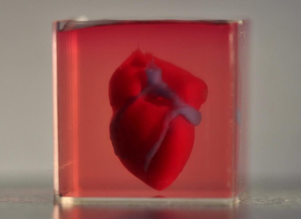
First 3D heart printed using patient’s biological materials

This is very exciting news and it’s likely be at least 10 years before this technology could be made available to the public.

Caption: A 3D-printed, small-scaled human heart engineered from the patient’s own materials and cells. Credit: Advanced Science. © 2019 The Authors.

An April 15, 2019 news item on ScienceDaily makes a remarkable announcement,

In a major medical breakthrough, Tel Aviv University researchers have “printed” the world’s first 3D vascularised engineered heart using a patient’s own cells and biological materials. Their findings were published on April 15 [2019] in a study in Advanced Science.

Until now, scientists in regenerative medicine — a field positioned at the crossroads of biology and technology — have been successful in printing only simple tissues without blood vessels.

“This is the first time anyone anywhere has successfully engineered and printed an entire heart replete with cells, blood vessels, ventricles and chambers,” says Prof. Tal Dvir of TAU’s School of Molecular Cell Biology and Biotechnology, Department of Materials Science and Engineering, Center for Nanoscience and Nanotechnology and Sagol Center for Regenerative Biotechnology, who led the research for the study.

An April 15, 2019 Amricna Friends of Tel Aviv University (TAU) news release (also on EurekAlert), which originated the news item, provides more detail,

Heart disease is the leading cause of death among both men and women in the United States. Heart transplantation is currently the only treatment available to patients with end-stage heart failure. Given the dire shortage of heart donors, the need to develop new approaches to regenerate the diseased heart is urgent.

“This heart is made from human cells and patient-specific biological materials. In our process these materials serve as the bioinks, substances made of sugars and proteins that can be used for 3D printing of complex tissue models,” Prof. Dvir says. “People have managed to 3D-print the structure of a heart in the past, but not with cells or with blood vessels. Our results demonstrate the potential of our approach for engineering personalized tissue and organ replacement in the future.

Research for the study was conducted jointly by Prof. Dvir, Dr. Assaf Shapira of TAU’s Faculty of Life Sciences and Nadav Moor, a doctoral student in Prof. Dvir’s lab.

“At this stage, our 3D heart is small, the size of a rabbit’s heart, [emphasis mine] ” explains Prof. Dvir. “But larger human hearts require the same technology.”

For the research, a biopsy of fatty tissue was taken from patients. The cellular and a-cellular materials of the tissue were then separated. While the cells were reprogrammed to become pluripotent stem cells, the extracellular matrix (ECM), a three-dimensional network of extracellular macromolecules such as collagen and glycoproteins, were processed into a personalized hydrogel that served as the printing “ink.”

After being mixed with the hydrogel, the cells were efficiently differentiated to cardiac or endothelial cells to create patient-specific, immune-compatible cardiac patches with blood vessels and, subsequently, an entire heart.

According to Prof. Dvir, the use of “native” patient-specific materials is crucial to successfully engineering tissues and organs.

“The biocompatibility of engineered materials is crucial to eliminating the risk of implant rejection, which jeopardizes the success of such treatments,” Prof. Dvir says. “Ideally, the biomaterial should possess the same biochemical, mechanical and topographical properties of the patient’s own tissues. Here, we can report a simple approach to 3D-printed thick, vascularized and perfusable cardiac tissues that completely match the immunological, cellular, biochemical and anatomical properties of the patient.”

The researchers are now planning on culturing the printed hearts in the lab and “teaching them to behave” like hearts, Prof. Dvir says. They then plan to transplant the 3D-printed heart in animal models.

“We need to develop the printed heart further,” he concludes. “The cells need to form a pumping ability; they can currently contract, but we need them to work together. Our hope is that we will succeed and prove our method’s efficacy and usefulness.

“Maybe, in ten years, there will be organ printers in the finest hospitals around the world, and these procedures will be conducted routinely.”

Growing the heart to human size and getting the cells to work together so the heart will pump makes it seem like the 10 years Dvir imagines as the future date when there will be organ printers in hospitals routinely printing up hearts seems a bit optimistic. Regardless, I hope he’s right. Bravo to these Israeli researchers!

Here’s a link to and a citation for the paper,

3D Printing of Personalized Thick and Perfusable Cardiac Patches and Hearts by Nadav Noor, Assaf Shapira, Reuven Edri, Idan Gal, Lior Wertheim, Tal Dvir. Advanced Science DOI: https://doi.org/10.1002/advs.201900344 First published: 15 April 2019

This paper is open access.

Canadian researchers develop bone implant material from cellulose nanocrystals (CNC) while Russian scientists restore internal structure of bone with polycaprolactone nanofibers

Two research groups are working to the same end where bone marrow is concerned, encourage bone cell growth, but they are using different strategies.

University of British Columbia and McMaster University (Canada)

Caption: Researchers treated nanocrystals derived from plant cellulose so that they can link up and form a strong but lightweight sponge (an aerogel) that can compress or expand as needed to completely fill out a bone cavity. Credit: Clare Kiernan, UBC

The samples look a little like teeth, don’t they?

Before diving into the research news, there’s a terminology issue that should be noted as you’ll see when you read the news/press releases. Nanocrystal cellulose/nanocrystalline cellulose (NCC) is a term coined by Canadian researchers. Since those early day, most researchers, internationally, have adopted the term cellulose nanocrystals (CNC) as the standard term. It fits better with the naming conventions for other nnanocellulose materials such as cellulose nanofibrils, etc. By the way, a Canadian company (CelluForce) that produces CNC retained the term nanocrystalline cellulose (NCC) as a trademark for the product, CelluForce NCC®.

For anyone not familiar with aerogels, what the University of British Columbia (UBC) and McMaster University researchers are developing, are also popularly known known as ‘frozen smoke’ (see the Aerogel Wikipedia entry for more).

A March 19, 2019 news item on ScienceDaily announces the research,

Researchers from the University of British Columbia and McMaster University have developed what could be the bone implant material of the future: an airy, foamlike substance that can be injected into the body and provide scaffolding for the growth of new bone.

It’s made by treating nanocrystals derived from plant cellulose so that they link up and form a strong but lightweight sponge — technically speaking, an aerogel — that can compress or expand as needed to completely fill out a bone cavity.

A March 19, 2019 UBC news release (also on EurekAlert), which originated the news item, describes the research in more detail,

“Most bone graft or implants are made of hard, brittle ceramic that doesn’t always conform to the shape of the hole, and those gaps can lead to poor growth of the bone and implant failure,” said study author Daniel Osorio, a PhD student in chemical engineering at McMaster. “We created this cellulose nanocrystal aerogel as a more effective alternative to these synthetic materials.”

For their research, the team worked with two groups of rats, with the first group receiving the aerogel implants and the second group receiving none. Results showed that the group with implants saw 33 per cent more bone growth at the three-week mark and 50 per cent more bone growth at the 12-week mark, compared to the controls.

“These findings show, for the first time in a lab setting, that a cellulose nanocrystal aerogel can support new bone growth,” said study co-author Emily Cranston, a professor of wood science and chemical and biological engineering who holds the President’s Excellence Chair in Forest Bio-products at UBC. She added that the implant should break down into non-toxic components in the body as the bone starts to heal.

The innovation can potentially fill a niche in the $2-billion bone graft market in North America, said study co-author Kathryn Grandfield, a professor of materials science and engineering, and biomedical engineering at McMaster who supervised the work.

“We can see this aerogel being used for a number of applications including dental implants and spinal and joint replacement surgeries,” said Grandfield. “And it will be economical because the raw material, the nanocellulose, is already being produced in commercial quantities.”

The researchers say it will be some time before the aerogel makes it out of the lab and into the operating room.

“This summer, we will study the mechanisms between the bone and implant that lead to bone growth,” said Grandfield. “We’ll also look at how the implant degrades using advanced microscopes. After that, more biological testing will be required before it is ready for clinical trials.”

Here’s a link to and a citation for the paper,

Cross-linked cellulose nanocrystal aerogels as viable bone tissue scaffolds by Daniel A. Osorio, Bryan E. J. Lee, Jacek M. Kwiecien, Xiaoyue Wang, Iflah Shahid, Ariana L. Hurley, Emily D. Cranston and Kathryn Grandfield. Acta Biomaterialia Volume 87, 15 March 2019, Pages 152-165 DOI: https://doi.org/10.1016/j.actbio.2019.01.049

This paper is behind a paywall

Now for the Russian team.

National University of Science and Technology “MISIS” (formerly part of the Moscow Mining Academy)

These scientists have adopted a different strategy as you’ll see in the March 19, 2019 news item on Nanwerk, which, coincidentally, was published on the same day as the Canadian research,

Scientists from the National University of Science and Technology “MISIS” developed a nanomaterial, which will be able to rstore the internal structure of bones damaged due to osteoporosis and osteomyelitis. A special bioactive coating of the material helped to increase the rate of division of bone cells by 3 times. In the future, it can allow to abandon bone marrow transplantation and patients will no longer need to wait for suitable donor material.

A March 19, 2019 National University of Science and Technology (MISIS) press release (also on EurekAlert), which originated the news item, provides detail about the impetus for the research and the technique being developed,

Such diseases as osteoporosis and osteomyelitis cause irreversible degenerative changes in the bone structure. Such diseases require serious complex treatment and surgery and transplantation of the destroyed bone marrow in severe stages. Donor material should have a number of compatibility indicators and even close relationship with the donor cannot guarantee full compatibility.

Research group from the National University of Science and Technology “MISIS” (NUST MISIS), led by Anton Manakhov (Laboratory for Inorganic Nanomaterials) developed material that will allow to restore damaged internal bone structure without bone marrow transplantation.
It is based on nanofibers of polycaprolactone, which is biocompatible self-dissolvable material. Earlier, the same research group has already worked with this material: by adding antibiotics to the nanofibers, scientists have managed to create non-changeable healing bandages.

“If we want the implant to take, not only biocompatibility is needed, but also activation of the natural cell growth on the surface of the material. Polycaprolactone as such is a hydrophobic material, meaning, and cells feel uncomfortable on its surface. They gather on the smooth surface and divide extremely slow”, Elizaveta Permyakova, one of the co-authors and researcher at NUST MISIS Laboratory for Inorganic Nanomaterials, explains.

To increase the hydrophilicity of the material, a thin layer of bioactive film consisting of titanium, calcium, phosphorus, carbon, oxygen and nitrogen (TiCaPCON) was deposited on it. The structure of nanofibers identical to the cell surface was preserved. These films, when immersed in a special salt medium, which chemical composition is identical to human blood plasma, are able to form on its surface a special layer of calcium and phosphorus, which in natural conditions forms the main part of the bone. Due to the chemical similarity and the structure of nanofibers, new bone tissue begins to grow rapidly on this layer. Most importantly, polycaprolactone nanofibers dissolve, having fulfilled their functions. Only new “native” tissue remains in the bone.

In the experimental part of the study, the researchers compared the rate of division of osteoblastic bone cells on the surface of the modified and unmodified material. It was found that the modified material TiCaPCON has a high hydrophilicity. In contrast to the unmodified material, the cells on its surface felt clearly more comfortable, and divided three times faster.

According to scientists, such results open up great prospects for further work with modified polycaprolactone nanofibers as an alternative to bone marrow transplantation.

Here’s a link to and a citation for the paper,

Bioactive TiCaPCON-coated PCL nanofibers as a promising material for bone tissue engineering by Anton Manakhov, Elizaveta S. Permyakova, Sergey Ershov, Alexander Sheveyko, Andrey Kovalskii, Josef Polčák, Irina Y. Zhitnyak, Natalia A. Gloushankova, Lenka Zajíčková, Dmitry V. Shtansky. Applied Surface Science Volume 479, 15 June 2019, Pages 796-802 DOI: https://doi.org/10.1016/j.apsusc.2019.02.163

This paper is behind a paywall.

Cooking up a lung one way or the other

I have two stories about lungs and they are entirely different with the older one being a bioengineering story from the US and the more recent one being an artificial tissue story from the University of Toronto and the University of Ottawa (both in Canada).

Lab grown lungs

The Canadian Broadcasting Corporation’s Quirks and Quarks radio programme posted a December 29, 2018 news item (with embedded radio files) about bioengineered lunjgs,

There are two major components to building an organ: the structure and the right cells on that structure. A team led by Dr. Joan Nichols, a Professor of Internal Medicine, Microbiology and Immunology at the University of Texas Medical Branch in Galveston, were able to tackle both parts of the problem

In their experiment they used a donor organ for the structure. They took a lung from an unrelated pig, and stripped it of its cells, leaving a scaffold of collagen, a tough, flexible protein.  This provided a pre-made appropriate structure, though in future they think it may be possible to use 3-D printing technology to get the same result.

They then added cultured cells from the animal who would be receiving the transplant – so the lung was made of the animal’s own cells. Cultured lung and blood vessel cells were placed on the scaffold and it was  placed in a tank for 30 days with a cocktail of nutrients to help the cells stick to the scaffold and proliferate. The result was a kind of baby lung.

They then transplanted the bio-engineered, though immature, lung into the recipient animal where they hoped it would continue to develop and mature – growing to become a healthy, functioning organ.

The recipients of the bio-engineered lungs were four pigs adult pigs, which appeared to tolerate the transplants well. In order to study the development of the bio-engineered lungs, they euthanized the animals at different times: 10 hours, two weeks, one month and two months after transplantation.

They found that as early as two weeks, the bio-engineered lung had integrated into the recipient animals’ body, building a strong network of blood vessels essential for the lung to survive. There was no evidence of pulmonary edema, the build of fluid in the lungs, which is usually a sign of the blood vessels not working efficiently.  There was no sign of rejection of the transplanted organs, and the pigs were healthy up to the point where they were euthanized.

One lingering concern is how well the bio-engineered lungs delivered oxygen. The four pigs who received the trasplant [sic] had one original functioning lung, so they didn’t depend on their new bio-engineered lung for breathing. The scientists were not sure that the bio-engineered lung was mature enough to handle the full load of oxygen on its own.

You can hear Bob McDonald’s (host of Quirks & Quarks, a Canadian Broadcasting Corporation science radio programme) interview lead scientist, Dr. Joan Nichols if you go to here. (Note: I find he overmodulates his voice but some may find he has a ‘friendly’ voice.)

This is an image of the lung scaffold produced by the team,

Lung scaffold in the bioreactor chamber on Day 1 of the experiment, before the cells from the study pig were added. (Credit: Joan Nichols) [downloaded from https://www.cbc.ca/radio/quirks/dec-29-2018-water-on-mars-lab-grown-lungs-and-more-the-biggest-science-stories-of-2018-1.4940811/lab-grown-lungs-are-transplanted-in-pigs-today-they-may-help-humans-tomorrow-1.4940822]

Here’s more technical detail in an August 1, 2018i University of Texas Medical Branch (UTMB) news release (also on EurekAlert), which originally announced the research,

A research team at the University of Texas Medical Branch at Galveston have bioengineered lungs and transplanted them into adult pigs with no medical complication.

In 2014, Joan Nichols and Joaquin Cortiella from The University of Texas Medical Branch at Galveston were the first research team to successfully bioengineer human lungs in a lab. In a paper now available in Science Translational Medicine, they provide details of how their work has progressed from 2014 to the point no complications have occurred in the pigs as part of standard preclinical testing.

“The number of people who have developed severe lung injuries has increased worldwide, while the number of available transplantable organs have decreased,” said Cortiella, professor of pediatric anesthesia. “Our ultimate goal is to eventually provide new options for the many people awaiting a transplant,” said Nichols, professor of internal medicine and associate director of the Galveston National Laboratory at UTMB.

To produce a bioengineered lung, a support scaffold is needed that meets the structural needs of a lung. A support scaffold was created using a lung from an unrelated animal that was treated using a special mixture of sugar and detergent to eliminate all cells and blood in the lung, leaving only the scaffolding proteins or skeleton of the lung behind. This is a lung-shaped scaffold made totally from lung proteins.

The cells used to produce each bioengineered lung came from a single lung removed from each of the study animals. This was the source of the cells used to produce a tissue-matched bioengineered lung for each animal in the study. The lung scaffold was placed into a tank filled with a carefully blended cocktail of nutrients and the animals’ own cells were added to the scaffold following a carefully designed protocol or recipe. The bioengineered lungs were grown in a bioreactor for 30 days prior to transplantation. Animal recipients were survived for 10 hours, two weeks, one month and two months after transplantation, allowing the research team to examine development of the lung tissue following transplantation and how the bioengineered lung would integrate with the body.

All of the pigs that received a bioengineered lung stayed healthy. As early as two weeks post-transplant, the bioengineered lung had established the strong network of blood vessels needed for the lung to survive.

“We saw no signs of pulmonary edema, which is usually a sign of the vasculature not being mature enough,” said Nichols and Cortiella. “The bioengineered lungs continued to develop post-transplant without any infusions of growth factors, the body provided all of the building blocks that the new lungs needed.”

Nichols said that the focus of the study was to learn how well the bioengineered lung adapted and continued to mature within a large, living body. They didn’t evaluate how much the bioengineered lung provided oxygenation to the animal.

“We do know that the animals had 100 percent oxygen saturation, as they had one normal functioning lung,” said Cortiella. “Even after two months, the bioengineered lung was not yet mature enough for us to stop the animal from breathing on the normal lung and switch to just the bioengineered lung.”

For this reason, future studies will look at long-term survival and maturation of the tissues as well as gas exchange capability.

The researchers said that with enough funding, they could grow lungs to transplant into people in compassionate use circumstances within five to 10 years.

“It has taken a lot of heart and 15 years of research to get us this far, our team has done something incredible with a ridiculously small budget and an amazingly dedicated group of people,” Nichols and Cortiella said.

Here’s a citation and another link for the paper,

Production and transplantation of bioengineered lung into a large-animal model by Joan E. Nichols, Saverio La Francesca, Jean A. Niles, Stephanie P. Vega, Lissenya B. Argueta, Luba Frank, David C. Christiani, Richard B. Pyles, Blanca E. Himes, Ruyang Zhang, Su Li, Jason Sakamoto, Jessica Rhudy, Greg Hendricks, Filippo Begarani, Xuewu Liu, Igor Patrikeev, Rahul Pal, Emiliya Usheva, Grace Vargas, Aaron Miller, Lee Woodson, Adam Wacher, Maria Grimaldo, Daniil Weaver, Ron Mlcak, and Joaquin Cortiella. Science Translational Medicine 01 Aug 2018: Vol. 10, Issue 452, eaao3926 DOI: 10.1126/scitranslmed.aao3926

This paper is behind a paywall.

Artificial lung cancer tissue

The research teams at the University of Toronto and the University of Ottawa worked on creating artificial lung tissue but other applications are possible too. First, there’s the announcement in a February 25, 2019 news item on phys.org,

A 3-D hydrogel created by researchers in U of T Engineering Professor Molly Shoichet’s lab is helping University of Ottawa researchers to quickly screen hundreds of potential drugs for their ability to fight highly invasive cancers.

Cell invasion is a critical hallmark of metastatic cancers, such as certain types of lung and brain cancer. Fighting these cancers requires therapies that can both kill cancer cells as well as prevent cell invasion of healthy tissue. Today, most cancer drugs are only screened for their ability to kill cancer cells.

“In highly invasive diseases, there is a crucial need to screen for both of these functions,” says Shoichet. “We now have a way to do this.”

A February 25, 2019 University of Toronto news release (also on EurekAlert), which originated the news item, offers more detail ,

In their latest research, the team used hydrogels to mimic the environment of lung cancer, selectively allowing cancer cells, and not healthy cells, to invade. In their latest research, the team used hydrogels to mimic the environment of lung cancer, selectively allowing cancer cells, and not healthy cells, to invade. This emulated environment enabled their collaborators in Professor Bill Stanford’s lab at University of Ottawa to screen for both cancer-cell growth and invasion. The study, led by Roger Y. Tam, a research associate in Shochet’s lab, was recently published in Advanced Materials.

“We can conduct this in a 384-well plate, which is no bigger than your hand. And with image-analysis software, we can automate this method to enable quick, targeted screenings for hundreds of potential cancer treatments,” says Shoichet.

One example is the researchers’ drug screening for lymphangioleiomyomatosis (LAM), a rare lung disease affecting women. Shoichet and her team were inspired by the work of Green Eggs and LAM, a Toronto-based organization raising awareness of the disease.

Using their hydrogels, they were able to automate and screen more than 800 drugs, thereby uncovering treatments that could target disease growth and invasion.

In the ongoing collaboration, the researchers plan to next screen multiple drugs at different doses to gain greater insight into new treatment methods for LAM. The strategies and insights they gain could also help identify new drugs for other invasive cancers.

Shoichet, who was recently named a Distinguished Woman in Chemistry or Chemical Engineering, also plans to patent the hydrogel technology.

“This has, and continues to be, a great collaboration that is advancing knowledge at the intersection of engineering and biology,” says Shoichet.

I note that Shoichet (pronounced ShoyKet) is getting ready to patent this work. I do have a question about this and it’s not up to Shoichet to answer as she didn’t create the system. Will the taxpayers who funded her work receive any financial benefits should the hydrogel prove to be successful or will we be paying double, both supporting her research and paying for the hydrogel through our healthcare costs?

Getting back to the research, here’s a link to and a citation for the paper,

Rationally Designed 3D Hydrogels Model Invasive Lung Diseases Enabling High‐Content Drug Screening by Roger Y. Tam, Julien Yockell‐Lelièvre, Laura J. Smith, Lisa M. Julian, Alexander E. G. Baker, Chandarong Choey, Mohamed S. Hasim, Jim Dimitroulakos, William L. Stanford, Molly S. Shoichet. Advanced Materials Volume 31, Issue 7 February 15, 2019 1806214 First published online: 27 December 2018 DOI: https://doi.org/10.1002/adma.201806214

This paper is behind a paywall.