Tag Archives: spinal cord implant

Two advances in the field of prosthetic implants

I have a story from New Zealand and another one from Spain.

Rats walk again

A June 28, 2025 news item on ScienceDaily announces spinal cord research from New Zealand,

Spinal cord injuries are currently incurable with devastating effects on people’s lives, but now a trial at Waipapa Taumata Rau, University of Auckland offers hope for an effective treatment.

Spinal cord injuries shatter the signal between the brain and body, often resulting in a loss of function.”Unlike a cut on the skin, which typically heals on its own, the spinal cord does not regenerate effectively, making these injuries devastating and currently incurable,” says lead researcher Dr Bruce Harland, a senior research fellow in the School of Pharmacy at Waipapa Taumata Rau, University of Auckland.

Before birth, and to a lesser extent afterwards, naturally occurring electric fields play a vital role in early nervous system development, encouraging and guiding the growth of nerve tissue along the spinal cord. Scientists are now harnessing this same electrical guidance system in the lab.An implantable electronic device has restored movement following spinal cord injury in an animal study, raising hopes for an effective treatment for humans and even their pets.

A June 27, 2025 University of Auckland press release, which originated the news item, describes the implantable device in more detail, Note: A link has been removed,

“We developed an ultra-thin implant designed to sit directly on the spinal cord, precisely positioned over the injury site in rats,” Dr Harland says.

The device delivers a carefully controlled electrical current across the injury site.

“The aim is to stimulate healing so people can recover functions lost through spinal-cord injury,” Professor Darren Svirskis, director of the CatWalk Cure Programme at the University’s School of Pharmacy says.

Unlike humans, rats have a greater capacity for spontaneous recovery after spinal cord injury, which allowed researchers to compare natural healing with healing supported by electrical stimulation.

After four weeks, animals that received daily electric field treatment showed improved movement compared with those who did not.

Throughout the 12-week study, they responded more quickly to gentle touch.

“This indicates that the treatment supported recovery of both movement and sensation,” Harland says.

“Just as importantly, our analysis confirmed that the treatment did not cause inflammation or other damage to the spinal cord, demonstrating that it was not only effective but also safe.”

This new study, published in a leading journal, has come out of a partnership between the University of Auckland and Chalmers University of Technology in Sweden. See Nature Communications.

“Long term, the goal is to transform this technology into a medical device that could benefit people living with these life-changing spinal-cord injuries,” says Professor Maria Asplund of Chalmers University of Technology.

“This study offers an exciting proof of concept showing that electric field treatment can support recovery after spinal cord injury,” says doctoral student Lukas Matter, also from Chalmers University.

The next step is to explore how different doses, including the strength, frequency, and duration of the treatment, affect recovery, to discover the most effective recipe for spinal-cord repair.

This approach is quite different to that used by the Israeli team featured in my August 22, 2025 posting “Walking again? Israeli team gears up to implant bioengineered spinal cord tissue into paralyzed patient.” It would also appear that at least a few years will pass before the team in New Zealand is ready for human clinical trials.

Here’s a link to and a citation to the New Zealand team’s paper,

Daily electric field treatment improves functional outcomes after thoracic contusion spinal cord injury in rats by Bruce Harland, Lukas Matter, Salvador Lopez, Barbara Fackelmeier, Brittany Hazelgrove, Svenja Meissner, Simon O’Carroll, Brad Raos, Maria Asplund & Darren Svirskis. Nature Communications volume 16, Article number: 5372 (2025) DOI: https://doi.org/10.1038/s41467-025-60332-0 Published: 26 June 2025

Thia paper is open access.

Improving tolerance for prosthetic implants

A June 30, 2025 Universitat Autonoma de Barcelona press release (also on EurekAert) announces development of a new coating for prosthetic devices,

An international research team, including scientists from the Institut de Neurociències at the Universitat Autònoma de Barcelona (UAB), has developed a new solution to reduce the immune response triggered by neural prosthetics used after limb amputations or severe nerve injuries. The approach consists of coating the electronic implants (which connect the prosthetic device to the patient’s nervous system) with a potent anti-inflammatory drug. This coating helps the body better tolerate the implant, improving its long-term performance and stability.

Neural electrode implants are commonly used in prosthetics to restore communication between the device and the nervous system. However, their long-term effectiveness can be compromised by the body’s natural immune reaction to foreign objects, which leads to the formation of scar tissue around the implant and can impair its function.

Now, a recent study published in Advanced Healthcare Materials by researchers from the Universitat Autònoma de Barcelona, the Università di Ferrara, the University of Freiburg, and Chalmers University of Technology, conducted as part of the European collaborative project BioFINE, reports a novel method to improve the biocompatibility and chronic stability of these electrodes.

The technique involves activating and modifying the surface of polyimide (a material commonly used for implanted electrodes) using a chemical strategy that enables the covalent binding of the anti-inflammatory drug dexamethasone. This innovation allows the drug to be released at the implant site slowly over at least two months, a critical period when the immune system typically mounts its strongest response.

Biological tests showed that this approach reduces inflammation-related signals in immune cells, while maintaining the material’s biocompatibility and mechanical integrity. Animal testing further confirmed that the dexamethasone-releasing implants significantly reduce immune reactions and scar tissue formation around the device.

These findings suggest that the slow and localized release of dexamethasone from the implant surface could extend the functional lifespan of neural prostheses, offering a promising step forward in addressing the long-term challenges of implantable neurotechnology.

“This is a main step that has to be complemented by the demonstration in vivo that this coating improves the functional performance of chronically implanted electrodes in the peripheral nerves, for stimulating and recording nerve signals”, says Dr. Xavier Navarro, principal investigator of the UAB team in the BioFINE project.

Here’s a link to and a citation for the paper,

Covalent Binding of Dexamethasone to Polyimide Improves Biocompatibility of Neural Implantable Devices by Giulia Turrin, Jose Crugeiras, Chiara Bisquoli, Davide Barboni, Martina Catani, Bruno Rodríguez-Meana, Rita Boaretto, Michele Albicini, Stefano Caramori, Claudio Trapella, Thomas Stieglitz, Yara Baslan, Hanna Karlsson-Fernberg, Fernanda L. Narvaez-Chicaiza, Edoardo Marchini, Alberto Cavazzini, Ruben López-Vales, Maria Asplund, Xavier Navarro, Stefano Carli. Advanced Healthcare Materials Volume 14, Issue 21 August 19, 2025 2405004 First published online: 17 June 2025 OI: https://doi.org/10.1002/adhm.202405004

This paper is open access.

Walking again? Israeli team gears up to implant bioengineered spinal cord tissue into paralyzed patient

The Israeli team working on this regenerative medicine project has already (in 2022) been successful with mice. Diana Bletter’s August 21, 2025 Times of Israel article, excerpts of which can be found later in this posting, added some details that I appreciated. That said, the press release is quite accessible and informative.

An August 19, 2025 Tel Aviv University (TAU) press release (also on EurekAlert but edited and published on August 20, 2025) describes the upcoming human trial, Note: Links have been removed,

What if we could restore the ability to walk to people paralyzed by injury or illness? This vision is now moving closer to reality. Three years ago, Tel Aviv University researchers succeeded in engineering a human spinal cord in the lab for the first time. Since then, progress has been rapid, with animal trials showing unprecedented success. Now, for the first time, the technology is set to be tested in human patients.

Prof. Tal Dvir, of TAU’s Sagol Center for Regenerative Biotechnology, head of the Nanotechnology Center, and Chief Scientist of the biotech company Matricelf, explains: “The spinal cord is made up of nerve cells that transmit electrical signals from the brain to every part of the body. When the spinal cord is torn due to trauma — from a car accident, a fall, or a battlefield injury — this chain is broken. Think of it like an electrical cable that’s been cut: if the two parts don’t touch, the electrical signal can’t pass. The cable won’t carry electricity, and in the same way, the person can’t transmit the signal beyond the site of the injury.”

This is one of the few injuries in the human body with no natural ability to regenerate. “Neurons are cells that do not divide and do not renew themselves. They are not like skin cells, which can repair themselves after injury. They are more similar to heart cells: once damage occurs, the body cannot restore them,” notes Prof. Dvir.

 Engineering a Personalized Implant

To overcome this challenge, the TAU researchers developed a fully personalized process. Blood cells are taken from the patient and reprogrammed through genetic engineering to behave like embryonic stem cells, capable of becoming any type of cell in the body.

Meanwhile, fat tissue from the same patient is used to extract substances such as collagen and sugars. These are used to produce a unique hydrogel. “The beauty of this gel is that it’s also personalized, just like the cells. We take the cells that we’ve reprogrammed into embryonic-like stem cells, place them inside the gel, and mimic the embryonic development of the spinal cord,” says Prof. Dvir.

The result is a complete three-dimensional implant. “At the end of the process, we don’t just turn the cells into motor neurons — because cells alone won’t help us — but into three-dimensional tissue: neuronal networks of the spinal cord. After about a month, we obtain a 3D implant with many neurons that transmit electrical signals. These 3D tissues are then implanted into the damaged area.”

Visualization of the next stage of the research – human spinal cord implants for treating paralysis (Photo: Sagol Center for Regenerative Biotechnology)

From Animals to Human Patients

The researchers first tested the implant in lab animals. “We showed that we can treat animals with chronic injuries. Not animals that were injured just recently, but those we allowed enough time to pass — like a person more than a year after an injury. More than 80% of the animals regained full walking ability,” Prof. Dvir explains.

Encouraged by these results, the team submitted the findings to Israel’s Ministry of Health. “About six months ago we received preliminary approval to begin compassionate-use trials with eight patients. We decided, of course, that the first patient would be Israeli. This is undoubtedly a matter of national pride. The technology was developed here in Israel, at Tel Aviv University and at Matricelf, and from the very beginning it was clear to us that the first-ever surgery would be performed in Israel, with an Israeli patient.” he says.

Looking Ahead

The first implant in a human patient is expected within about a year. For the initial trials, the team will focus on patients whose paralysis is relatively recent — within about a year of injury. “Once we prove that the treatment works — everything is open, and we’ll be able to treat any injury,” says Prof. Dvir.

Behind the initiative are key figures from both academia and industry. Prof. Dvir founded Matricelf in 2019 together with Dr. Alon Sinai, based on the revolutionary organ engineering technology developed at TAU under a licensing agreement through Ramot, the University’s technology transfer company. The company’s CEO is Gil Hakim, while the scientific development is led by Dr. Tamar Harel-Adar and her team.

“They managed to get us to the stage of regulatory approvals so quickly — and that’s amazing,” says Prof. Dvir.

Gil Hakim, CEO of Matricelf , concludes: “This milestone marks the shift from pioneering research to patient treatment. For the first time, we are translating years of successful preclinical work into a procedure for people living with paralysis. Our approach, using each patient’s own cells to engineer a new spinal cord, eliminates key safety risks and positions Matricelf at the forefront of regenerative medicine. If successful, this therapy has the potential to define a new standard of care in spinal cord repair, addressing a multi-billion-dollar market with no effective solutions today. This first procedure is more than a scientific breakthrough, it is a value-inflection point for Matricelf and a step toward transforming an area of medicine long considered untreatable. We are proud that Israel is leading this global effort and are fully committed to bringing this innovation to patients worldwide.”

Diana Bletter’s August 21, 2025 article for The Times of Israel (h/t August 21, 2025 Google alert) covers much of the same ground as the press release but there are some new details, Note: Links have been removed,

Prof. Tal Dvir, head of the Sagol Center for Regenerative Biotechnology and the Nanotechnology Center at Tel Aviv University, said his research team is now able to engineer a spinal cord that functions exactly like a natural one by implanting 3D-engineered tissue into the damaged area.

Fusion then occurs between the new tissue and the healthy areas above and below the injury that will end the paralysis.

The upcoming spinal cord implant surgery marks the next stage in a process that began about three years ago, when Dvir’s lab at Tel Aviv University succeeded in engineering a personalized 3D spinal cord in the laboratory.

The groundbreaking findings, published in the prestigious journal Advanced Science, demonstrated for the first time ever that mice suffering from chronic paralysis that were treated with these engineered implants started to walk — and even scamper — again.


The success rate with the engineered spinal cord was 80 percent for mice with chronic paralysis. Among those with recent or short-term paralysis, 100% of the mice walked.

Patients remain paralyzed because neurons do not renew

Around the world, there are over 15 million people who have suffered spinal cord injuries. Professionals can help stabilize the injury but not much else.

Dvir said that as a result, the damage only worsens. Over time, the damaged area becomes scar tissue.

“The patient remains paralyzed below the site of injury,” he said. “If the injury is in the neck, all four limbs may be paralyzed. If in the lower back, the legs will not move, and so on.”

Spinal cord injuries are one of the very few injuries in the human body that are not impacted by natural regenerative ability, Dvir explained.

“The neurons do not divide and do not renew themselves,” he said. “These cells are not like skin cells, which can heal after injury, but are more like heart cells: Once damaged, the body cannot repair them.”

“The spinal cord is composed of nerve cells that transmit electrical signals from the brain to all parts of the body,” Dvir said. “The decision is made in the brain, the electrical signal passes through the spinal cord, and from there, neurons activate the muscles throughout the body.”

When the spinal cord is severed due to trauma, such as a car accident, a fall, or a combat injury, this chain is broken.

“Think of an electrical cable that has been cut,” Dvir said. “When the two ends no longer touch, the electrical signal cannot pass. The cable will not transmit electricity, and the person cannot transmit the signal beyond the injury.”

Dvir’s team aims to fix that.

Implanting an engineered human spinal cord

Dvir said that the researchers start the process with a small biopsy from the belly.

They then take these blood cells and perform a process known as reprogramming — genetic engineering that transforms the cells into embryonic stem cell-like cells, capable of developing into any cell type in the body.

In the next step, the scientists take fatty tissue from the patient, extract key components such as collagens and sugars, and build a customized hydrogel. The embryonic stem cell-like cells are placed in this gel, and the embryonic development of a spinal cord is mimicked.

This spinal cord will then be transplanted into the human body, restoring the body’s abilities.

I have a link to Dvir’s company, Matricelf and a link to and a citation to the Dvir team’s 2022 study,

Regenerating the Injured Spinal Cord at the Chronic Phase by Engineered iPSCs-Derived 3D Neuronal Networks by Lior Wertheim, Reuven Edri, Yona Goldshmit, Tomer Kagan, Nadav Noor, Angela Ruban, Assaf Shapira, Irit Gat-Viks, Yaniv Assaf, Tal Dvir. Advanced Science Volume9, Issue 11 April 14, 2022 2105694 DOI: https://doi.org/10.1002/advs.202105694 First published online: 07 February 2022

This paper is open access.

One more note, there is other work devoted to enable paralyzed people to walk again such as the Walk Again Project (Wikipedia entry), Note: Links have been removed,

Walk Again Project is an international, non-profit consortium led by Miguel Nicolelis, created in 2009 in a partnership between Duke University and the IINN/ELS [International Institute for Neurosciences of Natal – Edmond and Lily Safra or Instituto Internacional de Neurociências Edmond e Lily Safra; (INN-ELS)], where researchers come together to find neuro-rehabilitation treatments for spinal cord injuries,[1][2][3] which pioneered the development and use of the brain–machine interface, including its non-invasive version,[4] with an EEG.[5]

My May 15, 2019 posting “Walking again with exoskeletons and brain-controlled, non-invasive muscle stimulation enabling people to walk” features more information about the Walk Again Project (scroll down to the ‘Brazil and Walk Again” subhead and a Canadian project (Note: The CBC has removed access to a video that I’d embedded in the posting.)

I wish all the best for everyone involved in the upcoming human trial.

Asparagus spinal cord?

I love this picture,

Pelling in the kitchen with asparagus, the veggie that inspired his work on spinal cord injuries. Credit: Andrew Pelling?

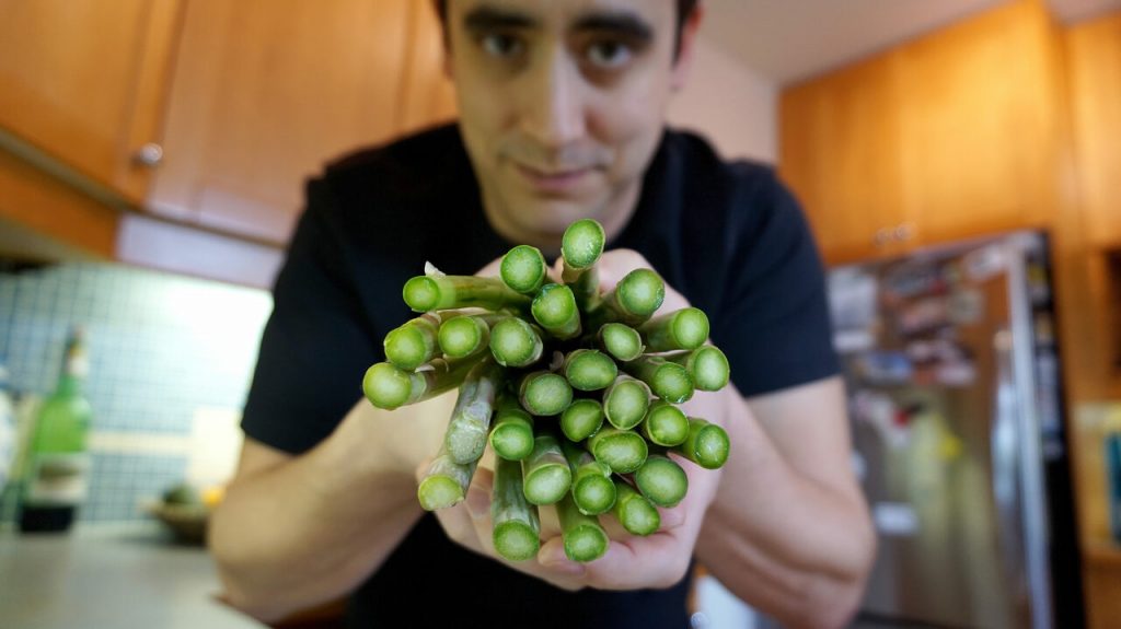
The image accompanies Cari Shane’s August 4, 2021 article for Atlas Obscura’s Gastro Obscura about Andrew Pelling and his asparagus-based scaffolds for spinal cord stem cells (Note: A link has been removed),

Around 10 years ago, Pelling [Dr. Andrew Pelling at the University of Ottawa], a biophysicist, started thinking with his team about materials that could be used to reconstruct damaged or diseased human tissues. Surrounded by a rainbow of fresh fruits and vegetables at his University of Ottawa lab, Pelling and his team dismantle biological systems, mixing and matching parts, and put them back together in new and creative ways. It’s a little bit like a hacker who takes parts from a phone, a computer, and a car to build a robotic arm. Or like Mary Shelley’s Dr. Frankenstein, who built a monster out of cadavers. Except Pelling’s team has turned an apple into an ear and, most recently, a piece of asparagus into a scaffold for spinal-cord implants.

Pelling believes the future of regenerative medicine—which uses external therapies to help the body heal, the same way a cut heals by itself or a broken bone can mend without surgery—is in the supermarket produce aisle. He calls it “augmented biology,” and it’s a lot less expensive—by thousands and thousands of dollars—than implanting organs donated by humans, taken from animals, or manmade or bioengineered from animal tissue.

Decellularization as a process for implantation is fairly new, developed in the mid 1990s primarily by Doris Taylor. By washing out the genetic materials that make an apple an apple, for example, you are left with plant tissue, or a “cellulose mesh,” explains Pelling. “What we’re doing is washing out all the plant DNA, RNA proteins, all that sort of stuff that can cause immune responses, and rejection. And we’re just leaving behind the fiber in a plant—like literally the stuff that gets stuck in your teeth.”

When Pelling noticed the resemblance between a decellularized apple slice and an ear, he saw the true potential of his lab games. If he implanted the apple scaffolding into a living animal, he wondered, would it “be accepted” and vascularize? That is, would the test animal’s body glom onto the plant cells as if they weren’t a dangerous, foreign body and instead send out signals to create a blood supply, allowing the plant tissue to become a living part of the animal’s body? The answer was yes. “Suddenly, and by accident, we developed a material that has huge therapeutic and regenerative potential,” says Pelling. The apple ear does not enable hearing, and it remains in the animal-testing phase, but it may have applications for aesthetic implantation.

Soon after his breakthrough apple experiment, which was published in 2016 and earned him the moniker of “mad scientist,” Pelling shifted his focus to asparagus. The idea hit him when he was cooking. Looking at the end of a spear, he thought, “Hey, it looks like a spinal cord. What the hell? Maybe we can do something,” he says.

… Pelling implanted decellularized asparagus tissue under the skin of a lab rat. In just a few weeks, blood vessels flowed through the asparagus scaffolding; healthy cells from the animal moved into the tissue and turned the scaffold into living tissue. “The surprise here was that the body, instead of rejecting this material, it actually integrated into the material,” says Pelling. In the bioengineering world, getting that to happen has typically been a major challenge.

And then came the biggest surprise of all. Rats with severed spinal cords that had been implanted with the asparagus tissue were able to walk again, just a few weeks after implantation. …

While using asparagus tissue as scaffolding to repair spinal cords is not a “miracle cure,” says Pelling, it’s unlike the kinds of implants that have come before. Donated or manufactured organs are historically both more complicated and more expensive. Pelling’s implants were “done without stem cells or electrical stimulation or exoskeletons, or any of the usual approaches, but rather using very low cost, accessible materials that we honestly just bought at the grocery store,” he says, “and, we achieved the same level of recovery.” (At least in animal tests.) Plus, whereas patients usually need lifelong immunosuppressants, which can have negative side effects, to prevent their body from rejecting an implant, that doesn’t seem necessary with Pelling’s plant-based implants. And, so far, the plant-based implants don’t seem to break down over time like traditional spinal-cord implants. “The inertness of plant tissue is exactly why it’s so biocompatible,” says Pelling.

In October 2020, the asparagus implant was designated as a “breakthrough device” by the FDA [US Food and Drug Administration]. The designation means human trials will be fast-tracked and likely begin in a few years. …

Shane’s August 4, 2021 article is fascinating and well illustrated with a number of embedded images. If you have the time and the inclination, do read it.

More of Pelling’s work can be found here at the Pelling Lab website. He was mentioned (by name only as a participant in the second Canadian DIY Biology Summit organized by the Public Health Agency of Canada [PHAC]) here in an April 21, 2020 posting (my 10 year review of science culture in Canada). You’ll find the Pelling mention under the DIY Biology subhead about 20% of the way down the screen.