Tag Archives: bioengineering

Purifying DNA origami nanostructures with a LEGO robot

This July 20, 2023 article by Bob Yirka for phys.org highlights some frugal science, Note: A link has been removed,

A team of bioengineers at Arizona State University has found a way to use a LEGO robot as a gradient mixer in one part of a process to create DNA origami nanostructures. In their paper published on the open-access site PLOS [Public Library of Science] ONE, the group describes how they made their mixer and its performance.

To create DNA origami structures, purification of DNA [deoxyribonucleic acid] origami nanostructures is required. This is typically done using rate-zone centrifugation, which involves the use of a relatively expensive piece of a machinery, a gradient mixer. In this new effort, the team at ASU has found that it is possible to build such a mixer using off-the-shelf LEGO kits.

I found a video provided by MindSpark Media describing the process on YouTube,

I’d love to know who paid for the video and why. This is pretty slick and it’s not from the Arizona State University’s (ASU) media team.

It gets more interesting on the MindSpark Media About webpage,

MindSpark Media is an independent media unit focusing on all major Media & Marketing services that includes Media Buying and Selling activities, bringing out special features on various supplements/country reports and international features on topics of interest in association with various leading English & Arabic vernaculars in the UAE [United Arab Emirates] and across MENA [Middle East and North Africa].

MindSpark Media is a complete media-selling experience that offers its clientele a wholesome exposure to the best media brands in the country. We also offer an opportunity to meet up and interact with the top brass of the industry & corporates for their advertorial packages including one-to-one interviews with photo-shoot sessions etc.

MindSpark Media delivers client-tailored advertorials that includes their product advertisements, features and interviews published in the form of special reports, supplements & special features, which are released and distributed with top-notch publications in the UAE.

We also focus on advertising activities in the media-buying sector such as Print, Outdoor, TV, Radio and Corporate Video, e-commerce & web-designing for clients in the UAE, MENA and beyond.

Perhaps the researchers are hoping to commercialize the work in some fashion? I couldn’t find any mention of a startup or other commercial entity but it’s a common practice these days in the US and, increasingly, many other countries.

Getting back to the research, here’s a link to and a citation for the paper,

Gradient-mixing LEGO robots for purifying DNA origami nanostructures of multiple components by rate-zonal centrifugation by Jason Sentosa, Franky Djutanta, Brian Horne, Dominic Showkeir, Robert Rezvani, Chloe Leff, Swechchha Pradhan, Rizal F. Hariadi. PLOS ONE (2023). DOI: 10.1371/journal.pone.0283134 Published: July 19, 2023

This paper is open access.

The art and science of architecture that is ‘living-like’

Biology in the service of architecture, from a June 21, 2023 news item on phys.org, Note: Links have been removed,

“This technology is not alive,” says Laia Mogas-Soldevila. “It is living-like.”

The distinction is an important one for the assistant professor at the Stuart Weitzman School of Design [University of Pennsylvania], for reasons both scientific and artistic. With a doctorate in biomedical engineering, several degrees in architecture, and a devotion to sustainable design, Mogas-Soldevila brings biology to everyday life, creating materials for a future built halfway between nature and artifice.

A June 21, 2023 University of Pennsylvania news release (from a Penn Engineering Today blog posting by Devorah Fischler; also on EurekAlert), which originated the news item, provides more details, Note: Links have been removed,

The architectural technology she describes is unassuming at first look: A freeze-dried pellet, small enough to get lost in your pocket. But this tiny lump of matter, the result of more than a year’s collaboration between designers, engineers and biologists, is a biomaterial that contains a “living-like” system.

When touched by water, the pellet activates and expresses a glowing protein, its fluorescence demonstrating that life and art can harmonize into a third and very different thing, as ready to please as to protect. Woven into lattices made of flexible natural materials promoting air and moisture flow, the pellets form striking interior design elements that could one day keep us healthy.

“We envision them as sensors,” explains Mogas-Soldevila. “They may detect pathogens, such as bacteria or viruses, or alert people to toxins inside their home. The pellets are designed to interact with air. With development, they could monitor or even clean it.”

For now, they glow, a triumphant first stop on the team’s roadmap to the future. The fluorescence establishes that the lab’s biomaterial manufacturing process is compatible with the leading-edge cell-free engineering that gives the pellets their life-like properties.

A rapidly expanding technology, cell-free protein expression systems allow researchers to manufacture proteins without the use of living cells.

Gabrielle Ho, Ph.D. candidate in the Department of Bioengineering and co-leader of the project, explains how the team’s design work came to be cell-free, a technique rarely explored outside of lab study or medical applications.

“Typically, we’d use living E. coli cells to make a protein,” says Ho. “E. coli is a biological workhorse, accessible and very productive. We’d introduce DNA to the cell to encourage expression of specific proteins. But this traditional method was not an option for this project. You can’t have engineered E. coli hanging on your walls.”

Cell-free systems contain all the components a living cell requires to manufacture protein —energy, enzymes and amino acids — and not much else. These systems are therefore not alive. They do not replicate, and neither can they cause infection. They are “living-like,” designed to take in DNA and push out protein in ways that previously were only possible using living cells.

“One of the nicest things about these materials not being alive,” says Mogas-Soldevila, “is that we don’t need to worry about keeping them that way.”

Unlike living cells, cell-free materials don’t need a wet environment or constant monitoring in a lab. The team’s research has established a process for making these dry pellets that preserves bioactivity throughout manufacturing, storage and use.

Bioactive, expressive and programmable, this technology is designed to capitalize on the unique properties of organic materials.

Mogas-Soldevila, whose lab focuses exclusively on biodegradable architecture, understands the value of biomaterials as both environmentally responsible and aesthetically rich.

“Architects are coming to the realization that conventional materials — concrete, steel, glass, ceramic, etc. — are environmentally damaging and they are becoming more and more interested in alternatives to replace at least some of them. Because we use so much, even being able to replace a small percentage would result in a significant reduction in waste and pollution.”

Her lab’s signature materials — biopolymers made from shrimp shells, wood pulp, sand and soil, silk cocoons, and algae gums — lend qualities over and above their sustainable advantages.

“My obsession is diagnostic, but my passion is playfulness,” says Mogas-Soldevila. “Biomaterials are the only materials that can encapsulate this double function observed in nature.”

This multivalent approach benefited from the help of Penn Engineering’s George H. Stephenson Foundation Educational Laboratory & Bio-MakerSpace, and the support of its director, Sevile Mannickarottu. In addition to contributing essential equipment and research infrastructure to the team, Mannickarottu was instrumental in enabling the interdisciplinary relationships that led the team to success, introducing Ho to the DumoLab Research team collaborators. These include Mogas-Soldevila, Camila Irabien, a Penn Biology major who provided crucial contributions to experimental work, and Fulbright design fellow Vlasta Kubušová, who co-led the project during her time at Penn and who will continue fueling the project’s next steps.

The cell-free manufacturing and design research required unique dialogues between science and art, categories that Ho believed to be entirely separate before embarking on this project.

“I learned so much from the approach the designers brought to the lab,” says Ho. “Usually, in science, we have a specific problem or hypothesis that we systematically work towards.”

But in this collaboration, things were different. Open-ended. The team sought a living-like platform that does sensing and tells people about interactive matter. They needed to explore, step by step, how to get there.

“Design is only limited by imagination. We sought a technology that could help build towards a vision, and that turned out to be cell-free” says Ho.

“For my part,” says Mogas-Soldevila, “it was inspiring to witness the rigor and attention to constraints that bioengineering brings.”

The constraints were many — machine constraints, biological constraints, financial constraints and space constraints.

“But as we kept these restrictions in play,” she continues, “we asked our most pressing creative questions. Can materials warn us of invisible threats? How will humans react to these bioactive sites? Will they be beautiful? Will they be weird? Most importantly, will they enable a new aesthetic relationship with the potential of bio-based and bioactive matter?”

Down the line, the cell-free pellets and biopolymer lattices could drape protectively over our interior lives, caring for our mental and physical health. For now, research is ongoing, the poetry of design energized by constraint, the constraint of engineering energized by poetry. [emphases mine]

The “poetry of design” and “engineering energized by poetry,” eh? (I have a few comments about science, in my September 11, 2023 posting; scroll down to the ‘Poetry and physics’ subhead.)

Back on topic, here’s a link to and a citation for the paper,

Multiscale design of cell-free biologically active architectural structures by G. Ho, V. Kubušová, C. Irabien, V. Li, A. Weinstein, Sh. Chawla, D. Yeung, A. Mershin, K. Zolotovsky, L. Mogas-Soldevila. Front. Bioeng. Biotechnol., 28 March 2023 Volume 11 – 2023 DOI: https://doi.org/10.3389/fbioe.2023.1125156

This paper appears to be open access.

Inspired by Picasso (or Schumpeter, Shiva, and others?), Université de Montréal researchers employ creative destruction to create new nanomachines

I associate the idea of ‘creative destruction’ with economics and Joseph Schumpeter but it is more widespread and has a much longer history (see more at the end of this posting).

Here we have Université de Montréal researchers being inspired by the idea from (what was to me) an unexpected source, from a February 9, 2023 news item on Nanowerk,

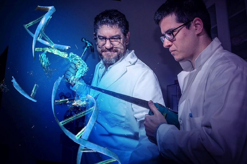
“Every act of creation,” Picasso famously noted, “is first an act of destruction.”

Taking this concept literally, researchers in Canada have now discovered that “breaking” molecular nanomachines basic to life can create new ones that work even better.

I love this image. Bravo!

Researchers Dominic Lauzon and Alexis Vallée-Bélisle Credit: Amélie Philibert & Benoit Gougeon | Université de Montréal

A February 9, 2023 Université de Montréal news release, which originated the news item, delves further into this act of creative destruction,

Evolved over millions of years

Life on Earth is made possible by tens of thousands of nanomachines that have evolved over millions of years. Often made of proteins or nucleic acids, they typically contain thousands of atoms and are less than 10,000 times the size of a human hair.

“These nanomachines control all molecular activities in our body, and problems with their regulation or structure are at the origin of most human diseases,” said the new study’s principal investigator Alexis Vallée-Bélisle, a chemistry professor at Université de Montréal.

Studying the way these nanomachines are built, Vallée-Bélisle, holder of the Canada Research Chair in Bioengineering and Bio-Nanotechnology, noticed that while some are made using a single component or part (often long biopolymers), others use several components that spontaneously assemble.

“Since most of my students spend their lives creating nanomachines, we started to wonder if it is more beneficial to create them using one or more self-assembling molecular components,” said Vallée-Bélisle.

A ‘destructive’ idea

To explore this question, his doctoral student Dominic Lauzon, had the “destructive” idea of breaking up some nanomachines to see if they could be reassembled. To do so, he made artificial DNA-based nanomachines that could be “destroyed” by breaking them up.

“DNA is a remarkable molecule that offers simple, programmable and easy-to-use chemistry,” said Lauzon, the study’s first author. “We believed that DNA-based nanomachines could help answer fundamental questions about the creation and evolution of natural and human-made nanomachines.”

Lauzon and Vallée-Bélisle spent years performing the experimental validations. They were able to demonstrate that nanomachines could easily withstand fragmentation, but more importantly, that such a destructive event allowed for the creation of various novel functionalities, including different sensitivity levels towards variation in component concentration, temperature and mutations.

What the researchers found is that these functionalities could arise simply by controlling the concentration of each individual component. For example, when cutting a nanomachine in three components, nanomachines were found to activate more sensitively at high concentration of components. In contrast, at low concentration of components, nanomachines could be programmed to activate or deactivate at specific moment in time or to simply inhibit their function.

“Overall, these novel functionalities were created  by simply cutting up, or destroying, the structure of an existing nanomachine,” said Lauzon. “These functionalities could drastically improve human-based nanotechnologies such as sensors, drug carriers and even molecular computers”.

Evolving new functionalities

Just as Picasso typically destroyed dozens of unfinished works to create his famous artworks, and just like muscles need to break down to get stronger, and innovative new companies are born by eliminating older competitors from the market, nanoscale machines can evolve new functionalities by being taken apart.

Unlike common machines like cell phones, televisions and cars, which are made by combining components using screws and bolts, glue, solder or electronics, “nanomachines rely on thousands of weak dynamic intermolecular forces that can spontaneously reform, enabling broken nanomachines to re-assemble,” said Vallée-Bélisle.

In addition to providing nanotechnology researchers with a simple design strategy to create the next generation of nanomachines, the UdeM team’s findings also shed light on how natural molecular nanomachines may have evolved.

“Biologists have recently discovered that about 20 per cent of biological nanomachines may have evolved through the fragmentation of their genes,” said Vallée-Bélisle. “With our results, biologists now have a rational basis for understanding how the fragmentation of these ancestral proteins could have created new molecular functionalities for life on Earth.”

Here’s a link to and a citation for the paper,

Functional advantages of building nanosystems using multiple molecular components by D. Lauzon & A. Vallée-Bélisle. Nature Chemistry volume 15, pages 458–467 (2023) DOI: https://doi.org/10.1038/s41557-022-01127-4 Published online: 09 February 2023 Issue Date: April 2023

This paper is behind a paywall.

Creative destruction

The Wikipedia entry for ‘Creative destruction’ is primarily on economic theory and various philosophies with no mention of Picasso. However, there is a fascinating segue into Eastern mysticism,

Other early usage

Hugo Reinert has argued that Sombart’s formulation of the concept was influenced by Eastern mysticism, specifically the image of the Hindu god Shiva, who is presented in the paradoxical aspect of simultaneous destroyer and creator.

On that note, have a lovely weekend.

Asparagus spinal cord?

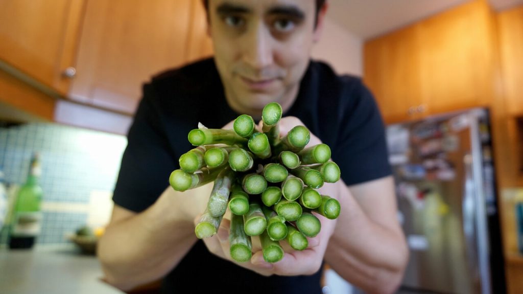
I love this picture,

Pelling in the kitchen with asparagus, the veggie that inspired his work on spinal cord injuries. Credit: Andrew Pelling?

The image accompanies Cari Shane’s August 4, 2021 article for Atlas Obscura’s Gastro Obscura about Andrew Pelling and his asparagus-based scaffolds for spinal cord stem cells (Note: A link has been removed),

Around 10 years ago, Pelling [Dr. Andrew Pelling at the University of Ottawa], a biophysicist, started thinking with his team about materials that could be used to reconstruct damaged or diseased human tissues. Surrounded by a rainbow of fresh fruits and vegetables at his University of Ottawa lab, Pelling and his team dismantle biological systems, mixing and matching parts, and put them back together in new and creative ways. It’s a little bit like a hacker who takes parts from a phone, a computer, and a car to build a robotic arm. Or like Mary Shelley’s Dr. Frankenstein, who built a monster out of cadavers. Except Pelling’s team has turned an apple into an ear and, most recently, a piece of asparagus into a scaffold for spinal-cord implants.

Pelling believes the future of regenerative medicine—which uses external therapies to help the body heal, the same way a cut heals by itself or a broken bone can mend without surgery—is in the supermarket produce aisle. He calls it “augmented biology,” and it’s a lot less expensive—by thousands and thousands of dollars—than implanting organs donated by humans, taken from animals, or manmade or bioengineered from animal tissue.

Decellularization as a process for implantation is fairly new, developed in the mid 1990s primarily by Doris Taylor. By washing out the genetic materials that make an apple an apple, for example, you are left with plant tissue, or a “cellulose mesh,” explains Pelling. “What we’re doing is washing out all the plant DNA, RNA proteins, all that sort of stuff that can cause immune responses, and rejection. And we’re just leaving behind the fiber in a plant—like literally the stuff that gets stuck in your teeth.”

When Pelling noticed the resemblance between a decellularized apple slice and an ear, he saw the true potential of his lab games. If he implanted the apple scaffolding into a living animal, he wondered, would it “be accepted” and vascularize? That is, would the test animal’s body glom onto the plant cells as if they weren’t a dangerous, foreign body and instead send out signals to create a blood supply, allowing the plant tissue to become a living part of the animal’s body? The answer was yes. “Suddenly, and by accident, we developed a material that has huge therapeutic and regenerative potential,” says Pelling. The apple ear does not enable hearing, and it remains in the animal-testing phase, but it may have applications for aesthetic implantation.

Soon after his breakthrough apple experiment, which was published in 2016 and earned him the moniker of “mad scientist,” Pelling shifted his focus to asparagus. The idea hit him when he was cooking. Looking at the end of a spear, he thought, “Hey, it looks like a spinal cord. What the hell? Maybe we can do something,” he says.

… Pelling implanted decellularized asparagus tissue under the skin of a lab rat. In just a few weeks, blood vessels flowed through the asparagus scaffolding; healthy cells from the animal moved into the tissue and turned the scaffold into living tissue. “The surprise here was that the body, instead of rejecting this material, it actually integrated into the material,” says Pelling. In the bioengineering world, getting that to happen has typically been a major challenge.

And then came the biggest surprise of all. Rats with severed spinal cords that had been implanted with the asparagus tissue were able to walk again, just a few weeks after implantation. …

While using asparagus tissue as scaffolding to repair spinal cords is not a “miracle cure,” says Pelling, it’s unlike the kinds of implants that have come before. Donated or manufactured organs are historically both more complicated and more expensive. Pelling’s implants were “done without stem cells or electrical stimulation or exoskeletons, or any of the usual approaches, but rather using very low cost, accessible materials that we honestly just bought at the grocery store,” he says, “and, we achieved the same level of recovery.” (At least in animal tests.) Plus, whereas patients usually need lifelong immunosuppressants, which can have negative side effects, to prevent their body from rejecting an implant, that doesn’t seem necessary with Pelling’s plant-based implants. And, so far, the plant-based implants don’t seem to break down over time like traditional spinal-cord implants. “The inertness of plant tissue is exactly why it’s so biocompatible,” says Pelling.

In October 2020, the asparagus implant was designated as a “breakthrough device” by the FDA [US Food and Drug Administration]. The designation means human trials will be fast-tracked and likely begin in a few years. …

Shane’s August 4, 2021 article is fascinating and well illustrated with a number of embedded images. If you have the time and the inclination, do read it.

More of Pelling’s work can be found here at the Pelling Lab website. He was mentioned (by name only as a participant in the second Canadian DIY Biology Summit organized by the Public Health Agency of Canada [PHAC]) here in an April 21, 2020 posting (my 10 year review of science culture in Canada). You’ll find the Pelling mention under the DIY Biology subhead about 20% of the way down the screen.

‘What becomes of the broken-hearted?’ Trinity College Dublin scientists may have an answer

While Valentine’s Day as celebrated here in Canada and elsewhere (but not everywhere) on February 14 of each year is usually marked in a purely joyous fashion,I’m going to focus on heartbreak. Here is one of the greatest versions of ‘What becomes of the broken-hearted?’ Then, repair follows in the context of some cardiac research coming out of Ireland,

Thank you Joan Osborne and the Funk Brothers. If you haven’t seen ‘Standing in the shadows of Motown’, you may want to make a point of it.

As for the musical question in the headline, researchers at Trinity College Dublin may have an answer of sorts. A February 13, 2020 Trinity College Dublin press release (also on EurekAlert) describes how broken hearts can be mended,

Bioengineers from Trinity College Dublin, Ireland, have developed a prototype patch that does the same job as crucial aspects of heart tissue.

Their patch withstands the mechanical demands and mimics the electrical signalling properties that allow our hearts to pump blood rhythmically round our bodies.

Their work essentially takes us one step closer to a functional design that could mend a broken heart.

One in six men and one in seven women in the EU will suffer a heart attack at some point in their lives. Worldwide, heart disease kills more women and men – regardless of race, than any other disease.

Cardiac patches lined with heart cells can be applied surgically to restore heart tissue in patients who have had damaged tissue removed after a heart attack and to repair congenital heart defects in infants and children. Ultimately, though, the goal is to create cell-free patches that can restore the synchronous beating of the heart cells, without impairing the heart muscle movement.

The bioengineers report their work, which takes us one step closer to such a reality, in the journal Advanced Functional Materials.

Michael Monaghan, ussher assistant professor in biomedical engineering at Trinity, and senior author on the paper, said:

“Despite some advances in the field, heart disease still places a huge burden on our healthcare systems and the life quality of patients worldwide. It affects all of us either directly or indirectly through family and friends. As a result, researchers are continuously looking to develop new treatments which can include stem cell treatments, biomaterial gel injections and assistive devices.”

“Ours is one of few studies that looks at a traditional material, and through effective design allows us to mimic the direction-dependent mechanical movement of the heart, which can be sustained repeatably. This was achieved through a novel method called ‘melt electrowriting’ and through close collaboration with the suppliers located nationally we were able to customise the process to fit our design needs.”

This work was performed in the Trinity Centre for Biomedical Engineering, based in the Trinity Biomedical Sciences Institute in collaboration with Spraybase®, a subsidiary of Avectas Ltd. It was funded by Enterprise Ireland through the Innovation Partnership Program (IPP).

Dr Gillian Hendy, director of Spraybase® is a co-author on the paper. Dr Hendy commended the team at Trinity on the work completed and advancements made on the Spraybase® Melt Electrowriting (MEW) System. The success achieved by the team highlights the potential applications of this novel technology in the cardiac field and succinctly captures the benefits of industry and academic collaboration, through platforms such as the IPP.

Engineering replacement materials for heart tissue is challenging since it is an organ that is constantly moving and contracting. The mechanical demands of heart muscle (myocardium) cannot be met using polyester-based thermoplastic polymers, which are predominantly the approved options for biomedical applications.

However, the functionality of thermoplastic polymers could be leveraged by its structural geometry. The bioengineers then set about making a patch that could control the expansion of a material in multiple directions and tune this using an engineering design approach.

The patches were manufactured via melt electrowriting – a core technology of Spraybase® – which is reproducible, accurate, and scalable. The patches were also coated with the electroconductive polymer polypyrrole to provide electrical conductivity while maintaining cell compatibility.

The patch withstood repeated stretching, which is a dominant concern for cardiac biomaterials, and showed good elasticity, to accurately mimic that key property of heart muscle.

Professor Monaghan added:

“Essentially, our material addresses a lot of requirements. The bulk material is currently approved for medical device use, the design accommodates the movement of the pumping heart, and has been functionalised to accommodate signaling between isolated contractile tissues.”

“This study currently reports the development of our method and design, but we are now looking forward to furthering the next generation of designs and materials with the eventual aim of applying this patch as a therapy for a heart attack.”

Dr Dinorath Olvera, Trinity, first author on the paper, added:

“Our electroconductive patches support electrical conduction between biological tissue in an ex vivo model. These results therefore represent a significant step towards generating a bioengineered patch capable of recapitulating aspects of heart tissue – namely its mechanical movement and electrical signalling.”

Here’s a link to and a citation for the paper,

Electroconductive Melt Electrowritten Patches Matching the Mechanical Anisotropy of Human Myocardium by Dinorath Olvera, Mina Sohrabi Molina, Gillian Hendy, Michael G. Monaghan. Advanced Functional Materials DOI: https://doi.org/10.1002/adfm.201909880 First published: 12 February 2020

This paper is behind a paywall.

Here are links, should you be interested in the company partnering with the researchers, Spraybase®, or its parent company, Avectas Ltd.

Finally, the singer who made ‘What becomes of the broken-hearted?’ a hit in 1965 was Jimmy Ruffin,

Enjoy.

Xenotransplantation—organs for transplantation in human patients—it’s a business and a science

The last time (June 18, 2018 post) I mentioned xenotransplantation (transplanting organs from one species into another species; see more here), it was in the context of an art/sci (or sciart) event coming to Vancouver (Canada).,
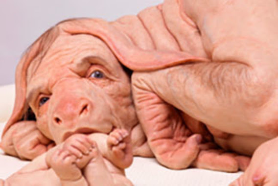
Patricia Piccinini’s Curious Imaginings Courtesy: Vancouver Biennale [downloaded from http://dailyhive.com/vancouver/vancouver-biennale-unsual-public-art-2018/]

The latest edition of the Vancouver Biennale was featured in a June 6, 2018 news item on the Daily Hive (Vancouver),

Melbourne artist Patricia Piccinini’s Curious Imaginings is expected to be one of the most talked about installations of the exhibit. Her style of “oddly captivating, somewhat grotesque, human-animal hybrid creature” is meant to be shocking and thought-provoking.

Piccinini’s interactive [emphasis mine] experience will “challenge us to explore the social impacts of emerging biotechnology and our ethical limits in an age where genetic engineering and digital technologies are already pushing the boundaries of humanity.”

Piccinini’s work will be displayed in the 105-year-old Patricia Hotel in Vancouver’s Strathcona neighbourhood. The 90-day ticketed exhibition [emphasis mine] is scheduled to open this September [2018].

(The show opens on Sept. 14, 2018.)

At the time, I had yet to stumble across Ingfei Chen’s thoughtful dive into the topic in her May 9, 2018 article for Slate.com,

In the United States, the clock is ticking for more than 114,700 adults and children waiting for a donated kidney or other lifesaving organ, and each day, nearly 20 of them die. Researchers are devising a new way to grow human organs inside other animals, but the method raises potentially thorny ethical issues. Other conceivable futuristic techniques sound like dystopian science fiction. As we envision an era of regenerative medicine decades from now, how far is society willing to go to solve the organ shortage crisis?

I found myself pondering this question after a discussion about the promises of stem cell technologies veered from the intriguing into the bizarre. I was interviewing bioengineer Zev Gartner, co-director and research coordinator of the Center for Cellular Construction at the University of California, San Francisco, about so-called organoids, tiny clumps of organlike tissue that can self-assemble from human stem cells in a Petri dish. These tissue bits are lending new insights into how our organs form and diseases take root. Some researchers even hope they can nurture organoids into full-size human kidneys, pancreases, and other organs for transplantation.

Certain organoid experiments have recently set off alarm bells, but when I asked Gartner about it, his radar for moral concerns was focused elsewhere. For him, the “really, really thought-provoking” scenarios involve other emerging stem cell–based techniques for engineering replacement organs for people, he told me. “Like blastocyst complementation,” he said.

Never heard of it? Neither had I. Turns out it’s a powerful new genetic engineering trick that researchers hope to use for growing human organs inside pigs or sheep—organs that could be genetically personalized for transplant patients, in theory avoiding immune-system rejection problems. The science still has many years to go, but if it pans out, it could be one solution to the organ shortage crisis. However, the prospect of creating hybrid animals with human parts and killing them to harvest organs has already raised a slew of ethical questions. In 2015, the National Institutes of Health placed a moratorium on federal funding of this nascent research area while it evaluated and discussed the issues.

As Gartner sees it, the debate over blastocyst complementation research—work that he finds promising—is just one of many conversations that society needs to have about the ethical and social costs and benefits of future technologies for making lifesaving transplant organs. “There’s all these weird ways that we could go about doing this,” he said, with a spectrum of imaginable approaches that includes organoids, interspecies organ farming, and building organs from scratch using 3D bioprinters. But even if it turns out we can produce human organs in these novel ways, the bigger issue, in each technological instance, may be whether we should.

Gartner crystallized things with a downright creepy example: “We know that the best bioreactor for tissues and organs for humans are human beings,” he said. Hypothetically, “the best way to get you a new heart would be to clone you, grow up a copy of yourself, and take the heart out.” [emphasis mine] Scientists could probably produce a cloned person with the technologies we already have, if money and ethics were of no concern. “But we don’t want to go there, right?” he added in the next breath. “The ethics involved in doing it are not compatible with who we want to be as a society.”

This sounds like Gartner may have been reading some science fiction, specifically, Lois McMaster Bujold and her Barrayar series where she often explored the ethics and possibilities of bioengineering. At this point, some of her work seems eerily prescient.

As for Chen’s article, I strongly encourage you to read it in its entirety if you have the time.

Medicine, healing, and big money

At about the same time, there was a May 31, 2018 news item on phys.org offering a perspective from some of the leaders in the science and the business (Note: Links have been removed),

Over the past few years, researchers led by George Church have made important strides toward engineering the genomes of pigs to make their cells compatible with the human body. So many think that it’s possible that, with the help of CRISPR technology, a healthy heart for a patient in desperate need might one day come from a pig.

“It’s relatively feasible to change one gene in a pig, but to change many dozens—which is quite clear is the minimum here—benefits from CRISPR,” an acronym for clustered regularly interspaced short palindromic repeats, said Church, the Robert Winthrop Professor of Genetics at Harvard Medical School (HMS) and a core faculty member of Harvard’s Wyss Institute for Biologically Inspired Engineering. Xenotransplantation is “one of few” big challenges (along with gene drives and de-extinction, he said) “that really requires the ‘oomph’ of CRISPR.”

To facilitate the development of safe and effective cells, tissues, and organs for future medical transplantation into human patients, Harvard’s Office of Technology Development has granted a technology license to the Cambridge biotech startup eGenesis.

Co-founded by Church and former HMS doctoral student Luhan Yang in 2015, eGenesis announced last year that it had raised $38 million to advance its research and development work. At least eight former members of the Church lab—interns, doctoral students, postdocs, and visiting researchers—have continued their scientific careers as employees there.

“The Church Lab is well known for its relentless pursuit of scientific achievements so ambitious they seem improbable—and, indeed, [for] its track record of success,” said Isaac Kohlberg, Harvard’s chief technology development officer and senior associate provost. “George deserves recognition too for his ability to inspire passion and cultivate a strong entrepreneurial drive among his talented research team.”

The license from Harvard OTD covers a powerful set of genome-engineering technologies developed at HMS and the Wyss Institute, including access to foundational intellectual property relating to the Church Lab’s 2012 breakthrough use of CRISPR, led by Yang and Prashant Mali, to edit the genome of human cells. Subsequent innovations that enabled efficient and accurate editing of numerous genes simultaneously are also included. The license is exclusive to eGenesis but limited to the field of xenotransplantation.

A May 30, 2018 Harvard University news release by Caroline Petty, which originated the news item, explores some of the issues associated with incubating humans organs in other species,

The prospect of using living, nonhuman organs, and concerns over the infectiousness of pathogens either present in the tissues or possibly formed in combination with human genetic material, have prompted the Food and Drug Administration to issue detailed guidance on xenotransplantation research and development since the mid-1990s. In pigs, a primary concern has been that porcine endogenous retroviruses (PERVs), strands of potentially pathogenic DNA in the animals’ genomes, might infect human patients and eventually cause disease. [emphases mine]

That’s where the Church lab’s CRISPR expertise has enabled significant advances. In 2015, the lab published important results in the journal Science, successfully demonstrating the use of genome engineering to eliminate all 62 PERVs in porcine cells. Science later called it “the most widespread CRISPR editing feat to date.”

In 2017, with collaborators at Harvard, other universities, and eGenesis, Church and Yang went further. Publishing again in Science, they first confirmed earlier researchers’ fears: Porcine cells can, in fact, transmit PERVs into human cells, and those human cells can pass them on to other, unexposed human cells. (It is still unknown under what circumstances those PERVs might cause disease.) In the same paper, they corrected the problem, announcing the embryogenesis and birth of 37 PERV-free pigs. [Note: My July 17, 2018 post features research which suggests CRISPR-Cas9 gene editing may cause greater genetic damage than had been thought.]

“Taken together, those innovations were stunning,” said Vivian Berlin, director of business development in OTD, who manages the commercialization strategy for much of Harvard’s intellectual property in the life sciences. “That was the foundation they needed, to convince both the scientific community and the investment community that xenotransplantation might become a reality.”

“After hundreds of tests, this was a critical milestone for eGenesis — and the entire field — and represented a key step toward safe organ transplantation from pigs,” said Julie Sunderland, interim CEO of eGenesis. “Building on this study, we hope to continue to advance the science and potential of making xenotransplantation a safe and routine medical procedure.”

Genetic engineering may undercut human diseases, but also could help restore extinct species, researcher says. [Shades of the Jurassic Park movies!]

It’s not, however, the end of the story: An immunological challenge remains, which eGenesis will need to address. The potential for a patient’s body to outright reject transplanted tissue has stymied many previous attempts at xenotransplantation. Church said numerous genetic changes must be achieved to make porcine organs fully compatible with human patients. Among these are edits to several immune functions, coagulation functions, complements, and sugars, as well as the PERVs.

“Trying the straight transplant failed almost immediately, within hours, because there’s a huge mismatch in the carbohydrates on the surface of the cells, in particular alpha-1-3-galactose, and so that was a showstopper,” Church explained. “When you delete that gene, which you can do with conventional methods, you still get pretty fast rejection, because there are a lot of other aspects that are incompatible. You have to take care of each of them, and not all of them are just about removing things — some of them you have to humanize. There’s a great deal of subtlety involved so that you get normal pig embryogenesis but not rejection.

“Putting it all together into one package is challenging,” he concluded.

In short, it’s the next big challenge for CRISPR.

Not unexpectedly, there is no mention of the CRISPR patent fight between Harvard/MIT’s (Massachusetts Institute of Technology) Broad Institute and the University of California at Berkeley (UC Berkeley). My March 15, 2017 posting featured an outcome where the Broad Institute won the first round of the fight. As I recall, it was a decision based on the principles associated with King Solomon, i.e., the US Patent Office, divided the baby and UCBerkeley got the less important part of the baby. As you might expect the decision has been appealed. In an April 30, 2018 piece, Scientific American reprinted an article about the latest round in the fight written by Sharon Begley for STAT (Note: Links have been removed),

All You Need to Know for Round 2 of the CRISPR Patent Fight

It’s baaaaack, that reputation-shredding, stock-moving fight to the death over key CRISPR patents. On Monday morning in Washington, D.C., the U.S. Court of Appeals for the Federal Circuit will hear oral arguments in University of California v. Broad Institute. Questions?

How did we get here? The patent office ruled in February 2017 that the Broad’s 2014 CRISPR patent on using CRISPR-Cas9 to edit genomes, based on discoveries by Feng Zhang, did not “interfere” with a patent application by UC based on the work of UC Berkeley’s Jennifer Doudna. In plain English, that meant the Broad’s patent, on using CRISPR-Cas9 to edit genomes in eukaryotic cells (all animals and plants, but not bacteria), was different from UC’s, which described Doudna’s experiments using CRISPR-Cas9 to edit DNA in a test tube—and it was therefore valid. The Patent Trial and Appeal Board concluded that when Zhang got CRISPR-Cas9 to work in human and mouse cells in 2012, it was not an obvious extension of Doudna’s earlier research, and that he had no “reasonable expectation of success.” UC appealed, and here we are.

For anyone who may not realize what the stakes are for these institutions, Linda Williams in a March 16, 1999 article for the LA Times had this to say about universities, patents, and money,

The University of Florida made about $2 million last year in royalties on a patent for Gatorade Thirst Quencher, a sports drink that generates some $500 million to $600 million a year in revenue for Quaker Oats Co.

The payments place the university among the top five in the nation in income from patent royalties.

Oh, but if some people on the Gainesville, Fla., campus could just turn back the clock. “If we had done Gatorade right, we would be getting $5 or $6 million (a year),” laments Donald Price, director of the university’s office of corporate programs. “It is a classic example of how not to handle a patent idea,” he added.

Gatorade was developed in 1965 when many universities were ill equipped to judge the commercial potential of ideas emerging from their research labs. Officials blew the university’s chance to control the Gatorade royalties when they declined to develop a professor’s idea.

The Gatorade story does not stop there and, even though it’s almost 20 years old, this article stands the test of time. I strongly encourage you to read it if the business end of patents and academia interest you or if you would like to develop more insight into the Broad Institute/UC Berkeley situation.

Getting back to the science, there is that pesky matter of diseases crossing over from one species to another. While, Harvard and eGenesis claim a victory in this area, it seems more work needs to be done.

Infections from pigs

An August 29, 2018 University of Alabama at Birmingham news release (also on EurekAlert) by Jeff Hansen, describes the latest chapter in the quest to provide more organs for transplantion,

A shortage of organs for transplantation — including kidneys and hearts — means that many patients die while still on waiting lists. So, research at the University of Alabama at Birmingham and other sites has turned to pig organs as an alternative. [emphasis mine]

Using gene-editing, researchers have modified such organs to prevent rejection, and research with primates shows the modified pig organs are well-tolerated.

An added step is needed to ensure the safety of these inter-species transplants — sensitive, quantitative assays for viruses and other infectious microorganisms in donor pigs that potentially could gain access to humans during transplantation.

The U.S. Food and Drug Administration requires such testing, prior to implantation, of tissues used for xenotransplantation from animals to humans. It is possible — though very unlikely — that an infectious agent in transplanted tissues could become an emerging infectious disease in humans.

In a paper published in Xenotransplantation, Mark Prichard, Ph.D., and colleagues at UAB have described the development and testing of 30 quantitative assays for pig infectious agents. These assays had sensitivities similar to clinical lab assays for viral loads in human patients. After validation, the UAB team also used the assays on nine sows and 22 piglets delivered from the sows through caesarian section.

“Going forward, ensuring the safety of these organs is of paramount importance,” Prichard said. “The use of highly sensitive techniques to detect potential pathogens will help to minimize adverse events in xenotransplantation.”

“The assays hold promise as part of the screening program to identify suitable donor animals, validate and release transplantable organs for research purposes, and monitor transplant recipients,” said Prichard, a professor in the UAB Department of Pediatrics and director of the Department of Pediatrics Molecular Diagnostics Laboratory.

The UAB researchers developed quantitative polymerase chain reaction, or qPCR, assays for 28 viruses sometimes found in pigs and two groups of mycoplasmas. They established reproducibility, sensitivity, specificity and lower limit of detection for each assay. All but three showed features of good quantitative assays, and the lower limit of detection values ranged between one and 16 copies of the viral or bacterial genetic material.

Also, the pig virus assays did not give false positives for some closely related human viruses.

As a start to understanding the infectious disease load in normal healthy animals and ensuring the safety of pig tissues used in xenotransplantation research, the researchers then screened blood, nasal swab and stool specimens from nine adult sows and 22 of their piglets delivered by caesarian section.

Mycoplasma species and two distinct herpesviruses were the most commonly detected microorganisms. Yet 14 piglets that were delivered from three sows infected with either or both herpesviruses were not infected with the herpesviruses, showing that transmission of these viruses from sow to the caesarian-delivery piglet was inefficient.

Prichard says the assays promise to enhance the safety of pig tissues for xenotransplantation, and they will also aid evaluation of human specimens after xenotransplantation.

The UAB researchers say they subsequently have evaluated more than 300 additional specimens, and that resulted in the detection of most of the targets. “The detection of these targets in pig specimens provides reassurance that the analytical methods are functioning as designed,” said Prichard, “and there is no a priori reason some targets might be more difficult to detect than others with the methods described here.”

As is my custom, here’s a link to and a citation for the paper,

Xenotransplantation panel for the detection of infectious agents in pigs by Caroll B. Hartline, Ra’Shun L. Conner, Scott H. James, Jennifer Potter, Edward Gray, Jose Estrada, Mathew Tector, A. Joseph Tector, Mark N. Prichard. Xenotransplantaion Volume 25, Issue 4 July/August 2018 e12427 DOI: https://doi.org/10.1111/xen.12427 First published: 18 August 2018

This paper is open access.

All this leads to questions about chimeras. If a pig is incubating organs with human cells it’s a chimera but then means the human receiving the organ becomes a chimera too. (For an example, see my Dec. 22, 2013 posting where there’s mention of a woman who received a trachea from a pig. Scroll down about 30% of the way.)

What is it to be human?

A question much beloved of philosophers and others, the question seems particularly timely with xenotransplantion and other developments such neuroprosthetics (cyborgs) and neuromorphic computing (brainlike computing).

As I’ve noted before, although not recently, popular culture offers a discourse on these issues. Take a look at the superhero movies and the way in which enhanced humans and aliens are presented. For example, X-Men comics and movies present mutants (humans with enhanced abilities) as despised and rejected. Video games (not really my thing but there is the Deus Ex series which has as its hero, a cyborg also offer insight into these issues.

Other than popular culture and in the ‘bleeding edge’ arts community, I can’t recall any public discussion on these matters arising from the extraordinary set of technologies which are being deployed or prepared for deployment in the foreseeable future.

(If you’re in Vancouver (Canada) from September 14 – December 15, 2018, you may want to check out Piccinini’s work. Also, there’s ” NCSU [North Carolina State University] Libraries, NC State’s Genetic Engineering and Society (GES) Center, and the Gregg Museum of Art & Design have issued a public call for art for the upcoming exhibition Art’s Work in the Age of Biotechnology: Shaping our Genetic Futures.” from my Sept. 6, 2018 posting. Deadline: Oct. 1, 2018.)

At a guess, there will be pushback from people who have no interest in debating what it is to be human as they already know, and will find these developments, when they learn about them, to be horrifying and unnatural.
A transatlantic report highlighting the risks and opportunities associated with synthetic biology and bioengineering

I love e-Life, the open access journal where its editors noted that a submitted synthetic biology and bioengineering report was replete with US and UK experts (along with a European or two) but no expert input from other parts of the world. In response the authors added ‘transatlantic’ to the title. It was a good decision since it was too late to add any new experts if the authors planned to have their paper published in the foreseeable future.

I’ve commented many times here when panels of experts include only Canadian, US, UK, and, sometimes, European or Commonwealth (Australia/New Zealand) experts that we need to broaden our perspectives and now I can add: or at least acknowledge (e.g. transatlantic) that the perspectives taken are reflective of a rather narrow range of countries.

Now getting to the report, here’s more from a November 21, 2017 University of Cambridge press release,

Human genome editing, 3D-printed replacement organs and artificial photosynthesis – the field of bioengineering offers great promise for tackling the major challenges that face our society. But as a new article out today highlights, these developments provide both opportunities and risks in the short and long term.

Rapid developments in the field of synthetic biology and its associated tools and methods, including more widely available gene editing techniques, have substantially increased our capabilities for bioengineering – the application of principles and techniques from engineering to biological systems, often with the goal of addressing ‘real-world’ problems.

In a feature article published in the open access journal eLife, an international team of experts led by Dr Bonnie Wintle and Dr Christian R. Boehm from the Centre for the Study of Existential Risk at the University of Cambridge, capture perspectives of industry, innovators, scholars, and the security community in the UK and US on what they view as the major emerging issues in the field.

Dr Wintle says: “The growth of the bio-based economy offers the promise of addressing global environmental and societal challenges, but as our paper shows, it can also present new kinds of challenges and risks. The sector needs to proceed with caution to ensure we can reap the benefits safely and securely.”

The report is intended as a summary and launching point for policy makers across a range of sectors to further explore those issues that may be relevant to them.

Among the issues highlighted by the report as being most relevant over the next five years are:

Artificial photosynthesis and carbon capture for producing biofuels

If technical hurdles can be overcome, such developments might contribute to the future adoption of carbon capture systems, and provide sustainable sources of commodity chemicals and fuel.

Enhanced photosynthesis for agricultural productivity

Synthetic biology may hold the key to increasing yields on currently farmed land – and hence helping address food security – by enhancing photosynthesis and reducing pre-harvest losses, as well as reducing post-harvest and post-consumer waste.

Synthetic gene drives

Gene drives promote the inheritance of preferred genetic traits throughout a species, for example to prevent malaria-transmitting mosquitoes from breeding. However, this technology raises questions about whether it may alter ecosystems [emphasis mine], potentially even creating niches where a new disease-carrying species or new disease organism may take hold.

Human genome editing

Genome engineering technologies such as CRISPR/Cas9 offer the possibility to improve human lifespans and health. However, their implementation poses major ethical dilemmas. It is feasible that individuals or states with the financial and technological means may elect to provide strategic advantages to future generations.

Defence agency research in biological engineering

The areas of synthetic biology in which some defence agencies invest raise the risk of ‘dual-use’. For example, one programme intends to use insects to disseminate engineered plant viruses that confer traits to the target plants they feed on, with the aim of protecting crops from potential plant pathogens – but such technologies could plausibly also be used by others to harm targets.

In the next five to ten years, the authors identified areas of interest including:

Regenerative medicine: 3D printing body parts and tissue engineering

While this technology will undoubtedly ease suffering caused by traumatic injuries and a myriad of illnesses, reversing the decay associated with age is still fraught with ethical, social and economic concerns. Healthcare systems would rapidly become overburdened by the cost of replenishing body parts of citizens as they age and could lead new socioeconomic classes, as only those who can pay for such care themselves can extend their healthy years.

Microbiome-based therapies

The human microbiome is implicated in a large number of human disorders, from Parkinson’s to colon cancer, as well as metabolic conditions such as obesity and type 2 diabetes. Synthetic biology approaches could greatly accelerate the development of more effective microbiota-based therapeutics. However, there is a risk that DNA from genetically engineered microbes may spread to other microbiota in the human microbiome or into the wider environment.

Intersection of information security and bio-automation

Advancements in automation technology combined with faster and more reliable engineering techniques have resulted in the emergence of robotic ‘cloud labs’ where digital information is transformed into DNA then expressed in some target organisms. This opens the possibility of new kinds of information security threats, which could include tampering with digital DNA sequences leading to the production of harmful organisms, and sabotaging vaccine and drug production through attacks on critical DNA sequence databases or equipment.

Over the longer term, issues identified include:

New makers disrupt pharmaceutical markets

Community bio-labs and entrepreneurial startups are customizing and sharing methods and tools for biological experiments and engineering. Combined with open business models and open source technologies, this could herald opportunities for manufacturing therapies tailored to regional diseases that multinational pharmaceutical companies might not find profitable. But this raises concerns around the potential disruption of existing manufacturing markets and raw material supply chains as well as fears about inadequate regulation, less rigorous product quality control and misuse.

Platform technologies to address emerging disease pandemics

Emerging infectious diseases—such as recent Ebola and Zika virus disease outbreaks—and potential biological weapons attacks require scalable, flexible diagnosis and treatment. New technologies could enable the rapid identification and development of vaccine candidates, and plant-based antibody production systems.

Shifting ownership models in biotechnology

The rise of off-patent, generic tools and the lowering of technical barriers for engineering biology has the potential to help those in low-resource settings, benefit from developing a sustainable bioeconomy based on local needs and priorities, particularly where new advances are made open for others to build on.

Dr Jenny Molloy comments: “One theme that emerged repeatedly was that of inequality of access to the technology and its benefits. The rise of open source, off-patent tools could enable widespread sharing of knowledge within the biological engineering field and increase access to benefits for those in developing countries.”

Professor Johnathan Napier from Rothamsted Research adds: “The challenges embodied in the Sustainable Development Goals will require all manner of ideas and innovations to deliver significant outcomes. In agriculture, we are on the cusp of new paradigms for how and what we grow, and where. Demonstrating the fairness and usefulness of such approaches is crucial to ensure public acceptance and also to delivering impact in a meaningful way.”

Dr Christian R. Boehm concludes: “As these technologies emerge and develop, we must ensure public trust and acceptance. People may be willing to accept some of the benefits, such as the shift in ownership away from big business and towards more open science, and the ability to address problems that disproportionately affect the developing world, such as food security and disease. But proceeding without the appropriate safety precautions and societal consensus—whatever the public health benefits—could damage the field for many years to come.”

The research was made possible by the Centre for the Study of Existential Risk, the Synthetic Biology Strategic Research Initiative (both at the University of Cambridge), and the Future of Humanity Institute (University of Oxford). It was based on a workshop co-funded by the Templeton World Charity Foundation and the European Research Council under the European Union’s Horizon 2020 research and innovation programme.

Here’s a link to and a citation for the paper,

A transatlantic perspective on 20 emerging issues in biological engineering by Bonnie C Wintle, Christian R Boehm, Catherine Rhodes, Jennifer C Molloy, Piers Millett, Laura Adam, Rainer Breitling, Rob Carlson, Rocco Casagrande, Malcolm Dando, Robert Doubleday, Eric Drexler, Brett Edwards, Tom Ellis, Nicholas G Evans, Richard Hammond, Jim Haseloff, Linda Kahl, Todd Kuiken, Benjamin R Lichman, Colette A Matthewman, Johnathan A Napier, Seán S ÓhÉigeartaigh, Nicola J Patron, Edward Perello, Philip Shapira, Joyce Tait, Eriko Takano, William J Sutherland. eLife; 14 Nov 2017; DOI: 10.7554/eLife.30247

This paper is open access and the editors have included their notes to the authors and the authors’ response.

You may have noticed that I highlighted a portion of the text concerning synthetic gene drives. Coincidentally I ran across a November 16, 2017 article by Ed Yong for The Atlantic where the topic is discussed within the context of a project in New Zealand, ‘Predator Free 2050’ (Note: A link has been removed),

Until the 13th century, the only land mammals in New Zealand were bats. In this furless world, local birds evolved a docile temperament. Many of them, like the iconic kiwi and the giant kakapo parrot, lost their powers of flight. Gentle and grounded, they were easy prey for the rats, dogs, cats, stoats, weasels, and possums that were later introduced by humans. Between them, these predators devour more than 26 million chicks and eggs every year. They have already driven a quarter of the nation’s unique birds to extinction.

Many species now persist only in offshore islands where rats and their ilk have been successfully eradicated, or in small mainland sites like Zealandia where they are encircled by predator-proof fences. The songs in those sanctuaries are echoes of the New Zealand that was.

But perhaps, they also represent the New Zealand that could be.

In recent years, many of the country’s conservationists and residents have rallied behind Predator-Free 2050, an extraordinarily ambitious plan to save the country’s birds by eradicating its invasive predators. Native birds of prey will be unharmed, but Predator-Free 2050’s research strategy, which is released today, spells doom for rats, possums, and stoats (a large weasel). They are to die, every last one of them. No country, anywhere in the world, has managed such a task in an area that big. The largest island ever cleared of rats, Australia’s Macquarie Island, is just 50 square miles in size. New Zealand is 2,000 times bigger. But, the country has committed to fulfilling its ecological moonshot within three decades.

In 2014, Kevin Esvelt, a biologist at MIT, drew a Venn diagram that troubles him to this day. In it, he and his colleagues laid out several possible uses for gene drives—a nascent technology for spreading designer genes through groups of wild animals. Typically, a given gene has a 50-50 chance of being passed to the next generation. But gene drives turn that coin toss into a guarantee, allowing traits to zoom through populations in just a few generations. There are a few natural examples, but with CRISPR, scientists can deliberately engineer such drives.

Suppose you have a population of rats, roughly half of which are brown, and the other half white. Now, imagine there is a gene that affects each rat’s color. It comes in two forms, one leading to brown fur, and the other leading to white fur. A male with two brown copies mates with a female with two white copies, and all their offspring inherit one of each. Those offspring breed themselves, and the brown and white genes continue cascading through the generations in a 50-50 split. This is the usual story of inheritance. But you can subvert it with CRISPR, by programming the brown gene to cut its counterpart and replace it with another copy of itself. Now, the rats’ children are all brown-furred, as are their grandchildren, and soon the whole population is brown.

Forget fur. The same technique could spread an antimalarial gene through a mosquito population, or drought-resistance through crop plants. The applications are vast, but so are the risks. In theory, gene drives spread so quickly and relentlessly that they could rewrite an entire wild population, and once released, they would be hard to contain. If the concept of modifying the genes of organisms is already distasteful to some, gene drives magnify that distaste across national, continental, and perhaps even global scales.

These excerpts don’t do justice to this thought-provoking article. If you have time, I recommend reading it in its entirety  as it provides some insight into gene drives and, with some imagination on the reader’s part, the potential for the other technologies discussed in the report.

One last comment, I notice that Eric Drexler is cited as on the report’s authors. He’s familiar to me as K. Eric Drexler, the author of the book that popularized nanotechnology in the US and other countries, Engines of Creation (1986) .

A bioengineered robot hand with its own nervous system: machine/flesh and a job opening

A November 14, 2017 news item on phys.org announces a grant for a research project which will see engineered robot hands combined with regenerative medicine to imbue neuroprosthetic hands with the sense of touch,

The sense of touch is often taken for granted. For someone without a limb or hand, losing that sense of touch can be devastating. While highly sophisticated prostheses with complex moving fingers and joints are available to mimic almost every hand motion, they remain frustratingly difficult and unnatural for the user. This is largely because they lack the tactile experience that guides every movement. This void in sensation results in limited use or abandonment of these very expensive artificial devices. So why not make a prosthesis that can actually “feel” its environment?

That is exactly what an interdisciplinary team of scientists from Florida Atlantic University and the University of Utah School of Medicine aims to do. They are developing a first-of-its-kind bioengineered robotic hand that will grow and adapt to its environment. This “living” robot will have its own peripheral nervous system directly linking robotic sensors and actuators. FAU’s College of Engineering and Computer Science is leading the multidisciplinary team that has received a four-year, $1.3 million grant from the National Institute of Biomedical Imaging and Bioengineering of the [US] National Institutes of Health for a project titled “Virtual Neuroprosthesis: Restoring Autonomy to People Suffering from Neurotrauma.”

A November14, 2017 Florida Atlantic University (FAU) news release by Gisele Galoustian, which originated the news item, goes into more detail,

With expertise in robotics, bioengineering, behavioral science, nerve regeneration, electrophysiology, microfluidic devices, and orthopedic surgery, the research team is creating a living pathway from the robot’s touch sensation to the user’s brain to help amputees control the robotic hand. A neuroprosthesis platform will enable them to explore how neurons and behavior can work together to regenerate the sensation of touch in an artificial limb.

At the core of this project is a cutting-edge robotic hand and arm developed in the BioRobotics Laboratory in FAU’s College of Engineering and Computer Science. Just like human fingertips, the robotic hand is equipped with numerous sensory receptors that respond to changes in the environment. Controlled by a human, it can sense pressure changes, interpret the information it is receiving and interact with various objects. It adjusts its grip based on an object’s weight or fragility. But the real challenge is figuring out how to send that information back to the brain using living residual neural pathways to replace those that have been damaged or destroyed by trauma.

“When the peripheral nerve is cut or damaged, it uses the rich electrical activity that tactile receptors create to restore itself. We want to examine how the fingertip sensors can help damaged or severed nerves regenerate,” said Erik Engeberg, Ph.D., principal investigator, an associate professor in FAU’s Department of Ocean and Mechanical Engineering, and director of FAU’s BioRobotics Laboratory. “To accomplish this, we are going to directly connect these living nerves in vitro and then electrically stimulate them on a daily basis with sensors from the robotic hand to see how the nerves grow and regenerate while the hand is operated by limb-absent people.”

For the study, the neurons will not be kept in conventional petri dishes. Instead, they will be placed in  biocompatible microfluidic chambers that provide a nurturing environment mimicking the basic function of living cells. Sarah E. Du, Ph.D., co-principal investigator, an assistant professor in FAU’s Department of Ocean and Mechanical Engineering, and an expert in the emerging field of microfluidics, has developed these tiny customized artificial chambers with embedded micro-electrodes. The research team will be able to stimulate the neurons with electrical impulses from the robot’s hand to help regrowth after injury. They will morphologically and electrically measure in real-time how much neural tissue has been restored.

Jianning Wei, Ph.D., co-principal investigator, an associate professor of biomedical science in FAU’s Charles E. Schmidt College of Medicine, and an expert in neural damage and regeneration, will prepare the neurons in vitro, observe them grow and see how they fare and regenerate in the aftermath of injury. This “virtual” method will give the research team multiple opportunities to test and retest the nerves without any harm to subjects.

Using an electroencephalogram (EEG) to detect electrical activity in the brain, Emmanuelle Tognoli, Ph.D., co-principal investigator, associate research professor in FAU’s Center for Complex Systems and Brain Sciences in the Charles E. Schmidt College of Science, and an expert in electrophysiology and neural, behavioral, and cognitive sciences, will examine how the tactile information from the robotic sensors is passed onto the brain to distinguish scenarios with successful or unsuccessful functional restoration of the sense of touch. Her objective: to understand how behavior helps nerve regeneration and how this nerve regeneration helps the behavior.

Once the nerve impulses from the robot’s tactile sensors have gone through the microfluidic chamber, they are sent back to the human user manipulating the robotic hand. This is done with a special device that converts the signals coming from the microfluidic chambers into a controllable pressure at a cuff placed on the remaining portion of the amputated person’s arm. Users will know if they are squeezing the object too hard or if they are losing their grip.

Engeberg also is working with Douglas T. Hutchinson, M.D., co-principal investigator and a professor in the Department of Orthopedics at the University of Utah School of Medicine, who specializes in hand and orthopedic surgery. They are developing a set of tasks and behavioral neural indicators of performance that will ultimately reveal how to promote a healthy sensation of touch in amputees and limb-absent people using robotic devices. The research team also is seeking a post-doctoral researcher with multi-disciplinary experience to work on this breakthrough project.

Here’s more about the job opportunity from the FAU BioRobotics Laboratory job posting, (I checked on January 30, 2018 and it seems applications are still being accepted.)

Post-doctoral Opportunity

Dated Posted: Oct. 13, 2017

The BioRobotics Lab at Florida Atlantic University (FAU) invites applications for a NIH NIBIB-funded Postdoctoral position to develop a Virtual Neuroprosthesis aimed at providing a sense of touch in amputees and limb-absent people.

Candidates should have a Ph.D. in one of the following degrees: mechanical engineering, electrical engineering, biomedical engineering, bioengineering or related, with interest and/or experience in transdisciplinary work at the intersection of robotic hands, biology, and biomedical systems. Prior experience in the neural field will be considered an advantage, though not a necessity. Underrepresented minorities and women are warmly encouraged to apply.

The postdoctoral researcher will be co-advised across the department of Mechanical Engineering and the Center for Complex Systems & Brain Sciences through an interdisciplinary team whose expertise spans Robotics, Microfluidics, Behavioral and Clinical Neuroscience and Orthopedic Surgery.

The position will be for one year with a possibility of extension based on performance. Salary will be commensurate with experience and qualifications. Review of applications will begin immediately and continue until the position is filled.

The application should include:

  1. a cover letter with research interests and experiences,
  2. a CV, and
  3. names and contact information for three professional references.

Qualified candidates can contact Erik Engeberg, Ph.D., Associate Professor, in the FAU Department of Ocean and Mechanical Engineering at eengeberg@fau.edu. Please reference AcademicKeys.com in your cover letter when applying for or inquiring about this job announcement.

You can find the apply button on this page. Good luck!

Prawn (shrimp) shopping bags and saving the earth

Using a material (shrimp shells) that is disposed of as waste to create a biodegradable product (shopping bags) can only be described as a major win. A Jan. 10, 2017 news item on Nanowerk makes the announcement,

Bioengineers at The University of Nottingham are trialling how to use shrimp shells to make biodegradable shopping bags, as a ‘green’ alternative to oil-based plastic, and as a new food packaging material to extend product shelf life.

The new material for these affordable ‘eco-friendly’ bags is being optimised for Egyptian conditions, as effective waste management is one of the country’s biggest challenges.

An expert in testing the properties of materials, Dr Nicola Everitt from the Faculty of Engineering at Nottingham, is leading the research together with academics at Nile University in Egypt.

“Non-degradable plastic packaging is causing environmental and public health problems in Egypt, including contamination of water supplies which particularly affects living conditions of the poor,” explains Dr Everitt.

Natural biopolymer products made from plant materials are a ‘green’ alternative growing in popularity, but with competition for land with food crops, it is not a viable solution in Egypt.

A Jan. 10, 2017 University of Nottingham press release, which originated the news item,expands on the theme,

This new project aims to turn shrimp shells, which are a part of the country’s waste problem into part of the solution.

Dr Everitt said: “Use of a degradable biopolymer made of prawn shells for carrier bags would lead to lower carbon emissions and reduce food and packaging waste accumulating in the streets or at illegal dump sites. It could also make exports more acceptable to a foreign market within a 10-15-year time frame. All priorities at a national level in Egypt.”

Degradable nanocomposite material

The research is being undertaken to produce an innovative biopolymer nanocomposite material which is degradable, affordable and suitable for shopping bags and food packaging.

Chitosan is a man-made polymer derived from the organic compound chitin, which is extracted from shrimp shells, first using acid (to remove the calcium carbonate “backbone” of the crustacean shell) and then alkali (to produce the long molecular chains which make up the biopolymer).

The dried chitosan flakes can then be dissolved into solution and polymer film made by conventional processing techniques.

Chitosan was chosen because it is a promising biodegradable polymer already used in pharmaceutical packaging due to its antimicrobial, antibacterial and biocompatible properties. The second strand of the project is to develop an active polymer film that absorbs oxygen.

Enhancing food shelf life and cutting food waste

This future generation food packaging could have the ability to enhance food shelf life with high efficiency and low energy consumption, making a positive impact on food wastage in many countries.

If successful, Dr Everitt plans to approach UK packaging manufacturers with the product.

Additionally, the research aims to identify a production route by which these degradable biopolymer materials for shopping bags and food packaging could be manufactured.

I also found the funding for this project to be of interest (from the press release),

The project is sponsored by the Newton Fund and the Newton-Mosharafa Fund grant and is one of 13 Newton-funded collaborations for The University of Nottingham.

The collaborations, which are designed to tackle community issues through science and innovation, with links formed with countries such as Brazil, Egypt, Philippines and Indonesia.

Since the Newton Fund was established in 2014, the University has been awarded a total of £4.5m in funding. It also boasts the highest number of institutional-led collaborations.

Professor Nick Miles Pro-Vice-Chancellor for Global Engagement said: “The University of Nottingham has a long and established record in global collaboration and research.

The Newton Fund plays to these strengths and enables us to work with institutions around the world to solve some of the most pressing issues facing communities.”

From a total of 68 universities, The University of Nottingham has emerged as the top awardee of British Council Newton Fund Institutional Links grants (13) and is joint top awardee from a total of 160 institutions competing for British Council Newton Fund Researcher Links Workshop awards (6).

Professor Miles added: “This is testament to the incredible research taking place across the University – both here in the UK and in the campuses in Malaysia and China – and underlines the strength of our research partnerships around the world.”

That’s it!

Better technique for growing organoids taking them from the lab to the clinic

A Nov. 16, 2016 École Polytechnique Fédérale de Lausanne (EPFL) press release (also on EurekAlert) describes a new material for growing organoids,

Organoids are miniature organs that can be grown in the lab from a person’s stem cells. They can be used to model diseases, and in the future could be used to test drugs or even replace damaged tissue in patients. But currently organoids are very difficult to grow in a standardized and controlled way, which is key to designing and using them. EPFL scientists have now solved the problem by developing a patent-pending “hydrogel” that provides a fully controllable and tunable way to grow organoids. …

Organoids need a 3D scaffold

Growing organoids begins with stem cells — immature cells that can grow into any cell type of the human body and that play key roles in tissue function and regeneration. To form an organoid, the stem cells are grown inside three-dimensional gels that contain a mix of biomolecules that promote stem cell renewal and differentiation.

The role of these gels is to mimic the natural environment of the stem cells, which provides them with a protein- and sugar-rich scaffold called the “extracellular matrix”, upon which the stem cells build specific body tissues. The stem cells stick to the extracellular matrix gel, and then “self-organize” into miniature organs like retinas, kidneys, or the gut. These tiny organs retain key aspects of their real-life biology, and can be used to study diseases or test drugs before moving on to human trials.

But the current gels used for organoid growth are derived from mice, and have problems. First, it is impossible to control their makeup from batch to batch, which can cause stem cells to behave inconsistently. Second, their biochemical complexity makes them very difficult to fine-tune for studying the effect of different parameters (e.g. biological molecules, mechanical properties, etc.) on the growth of organoids. Finally, the gels can carry pathogens or immunogens, which means that they are not suitable for growing organoids to be used in the clinic.

A hydrogel solution

The lab of Matthias Lütolf at EPFL’s Institute of Bioengineering has developed a synthetic “hydrogel” that eschews the limitations of conventional, naturally derived gels. The patent-pending gel is made of water and polyethylene glycol, a substance used widely today in various forms, from skin creams and toothpastes to industrial applications and, as in this case, bioengineering.

Nikolce Gjorevski, the first author of the study, and his colleagues used the hydrogel to grow stem cells of the gut into a miniature intestine. The functional hydrogel was not only a goal in and of itself, but also a means to identify the factors that influence the stem cells’ ability to expand and form organoids. By carefully tweaking the hydrogel’s properties, they discovered that separate stages of the organoid formation process require different mechanical environments and biological components.

One such factor is a protein called fibronectin, which helps the stem cells attach to the hydrogel. Lütolf’s lab found that this attachment itself is immensely important for growing organoids, as it triggers a whole host of signals to the stem cell that tell it to grow and build an intestine-like structure. The researchers also discovered an essential role for the mechanical properties, i.e. the physical stiffness, of the gel in regulating intestinal stem cell behavior, shedding light on how cells are able to sense, process and respond to physical stimuli. This insight is particularly valuable – while the influence of biochemical signals on stem cells is well-understood, the effect of physical factors has been more mysterious.

Because the hydrogel is man-made, it is easy to control its chemical composition and key properties, and ensure consistency from batch to batch. And because it is artificial, it does not carry any risk of infection or triggering immune responses. As such, it provides a means of moving organoids from basic research to actual pharmaceutical and clinical applications in the future.

Lütolf’s lab is now researching other types of stem cells in order to extend the capacities of their hydrogel into other tissues.

Here’s a link to and a citation for the paper,

Designer matrices for intestinal stem cell and organoid culture by Nikolce Gjorevski, Norman Sachs, Andrea Manfrin, Sonja Giger, Maiia E. Bragina, Paloma Ordóñez-Morán, Hans Clevers, & Matthias P. Lutolf.  Nature (2016) doi:10.1038/nature20168 Published online 16 November 2016

This paper is behind a paywall.