Tag Archives: US National Institutes of Health

Of puke, CRISPR, fruit flies, and monarch butterflies

I’ve never seen an educational institution use a somewhat vulgar slang term such as ‘puke’ before. Especially not in a news release. You’ll find that elsewhere online ‘puke’ has been replaced, in the headline, with the more socially acceptable ‘vomit’.

Since I wanted to catch this historic moment amid concerns that the original version of the news release will disappear, I’m including the entire news release as i saw it on EurekAlert.com (from an October 2, 2019 University of California at Berkeley news release),

News Release 2-Oct-2019

CRISPRed fruit flies mimic monarch butterfly — and could make you puke
Scientists recreate in flies the mutations that let monarch butterfly eat toxic milkweed with impunity

University of California – Berkeley

The fruit flies in Noah Whiteman’s lab may be hazardous to your health.

Whiteman and his University of California, Berkeley, colleagues have turned perfectly palatable fruit flies — palatable, at least, to frogs and birds — into potentially poisonous prey that may cause anything that eats them to puke. In large enough quantities, the flies likely would make a human puke, too, much like the emetic effect of ipecac syrup.

That’s because the team genetically engineered the flies, using CRISPR-Cas9 gene editing, to be able to eat milkweed without dying and to sequester its toxins, just as America’s most beloved butterfly, the monarch, does to deter predators.

This is the first time anyone has recreated in a multicellular organism a set of evolutionary mutations leading to a totally new adaptation to the environment — in this case, a new diet and new way of deterring predators.

Like monarch caterpillars, the CRISPRed fruit fly maggots thrive on milkweed, which contains toxins that kill most other animals, humans included. The maggots store the toxins in their bodies and retain them through metamorphosis, after they turn into adult flies, which means the adult “monarch flies” could also make animals upchuck.

The team achieved this feat by making three CRISPR edits in a single gene: modifications identical to the genetic mutations that allow monarch butterflies to dine on milkweed and sequester its poison. These mutations in the monarch have allowed it to eat common poisonous plants other insects could not and are key to the butterfly’s thriving presence throughout North and Central America.

Flies with the triple genetic mutation proved to be 1,000 times less sensitive to milkweed toxin than the wild fruit fly, Drosophila melanogaster.

Whiteman and his colleagues will describe their experiment in the Oct. 2 [2019] issue of the journal Nature.

Monarch flies

The UC Berkeley researchers created these monarch flies to establish, beyond a shadow of a doubt, which genetic changes in the genome of monarch butterflies were necessary to allow them to eat milkweed with impunity. They found, surprisingly, that only three single-nucleotide substitutions in one gene are sufficient to give fruit flies the same toxin resistance as monarchs.

“All we did was change three sites, and we made these superflies,” said Whiteman, an associate professor of integrative biology. “But to me, the most amazing thing is that we were able to test evolutionary hypotheses in a way that has never been possible outside of cell lines. It would have been difficult to discover this without having the ability to create mutations with CRISPR.”

Whiteman’s team also showed that 20 other insect groups able to eat milkweed and related toxic plants – including moths, beetles, wasps, flies, aphids, a weevil and a true bug, most of which sport the color orange to warn away predators – independently evolved mutations in one, two or three of the same amino acid positions to overcome, to varying degrees, the toxic effects of these plant poisons.

In fact, his team reconstructed the one, two or three mutations that led to each of the four butterfly and moth lineages, each mutation conferring some resistance to the toxin. All three mutations were necessary to make the monarch butterfly the king of milkweed.
Resistance to milkweed toxin comes at a cost, however. Monarch flies are not as quick to recover from upsets, such as being shaken — a test known as “bang” sensitivity.

“This shows there is a cost to mutations, in terms of recovery of the nervous system and probably other things we don’t know about,” Whiteman said. “But the benefit of being able to escape a predator is so high … if it’s death or toxins, toxins will win, even if there is a cost.”

Plant vs. insect

Whiteman is interested in the evolutionary battle between plants and parasites and was intrigued by the evolutionary adaptations that allowed the monarch to beat the milkweed’s toxic defense. He also wanted to know whether other insects that are resistant — though all less resistant than the monarch — use similar tricks to disable the toxin.

“Since plants and animals first invaded land 400 million years ago, this coevolutionary arms race is thought to have given rise to a lot of the plant and animal diversity that we see, because most animals are insects, and most insects are herbivorous: they eat plants,” he said.

Milkweeds and a variety of other plants, including foxglove, the source of digitoxin and digoxin, contain related toxins — called cardiac glycosides — that can kill an elephant and any creature with a beating heart. Foxglove’s effect on the heart is the reason that an extract of the plant, in the genus Digitalis, has been used for centuries to treat heart conditions, and why digoxin and digitoxin are used today to treat congestive heart failure.

These plants’ bitterness alone is enough to deter most animals, but a small minority of insects, including the monarch (Danaus plexippus) and its relative, the queen butterfly (Danaus gilippus), have learned to love milkweed and use it to repel predators.

Whiteman noted that the monarch is a tropical lineage that invaded North America after the last ice age, in part enabled by the three mutations that allowed it to eat a poisonous plant other animals could not, giving it a survival edge and a natural defense against predators.

“The monarch resists the toxin the best of all the insects, and it has the biggest population size of any of them; it’s all over the world,” he said.

The new paper reveals that the mutations had to occur in the right sequence, or else the flies would never have survived the three separate mutational events.

Thwarting the sodium pump

The poisons in these plants, most of them a type of cardenolide, interfere with the sodium/potassium pump (Na+/K+-ATPase) that most of the body’s cells use to move sodium ions out and potassium ions in. The pump creates an ion imbalance that the cell uses to its favor. Nerve cells, for example, transmit signals along their elongated cell bodies, or axons, by opening sodium and potassium gates in a wave that moves down the axon, allowing ions to flow in and out to equilibrate the imbalance. After the wave passes, the sodium pump re-establishes the ionic imbalance.

Digitoxin, from foxglove, and ouabain, the main toxin in milkweed, block the pump and prevent the cell from establishing the sodium/potassium gradient. This throws the ion concentration in the cell out of whack, causing all sorts of problems. In animals with hearts, like birds and humans, heart cells begin to beat so strongly that the heart fails; the result is death by cardiac arrest.

Scientists have known for decades how these toxins interact with the sodium pump: they bind the part of the pump protein that sticks out through the cell membrane, clogging the channel. They’ve even identified two specific amino acid changes or mutations in the protein pump that monarchs and the other insects evolved to prevent the toxin from binding.

But Whiteman and his colleagues weren’t satisfied with this just so explanation: that insects coincidentally developed the same two identical mutations in the sodium pump 14 separate times, end of story. With the advent of CRISPR-Cas9 gene editing in 2012, coinvented by UC Berkeley’s Jennifer Doudna, Whiteman and colleagues Anurag Agrawal of Cornell University and Susanne Dobler of the University of Hamburg in Germany applied to the Templeton Foundation for a grant to recreate these mutations in fruit flies and to see if they could make the flies immune to the toxic effects of cardenolides.

Seven years, many failed attempts and one new grant from the National Institutes of Health later, along with the dedicated CRISPR work of GenetiVision of Houston, Texas, they finally achieved their goal. In the process, they discovered a third critical, compensatory mutation in the sodium pump that had to occur before the last and most potent resistance mutation would stick. Without this compensatory mutation, the maggots died.

Their detective work required inserting single, double and triple mutations into the fruit fly’s own sodium pump gene, in various orders, to assess which ones were necessary. Insects having only one of the two known amino acid changes in the sodium pump gene were best at resisting the plant poisons, but they also had serious side effects — nervous system problems — consistent with the fact that sodium pump mutations in humans are often associated with seizures. However, the third, compensatory mutation somehow reduces the negative effects of the other two mutations.

“One substitution that evolved confers weak resistance, but it is always present and allows for substitutions that are going to confer the most resistance,” said postdoctoral fellow Marianna Karageorgi, a geneticist and evolutionary biologist. “This substitution in the insect unlocks the resistance substitutions, reducing the neurological costs of resistance. Because this trait has evolved so many times, we have also shown that this is not random.”

The fact that one compensatory mutation is required before insects with the most resistant mutation could survive placed a constraint on how insects could evolve toxin resistance, explaining why all 21 lineages converged on the same solution, Whiteman said. In other situations, such as where the protein involved is not so critical to survival, animals might find different solutions.

“This helps answer the question, ‘Why does convergence evolve sometimes, but not other times?'” Whiteman said. “Maybe the constraints vary. That’s a simple answer, but if you think about it, these three mutations turned a Drosophila protein into a monarch one, with respect to cardenolide resistance. That’s kind of remarkable.”

###

The research was funded by the Templeton Foundation and the National Institutes of Health. Co-authors with Whiteman and Agrawal are co-first authors Marianthi Karageorgi of UC Berkeley and Simon Groen, now at New York University; Fidan Sumbul and Felix Rico of Aix-Marseille Université in France; Julianne Pelaez, Kirsten Verster, Jessica Aguilar, Susan Bernstein, Teruyuki Matsunaga and Michael Astourian of UC Berkeley; Amy Hastings of Cornell; and Susanne Dobler of Universität Hamburg in Germany.

Robert Sanders’ Oct. 2, 2019′ news release for the University of California at Berkeley (it’s also been republished as an Oct. 2, 2019 news item on ScienceDaily) has had its headline changed to ‘vomit’ but you’ll find the more vulgar word remains in two locations of the second paragraph of the revised new release.

If you have time, go to the news release on the University of California at Berkeley website just to admire the images that have been embedded in the news release. Here’s one,
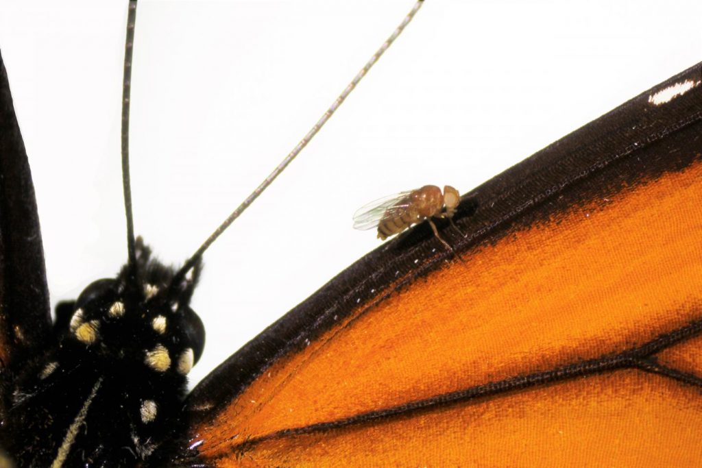
Caption: A Drosophila melanogaster “monarch fly” with mutations introduced by CRISPR-Cas9 genome editing (V111, S119 and H122) to the sodium potassium pump, on a wing of a monarch butterfly (Danaus plexippus). Credit & Ccpyright: Julianne Pelaez

Here’s a link to and a citation for the paper,

Genome editing retraces the evolution of toxin resistance in the monarch butterfly by Marianthi Karageorgi, Simon C. Groen, Fidan Sumbul, Julianne N. Pelaez, Kirsten I. Verster, Jessica M. Aguilar, Amy P. Hastings, Susan L. Bernstein, Teruyuki Matsunaga, Michael Astourian, Geno Guerra, Felix Rico, Susanne Dobler, Anurag A. Agrawal & Noah K. Whiteman. Nature (2019) DOI: https://doi.org/10.1038/s41586-019-1610-8 Published 02 October 2019

This paper is behind a paywall.

Words about a word

I’m glad they changed the headline and substituted vomit for puke. I think we need vulgar and/or taboo words to release anger or disgust or other difficult emotions. Incorporating those words into standard language deprives them of that power.

The last word: Genetivision

The company mentioned in the new release, Genetivision, is the place to go for transgenic flies. Here’s a sampling from the their Testimonials webpage,

GenetiVision‘s service has been excellent in the quality and price. The timeliness of its international service has been a big plus. We are very happy with its consistent service and the flies it generates.”
Kwang-Wook Choi, Ph.D.
Department of Biological Sciences
Korea Advanced Institute of Science and Technology


“We couldn’t be happier with GenetiVision. Great prices on both standard P and PhiC31 transgenics, quick turnaround time, and we’re still batting 1000 with transformant success. We used to do our own injections but your service makes it both faster and more cost-effective. Thanks for your service!”
Thomas Neufeld, Ph.D.
Department of Genetics, Cell Biology and Development
University of Minnesota

You can find out more here at the Genetivision website.

Revival of dead pig brains raises moral questions about life and death

The line between life and death may not be what we thought it was according to some research that was reported in April 2019. Ed Wong’s April 17, 2019 article (behind a paywall) for The Atlantic was my first inkling about the life-death questions raised by some research performed at Yale University, (Note: Links have been removed)

The brain, supposedly, cannot long survive without blood. Within seconds, oxygen supplies deplete, electrical activity fades, and unconsciousness sets in. If blood flow is not restored, within minutes, neurons start to die in a rapid, irreversible, and ultimately fatal wave.

But maybe not? According to a team of scientists led by Nenad Sestan at Yale School of Medicine, this process might play out over a much longer time frame, and perhaps isn’t as inevitable or irreparable as commonly believed. Sestan and his colleagues showed this in dramatic fashion—by preserving and restoring signs of activity in the isolated brains of pigs that had been decapitated four hours earlier.

The team sourced 32 pig brains from a slaughterhouse, placed them in spherical chambers, and infused them with nutrients and protective chemicals, using pumps that mimicked the beats of a heart. This system, dubbed BrainEx, preserved the overall architecture of the brains, preventing them from degrading. It restored flow in their blood vessels, which once again became sensitive to dilating drugs. It stopped many neurons and other cells from dying, and reinstated their ability to consume sugar and oxygen. Some of these rescued neurons even started to fire. “Everything was surprising,” says Zvonimir Vrselja, who performed most of the experiments along with Stefano Daniele.

… “I don’t see anything in this report that should undermine confidence in brain death as a criterion of death,” says Winston Chiong, a neurologist at the University of California at San Francisco. The matter of when to declare someone dead has become more controversial since doctors began relying more heavily on neurological signs, starting around 1968, when the criteria for “brain death” were defined. But that diagnosis typically hinges on the loss of brainwide activity—a line that, at least for now, is still final and irreversible. After MIT Technology Review broke the news of Sestan’s work a year ago, he started receiving emails from people asking whether he could restore brain function to their loved ones. He very much cannot. BrainEx isn’t a resurrection chamber.

“It’s not going to result in human brain transplants,” adds Karen Rommelfanger, who directs Emory University’s neuroethics program. “And I don’t think this means that the singularity is coming, or that radical life extension is more possible than before.”

So why do the study? “There’s potential for using this method to develop innovative treatments for patients with strokes or other types of brain injuries, and there’s a real need for those kinds of treatments,” says L. Syd M Johnson, a neuroethicist at Michigan Technological University. The BrainEx method might not be able to fully revive hours-dead brains, but Yama Akbari, a critical-care neurologist at the University of California at Irvine, wonders whether it would be more successful if applied minutes after death. Alternatively, it could help to keep oxygen-starved brains alive and intact while patients wait to be treated. “It’s an important landmark study,” Akbari says.

Yong notes that the study still needs to be replicated in his article which also probes some of the ethical issues associated with the latest neuroscience research.

Nature published the Yale study,

Restoration of brain circulation and cellular functions hours post-mortem by Zvonimir Vrselja, Stefano G. Daniele, John Silbereis, Francesca Talpo, Yury M. Morozov, André M. M. Sousa, Brian S. Tanaka, Mario Skarica, Mihovil Pletikos, Navjot Kaur, Zhen W. Zhuang, Zhao Liu, Rafeed Alkawadri, Albert J. Sinusas, Stephen R. Latham, Stephen G. Waxman & Nenad Sestan. Nature 568, 336–343 (2019) DOI: https://doi.org/10.1038/s41586-019-1099-1 Published 17 April 2019 Issue Date 18 April 2019

This paper is behind a paywall.

Two neuroethicists had this to say (link to their commentary in Nature follows) as per an April 71, 2019 news release from Case Western Reserve University (also on EurekAlert), Note: Links have been removed,

The brain is more resilient than previously thought. In a groundbreaking experiment published in this week’s issue of Nature, neuroscientists created an artificial circulation system that successfully restored some functions and structures in donated pig brains–up to four hours after the pigs were butchered at a USDA food processing facility. Though there was no evidence of restored consciousness, brains from the pigs were without oxygen for hours, yet could still support key functions provided by the artificial system. The result challenges the notion that mammalian brains are fully and irreversibly damaged by a lack of oxygen.

“The assumptions have always been that after a couple minutes of anoxia, or no oxygen, the brain is ‘dead,'” says Stuart Youngner, MD, who co-authored a commentary accompanying the study with Insoo Hyun, PhD, both professors in the Department of Bioethics at Case Western Reserve University School of Medicine. “The system used by the researchers begs the question: How long should we try to save people?”

In the pig experiment, researchers used an artificial perfusate (a type of cell-free “artificial blood”), which helped brain cells maintain their structure and some functions. Resuscitative efforts in humans, like CPR, are also designed to get oxygen to the brain and stave off brain damage. After a period of time, if a person doesn’t respond to resuscitative efforts, emergency medical teams declare them dead.

The acceptable duration of resuscitative efforts is somewhat uncertain. “It varies by country, emergency medical team, and hospital,” Youngner said. Promising results from the pig experiment further muddy the waters about the when to stop life-saving efforts.

At some point, emergency teams must make a critical switch from trying to save a patient, to trying to save organs, said Youngner. “In Europe, when emergency teams stop resuscitation efforts, they declare a patient dead, and then restart the resuscitation effort to circulate blood to the organs so they can preserve them for transplantation.”

The switch can involve extreme means. In the commentary, Youngner and Hyun describe how some organ recovery teams use a balloon to physically cut off blood circulation to the brain after declaring a person dead, to prepare the organs for transplantation.

The pig experiment implies that sophisticated efforts to perfuse the brain might maintain brain cells. If technologies like those used in the pig experiment could be adapted for humans (a long way off, caution Youngner and Hyun), some people who, today, are typically declared legally dead after a catastrophic loss of oxygen could, tomorrow, become candidates for brain resuscitation, instead of organ donation.

Said Youngner, “As we get better at resuscitating the brain, we need to decide when are we going to save a patient, and when are we going to declare them dead–and save five or more who might benefit from an organ.”

Because brain resuscitation strategies are in their infancy and will surely trigger additional efforts, the scientific and ethics community needs to begin discussions now, says Hyun. “This study is likely to raise a lot of public concerns. We hoped to get ahead of the hype and offer an early, reasoned response to this scientific advance.”

Both Youngner and Hyun praise the experiment as a “major scientific advancement” that is overwhelmingly positive. It raises the tantalizing possibility that the grave risks of brain damage caused by a lack of oxygen could, in some cases, be reversible.
“Pig brains are similar in many ways to human brains, which makes this study so compelling,” Hyun said. “We urge policymakers to think proactively about what this line of research might mean for ongoing debates around organ donation and end of life care.”

Here’s a link to and a citation to the Nature commentary,

Pig experiment challenges assumptions around brain damage in people by Stuart Youngner and Insoo Hyun. Nature 568, 302-304 (2019) DOI: 10.1038/d41586-019-01169-8 April 17, 2019

This paper is open access.

I was hoping to find out more about BrainEx, but this April 17, 2019 US National Institute of Mental Health news release is all I’ve been able to find in my admittedly brief online search. The news release offers more celebration than technical detail.

Quick comment

Interestingly, there hasn’t been much of a furor over this work. Not yet.

Detecting off-target effects of CRISPR gene-editing

In amidst all the hyperbole about CRISPR (clustered regularly interspaced short palindromic repeats), the gene editing technology, you will sometimes find a mild cautionary note. It seems that CRISPR is not as precise as you might think.

Some months ago there was a story about research into detecting possible unanticipated (off target) effects from using CRISPR, from an April 19, 2019 news item on ScienceDaily,

Since the CRISPR genome editing technology was invented in 2012, it has shown great promise to treat a number of intractable diseases. However, scientists have struggled to identify potential off-target effects in therapeutically relevant cell types, which remains the main barrier to moving therapies to the clinic. Now, a group of scientists at the Gladstone Institutes and the Innovative Genomics Institute (IGI), with collaborators at AstraZeneca, have developed a reliable method to do just that.

An April 19, 2019 Gladstone Institutes press release by Julie Langelier, which originated the press release, provides details,

CRISPR edits a person’s genome by cutting the DNA at a specific location. The challenge is to ensure the tool doesn’t also make cuts elsewhere along the DNA—damage referred to as “off-target effects,” which could have unforeseen consequences.

In a study published in the journal Science, the two first authors, Beeke Wienert and Stacia Wyman, found a new way to approach the problem.

“When CRISPR makes a cut, the DNA is broken,” says Wienert, PhD, who began the work in Jacob E. Corn’s IGI laboratory and who is now a postdoctoral scholar in Bruce R. Conklin’s laboratory at Gladstone. “So, in order to survive, the cell recruits many different DNA repair factors to that particular site in the genome to fix the break and join the cut ends back together. We thought that if we could find the locations of these DNA repair factors, we could identify the sites that have been cut by CRISPR.”

To test their idea, the researchers studied a panel of different DNA repair factors. They found that one of them, called MRE11, is one of the first responders to the site of the cut. Using MRE11, the scientists developed a new technique, named DISCOVER-Seq, that can identify the exact sites in the genome where a cut has been made by CRISPR.

“The human genome is extremely large—if you printed the entire DNA sequence, you would end up with a novel as tall as a 16-story building,” explains Conklin, MD, senior investigator at Gladstone and deputy director at IGI. “When we want to cut DNA with CRISPR, it’s like we’re trying to remove one specific word on a particular page in that novel.”

“You can think of the DNA repair factors as different types of bookmarks added to the book,” Conklin adds. “While some may bookmark an entire chapter, MRE11 is a bookmark that drills down to the exact letter than has been changed.”

Different methods currently exist to detect CRISPR off-target effects. However, they come with limitations that range from producing false-positive results to killing the cells they’re examining. In addition, the most common method used to date is currently limited to cultured cells in the laboratory, excluding its use in patient-derived stem cells or animal tissue.

“Because our method relies on the cell’s natural repair process to identify cuts, it has proven to be much less invasive and much more reliable,” says Corn, PhD, who now runs a laboratory at ETH Zurich. “We were able to test our new DISCOVER-Seq method in induced pluripotent stem cells, patient cells, and mice, and our findings indicate that this method could potentially be used in any system, rather than just in the lab.”

The DISCOVER-Seq method, by being applied to new cell types and systems, has also revealed new insights into the mechanisms used by CRISPR to edit the genome, which will lead to a better understanding of the biology of how this tool works.

“The new method greatly simplifies the process of identifying off-target effects while also increasing the accuracy of the results,” says Conklin, who is also a professor of medical genetics and molecular pharmacology at UC San Francisco (UCSF). “This could allow us to better predict how genome editing would work in a clinical setting. As a result, it represents an essential step in improving pre-clinical studies and bringing CRISPR-based therapies closer to the patients in need.”

###

About the Study

The paper “Unbiased detection of CRISPR off-targets in vivo 1 using DISCOVER-Seq” was published by the journal Science on April 19, 2019. Gladstone’s Hannah L. Watry and Luke M. Judge (who is also at UCSF) contributed to this study. Other authors also include Christopher D. Richardson, Jonathan T. Vu, and Katelynn R. Kazane from IGI, Charles D. Yeh from ETH Zurich, as well as Pinar Akcakaya, Michelle J. Porritt, and Michaela Morlock from AstraZeneca.

The work was supported by Gladstone, the National Institutes of Health (grants EY028249 and HL13535801), the Li Ka Shing Foundation, the Heritage Medical Research Institute, the Fanconi Anemia Research Foundation, a Sir Keith Murdoch Fellowship from the American Australian Association, and an Early Career Fellowship from the National Health and Medical Research Council.

About the Gladstone Institute

To ensure our work does the greatest good, the Gladstone Institutes focuses on conditions with profound medical, economic, and social impact—unsolved diseases. Gladstone is an independent, nonprofit life science research organization that uses visionary science and technology to overcome disease. It has an academic affiliation with the University of California, San Francisco.

Before getting to the link and citation that I usually offer you might find this July 17, 2018 posting, The CRISPR ((clustered regularly interspaced short palindromic repeats)-CAS9 gene-editing technique may cause new genetic damage kerfuffle of interest. I wonder if this latest news affected the CRISPR market as the did the news in 2018.

In addition to the link in the press release, I am including a link and a citation for the study,

Unbiased detection of CRISPR off-targets in vivo using DISCOVER-Seq by Beeke Wienert, Stacia K. Wyman, Christopher D. Richardson, Charles D. Yeh, Pinar Akcakaya, Michelle J. Porritt, Michaela Morlock, Jonathan T. Vu, Katelynn R. Kazane, Hannah L. Watry, Luke M. Judge, Bruce R. Conklin, Marcello Maresca, Jacob E. Corn. Science 19 Apr 2019: Vol. 364, Issue 6437, pp. 286-289 DOI: 10.1126/science.aav9023

This paper is behind a paywall.

Money

Over the last 10 or more years, I have, on occasion made a point, of finding out about the funding for various non-profit agencies and projects. I find that sort of thing interesting and have hoped that my readers might feel the same way.

It seems that my readers and I might not be the only ones to care about the source of funding. Joi Ito who held appointments with Harvard University and the Massachusetts Institute of Technology (MIT) resigned from his various appointments on Sept. 7, 2019 after news of major donations from Jeffrey Epstein (a disgraced financier and sex offender) to MIT were revealed. From the Joi Ito’s entry on Wikipedia (Note: Links have been removed),

Joichi “Joi” Ito (伊藤 穰一 Itō Jōichi, born June 19, 1966) is a Japanese activist, entrepreneur and venture capitalist. He is the former director of the MIT Media Lab, and a former professor of the practice of media arts and sciences at MIT. He is a former visiting professor of practice at the Harvard Law School.[1][2]

Ito has received recognition for his role as an entrepreneur focused on Internet and technology companies and has founded, among other companies, PSINet Japan, Digital Garage and Infoseek Japan. Ito is a strategic advisor to Sony Corporation[3] and general partner of Neoteny Labs.[4] Ito writes a monthly column in the Ideas section of Wired.[5]

Ito resigned from his roles at MIT, Harvard, the John D. and Catherine T. MacArthur Foundation, the Knight Foundation, PureTech Health and The New York Times Company on September 7, 2019, following allegations of financial ties to sex offender and financier Jeffrey Epstein.[2][6][7]

Many, many institutions have accepted funds from sketchy characters and orgnaizations. It’s not new to academia, the sciences, or the arts. For a contemporary view of how some of this works, take a look at Anand Giridharadas’s 2018 book, Winners Take All. From the webepage for the book,

WINNERS TAKE ALL
The Elite Charade of Changing the World
 
An insider’s groundbreaking investigation of how the global elite’s efforts to “change the world” preserve the status quo and obscure their role in causing the problems they later seek to solve.

Former New York Times columnist Anand Giridharadas takes us into the inner sanctums of a new gilded age, where the rich and powerful fight for equality and justice any way they can–except ways that threaten the social order and their position atop it. We see how they rebrand themselves as saviors of the poor; how they lavishly reward “thought leaders” who redefine “change” in winner-friendly ways; and how they constantly seek to do more good, but never less harm. We hear the limousine confessions of a celebrated foundation boss; witness an American president hem and haw about his plutocratic benefactors; and attend a cruise-ship conference where entrepreneurs celebrate their own self-interested magnanimity.

I don’t recall any mention of Epstein in Giridharadas’s book but he did have this to say on Twitter about Epstein,

Anand Giridharadas‏Verified account @AnandWrites


Everything that made Epstein’s life possible remains in place after his arrest: the Caribbean tax havens, the hidden real-estate deals, the buying of politicians, the nonprofits that sell reputational glow, the editors who cover for people of their class.

7:34 PM – 8 Jul 2019

it can’t be easy to withstand the temptation to take the money and hope that the misdoings have been exaggerated or that they have stopped. I imagine Ito and others are under constant pressure to get funds.

AstraZeneca

One of the partners in this research about CRISPR, AstraZeneca, is a pharmaceutical company. In fact, it’s one of the largest in the world (from the AstraZeneca Wikipedia entry; Note: Links have been removed),

AstraZeneca plc[4] is a British-Swedish multinational pharmaceutical and biopharmaceutical company. In 2013, it moved its headquarters to Cambridge, UK, and concentrated its R&D in three sites: Cambridge; Gaithersburg, Maryland, USA (location of MedImmune) for work on biopharmaceuticals; and Mölndal (near Gothenburg) in Sweden, for research on traditional chemical drugs.[5] AstraZeneca has a portfolio of products for major disease areas including cancer, cardiovascular, gastrointestinal, infection, neuroscience, respiratory and inflammation.[6]

The company was founded in 1999 through the merger of the Swedish Astra AB and the British Zeneca Group[7][8] (itself formed by the demerger of the pharmaceutical operations of Imperial Chemical Industries in 1993). Since the merger it has been among the world’s largest pharmaceutical companies and has made numerous corporate acquisitions, including Cambridge Antibody Technology (in 2006), MedImmune (in 2007), Spirogen (in 2013) and Definiens (by MedImmune in 2014).

Controversies

Seroquel
In April 2010 AstraZeneca settled a qui tam lawsuit brought by Stefan P. Kruszewski for $520 million to settle allegations that the company defrauded Medicare, Medicaid, and other government-funded health care programs in connection with its marketing and promotional practices for the blockbuster atypical antipsychotic, Seroquel.[76]
In March 2011, AstraZeneca settled a lawsuit in the United States totalling $68.5 million to be divided up to 38 states.[77]
Nexium
The company’s most commercially successful medication is esomeprazole (Nexium). The primary uses are treatment of gastroesophageal reflux disease, treatment and maintenance of erosive esophagitis, treatment of duodenal ulcers caused by Helicobacter pylori, prevention of gastric ulcers in those on chronic NSAID therapy, and treatment of gastrointestinal ulcers associated with Crohn’s disease. When it is manufactured the result is a mixture of two mirror-imaged molecules, R and S. Two years before the omeprazole patent expired, AstraZeneca patented S-omeprazole in pure form, pointing out that since some people metabolise R-omeprazole slowly, pure S-omeprazole treatment would give higher dose efficiency and less variation between individuals.[78] In March 2001, the company began to market Nexium, as it would a brand new drug.[79]

In 2007, Marcia Angell, former editor-in-chief of the New England Journal of Medicine and a lecturer in social medicine at the Harvard Medical School, said in Stern, a German-language weekly newsmagazine, that AstraZeneca’s scientists had misrepresented their research on the drug’s efficiency, saying “Instead of using presumably comparable doses [of each drug], the company’s scientists used Nexium in higher dosages. They compared 20 and 40 mg Nexium with 20 mg Prilosec. With the cards having been marked in that way, Nexium looked like an improvement – which however was only small and shown in only two of the three studies.”[83]
Bildman fraud, and faithless servant clawback

Study
In 2004, University of Minnesota research participant Dan Markingson committed suicide while enrolled in an industry-sponsored pharmaceutical trial comparing three FDA-approved atypical antipsychotics: Seroquel (quetiapine), Zyprexa (olanzapine), and Risperdal (risperidone). University of Minnesota Professor of Bioethics Carl Elliott noted that Markingson was enrolled in the study against the wishes of his mother, Mary Weiss, and that he was forced to choose between enrolling in the study or being involuntarily committed to a state mental institution.[89] Further investigation revealed financial ties to AstraZeneca by Markingson’s psychiatrist, Stephen C. Olson, oversights and biases in AstraZeneca’s trial design, and the inadequacy of university Institutional Review Board (IRB) protections for research subjects.[90][unreliable source?] A 2005 FDA investigation cleared the university. Nonetheless, controversy around the case has continued. A Mother Jones article[89] resulted in a group of university faculty members sending a public letter to the university Board of Regents urging an external investigation into Markingson’s death.[91]

Is it ok to take money and/or other goods and services from them?

Innovative Genomics Institute (IGI)

Also mentioned as a partner in the research, is the Innovative Genomics Institute (IGI). Here’s more from the company’s Overview webpage (Note: Links have been removed),,

The IGI began in 2014 through the Li Ka Shing Center for Genetic Engineering, which was created thanks to a generous donation from the Li Ka Shing Foundation. [emphasis mine] The Innovative Genomics Initiative formed as a partnership between the University of California, Berkeley and the University of California, San Francisco. Combining the fundamental research expertise and the biomedical talent at UCB and UCSF, the Innovative Genomics Initiative focused on unraveling the mechanisms underlying CRISPR-based genome editing and applying this technology to improve human health. Early achievements include improving the efficiency of gene replacement and foundational work toward a treatment for sickle cell disease.

In late 2015, generous philanthropic donations enabled a bolder vision and broader mission for the IGI. With this expansion came a significant enhancement of the organization, and in January 2017, the IGI officially re-launched as the Innovative Genomics Institute.

As it turns out, there is a Li Ka-shing and he has a bit of a history with Vancouver (Canada). First, here’s more about him from the Li Ka-shing Wikipedia entry,(Note: Links have been removed),

Sir Li Ka-shing GBM KBE JP[4] (born 13 June 1928)[5][6] is a Hong Kong business magnate, investor, and philanthropist. As of June 2019, Li is the 30th richest person in the world, with an estimated net wealth of US$29.4 billion.[3] He is the senior advisor for CK Hutchison Holdings,[7] after he retired from the Chairman of the Board in May 2018;[8] through it, he is the world’s leading port investor, developer, and operator of the largest health and beauty retailer in Asia and Europe.[9]

Besides business through his flagship companies Cheung Kong Property Holdings and CK Hutchison Holdings Limited, Li Ka-shing has also personally invested extensively in real estate in Singapore and Canada. He was the single largest shareholder of Canadian Imperial Bank of Commerce (CIBC), the fifth largest bank in Canada, until the sale of his share in 2005 (with all proceedings donated, see below). He is also the majority shareholder of a major energy company, Husky Energy, based in Alberta, Canada.[48]

In January 2005, Li announced plans to sell his $1.2 billion CAD stake in the Canadian Imperial Bank of Commerce, with all proceeds going to private charitable foundations established by Li, including the Li Ka Shing Foundation in Hong Kong and the Li Ka Shing (Canada) Foundation based in Toronto, Ontario.[49]

His son Victor Li was kidnapped in 1996 on his way home after work by gangster “Big Spender” Cheung Tze-keung. Li Ka-shing paid a ransom of HK$1 billion, directly to Cheung who had come to his house.[53] A report was never filed with Hong Kong police. Instead the case was pursued by Mainland authorities, leading to Cheung’s execution in 1998, an outcome not possible under Hong Kong law. Rumours circulated of a deal between Li and the Mainland.[53] In interviews, when this rumor was brought up, Li brushed it off and dismissed it completely.

Li Ka-shing was well known here in Vancouver due to his purchase of a significant chunk of land in the city. This January 9, 2015 article by Glen Korstrum for Business in Vancouver notes some rather interesting news and contextualizes with Li’s Vancouver history,

Hong Kong billionaire Li Ka-shing is restructuring his empire and shifting his base to the Cayman Islands and away from the Chinese special administrative region.

His January 9 [2015] announcement came the same day that Forbes ranked him as Hong Kong’s richest man for the 17th consecutive year, with a total wealth of US$33.5 billion.

Li is best known in Vancouver for buying an 82.5-hectare parcel of land around False Creek for $328 million in 1988 along with partners, who included fellow Hong Kong tycoons, Lee Shau Kee and Cheng Yu Tung.

The group formed Concord Pacific, which redeveloped the site that had been home to Vancouver’s 1986 world’s fair, Expo ’86.

Li cashed out of Concord Pacific in the late 1990s and, in 2007, invested in Deltaport through his Hutchison Port Holdings.

Li’s biggest Canadian holding is his controlling stake in Husky Energy. …

Intriguing, yes? It also makes the prospect of deciding whose money you’re going to accept a bit more complicated than it might seem.

Gladstone Institutes

In what seems to be a decided contrast to the previous two partners, here’s more from the Gladstone Institutes, About Us, History webpage,

Born in London in 1910, J. David Gladstone was orphaned as a boy and came to North America at age 10. He began a career in real estate in Southern California at age 28, eventually making his fortune as the first developer to create the region’s enclosed shopping malls (such as the Northridge Fashion Center mall). His accidental death in 1971 left an estate valued at about $8 million to support medical students interested in research.

It soon became clear to the three trustees administering Mr. Gladstone’s trust that his legacy could support a far more substantial philanthropic enterprise. In 1979, they launched The J. David Gladstone Institutes under the leadership of Robert W. Mahley, MD, PhD, a leading cardiovascular scientist who at the time was working at the National Institutes of Health.

In 2010, after three decades of leading Gladstone, Dr. Mahley stepped down in order to return to more active research. That same year, R. Sanders “Sandy” Williams, MD, left Duke University, where he had been Dean of the School of Medicine—as well as Senior Vice Chancellor and Senior Advisor for International Strategy—to become Gladstone’s new president. The following year, the S.D. Bechtel, Jr. Foundation [emphasis mine] helped launch the Center for Comprehensive Alzheimer’s Disease Research with a generous $6M lead gift, while the Roddenberry Foundation [emphasis mine] gave $5 million to launch the Roddenberry Center for Stem Cell Biology and Medicine. Also in 2011, the independent and philanthropic Gladstone Foundation formed with the mission of expanding the financial resources available to drive’s Gladstone’s mission.

The S. D. Bechtel jr. mentioned is associated with Bechtel, an international engineering firm. I did not find any scandals or controversies in the Bechtel Wikipedia entry. That seemed improbable so I did a little digging and found a January 30, 2015 (?) article by Matthew Brunwasser for foreignpolicy.com (Note: A link has been removed),

Steamrolled; A special investigation into the diplomacy of doing business abroad.

One of Europe’s poorest countries wanted a road, so U.S. mega-contractor Bechtel sold it a $1.3 billion highway, with the backing of a powerful American ambassador. Funny thing is, the highway is barely being used—and the ambassador is now working for Bechtel.

Bechtel, the largest contractor by revenue in the United States and the third-largest internationally, according to an annual list compiled by the Engineering News-Record, has in recent years constructed expensive highways in Kosovo, Croatia, Romania, and Albania. A six-month investigation by the Investigative Reporting Program at the University of California at Berkeley Graduate School of Journalism has found that these highways were boondoggles for the countries in which they were constructed, and that members of governments and international institutions often saw problems coming before Bechtel (along with its Turkish joint venture partner, Enka) even began work on the roads.

My other source is a May 8, 1988 article by Walter Russell Mead for the Los Angeles Time,s

From San Francisco to Saudi Arabia, the Bechtel Group Inc. has left its mark around the world. Yet the privately owned Bechtel Group is one of the country’s most mysterious operations–or was, until the publication of Laton McCartney’s critical and controversial “Friends in High Places.”

Those who believe that “Dynasty” and “Falcon Crest” describe life at the top of America’s corporate pyramids will find a picture here that makes the most far-fetched TV plots look dull. One Bechtel executive was torn to pieces by an angry mob; another, kidnaped, survived two days in the trunk of a Mercedes that had been driven over the edge of a cliff but caught on an obstacle half way down. Wheeling and dealing from Beirut to the Bohemian Grove, Bechtel executives fought off Arab and Jewish nationalists, angry senators, bitter business rivals, and furious consumer groups to build the world’s largest construction and engineering firm.

Poor Bechtel sometimes seems damned if it does and damned if it doesn’t. No major corporation could undertake foreign operations on Bechtel’s scale without some cooperation from the U.S. government–and few companies could refuse a government request that, in return, they provide cover for intelligence agents. Given the enormous scope of Bechtel’s operations in global trouble spots–a $20-billion industrial development in Saudi Arabia, for example–it could only proceed with assurances that its relations with both Saudi and American governments were good. Where, exactly, is the line between right and wrong? [emphasis mine]

… The white elephants Bechtel scattered across the American landscape–particularly the nuclear power plants that threaten to bankrupt some of the country’s largest utility systems–are monuments to wasted talent and misdirected resources.

Finally, I get to the Roddenberry Foundation, which was founded by Gene Roddenberry’s (Star Trek) son. Here’s more from the About Us, Origin webpage,

Gene Roddenberry, creator of the Star Trek series, brought to his audiences meaningful and thought-provoking science fiction to “think, question, and challenge the status quo” with the intention of creating “a brighter future”. His work has touched countless lives and continues to entertain and inspire audiences worldwide. In 2010, Gene’s son Rod established the Roddenberry Foundation to build on his father’s legacy and philosophy of inclusion, diversity, and respect for life to drive social change and meaningfully improve the lives of people around the world.

While there are many criticisms of Mr. Roddenberry, there doesn’t seem to be anything that would be considered a serious scandal on the order of a Jeffrey Epstein or the whisper of scandal on the order of Sir Li Ka-shing or Bechtel.

Final comments

It’s a good thing when research is funded and being able to detect off-target effects from CRISPR is very good, assuming the research holds up to closer scrutiny.

As for vetting your donors, that’s tricky. Of course, Epstein was already a convicted sex offender when Ito accepted his funding for MIT but I cannot emphasize enough the amount of pressure these folks are under. Academia is always hungry for money. Hopefully this incident will introduce checks and balances in the donor process.

Unusual appetite for gold

This bacterium (bacteria being the plural) loves gold, which is lucky for anyone trying to develop artificial photosynthesis.From an October 9, 2018 news item on ScienceDaily,

A bacterium named Moorella thermoacetica won’t work for free. But UC Berkeley [University of California at Berkeley] researchers have figured out it has an appetite for gold. And in exchange for this special treat, the bacterium has revealed a more efficient path to producing solar fuels through artificial photosynthesis.

An October 5, 2018 UC Berkeley news release by Theresa Duque (also on EurekAlert but published on October 9, 2018), which originated the news item, expands on the theme,

M. thermoacetica first made its debut as the first non-photosensitive bacterium to carry out artificial photosynthesis (link is external) in a study led by Peidong Yang, a professor in UC Berkeley’s College of Chemistry. By attaching light-absorbing nanoparticles made of cadmium sulfide (CdS) to the bacterial membrane exterior, the researchers turned M. thermoacetica into a tiny photosynthesis machine, converting sunlight and carbon dioxide into useful chemicals.

Now Yang and his team of researchers have found a better way to entice this CO2-hungry bacterium into being even more productive. By placing light-absorbing gold nanoclusters inside the bacterium, they have created a biohybrid system that produces a higher yield of chemical products than previously demonstrated. The research, funded by the National Institutes of Health, was published on Oct. 1 in Nature Nanotechnology (link is external).

For the first hybrid model, M. thermoacetica-CdS, the researchers chose cadmium sulfide as the semiconductor for its ability to absorb visible light. But because cadmium sulfide is toxic to bacteria, the nanoparticles had to be attached to the cell membrane “extracellularly,” or outside the M. thermoacetica-CdS system. Sunlight excites each cadmium-sulfide nanoparticle into generating a charged particle known as an electron. As these light-generated electrons travel through the bacterium, they interact with multiple enzymes in a process known as “CO2 reduction,” triggering a cascade of reactions that eventually turns CO2 into acetate, a valuable chemical for making solar fuels.

But within the extracellular model, the electrons end up interacting with other chemicals that have no part in turning CO2 into acetate. And as a result, some electrons are lost and never reach the enzymes. So to improve what’s known as “quantum efficiency,” or the bacterium’s ability to produce acetate each time it gains an electron, the researchers found another semiconductor: nanoclusters made of 22 gold atoms (Au22), a material that M. thermoacetica took a surprising shine to.

A single nanocluster of 22 gold atoms

Figure: A single nanocluster of 22 gold atoms – Au22 – is only 1 nanometer in diameter, allowing it to easily slip through the bacterial cell wall.

“We selected Au22 because it’s ideal for absorbing visible light and has the potential for driving the CO2 reduction process, but we weren’t sure whether it would be compatible with the bacteria,” Yang said. “When we inspected them under the microscope, we discovered that the bacteria were loaded with these Au22 clusters – and were still happily alive.”

Imaging of the M. thermoacetica-Au22 system was done at UC Berkeley’s Molecular Imaging Center (link is external).

The researchers also selected Au22 ­– dubbed by the researchers as “magic” gold nanoclusters – for its ultrasmall size: A single Au22nanocluster is only 1 nanometer in diameter, allowing each nanocluster to easily slip through the bacterial cell wall.

“By feeding bacteria with Au22 nanoclusters, we’ve effectively streamlined the electron transfer process for the CO2 reduction pathway inside the bacteria, as evidenced by a 2.86 percent quantum efficiency – or 33 percent more acetate produced within the M. thermoacetica-Au22 system than the CdS model,” Yang said.

The magic gold nanocluster is the latest discovery coming out of Yang’s lab, which for the past six years has focused on using biohybrid nanostructures to convert CO2 into useful chemicals as part of an ongoing effort to find affordable, abundant resources for renewable fuels, and potential solutions to thwart the effects of climate change.

“Next, we’d like to find a way to reduce costs, improve the lifetimes for these biohybrid systems, and improve quantum efficiency,” Yang said. “By continuing to look at the fundamental aspect of how gold nanoclusters are being photoactivated, and by following the electron transfer process within the CO2 reduction pathway, we hope to find even better solutions.”

Co-authors with Yang are UC Berkeley graduate student Hao Zhang and former postdoctoral fellow Hao Liu, now at Donghua University in Shanghai, China.

Here’s a link to and a citation for the paper,

Bacteria photosensitized by intracellular gold nanoclusters for solar fuel production by Hao Zhang, Hao Liu, Zhiquan Tian, Dylan Lu, Yi Yu, Stefano Cestellos-Blanco, Kelsey K. Sakimoto, & Peidong Yang. Nature Nanotechnologyvolume 13, pages900–905 (2018). DOI: https://doi.org/10.1038/s41565-018-0267-z Published: 01 October 2018

This paper is behind a paywall.

For lovers of animation, the folks at UC Berkeley have produced this piece about the ‘gold-loving’ bacterium,

Change the shape of water with nanotubes

An August 24, 2018 news item on ScienceDaily describes a ‘shapeshifting’ water technique,

First, according to Rice University engineers, get a nanotube hole. Then insert water. If the nanotube is just the right width, the water molecules will align into a square rod.

Rice materials scientist Rouzbeh Shahsavari and his team used molecular models to demonstrate their theory that weak van der Waals forces between the inner surface of the nanotube and the water molecules are strong enough to snap the oxygen and hydrogen atoms into place.

Shahsavari referred to the contents as two-dimensional “ice,” because the molecules freeze regardless of the temperature. He said the research provides valuable insight on ways to leverage atomic interactions between nanotubes and water molecules to fabricate nanochannels and energy-storing nanocapacitors.

An August 24, 2018 Rice University news release (also on EurekAlert and received via email), which originated the news item, delves further,

Shahsavari and his colleagues built molecular models of carbon and boron nitride nanotubes with adjustable widths. They discovered boron nitride is best at constraining the shape of water when the nanotubes are 10.5 angstroms wide. (One angstrom is one hundred-millionth of a centimeter.)

The researchers already knew that hydrogen atoms in tightly confined water take on interesting structural properties. Recent experiments by other labs showed strong evidence for the formation of nanotube ice and prompted the researchers to build density functional theory models to analyze the forces responsible.

Shahsavari’s team modeled water molecules, which are about 3 angstroms wide, inside carbon and boron nitride nanotubes of various chiralities (the angles of their atomic lattices) and between 8 and 12 angstroms in diameter. They discovered that nanotubes in the middle diameters had the most impact on the balance between molecular interactions and van der Waals pressure that prompted the transition from a square water tube to ice.

“If the nanotube is too small and you can only fit one water molecule, you can’t judge much,” Shahsavari said. “If it’s too large, the water keeps its amorphous shape. But at about 8 angstroms, the nanotubes’ van der Waals force [if you’re not familiar with the term, see below the link and citation for my brief explanation] starts to push water molecules into organized square shapes.”

He said the strongest interactions were found in boron nitride nanotubes due to the particular polarization of their atoms.

Shahsavari said nanotube ice could find use in molecular machines or as nanoscale capillaries, or foster ways to deliver a few molecules of water or sequestered drugs to targeted cells, like a nanoscale syringe.

Lead author Farzaneh Shayeganfar, a former visiting scholar at Rice, is an instructor at Shahid Rajaee Teacher Training University in Tehran, Iran. Co-principal investigator Javad Beheshtian is a professor at Amirkabir University, Tehran.

Supercomputer resources were provided with support from the [US] National Institutes of Health and an IBM Shared Rice University Research grant.

Here’s a link to and a citation for the paper,

First Principles Study of Water Nanotubes Captured Inside Carbon/Boron Nitride Nanotubes by Farzaneh Shayeganfar, Javad Beheshtian, and Rouzbeh Shahsavari. Langmuir, DOI: 10.1021/acs.langmuir.8b00856 Publication Date (Web): August 23, 2018

Copyright © 2018 American Chemical Society

This paper is behind a paywall.

For the purposes of the posting, van der Waals force(s) are weak adhesive forces measured at the nanoscale. Humans don’t feel them (we’re too big) but gecko lizards can exploit those forces which is why they are able to hang from the ceiling by a single toe.  There’s a more informed description here in the van der Waals force entry on Wikipedia.

Xenotransplantation—organs for transplantation in human patients—it’s a business and a science

The last time (June 18, 2018 post) I mentioned xenotransplantation (transplanting organs from one species into another species; see more here), it was in the context of an art/sci (or sciart) event coming to Vancouver (Canada).,

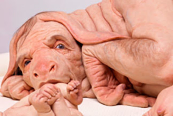
Patricia Piccinini’s Curious Imaginings Courtesy: Vancouver Biennale [downloaded from http://dailyhive.com/vancouver/vancouver-biennale-unsual-public-art-2018/]

The latest edition of the Vancouver Biennale was featured in a June 6, 2018 news item on the Daily Hive (Vancouver),

Melbourne artist Patricia Piccinini’s Curious Imaginings is expected to be one of the most talked about installations of the exhibit. Her style of “oddly captivating, somewhat grotesque, human-animal hybrid creature” is meant to be shocking and thought-provoking.

Piccinini’s interactive [emphasis mine] experience will “challenge us to explore the social impacts of emerging biotechnology and our ethical limits in an age where genetic engineering and digital technologies are already pushing the boundaries of humanity.”

Piccinini’s work will be displayed in the 105-year-old Patricia Hotel in Vancouver’s Strathcona neighbourhood. The 90-day ticketed exhibition [emphasis mine] is scheduled to open this September [2018].

(The show opens on Sept. 14, 2018.)

At the time, I had yet to stumble across Ingfei Chen’s thoughtful dive into the topic in her May 9, 2018 article for Slate.com,

In the United States, the clock is ticking for more than 114,700 adults and children waiting for a donated kidney or other lifesaving organ, and each day, nearly 20 of them die. Researchers are devising a new way to grow human organs inside other animals, but the method raises potentially thorny ethical issues. Other conceivable futuristic techniques sound like dystopian science fiction. As we envision an era of regenerative medicine decades from now, how far is society willing to go to solve the organ shortage crisis?

I found myself pondering this question after a discussion about the promises of stem cell technologies veered from the intriguing into the bizarre. I was interviewing bioengineer Zev Gartner, co-director and research coordinator of the Center for Cellular Construction at the University of California, San Francisco, about so-called organoids, tiny clumps of organlike tissue that can self-assemble from human stem cells in a Petri dish. These tissue bits are lending new insights into how our organs form and diseases take root. Some researchers even hope they can nurture organoids into full-size human kidneys, pancreases, and other organs for transplantation.

Certain organoid experiments have recently set off alarm bells, but when I asked Gartner about it, his radar for moral concerns was focused elsewhere. For him, the “really, really thought-provoking” scenarios involve other emerging stem cell–based techniques for engineering replacement organs for people, he told me. “Like blastocyst complementation,” he said.

Never heard of it? Neither had I. Turns out it’s a powerful new genetic engineering trick that researchers hope to use for growing human organs inside pigs or sheep—organs that could be genetically personalized for transplant patients, in theory avoiding immune-system rejection problems. The science still has many years to go, but if it pans out, it could be one solution to the organ shortage crisis. However, the prospect of creating hybrid animals with human parts and killing them to harvest organs has already raised a slew of ethical questions. In 2015, the National Institutes of Health placed a moratorium on federal funding of this nascent research area while it evaluated and discussed the issues.

As Gartner sees it, the debate over blastocyst complementation research—work that he finds promising—is just one of many conversations that society needs to have about the ethical and social costs and benefits of future technologies for making lifesaving transplant organs. “There’s all these weird ways that we could go about doing this,” he said, with a spectrum of imaginable approaches that includes organoids, interspecies organ farming, and building organs from scratch using 3D bioprinters. But even if it turns out we can produce human organs in these novel ways, the bigger issue, in each technological instance, may be whether we should.

Gartner crystallized things with a downright creepy example: “We know that the best bioreactor for tissues and organs for humans are human beings,” he said. Hypothetically, “the best way to get you a new heart would be to clone you, grow up a copy of yourself, and take the heart out.” [emphasis mine] Scientists could probably produce a cloned person with the technologies we already have, if money and ethics were of no concern. “But we don’t want to go there, right?” he added in the next breath. “The ethics involved in doing it are not compatible with who we want to be as a society.”

This sounds like Gartner may have been reading some science fiction, specifically, Lois McMaster Bujold and her Barrayar series where she often explored the ethics and possibilities of bioengineering. At this point, some of her work seems eerily prescient.

As for Chen’s article, I strongly encourage you to read it in its entirety if you have the time.

Medicine, healing, and big money

At about the same time, there was a May 31, 2018 news item on phys.org offering a perspective from some of the leaders in the science and the business (Note: Links have been removed),

Over the past few years, researchers led by George Church have made important strides toward engineering the genomes of pigs to make their cells compatible with the human body. So many think that it’s possible that, with the help of CRISPR technology, a healthy heart for a patient in desperate need might one day come from a pig.

“It’s relatively feasible to change one gene in a pig, but to change many dozens—which is quite clear is the minimum here—benefits from CRISPR,” an acronym for clustered regularly interspaced short palindromic repeats, said Church, the Robert Winthrop Professor of Genetics at Harvard Medical School (HMS) and a core faculty member of Harvard’s Wyss Institute for Biologically Inspired Engineering. Xenotransplantation is “one of few” big challenges (along with gene drives and de-extinction, he said) “that really requires the ‘oomph’ of CRISPR.”

To facilitate the development of safe and effective cells, tissues, and organs for future medical transplantation into human patients, Harvard’s Office of Technology Development has granted a technology license to the Cambridge biotech startup eGenesis.

Co-founded by Church and former HMS doctoral student Luhan Yang in 2015, eGenesis announced last year that it had raised $38 million to advance its research and development work. At least eight former members of the Church lab—interns, doctoral students, postdocs, and visiting researchers—have continued their scientific careers as employees there.

“The Church Lab is well known for its relentless pursuit of scientific achievements so ambitious they seem improbable—and, indeed, [for] its track record of success,” said Isaac Kohlberg, Harvard’s chief technology development officer and senior associate provost. “George deserves recognition too for his ability to inspire passion and cultivate a strong entrepreneurial drive among his talented research team.”

The license from Harvard OTD covers a powerful set of genome-engineering technologies developed at HMS and the Wyss Institute, including access to foundational intellectual property relating to the Church Lab’s 2012 breakthrough use of CRISPR, led by Yang and Prashant Mali, to edit the genome of human cells. Subsequent innovations that enabled efficient and accurate editing of numerous genes simultaneously are also included. The license is exclusive to eGenesis but limited to the field of xenotransplantation.

A May 30, 2018 Harvard University news release by Caroline Petty, which originated the news item, explores some of the issues associated with incubating humans organs in other species,

The prospect of using living, nonhuman organs, and concerns over the infectiousness of pathogens either present in the tissues or possibly formed in combination with human genetic material, have prompted the Food and Drug Administration to issue detailed guidance on xenotransplantation research and development since the mid-1990s. In pigs, a primary concern has been that porcine endogenous retroviruses (PERVs), strands of potentially pathogenic DNA in the animals’ genomes, might infect human patients and eventually cause disease. [emphases mine]

That’s where the Church lab’s CRISPR expertise has enabled significant advances. In 2015, the lab published important results in the journal Science, successfully demonstrating the use of genome engineering to eliminate all 62 PERVs in porcine cells. Science later called it “the most widespread CRISPR editing feat to date.”

In 2017, with collaborators at Harvard, other universities, and eGenesis, Church and Yang went further. Publishing again in Science, they first confirmed earlier researchers’ fears: Porcine cells can, in fact, transmit PERVs into human cells, and those human cells can pass them on to other, unexposed human cells. (It is still unknown under what circumstances those PERVs might cause disease.) In the same paper, they corrected the problem, announcing the embryogenesis and birth of 37 PERV-free pigs. [Note: My July 17, 2018 post features research which suggests CRISPR-Cas9 gene editing may cause greater genetic damage than had been thought.]

“Taken together, those innovations were stunning,” said Vivian Berlin, director of business development in OTD, who manages the commercialization strategy for much of Harvard’s intellectual property in the life sciences. “That was the foundation they needed, to convince both the scientific community and the investment community that xenotransplantation might become a reality.”

“After hundreds of tests, this was a critical milestone for eGenesis — and the entire field — and represented a key step toward safe organ transplantation from pigs,” said Julie Sunderland, interim CEO of eGenesis. “Building on this study, we hope to continue to advance the science and potential of making xenotransplantation a safe and routine medical procedure.”

Genetic engineering may undercut human diseases, but also could help restore extinct species, researcher says. [Shades of the Jurassic Park movies!]

It’s not, however, the end of the story: An immunological challenge remains, which eGenesis will need to address. The potential for a patient’s body to outright reject transplanted tissue has stymied many previous attempts at xenotransplantation. Church said numerous genetic changes must be achieved to make porcine organs fully compatible with human patients. Among these are edits to several immune functions, coagulation functions, complements, and sugars, as well as the PERVs.

“Trying the straight transplant failed almost immediately, within hours, because there’s a huge mismatch in the carbohydrates on the surface of the cells, in particular alpha-1-3-galactose, and so that was a showstopper,” Church explained. “When you delete that gene, which you can do with conventional methods, you still get pretty fast rejection, because there are a lot of other aspects that are incompatible. You have to take care of each of them, and not all of them are just about removing things — some of them you have to humanize. There’s a great deal of subtlety involved so that you get normal pig embryogenesis but not rejection.

“Putting it all together into one package is challenging,” he concluded.

In short, it’s the next big challenge for CRISPR.

Not unexpectedly, there is no mention of the CRISPR patent fight between Harvard/MIT’s (Massachusetts Institute of Technology) Broad Institute and the University of California at Berkeley (UC Berkeley). My March 15, 2017 posting featured an outcome where the Broad Institute won the first round of the fight. As I recall, it was a decision based on the principles associated with King Solomon, i.e., the US Patent Office, divided the baby and UCBerkeley got the less important part of the baby. As you might expect the decision has been appealed. In an April 30, 2018 piece, Scientific American reprinted an article about the latest round in the fight written by Sharon Begley for STAT (Note: Links have been removed),

All You Need to Know for Round 2 of the CRISPR Patent Fight

It’s baaaaack, that reputation-shredding, stock-moving fight to the death over key CRISPR patents. On Monday morning in Washington, D.C., the U.S. Court of Appeals for the Federal Circuit will hear oral arguments in University of California v. Broad Institute. Questions?

How did we get here? The patent office ruled in February 2017 that the Broad’s 2014 CRISPR patent on using CRISPR-Cas9 to edit genomes, based on discoveries by Feng Zhang, did not “interfere” with a patent application by UC based on the work of UC Berkeley’s Jennifer Doudna. In plain English, that meant the Broad’s patent, on using CRISPR-Cas9 to edit genomes in eukaryotic cells (all animals and plants, but not bacteria), was different from UC’s, which described Doudna’s experiments using CRISPR-Cas9 to edit DNA in a test tube—and it was therefore valid. The Patent Trial and Appeal Board concluded that when Zhang got CRISPR-Cas9 to work in human and mouse cells in 2012, it was not an obvious extension of Doudna’s earlier research, and that he had no “reasonable expectation of success.” UC appealed, and here we are.

For anyone who may not realize what the stakes are for these institutions, Linda Williams in a March 16, 1999 article for the LA Times had this to say about universities, patents, and money,

The University of Florida made about $2 million last year in royalties on a patent for Gatorade Thirst Quencher, a sports drink that generates some $500 million to $600 million a year in revenue for Quaker Oats Co.

The payments place the university among the top five in the nation in income from patent royalties.

Oh, but if some people on the Gainesville, Fla., campus could just turn back the clock. “If we had done Gatorade right, we would be getting $5 or $6 million (a year),” laments Donald Price, director of the university’s office of corporate programs. “It is a classic example of how not to handle a patent idea,” he added.

Gatorade was developed in 1965 when many universities were ill equipped to judge the commercial potential of ideas emerging from their research labs. Officials blew the university’s chance to control the Gatorade royalties when they declined to develop a professor’s idea.

The Gatorade story does not stop there and, even though it’s almost 20 years old, this article stands the test of time. I strongly encourage you to read it if the business end of patents and academia interest you or if you would like to develop more insight into the Broad Institute/UC Berkeley situation.

Getting back to the science, there is that pesky matter of diseases crossing over from one species to another. While, Harvard and eGenesis claim a victory in this area, it seems more work needs to be done.

Infections from pigs

An August 29, 2018 University of Alabama at Birmingham news release (also on EurekAlert) by Jeff Hansen, describes the latest chapter in the quest to provide more organs for transplantion,

A shortage of organs for transplantation — including kidneys and hearts — means that many patients die while still on waiting lists. So, research at the University of Alabama at Birmingham and other sites has turned to pig organs as an alternative. [emphasis mine]

Using gene-editing, researchers have modified such organs to prevent rejection, and research with primates shows the modified pig organs are well-tolerated.

An added step is needed to ensure the safety of these inter-species transplants — sensitive, quantitative assays for viruses and other infectious microorganisms in donor pigs that potentially could gain access to humans during transplantation.

The U.S. Food and Drug Administration requires such testing, prior to implantation, of tissues used for xenotransplantation from animals to humans. It is possible — though very unlikely — that an infectious agent in transplanted tissues could become an emerging infectious disease in humans.

In a paper published in Xenotransplantation, Mark Prichard, Ph.D., and colleagues at UAB have described the development and testing of 30 quantitative assays for pig infectious agents. These assays had sensitivities similar to clinical lab assays for viral loads in human patients. After validation, the UAB team also used the assays on nine sows and 22 piglets delivered from the sows through caesarian section.

“Going forward, ensuring the safety of these organs is of paramount importance,” Prichard said. “The use of highly sensitive techniques to detect potential pathogens will help to minimize adverse events in xenotransplantation.”

“The assays hold promise as part of the screening program to identify suitable donor animals, validate and release transplantable organs for research purposes, and monitor transplant recipients,” said Prichard, a professor in the UAB Department of Pediatrics and director of the Department of Pediatrics Molecular Diagnostics Laboratory.

The UAB researchers developed quantitative polymerase chain reaction, or qPCR, assays for 28 viruses sometimes found in pigs and two groups of mycoplasmas. They established reproducibility, sensitivity, specificity and lower limit of detection for each assay. All but three showed features of good quantitative assays, and the lower limit of detection values ranged between one and 16 copies of the viral or bacterial genetic material.

Also, the pig virus assays did not give false positives for some closely related human viruses.

As a start to understanding the infectious disease load in normal healthy animals and ensuring the safety of pig tissues used in xenotransplantation research, the researchers then screened blood, nasal swab and stool specimens from nine adult sows and 22 of their piglets delivered by caesarian section.

Mycoplasma species and two distinct herpesviruses were the most commonly detected microorganisms. Yet 14 piglets that were delivered from three sows infected with either or both herpesviruses were not infected with the herpesviruses, showing that transmission of these viruses from sow to the caesarian-delivery piglet was inefficient.

Prichard says the assays promise to enhance the safety of pig tissues for xenotransplantation, and they will also aid evaluation of human specimens after xenotransplantation.

The UAB researchers say they subsequently have evaluated more than 300 additional specimens, and that resulted in the detection of most of the targets. “The detection of these targets in pig specimens provides reassurance that the analytical methods are functioning as designed,” said Prichard, “and there is no a priori reason some targets might be more difficult to detect than others with the methods described here.”

As is my custom, here’s a link to and a citation for the paper,

Xenotransplantation panel for the detection of infectious agents in pigs by Caroll B. Hartline, Ra’Shun L. Conner, Scott H. James, Jennifer Potter, Edward Gray, Jose Estrada, Mathew Tector, A. Joseph Tector, Mark N. Prichard. Xenotransplantaion Volume 25, Issue 4 July/August 2018 e12427 DOI: https://doi.org/10.1111/xen.12427 First published: 18 August 2018

This paper is open access.

All this leads to questions about chimeras. If a pig is incubating organs with human cells it’s a chimera but then means the human receiving the organ becomes a chimera too. (For an example, see my Dec. 22, 2013 posting where there’s mention of a woman who received a trachea from a pig. Scroll down about 30% of the way.)

What is it to be human?

A question much beloved of philosophers and others, the question seems particularly timely with xenotransplantion and other developments such neuroprosthetics (cyborgs) and neuromorphic computing (brainlike computing).

As I’ve noted before, although not recently, popular culture offers a discourse on these issues. Take a look at the superhero movies and the way in which enhanced humans and aliens are presented. For example, X-Men comics and movies present mutants (humans with enhanced abilities) as despised and rejected. Video games (not really my thing but there is the Deus Ex series which has as its hero, a cyborg also offer insight into these issues.

Other than popular culture and in the ‘bleeding edge’ arts community, I can’t recall any public discussion on these matters arising from the extraordinary set of technologies which are being deployed or prepared for deployment in the foreseeable future.

(If you’re in Vancouver (Canada) from September 14 – December 15, 2018, you may want to check out Piccinini’s work. Also, there’s ” NCSU [North Carolina State University] Libraries, NC State’s Genetic Engineering and Society (GES) Center, and the Gregg Museum of Art & Design have issued a public call for art for the upcoming exhibition Art’s Work in the Age of Biotechnology: Shaping our Genetic Futures.” from my Sept. 6, 2018 posting. Deadline: Oct. 1, 2018.)

At a guess, there will be pushback from people who have no interest in debating what it is to be human as they already know, and will find these developments, when they learn about them, to be horrifying and unnatural.

Yes! Art, genetic modifications, gene editing, and xenotransplantation at the Vancouver Biennale (Canada)

Patricia Piccinini’s Curious Imaginings Courtesy: Vancouver Biennale [downloaded from http://dailyhive.com/vancouver/vancouver-biennale-unsual-public-art-2018/]

Up to this point, I’ve been a little jealous of the Art/Sci Salon’s (Toronto, Canada) January 2018 workshops for artists and discussions about CRISPR ((clustered regularly interspaced short palindromic repeats))/Cas9 and its social implications. (See my January 10, 2018 posting for more about the events.) Now, it seems Vancouver may be in line for its ‘own’ discussion about CRISPR and the implications of gene editing. The image you saw (above) represents one of the installations being hosted by the 2018 – 2020 edition of the Vancouver Biennale.

While this posting is mostly about the Biennale and Piccinini’s work, there is a ‘science’ subsection featuring the science of CRISPR and xenotransplantation. Getting back to the Biennale and Piccinini: A major public art event since 1988, the Vancouver Biennale has hosted over 91 outdoor sculptures and new media works by more than 78 participating artists from over 25 countries and from 4 continents.

Quickie description of the 2018 – 2020 Vancouver Biennale

The latest edition of the Vancouver Biennale was featured in a June 6, 2018 news item on the Daily Hive (Vancouver),

The Vancouver Biennale will be bringing new —and unusual— works of public art to the city beginning this June.

The theme for this season’s Vancouver Biennale exhibition is “re-IMAGE-n” and it kicks off on June 20 [2018] in Vanier Park with Saudi artist Ajlan Gharem’s Paradise Has Many Gates.

Gharem’s architectural chain-link sculpture resembles a traditional mosque, the piece is meant to challenge the notions of religious orthodoxy and encourages individuals to image a space free of Islamophobia.

Melbourne artist Patricia Piccinini’s Curious Imaginings is expected to be one of the most talked about installations of the exhibit. Her style of “oddly captivating, somewhat grotesque, human-animal hybrid creature” is meant to be shocking and thought-provoking.

Piccinini’s interactive [emphasis mine] experience will “challenge us to explore the social impacts of emerging biotechnology and our ethical limits in an age where genetic engineering and digital technologies are already pushing the boundaries of humanity.”

Piccinini’s work will be displayed in the 105-year-old Patricia Hotel in Vancouver’s Strathcona neighbourhood. The 90-day ticketed exhibition [emphasis mine] is scheduled to open this September [2018].

Given that this blog is focused on nanotechnology and other emerging technologies such as CRISPR, I’m focusing on Piccinini’s work and its art/science or sci-art status. This image from the GOMA Gallery where Piccinini’s ‘Curious Affection‘ installation is being shown from March 24 – Aug. 5, 2018 in Brisbane, Queensland, Australia may give you some sense of what one of her installations is like,

Courtesy: Queensland Art Gallery | Gallery of Modern Art (QAGOMA)

I spoke with Serena at the Vancouver Biennale office and asked about the ‘interactive’ aspect of Piccinini’s installation. She suggested the term ‘immersive’ as an alternative. In other words, you won’t be playing with the sculptures or pressing buttons and interacting with computer screens or robots. She also noted that the ticket prices have not been set yet and they are currently developing events focused on the issues raised by the installation. She knew that 2018 is the 200th anniversary of the publication of Mary Shelley’s Frankenstein but I’m not sure how the Biennale folks plan (or don’t plan)  to integrate any recognition of the novle’s impact on the discussions about ‘new’ technologies .They expect Piccinini will visit Vancouver. (Note 1: Piccinini’s work can  also be seen in a group exhibition titled: Frankenstein’s Birthday Party at the Hosfselt Gallery in San Francisco (California, US) from June 23 – August 11, 2018.  Note 2: I featured a number of international events commemorating the 200th anniversary of the publication of Mary Shelley’s novel, Frankenstein, in my Feb. 26, 2018 posting. Note 3: The term ‘Frankenfoods’ helped to shape the discussion of genetically modified organisms and food supply on this planet. It was a wildly successful campaign for activists affecting legislation in some areas of research. Scientists have not been as enthusiastic about the effects. My January 15, 2009 posting briefly traces a history of the term.)

The 2018 – 2020 Vancouver Biennale and science

A June 7, 2018 Vancouver Biennale news release provides more detail about the current series of exhibitions,

The Biennale is also committed to presenting artwork at the cutting edge of discussion and in keeping with the STEAM (science, technology, engineering, arts, math[ematics]) approach to integrating the arts and sciences. In August [2018], Colombian/American visual artist Jessica Angel will present her monumental installation Dogethereum Bridge at Hinge Park in Olympic Village. Inspired by blockchain technology, the artwork’s design was created through the integration of scientific algorithms, new developments in technology, and the arts. This installation, which will serve as an immersive space and collaborative hub for artists and technologists, will host a series of activations with blockchain as the inspirational jumping-off point.

In what is expected to become one of North America’s most talked-about exhibitions of the year, Melbourne artist Patricia Piccinini’s Curious Imaginings will see the intersection of art, science, and ethics. For the first time in the Biennale’s fifteen years of creating transformative experiences, and in keeping with the 2018-2020 theme of “re-IMAGE-n,” the Biennale will explore art in unexpected places by exhibiting in unconventional interior spaces.  The hyperrealist “world of oddly captivating, somewhat grotesque, human-animal hybrid creatures” will be the artist’s first exhibit in a non-museum setting, transforming a wing of the 105-year-old Patricia Hotel. Situated in Vancouver’s oldest neighbourbood of Strathcona, Piccinini’s interactive experience will “challenge us to explore the social impacts of emerging bio-technology and our ethical limits in an age where genetic engineering and digital technologies are already pushing the boundaries of humanity.” In this intimate hotel setting located in a neighborhood continually undergoing its own change, Curious Imaginings will empower visitors to personally consider questions posed by the exhibition, including the promises and consequences of genetic research and human interference. …

There are other pieces being presented at the Biennale but my special interest is in the art/sci pieces and, at this point, CRISPR.

Piccinini in more depth

You can find out more about Patricia Piccinini in her biography on the Vancouver Biennale website but I found this Char Larsson April 7, 2018 article for the Independent (UK) more informative (Note: A link has been removed),

Patricia Piccinini’s sculptures are deeply disquieting. Walking through Curious Affection, her new solo exhibition at Brisbane’s Gallery of Modern Art, is akin to entering a science laboratory full of DNA experiments. Made from silicone, fibreglass and even human hair, her sculptures are breathtakingly lifelike, however, we can’t be sure what life they are like. The artist creates an exuberant parallel universe where transgenic experiments flourish and human evolution has given way to genetic engineering and DNA splicing.

Curious Affection is a timely and welcome recognition of Piccinini’s enormous contribution to reaching back to the mid-1990s. Working across a variety of mediums including photography, video and drawing, she is perhaps best known for her hyperreal creations.

As a genre, hyperrealism depends on the skill of the artist to create the illusion of reality. To be truly successful, it must convince the spectator of its realness. Piccinini acknowledges this demand, but with a delightful twist. The excruciating attention to detail deliberately solicits our desire to look, only to generate unease, as her sculptures are imbued with a fascinating otherness. Part human, part animal, the works are uncannily familiar, but also alarmingly “other”.

Inspired by advances in genetically modified pigs to generate replacement organs for humans [also known as xenotransplantation], we are reminded that Piccinini has always been at the forefront of debates concerning the possibilities of science, technology and DNA cloning. She does so, however, with a warm affection and sense of humour, eschewing the hysterical anxiety frequently accompanying these scientific developments.

Beyond the astonishing level of detail achieved by working with silicon and fibreglass, there is an ethics at work here. Piccinini is asking us not to avert our gaze from the other, and in doing so, to develop empathy and understanding through the encounter.

I encourage anyone who’s interested to read Larsson’s entire piece (April 7, 2018 article).

According to her Wikipedia entry, Piccinini works in a variety of media including video, sound, sculpture, and more. She also has her own website.

Gene editing and xenotransplantation

Sarah Zhang’s June 8, 2018 article for The Atlantic provides a peek at the extraordinary degree of interest and competition in the field of gene editing and CRISPR ((clustered regularly interspaced short palindromic repeats))/Cas9 research (Note: A link has been removed),

China Is Genetically Engineering Monkeys With Brain Disorders

Guoping Feng applied to college the first year that Chinese universities reopened after the Cultural Revolution. It was 1977, and more than a decade’s worth of students—5.7 million—sat for the entrance exams. Feng was the only one in his high school to get in. He was assigned—by chance, essentially—to medical school. Like most of his contemporaries with scientific ambitions, he soon set his sights on graduate studies in the United States. “China was really like 30 to 50 years behind,” he says. “There was no way to do cutting-edge research.” So in 1989, he left for Buffalo, New York, where for the first time he saw snow piled several feet high. He completed his Ph.D. in genetics at the State University of New York at Buffalo.

Feng is short and slim, with a monk-like placidity and a quick smile, and he now holds an endowed chair in neuroscience at MIT, where he focuses on the genetics of brain disorders. His 45-person lab is part of the McGovern Institute for Brain Research, which was established in 2000 with the promise of a $350 million donation, the largest ever received by the university. In short, his lab does not lack for much.

Yet Feng now travels to China several times a year, because there, he can pursue research he has not yet been able to carry out in the United States. [emphasis mine] …

Feng had organized a symposium at SIAT [Shenzhen Institutes of Advanced Technology], and he was not the only scientist who traveled all the way from the United States to attend: He invited several colleagues as symposium speakers, including a fellow MIT neuroscientist interested in tree shrews, a tiny mammal related to primates and native to southern China, and Chinese-born neuroscientists who study addiction at the University of Pittsburgh and SUNY Upstate Medical University. Like Feng, they had left China in the ’80s and ’90s, part of a wave of young scientists in search of better opportunities abroad. Also like Feng, they were back in China to pursue a type of cutting-edge research too expensive and too impractical—and maybe too ethically sensitive—in the United States.

Here’s what precipitated Feng’s work in China, (from Zhang’s article; Note: Links have been removed)

At MIT, Feng’s lab worked on genetically engineering a monkey species called marmosets, which are very small and genuinely bizarre-looking. They are cheaper to keep due to their size, but they are a relatively new lab animal, and they can be difficult to train on lab tasks. For this reason, Feng also wanted to study Shank3 on macaques in China. Scientists have been cataloging the social behavior of macaques for decades, making it an obvious model for studies of disorders like autism that have a strong social component. Macaques are also more closely related to humans than marmosets, making their brains a better stand-in for those of humans.

The process of genetically engineering a macaque is not trivial, even with the advanced tools of CRISPR. Researchers begin by dosing female monkeys with the same hormones used in human in vitro fertilization. They then collect and fertilize the eggs, and inject the resulting embryos with CRISPR proteins using a long, thin glass needle. Monkey embryos are far more sensitive than mice embryos, and can be affected by small changes in the pH of the injection or the concentration of CRISPR proteins. Only some of the embryos will have the desired mutation, and only some will survive once implanted in surrogate mothers. It takes dozens of eggs to get to just one live monkey, so making even a few knockout monkeys required the support of a large breeding colony.

The first Shank3 macaque was born in 2015. Four more soon followed, bringing the total to five.

To visit his research animals, Feng now has to fly 8,000 miles across 12 time zones. It would be a lot more convenient to carry out his macaque research in the United States, of course, but so far, he has not been able to.

He originally inquired about making Shank3 macaques at the New England Primate Research Center, one of eight national primate research centers then funded by the National Institutes of Health in partnership with a local institution (Harvard Medical School, in this case). The center was conveniently located in Southborough, Massachusetts, just 20 miles west of the MIT campus. But in 2013, Harvard decided to shutter the center.

The decision came as a shock to the research community, and it was widely interpreted as a sign of waning interest in primate research in the United States. While the national primate centers have been important hubs of research on HIV, Zika, Ebola, and other diseases, they have also come under intense public scrutiny. Animal-rights groups like the Humane Society of the United States have sent investigators to work undercover in the labs, and the media has reported on monkey deaths in grisly detail. Harvard officially made its decision to close for “financial” reasons. But the announcement also came after the high-profile deaths of four monkeys from improper handling between 2010 and 2012. The deaths sparked a backlash; demonstrators showed up at the gates. The university gave itself two years to wind down their primate work, officially closing the center in 2015.

“They screwed themselves,” Michael Halassa, the MIT neuroscientist who spoke at Feng’s symposium, told me in Shenzhen. Wei-Dong Yao, another one of the speakers, chimed in, noting that just two years later CRISPR has created a new wave of interest in primate research. Yao was one of the researchers at Harvard’s primate center before it closed; he now runs a lab at SUNY Upstate Medical University that uses genetically engineered mouse and human stem cells, and he had come to Shenzhen to talk about restarting his addiction research on primates.

Here’s comes the competition (from Zhang’s article; Note: Links have been removed),

While the U.S. government’s biomedical research budget has been largely flat, both national and local governments in China are eager to raise their international scientific profiles, and they are shoveling money into research. A long-rumored, government-sponsored China Brain Project is supposed to give neuroscience research, and primate models in particular, a big funding boost. Chinese scientists may command larger salaries, too: Thanks to funding from the Shenzhen local government, a new principal investigator returning from overseas can get 3 million yuan—almost half a million U.S. dollars—over his or her first five years. China is even finding success in attracting foreign researchers from top U.S. institutions like Yale.

In the past few years, China has seen a miniature explosion of genetic engineering in monkeys. In Kunming, Shanghai, and Guangzhou, scientists have created monkeys engineered to show signs of Parkinson’s, Duchenne muscular dystrophy, autism, and more. And Feng’s group is not even the only one in China to have created Shank3 monkeys. Another group—a collaboration primarily between researchers at Emory University and scientists in China—has done the same.

Chinese scientists’ enthusiasm for CRISPR also extends to studies of humans, which are moving much more quickly, and in some cases under less oversight, than in the West. The first studies to edit human embryos and first clinical trials for cancer therapies using CRISPR have all happened in China. [emphases mine]

Some ethical issues are also covered (from Zhang’s article),

Parents with severely epileptic children had asked him if it would be possible to study the condition in a monkey. Feng told them what he thought would be technically possible. “But I also said, ‘I’m not sure I want to generate a model like this,’” he recalled. Maybe if there were a drug to control the monkeys’ seizures, he said: “I cannot see them seizure all the time.”

But is it ethical, he continued, to let these babies die without doing anything? Is it ethical to generate thousands or millions of mutant mice for studies of brain disorders, even when you know they will not elucidate much about human conditions?

Primates should only be used if other models do not work, says Feng, and only if a clear path forward is identified. The first step in his work, he says, is to use the Shank3 monkeys to identify the changes the mutations cause in the brain. Then, researchers might use that information to find targets for drugs, which could be tested in the same monkeys. He’s talking with the Oregon National Primate Research Center about carrying out similar work in the United States. ….[Note: I have a three-part series about CRISPR and germline editing* in the US, precipitated by research coming out of Oregon, Part 1, which links to the other parts, is here.]

Zhang’s June 8, 2018 article is excellent and I highly recommend reading it.

I touched on the topic of xenotransplanttaion in a commentary on a book about the science  of the television series, Orphan Black in a January 31,2018 posting (Note: A chimera is what you use to incubate a ‘human’ organ for transplantation or, more accurately, xenotransplantation),

On the subject of chimeras, the Canadian Broadcasting Corporation (CBC) featured a January 26, 2017 article about the pig-human chimeras on its website along with a video,

The end

I am very excited to see Piccinini’s work come to Vancouver. There have been a number of wonderful art and art/science installations and discussions here but this is the first one (I believe) to tackle the emerging gene editing technologies and the issues they raise. (It also fits in rather nicely with the 200th anniversary of the publication of Mary Shelley’s Frankenstein which continues to raise issues and stimulate discussion.)

In addition to the ethical issues raised in Zhang’s article, there are some other philosophical questions:

  • what does it mean to be human
  • if we are going to edit genes to create hybrid human/animals, what are they and how do they fit into our current animal/human schema
  • are you still human if you’ve had an organ transplant where the organ was incubated in a pig

There are also going to be legal issues. In addition to any questions about legal status, there are also fights about intellectual property such as the one involving Harvard & MIT’s [Massachusetts Institute of Technology] Broad Institute vs the University of California at Berkeley (March 15, 2017 posting)..

While I’m thrilled about the Piccinini installation, it should be noted the issues raised by other artworks hosted in this version of the Biennale are important. Happily, they have been broached here in Vancouver before and I suspect this will result in more nuanced  ‘conversations’ than are possible when a ‘new’ issue is introduced.

Bravo 2018 – 2020 Vancouver Biennale!

* Germline editing is when your gene editing will affect subsequent generations as opposed to editing out a mutated gene for the lifetime of a single individual.

Art/sci and CRISPR links

This art/science posting may prove of some interest:

The connectedness of living things: an art/sci project in Saskatchewan: evolutionary biology (February 16, 2018)

A selection of my CRISPR posts:

CRISPR and editing the germline in the US (part 1 of 3): In the beginning (August 15, 2017)

NOTE: An introductory CRISPR video describing how CRISPR/Cas9 works was embedded in part1.

Why don’t you CRISPR yourself? (January 25, 2018)

Editing the genome with CRISPR ((clustered regularly interspaced short palindromic repeats)-carrying nanoparticles (January 26, 2018)

Immune to CRISPR? (April 10, 2018)

Santiago Ramón y Cajal and the butterflies of the soul

The Cajal exhibit of drawings was here in Vancouver (Canada) this last fall (2017) and I still carry the memory of that glorious experience (see my Sept. 11, 2017 posting for more about the show and associated events). It seems Cajal’s drawings had a similar response in New York city, from a January 18, 2018 article by Roberta Smith for the New York Times,

It’s not often that you look at an exhibition with the help of the very apparatus that is its subject. But so it is with “The Beautiful Brain: The Drawings of Santiago Ramón y Cajal” at the Grey Art Gallery at New York University, one of the most unusual, ravishing exhibitions of the season.

The show finished its run on March 31, 2018 and is now on its way to the Massachusetts Institute of Technology (MIT) in Boston, Massachusetts for its opening on May 3, 2018. It looks like they have an exciting lineup of events to go along with the exhibit (from MIT’s The Beautiful Brain: The Drawings of Santiago Ramón y Cajal exhibit and event page),

SUMMER PROGRAMS

ONGOING

Spotlight Tours
Explorations led by local and Spanish scientists, artists, and entrepreneurs who will share their unique perspectives on particular aspects of the exhibition. (2:00 pm on select Tuesdays and Saturdays)

Tue, May 8 – Mark Harnett, Fred and Carole Middleton Career Development Professor at MIT and McGovern Institute Investigator Sat, May 26 – Marion Boulicault, MIT Graduate Student and Neuroethics Fellow in the Center for Sensorimotor Neural Engineering Tue, June 5 – Kelsey Allen, Graduate researcher, MIT Center for Brains, Minds, and Machines Sat, Jun 23 – Francisco Martin-Martinez, Research Scientist in MIT’s Laboratory for Atomistic & Molecular Mechanics and President of the Spanish Foundation for Science and Technology Jul 21 – Alex Gomez-Marin, Principal Investigator of the Behavior of Organisms Laboratory in the Instituto de Neurociencias, Spain Tue, Jul 31– Julie Pryor, Director of Communications at the McGovern Institute for Brain Research at MIT Tue, Aug 28 – Satrajit Ghosh, Principal Research Scientist at the McGovern Institute for Brain Research at MIT, Assistant Professor in the Department of Otolaryngology at Harvard Medical School, and faculty member in the Speech and Hearing Biosciences and Technology program in the Harvard Division of Medical Sciences

Idea Hub
Drop in and explore expansion microscopy in our maker-space.

Visualizing Science Workshop
Experiential learning with micro-scale biological images. (pre-registration required)

Gallery Demonstrations
Researchers share the latest on neural anatomy, signal transmission, and modern imaging techniques.

EVENTS

Teen Science Café: Mindful Matters
MIT researchers studying the brain share their mind-blowing findings.

Neuron Paint Night
Create a painting of cerebral cortex neurons and learn about the EyeWire citizen science game.

Cerebral Cinema Series
Hear from researchers and then compare real science to depictions on the big screen.

Brainy Trivia
Test your brain power in a night of science trivia and short, snappy research talks.

Come back to see our exciting lineup for the fall!

If you don’t have a chance to see the show or if you’d like a preview, I encourage you to read Smith’s article as it has embedded several Cajal drawings and rendered them exceptionally well.

For those who like a little contemporary (and related) science with their art, there’s a March 30, 2018 Harvard Medical Schoo (HMS)l news release by Kevin Jang (also on EurekAlert), Note: All links save one have been removed,

Drawing of the cells of the chick cerebellum by Santiago Ramón y Cajal, from “Estructura de los centros nerviosos de las aves,” Madrid, circa 1905


Modern neuroscience, for all its complexity, can trace its roots directly to a series of pen-and-paper sketches rendered by Nobel laureate Santiago Ramón y Cajal in the late 19th and early 20th centuries.

His observations and drawings exposed the previously hidden composition of the brain, revealing neuronal cell bodies and delicate projections that connect individual neurons together into intricate networks.

As he explored the nervous systems of various organisms under his microscope, a natural question arose: What makes a human brain different from the brain of any other species?

At least part of the answer, Ramón y Cajal hypothesized, lay in a specific class of neuron—one found in a dazzling variety of shapes and patterns of connectivity, and present in higher proportions in the human brain than in the brains of other species. He dubbed them the “butterflies of the soul.”

Known as interneurons, these cells play critical roles in transmitting information between sensory and motor neurons, and, when defective, have been linked to diseases such as schizophrenia, autism and intellectual disability.

Despite more than a century of study, however, it remains unclear why interneurons are so diverse and what specific functions the different subtypes carry out.

Now, in a study published in the March 22 [2018] issue of Nature, researchers from Harvard Medical School, New York Genome Center, New York University and the Broad Institute of MIT and Harvard have detailed for the first time how interneurons emerge and diversify in the brain.

Using single-cell analysis—a technology that allows scientists to track cellular behavior one cell at a time—the team traced the lineage of interneurons from their earliest precursor states to their mature forms in mice. The researchers identified key genetic programs that determine the fate of developing interneurons, as well as when these programs are switched on or off.

The findings serve as a guide for efforts to shed light on interneuron function and may help inform new treatment strategies for disorders involving their dysfunction, the authors said.

“We knew more than 100 years ago that this huge diversity of morphologically interesting cells existed in the brain, but their specific individual roles in brain function are still largely unclear,” said co-senior author Gordon Fishell, HMS professor of neurobiology and a faculty member at the Stanley Center for Psychiatric Research at the Broad.

“Our study provides a road map for understanding how and when distinct interneuron subtypes develop, giving us unprecedented insight into the biology of these cells,” he said. “We can now investigate interneuron properties as they emerge, unlock how these important cells function and perhaps even intervene when they fail to develop correctly in neuropsychiatric disease.”

A hippocampal interneuron. Image: Biosciences Imaging Gp, Soton, Wellcome Trust via Creative CommonsA hippocampal interneuron. Image: Biosciences Imaging Gp, Soton, Wellcome Trust via Creative Commons

Origins and Fates

In collaboration with co-senior author Rahul Satija, core faculty member of the New York Genome Center, Fishell and colleagues analyzed brain regions in developing mice known to contain precursor cells that give rise to interneurons.

Using Drop-seq, a single-cell sequencing technique created by researchers at HMS and the Broad, the team profiled gene expression in thousands of individual cells at multiple time points.

This approach overcomes a major limitation in past research, which could analyze only the average activity of mixtures of many different cells.

In the current study, the team found that the precursor state of all interneurons had similar gene expression patterns despite originating in three separate brain regions and giving rise to 14 or more interneuron subtypes alone—a number still under debate as researchers learn more about these cells.

“Mature interneuron subtypes exhibit incredible diversity. Their morphology and patterns of connectivity and activity are so different from each other, but our results show that the first steps in their maturation are remarkably similar,” said Satija, who is also an assistant professor of biology at New York University.

“They share a common developmental trajectory at the earliest stages, but the seeds of what will cause them to diverge later—a handful of genes—are present from the beginning,” Satija said.

As they profiled cells at later stages in development, the team observed the initial emergence of four interneuron “cardinal” classes, which give rise to distinct fates. Cells were committed to these fates even in the early embryo. By developing a novel computational strategy to link precursors with adult subtypes, the researchers identified individual genes that were switched on and off when cells began to diversify.

For example, they found that the gene Mef2c—mutations of which are linked to Alzheimer’s disease, schizophrenia and neurodevelopmental disorders in humans—is an early embryonic marker for a specific interneuron subtype known as Pvalb neurons. When they deleted Mef2c in animal models, Pvalb neurons failed to develop.

These early genes likely orchestrate the execution of subsequent genetic subroutines, such as ones that guide interneuron subtypes as they migrate to different locations in the brain and ones that help form unique connection patterns with other neural cell types, the authors said.

The identification of these genes and their temporal activity now provide researchers with specific targets to investigate the precise functions of interneurons, as well as how neurons diversify in general, according to the authors.

“One of the goals of this project was to address an incredibly fascinating developmental biology question, which is how individual progenitor cells decide between different neuronal fates,” Satija said. “In addition to these early markers of interneuron divergence, we found numerous additional genes that increase in expression, many dramatically, at later time points.”

The association of some of these genes with neuropsychiatric diseases promises to provide a better understanding of these disorders and the development of therapeutic strategies to treat them, a particularly important notion given the paucity of new treatments, the authors said.

Over the past 50 years, there have been no fundamentally new classes of neuropsychiatric drugs, only newer versions of old drugs, the researchers pointed out.

“Our repertoire is no better than it was in the 1970s,” Fishell said.

“Neuropsychiatric diseases likely reflect the dysfunction of very specific cell types. Our study puts forward a clear picture of what cells to look at as we work to shed light on the mechanisms that underlie these disorders,” Fishell said. “What we will find remains to be seen, but we have new, strong hypotheses that we can now test.”

As a resource for the research community, the study data and software are open-source and freely accessible online.

A gallery of the drawings of Santiago Ramón y Cajal is currently on display in New York City, and will open at the MIT Museum in Boston in May 2018.

Christian Mayer, Christoph Hafemeister and Rachel Bandler served as co-lead authors on the study.

This work was supported by the National Institutes of Health (R01 NS074972, R01 NS081297, MH071679-12, DP2-HG-009623, F30MH114462, T32GM007308, F31NS103398), the European Molecular Biology Organization, the National Science Foundation and the Simons Foundation.

Here’s link to and a citation for the paper,

Developmental diversification of cortical inhibitory interneurons by Christian Mayer, Christoph Hafemeister, Rachel C. Bandler, Robert Machold, Renata Batista Brito, Xavier Jaglin, Kathryn Allaway, Andrew Butler, Gord Fishell, & Rahul Satija. Nature volume 555, pages 457–462 (22 March 2018) doi:10.1038/nature25999 Published: 05 March 2018

This paper is behind a paywall.

Colliding organic nanoparticles caught on camera for the first time

There is high excitement about this development in a November 17, 2017 news item on Nanowerk,

A Northwestern University research team is the first to capture on video organic nanoparticles colliding and fusing together. This unprecedented view of “chemistry in motion” will aid Northwestern nanoscientists developing new drug delivery methods as well as demonstrate to researchers around the globe how an emerging imaging technique opens a new window on a very tiny world.

A November 17, 2017 Northwestern University news release (also on EurekAlert) by Megan Fellman, which originated the news item, further illuminates the matter,

This is a rare example of particles in motion. The dynamics are reminiscent of two bubbles coming together and merging into one: first they join and have a membrane between them, but then they fuse and become one larger bubble.

“I had an image in my mind, but the first time I saw these fusing nanoparticles in black and white was amazing,” said professor Nathan C. Gianneschi, who led the interdisciplinary study and works at the intersection of nanotechnology and biomedicine.

“To me, it’s literally a window opening up to this world you have always known was there, but now you’ve finally got an image of it. I liken it to the first time I saw Jupiter’s moons through a telescope. Nothing compares to actually seeing,” he said.

Gianneschi is the Jacob and Rosaline Cohn Professor in the department of chemistry in the Weinberg College of Arts and Sciences and in the departments of materials science and engineering and of biomedical engineering in the McCormick School of Engineering.

The study, which includes videos of different nanoparticle fusion events, was published today (Nov. 1 [2017]7) by the Journal of the American Chemical Society.

The research team used liquid-cell transmission electron microscopy to directly image how polymer-based nanoparticles, or micelles, that Gianneschi’s lab is developing for treating cancer and heart attacks change over time. The powerful new technique enabled the scientists to directly observe the particles’ transformation and characterize their dynamics.

“We can see on the molecular level how the polymeric matter rearranges when the particles fuse into one object,” said Lucas R. Parent, first author of the paper and a National Institutes of Health Postdoctoral Fellow in Gianneschi’s research group. “This is the first study of many to come in which researchers will use this method to look at all kinds of dynamic phenomena in organic materials systems on the nanoscale.”

In the Northwestern study, organic particles in water bounce off each other, and some collide and merge, undergoing a physical transformation. The researchers capture the action by shining an electron beam through the sample. The tiny particles — the largest are only approximately 200 nanometers in diameter — cast shadows that are captured directly by a camera below.

“We’ve observed classical fusion behavior on the nanoscale,” said Gianneschi, a member of Northwestern’s International Institute for Nanotechnology. “Capturing the fundamental growth and evolution processes of these particles in motion will help us immensely in our work with synthetic materials and their interactions with biological systems.”

The National Institutes of Health, the National Science Foundation, the Air Force Office of Scientific Research and the Army Research Office supported the research.

Here’s a link to and a citation for the paper,

Directly Observing Micelle Fusion and Growth in Solution by Liquid-Cell Transmission Electron Microscopy by Lucas R. Parent, Evangelos Bakalis, Abelardo Ramírez-Hernández, Jacquelin K. Kammeyer, Chiwoo Park, Juan de Pablo, Francesco Zerbetto, Joseph P. Patterson, and Nathan C. Gianneschi. J. Am. Chem. Soc., Article ASAP DOI: 10.1021/jacs.7b09060 Publication Date (Web): November 17, 2017

Copyright © 2017 American Chemical Society

This paper is behind a paywall.

A bioengineered robot hand with its own nervous system: machine/flesh and a job opening

A November 14, 2017 news item on phys.org announces a grant for a research project which will see engineered robot hands combined with regenerative medicine to imbue neuroprosthetic hands with the sense of touch,

The sense of touch is often taken for granted. For someone without a limb or hand, losing that sense of touch can be devastating. While highly sophisticated prostheses with complex moving fingers and joints are available to mimic almost every hand motion, they remain frustratingly difficult and unnatural for the user. This is largely because they lack the tactile experience that guides every movement. This void in sensation results in limited use or abandonment of these very expensive artificial devices. So why not make a prosthesis that can actually “feel” its environment?

That is exactly what an interdisciplinary team of scientists from Florida Atlantic University and the University of Utah School of Medicine aims to do. They are developing a first-of-its-kind bioengineered robotic hand that will grow and adapt to its environment. This “living” robot will have its own peripheral nervous system directly linking robotic sensors and actuators. FAU’s College of Engineering and Computer Science is leading the multidisciplinary team that has received a four-year, $1.3 million grant from the National Institute of Biomedical Imaging and Bioengineering of the [US] National Institutes of Health for a project titled “Virtual Neuroprosthesis: Restoring Autonomy to People Suffering from Neurotrauma.”

A November14, 2017 Florida Atlantic University (FAU) news release by Gisele Galoustian, which originated the news item, goes into more detail,

With expertise in robotics, bioengineering, behavioral science, nerve regeneration, electrophysiology, microfluidic devices, and orthopedic surgery, the research team is creating a living pathway from the robot’s touch sensation to the user’s brain to help amputees control the robotic hand. A neuroprosthesis platform will enable them to explore how neurons and behavior can work together to regenerate the sensation of touch in an artificial limb.

At the core of this project is a cutting-edge robotic hand and arm developed in the BioRobotics Laboratory in FAU’s College of Engineering and Computer Science. Just like human fingertips, the robotic hand is equipped with numerous sensory receptors that respond to changes in the environment. Controlled by a human, it can sense pressure changes, interpret the information it is receiving and interact with various objects. It adjusts its grip based on an object’s weight or fragility. But the real challenge is figuring out how to send that information back to the brain using living residual neural pathways to replace those that have been damaged or destroyed by trauma.

“When the peripheral nerve is cut or damaged, it uses the rich electrical activity that tactile receptors create to restore itself. We want to examine how the fingertip sensors can help damaged or severed nerves regenerate,” said Erik Engeberg, Ph.D., principal investigator, an associate professor in FAU’s Department of Ocean and Mechanical Engineering, and director of FAU’s BioRobotics Laboratory. “To accomplish this, we are going to directly connect these living nerves in vitro and then electrically stimulate them on a daily basis with sensors from the robotic hand to see how the nerves grow and regenerate while the hand is operated by limb-absent people.”

For the study, the neurons will not be kept in conventional petri dishes. Instead, they will be placed in  biocompatible microfluidic chambers that provide a nurturing environment mimicking the basic function of living cells. Sarah E. Du, Ph.D., co-principal investigator, an assistant professor in FAU’s Department of Ocean and Mechanical Engineering, and an expert in the emerging field of microfluidics, has developed these tiny customized artificial chambers with embedded micro-electrodes. The research team will be able to stimulate the neurons with electrical impulses from the robot’s hand to help regrowth after injury. They will morphologically and electrically measure in real-time how much neural tissue has been restored.

Jianning Wei, Ph.D., co-principal investigator, an associate professor of biomedical science in FAU’s Charles E. Schmidt College of Medicine, and an expert in neural damage and regeneration, will prepare the neurons in vitro, observe them grow and see how they fare and regenerate in the aftermath of injury. This “virtual” method will give the research team multiple opportunities to test and retest the nerves without any harm to subjects.

Using an electroencephalogram (EEG) to detect electrical activity in the brain, Emmanuelle Tognoli, Ph.D., co-principal investigator, associate research professor in FAU’s Center for Complex Systems and Brain Sciences in the Charles E. Schmidt College of Science, and an expert in electrophysiology and neural, behavioral, and cognitive sciences, will examine how the tactile information from the robotic sensors is passed onto the brain to distinguish scenarios with successful or unsuccessful functional restoration of the sense of touch. Her objective: to understand how behavior helps nerve regeneration and how this nerve regeneration helps the behavior.

Once the nerve impulses from the robot’s tactile sensors have gone through the microfluidic chamber, they are sent back to the human user manipulating the robotic hand. This is done with a special device that converts the signals coming from the microfluidic chambers into a controllable pressure at a cuff placed on the remaining portion of the amputated person’s arm. Users will know if they are squeezing the object too hard or if they are losing their grip.

Engeberg also is working with Douglas T. Hutchinson, M.D., co-principal investigator and a professor in the Department of Orthopedics at the University of Utah School of Medicine, who specializes in hand and orthopedic surgery. They are developing a set of tasks and behavioral neural indicators of performance that will ultimately reveal how to promote a healthy sensation of touch in amputees and limb-absent people using robotic devices. The research team also is seeking a post-doctoral researcher with multi-disciplinary experience to work on this breakthrough project.

Here’s more about the job opportunity from the FAU BioRobotics Laboratory job posting, (I checked on January 30, 2018 and it seems applications are still being accepted.)

Post-doctoral Opportunity

Dated Posted: Oct. 13, 2017

The BioRobotics Lab at Florida Atlantic University (FAU) invites applications for a NIH NIBIB-funded Postdoctoral position to develop a Virtual Neuroprosthesis aimed at providing a sense of touch in amputees and limb-absent people.

Candidates should have a Ph.D. in one of the following degrees: mechanical engineering, electrical engineering, biomedical engineering, bioengineering or related, with interest and/or experience in transdisciplinary work at the intersection of robotic hands, biology, and biomedical systems. Prior experience in the neural field will be considered an advantage, though not a necessity. Underrepresented minorities and women are warmly encouraged to apply.

The postdoctoral researcher will be co-advised across the department of Mechanical Engineering and the Center for Complex Systems & Brain Sciences through an interdisciplinary team whose expertise spans Robotics, Microfluidics, Behavioral and Clinical Neuroscience and Orthopedic Surgery.

The position will be for one year with a possibility of extension based on performance. Salary will be commensurate with experience and qualifications. Review of applications will begin immediately and continue until the position is filled.

The application should include:

  1. a cover letter with research interests and experiences,
  2. a CV, and
  3. names and contact information for three professional references.

Qualified candidates can contact Erik Engeberg, Ph.D., Associate Professor, in the FAU Department of Ocean and Mechanical Engineering at eengeberg@fau.edu. Please reference AcademicKeys.com in your cover letter when applying for or inquiring about this job announcement.

You can find the apply button on this page. Good luck!