Monthly Archives: July 2020

July 2020 update on Dr. He Jiankui (the CRISPR twins) situation

This was going to be written for January 2020 but sometimes things happen (e.g., a two-part overview of science culture in Canada from 2010-19 morphed into five parts with an addendum and, then, a pandemic). By now (July 28, 2020), Dr. He’s sentencing to three years in jail announced by the Chinese government in January 2020 is old news.

Regardless, it seems a neat and tidy ending to an international scientific scandal concerned with germline-editing which resulted in at least one set of twins, Lulu and Nana. He claimed to have introduced a variant (“Delta 32” variation) of their CCR5 gene. This does occur naturally and scientists have noted that people with this mutation seem to be resistant to HIV and smallpox.

For those not familiar with the events surrounding the announcement, here’s a brief recap. News of the world’s first gene-edited twins’ birth was announced in November 2018 just days before an international meeting group of experts who had agreed on a moratorium in 2015 on exactly that kind of work. The scientist making the announcement about the twins was scheduled for at least one presentation at the meeting, which was to be held in Hong Kong. He did give his presentation but left the meeting shortly afterwards as shock was beginning to abate and fierce criticism was rising. My November 28, 2018 posting (First CRISPR gene-edited babies? Ethics and the science story) offers a timeline of sorts and my initial response.

I subsequently followed up with two mores posts as the story continued to develop. My May 17, 2019 posting (Genes, intelligence, Chinese CRISPR (clustered regularly interspaced short palindromic repeats) babies, and other children) featured news that Dr. He’s gene-editing may have resulted in the twins having improved cognitive skills. Then, more news broke. The title for my June 20, 2019 posting (Greater mortality for the CRISPR twins Lulu and Nana?) is self-explanatory.

I have roughly organized my sources for this posting into two narratives, which I’m contrasting with each other. First, there is one found in the mainstream media (English language), ‘The Popular Narrative’. Second, there is story where Dr. He is viewed more sympathetically and as part of a larger community where there isn’t nearly as much consensus over what should or shouldn’t be done as ‘the popular narrative’ insists.

The popular narrative: Dr. He was a rogue scientist

A December 30, 2019 article for Fast Company by Kristin Toussaint lays out the latest facts (Note: A link has been removed),

… Now, a court in China has sentenced He to three years in prison, according to Xinhua, China’s state-run press agency, for “illegal medical practices.”

The court in China’s southern city of Shenzhen says that He’s team, which included colleagues Zhang Renli and Qin Jinzhou from two medical institutes in Guangdong Province, falsified ethical approval documents and violated China’s “regulations and ethical principles” with their gene-editing work. Zhang was sentenced to two years in jail, and Qin to 18 months with a two-year reprieve, according to Xinhau.

Ian Sample’s December 31, 2020 article for the Guardian offers more detail (Note: Links have been removed),

The court in Shenzhen found He guilty of “illegal medical practices” and in addition to the prison sentence fined him 3m yuan (£327,360), according to the state news agency, Xinhua. Two others on He’s research team received lesser fines and sentences.

“The three accused did not have the proper certification to practise medicine, and in seeking fame and wealth, deliberately violated national regulations in scientific research and medical treatment,” the court said, according to Xinhua. “They’ve crossed the bottom line of ethics in scientific research and medical ethics.”

[…] the court found He had forged documents from an ethics review panel that were used to recruit couples for the research. The couples that enrolled had a man with HIV and a woman without and were offered IVF in return for taking part.

Zhang Renli, who worked with He, was sentenced to two years in prison and fined 1m yuan. Colleague Qin Jinzhou received an 18-month sentence, but with a two-year reprieve, and a 500,000 yuan fine.

He’s experiments, which were carried out on seven embryos in late 2018, sent shockwaves through the medical and scientific world. The work was swiftly condemned for deceiving vulnerable patients and using a risky, untested procedure with no medical justification. Earlier this month, MIT Technology Review released excerpts from an early manuscript of He’s work. It casts serious doubts on his claims to have made the children immune to HIV.

Even as the scientific community turned against He, the scientist defended his work and said he was proud of having created Lulu and Nana. A third child has since been born as a result of the experiments.

Robin Lovell-Badge at the Francis Crick Institute in London said it was “far too premature” for anyone to pursue genome editing on embryos that are intended to lead to pregnancies. “At this stage we do not know if the methods will ever be sufficiently safe and efficient, although the relevant science is progressing rapidly, and new methods can look promising. It is also important to have standards established, including detailed regulatory pathways, and appropriate means of governance.”

A December 30, 2019 article, by Carolyn Y. Johnson for the Washington Post, covers much the same ground although it does go on to suggest that there might be some blame to spread around (Note: Links have been removed),

The Chinese researcher who stunned and alarmed the international scientific community with the announcement that he had created the world’s first gene-edited babies has been sentenced to three years in prison by a court in China.

He Jiankui sparked a bioethical crisis last year when he claimed to have edited the DNA of human embryos, resulting in the birth of twins called Lulu and Nana as well as a possible third pregnancy. The gene editing, which was aimed at making the children immune to HIV, was excoriated by many scientists as a reckless experiment on human subjects that violated basic ethical principles.

The judicial proceedings were not public, and outside experts said it is hard to know what to make of the punishment without the release of the full investigative report or extensive knowledge of Chinese law and the conditions under which He will be incarcerated.

Jennifer Doudna, a biochemist at the University of California at Berkeley who co-invented CRISPR, the gene editing technology that He utilized, has been outspoken in condemning the experiments and has repeatedly said CRISPR is not ready to be used for reproductive purposes.

R. Alta Charo, a fellow at Stanford’s Center for Advanced Study in the Behavioral Sciences, was among a small group of experts who had dinner with He the night before he unveiled his controversial research in Hong Kong in November 2018.

“He Jiankui is an example of somebody who fundamentally didn’t understand, or didn’t want to recognize, what have become international norms around responsible research,” Charo said. “My impression is he allowed his personal ambition to completely cloud rational thinking and judgment.”

Scientists have been testing an array of powerful biotechnology tools to fix genetic diseases in adults. There is tremendous excitement about the possibility of fixing genes that cause serious disease, and the first U.S. patients were treated with CRISPR this year.

But scientists have long drawn a clear moral line between curing genetic diseases in adults and editing and implanting human embryos, which raises the specter of “designer babies.” Those changes and any unanticipated ones could be inherited by future generations — in essence altering the human species.

“The fact that the individual at the center of the story has been punished for his role in it should not distract us from examining what supporting roles were played by others, particularly in the international scientific community and also the environment that shaped and encouraged him to push the limits,” said Benjamin Hurlbut [emphasis mine], associate professor in the School of Life Sciences at Arizona State University.

Stanford University cleared its scientists, including He’s former postdoctoral adviser, Stephen Quake, finding that Quake and others did not participate in the research and had expressed “serious concerns to Dr. He about his work.” A Rice University spokesman said an investigation continues into bioengineering professor Michael Deem, He’s former academic adviser. Deem was listed as a co-author on a paper called “Birth of Twins After Genome Editing for HIV Resistance,” submitted to scientific journals, according to MIT Technology Review.

It’s interesting that it’s only the Chinese scientists who are seen to be punished, symbolically at least. Meanwhile, Stanford clears its scientists of any wrongdoing and Rice University continues to investigate.

Watch for the Hurlbut name (son, Benjamin and father, William) to come up again in the ‘complex narrative’ section.

Criticism of the ‘twins’ CRISPR editing’ research

Antonio Regalado’s December 3, 2020 article for the MIT (Massachusetts Institute of Technology) Technology Review features comments from various experts on an unpublished draft of Dr. He Jiankui’s research

Earlier this year a source sent us a copy of an unpublished manuscript describing the creation of the first gene-edited babies, born last year in China. Today, we are making excerpts of that manuscript public for the first time.

Titled “Birth of Twins After Genome Editing for HIV Resistance,” and 4,699 words long, the still unpublished paper was authored by He Jiankui, the Chinese biophysicist who created the edited twin girls. A second manuscript we also received discusses laboratory research on human and animal embryos.

The metadata in the files we were sent indicate that the two draft papers were edited by He in late November 2018 and appear to be what he initially submitted for publication. Other versions, including a combined manuscript, may also exist. After consideration by at least two prestigious journals, Nature and JAMA, his research remains unpublished.

The text of the twins paper is replete with expansive claims of a medical breakthrough that can “control the HIV epidemic.” It claims “success”—a word used more than once—in using a “novel therapy” to render the girls resistant to HIV. Yet surprisingly, it makes little attempt to prove that the twins really are resistant to the virus. And the text largely ignores data elsewhere in the paper suggesting that the editing went wrong.

We shared the unpublished manuscripts with four experts—a legal scholar, an IVF doctor, an embryologist, and a gene-editing specialist—and asked them for their reactions. Their views were damning. Among them: key claims that He and his team made are not supported by the data; the babies’ parents may have been under pressure to agree to join the experiment; the supposed medical benefits are dubious at best; and the researchers moved forward with creating living human beings before they fully understood the effects of the edits they had made.

1. Why aren’t the doctors among the paper’s authors?

The manuscript begins with a list of the authors—10 of them, mostly from He Jiankui’s lab at the Southern University of Science and Technology, but also including Hua Bai, director of an AIDS support network, who helped recruit couples, and Michael Deem, an American biophysicist whose role is under review by Rice University. (His attorney previously said Deem never agreed to submit the manuscript and sought to remove his name from it.)

It’s a small number of people for such a significant project, and one reason is that some names are missing—notably, the fertility doctors who treated the patients and the obstetrician who delivered the babies. Concealing them may be an attempt to obscure the identities of the patients. However, it also leaves unclear whether or not these doctors understood they were helping to create the first gene-edited babies.

To some, the question of whether the manuscript is trustworthy arises immediately.

Hank Greely, professor of law, Stanford University: We have no, or almost no, independent evidence for anything reported in this paper. Although I believe that the babies probably were DNA-edited and were born, there’s very little evidence for that. Given the circumstances of this case, I am not willing to grant He Jiankui the usual presumption of honesty. 

That last article by Regalado is the purest example I have of how fierce the criticism is and how almost all of it is focused on Dr. He and his Chinese colleagues.

A complex, measured narrative: multiple players in the game

The most sympathetic and, in many ways, the most comprehensive article is an August 1, 2019 piece by Jon Cohen for Science magazine (Note: Links have been removed),

On 10 June 2017, a sunny and hot Saturday in Shenzhen, China, two couples came to the Southern University of Science and Technology (SUSTech) to discuss whether they would participate in a medical experiment that no researcher had ever dared to conduct. The Chinese couples, who were having fertility problems, gathered around a conference table to meet with He Jiankui, a SUSTech biophysicist. Then 33, He (pronounced “HEH”) had a growing reputation in China as a scientist-entrepreneur but was little known outside the country. “We want to tell you some serious things that might be scary,” said He, who was trim from years of playing soccer and wore a gray collared shirt, his cuffs casually unbuttoned.

He simply meant the standard in vitro fertilization (IVF) procedures. But as the discussion progressed, He and his postdoc walked the couples through informed consent forms [emphasis mine] that described what many ethicists and scientists view as a far more frightening proposition. Seventeen months later, the experiment triggered an international controversy, and the worldwide scientific community rejected him. The scandal cost him his university position and the leadership of a biotech company he founded. Commentaries labeled He, who also goes by the nickname JK, a “rogue,” “China’s Frankenstein,” and “stupendously immoral.” [emphases mine]

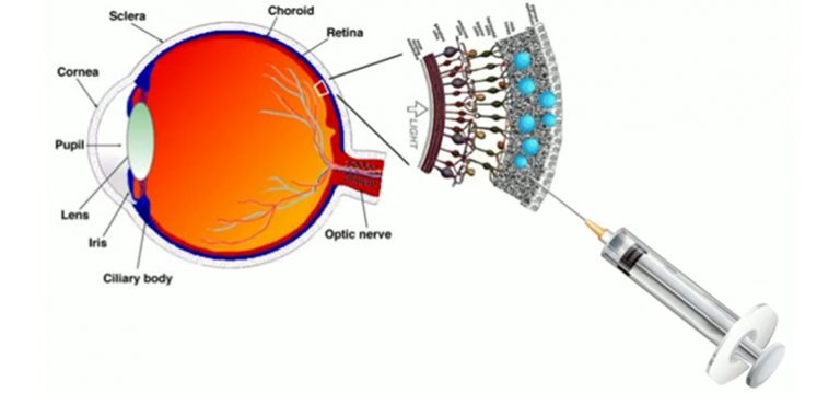
But that day in the conference room, He’s reputation remained untarnished. As the couples listened and flipped through the forms, occasionally asking questions, two witnesses—one American, the other Chinese—observed [emphasis mine]. Another lab member shot video, which Science has seen [emphasis mine], of part of the 50-minute meeting. He had recruited those couples because the husbands were living with HIV infections kept under control by antiviral drugs. The IVF procedure would use a reliable process called sperm washing to remove the virus before insemination, so father-to-child transmission was not a concern. Rather, He sought couples who had endured HIV-related stigma and discrimination and wanted to spare their children that fate by dramatically reducing their risk of ever becoming infected. [emphasis mine]

He, who for much of his brief career had specialized in sequencing DNA, offered a potential solution: CRISPR, the genome-editing tool that was revolutionizing biology, could alter a gene in IVF embryos to cripple production of an immune cell surface protein, CCR5, that HIV uses to establish an infection. “This technique may be able to produce an IVF baby naturally immunized against AIDS,” one consent form read.[emphasis mine]

The couples’ children could also pass the protective mutation to future generations. The prospect of this irrevocable genetic change is why, since the advent of CRISPR as a genome editor 5 years earlier, the editing of human embryos, eggs, or sperm has been hotly debated. The core issue is whether such germline editing would cross an ethical red line because it could ultimately alter our species. Regulations, some with squishy language, arguably prohibited it in many countries, China included.

Yet opposition was not unanimous. A few months before He met the couples, a committee convened by the U.S. National Academies of Sciences, Engineering, and Medicine (NASEM) concluded in a well-publicized report that human trials of germline editing “might be permitted” if strict criteria were met. The group of scientists, lawyers, bioethicists, and patient advocates spelled out a regulatory framework but cautioned that “these criteria are necessarily vague” because various societies, caregivers, and patients would view them differently. The committee notably did not call for an international ban, arguing instead for governmental regulation as each country deemed appropriate and “voluntary self-regulation pursuant to professional guidelines.”

[…] He hid his plans and deceived his colleagues and superiors, as many people have asserted? A preliminary investigation in China stated that He had forged documents, “dodged supervision,” and misrepresented blood tests—even though no proof of those charges was released [emphasis mine], no outsiders were part of the inquiry, and He has not publicly admitted to any wrongdoing. (CRISPR scientists in China say the He fallout has affected their research.) Many scientists outside China also portrayed He as a rogue actor. “I think there has been a failure of self-regulation by the scientific community because of a lack of transparency,” virologist David Baltimore, a Nobel Prize–winning researcher at the California Institute of Technology (Caltech) in Pasadena and co-chair of the Hong Kong summit, thundered at He after the biophysicist’s only public talk on the experiment.

Because the Chinese government has revealed little and He is not talking, key questions about his actions are hard to answer. Many of his colleagues and confidants also ignored Science‘s requests for interviews. But Ryan Ferrell, a public relations specialist He hired, has cataloged five dozen people who were not part of the study but knew or suspected what He was doing before it became public. Ferrell calls it He’s circle of trust. [emphasis mine]

That circle included leading scientists—among them a Nobel laureate—in China and the United States, business executives, an entrepreneur connected to venture capitalists, authors of the NASEM report, a controversial U.S. IVF specialist [John Zhang] who discussed opening a gene-editing clinic with He [emphasis mine], and at least one Chinese politician. “He had an awful lot of company to be called a ‘rogue,’” says geneticist George Church [emphases mine], a CRISPR pioneer at Harvard University who was not in the circle of trust and is one of the few scientists to defend at least some aspects of He’s experiment.

Some people sharply criticized He when he brought them into the circle; others appear to have welcomed his plans or did nothing. Several went out of their way to distance themselves from He after the furor erupted. For example, the two onlookers in that informed consent meeting were Michael Deem, He’s Ph.D. adviser at Rice University in Houston, Texas, and Yu Jun, a member of the Chinese Academy of Sciences (CAS) and co-founder of the Beijing Genomics Institute, the famed DNA sequencing company in Shenzhen. Deem remains under investigation by Rice for his role in the experiment and would not speak with Science. In a carefully worded statement, Deem’s lawyers later said he “did not meet the parents of the reported CCR5-edited children, or anyone else whose embryos were edited.” But earlier, Deem cooperated with the Associated Press (AP) for its exclusive story revealing the birth of the babies, which reported that Deem was “present in China when potential participants gave their consent and that he ‘absolutely’ thinks they were able to understand the risks. [emphasis mine]”

Yu, who works at CAS’s Beijing Institute of Genomics, acknowledges attending the informed consent meeting with Deem, but he told Science he did not know that He planned to implant gene-edited embryos. “Deem and I were chatting about something else,” says Yu, who has sequenced the genomes of humans, rice, silkworms, and date palms. “What was happening in the room was not my business, and that’s my personality: If it’s not my business, I pay very little attention.”

Some people who know He and have spoken to Science contend it is time for a more open discussion of how the biophysicist formed his circle of confidants and how the larger circle of trust—the one between the scientific community and the public—broke down. Bioethicist William Hurlbut at Stanford University [emphasis mine] in Palo Alto, California, who knew He wanted to conduct the embryo-editing experiment and tried to dissuade him, says that He was “thrown under the bus” by many people who once supported him. “Everyone ran for the exits, in both the U.S. and China. I think everybody would do better if they would just openly admit what they knew and what they did, and then collectively say, ‘Well, people weren’t clear what to do. We should all admit this is an unfamiliar terrain.’”

Steve Lombardi, a former CEO of Helicos, reacted far more charitably. Lombardi, who runs a consulting business in Bridgewater, Connecticut, says Quake introduced him to He to help find investors for Direct Genomics. “He’s your classic, incredibly bright, naïve entrepreneur—I run into them all the time,” Lombardi says. “He had the right instincts for what to do in China and just didn’t know how to do it. So I put him in front of as many people as I could.” Lombardi says He told him about his embryo-editing ambitions in August 2017, asking whether Lombardi could find investors for a new company that focused on “genetic medical tourism” and was based in China or, because of a potentially friendlier regulatory climate, Thailand. “I kept saying to him, ‘You know, you’ve got to deal with the ethics of this and be really sure that you know what you’re doing.’”

In April 2018, He asked Ferrell to handle his media full time. Ferrell was a good fit—he had an undergraduate degree in neuroscience, had spent a year in Beijing studying Chinese, and had helped another company using a pre-CRISPR genome editor. Now that a woman in the trial was pregnant, Ferrell says, He’s “understanding of the gravity of what he had done increased.” Ferrell had misgivings about the experiment, but he quit HDMZ and that August moved to Shenzhen. With the pregnancy already underway, Ferrell reasoned, “It was going to be the biggest science story of that week or longer, no matter what I did.”

MIT Technology Review had broken a story early that morning China time, saying human embryos were being edited and implanted, after reporter Antonio Regalado discovered descriptions of the project that He had posted online, without Ferrell’s knowledge, in an official Chinese clinical trial registry. Now, He gave AP the green light to post a detailed account, which revealed that twin girls—whom He, to protect their identifies, named Lulu and Nana—had been born. Ferrell and He also posted five unfinished YouTube videos explaining and justifying the unprecedented experiment.

“He was fearful that he’d be unable to communicate to the press and the onslaught in a way that would be in any way manageable for him,” Ferrell says. One video tried to forestall eugenics accusations, with He rejecting goals such as enhancing intelligence, changing skin color, and increasing sports performance as “not love.” Still, the group knew it had lost control of the news. [emphasis mine]

… On 7 March 2017, 5 weeks after the California gathering, He submitted a medical ethics approval application to the Shenzhen HarMoniCare Women and Children’s Hospital that outlined the planned CCR5 edit of human embryos. The babies, it claimed, would be resistant to HIV as well as to smallpox and cholera. (The natural CCR5 mutation may have been selected for because it helps carriers survive smallpox and plague, some studies suggest—but they don’t mention cholera.) “This is going to be a great science and medicine achievement ever since the IVF technology which was awarded the Nobel Prize in 2010, and will also bring hope to numerous genetic disease patients,” the application says. Seven people on the ethics committee, chaired by Lin Zhitong—a one-time Direct Genomics director and a HarMoniCare administrator—signed the application, indicating they approved it.

[…] John Zhang, […] [emphasis mine] earned his medical degree in China and a Ph.D. in reproductive biology at the University of Cambridge in the United Kingdom. Zhang had made international headlines himself in September 2016, when New Scientist revealed that he had created the world’s first “three-parent baby” by using mitochondrial DNA from a donor egg to revitalize the egg of a woman with infertility and then inseminating the resulting egg. “This technology holds great hope for ladies with advanced maternal age to have their own children with their own eggs,” Zhang explains in the center’s promotional video, which alternates between Chinese and English. It does not mention that Zhang did the IVF experiment in Mexico because it is not now allowed in the United States. [emphasis mine]

When Science contacted Zhang, the physician initially said he barely knew He: [emphases mine] “I know him just like many people know him, in an academic meeting.”

After his talk [November 2018 at Hong Kong meeting], He immediately drove back to Shenzhen, and his circle of trust began to disintegrate. He has not spoken publicly since. “I don’t think he can recover himself through PR,” says Ferrell, who no longer works for He but recently started to do part-time work for He’s wife. “He has to do other service to the world.”

Calls for a moratorium on human germline editing have increased, although at the end of the Hong Kong summit, the organizing committee declined in its consensus to call for a ban. China has stiffened its regulations on work with human embryos, and Chinese bioethicists in a Nature editorial about the incident urged the country to confront “the eugenic thinking that has persisted among a small proportion of Chinese scholars.”

Church, who has many CRISPR collaborations in China, finds it inconceivable that He’s work surprised the Chinese government. China has “the best surveillance system in the world,” he says. “I conclude that they were totally aware of what he was doing at every step of the way, especially because he wasn’t particularly secretive about it.”

Benjamin Hurlbut, William’s son and a historian of biomedicine at Arizona State University in Tempe, says leaders in the scientific community should take a hard look at their actions, too. [emphases mine] He thinks the 2017 NASEM report helped give rise to He by following a well-established approach to guiding science: appointing an elite group to decide how scientists should be regulated. Benjamin Hurlbut, whose book Experiments in Democracy explores the governance of embryo research and bioethics, questions why small, scientist-led groups—à la the totemic Asilomar conference held in 1975 to discuss the future of recombinant DNA research—are seen as the best way to shape thinking about new technologies. Hurlbut has called for a “global observatory for gene editing” to convene meetings with diverse perspectives.

The prevailing notion that the scientific community simply “failed to see the rogue among the responsible,” Hurlbut says, is a convenient narrative for those scientific leaders and inhibits their ability to learn from such failures. [emphases mine] “It puts them on the right side of history,” he says. They failed to paint a bright enough red line, Hurlbut contends. “They are not on the right side of history because they contributed to this.”

If you have the time, I strongly recommend reading Cohen’s piece in its entirety. You’ll find links to the reports and more articles with in-depth reporting on this topic.

A little kindness and no regrets

William Hurlbut was interviewed in an As it happens (Canadian Broadcasting Corporation’ CBC) radio programme segment on December 30, 2020. This is an excerpt from the story transcript written by Sheena Goodyear (Note: A link has been removed),

Dr. William Hurlbut, a physician and professor of neural-biology at Stanford University, says he tried to warn He to slow down before it was too late. Here is part of his conversation with As It Happens guest host Helen Mann.

What was your reaction to the news that Dr. He had been sentenced to three years in prison?

My first reaction was one of sadness because I know Dr. He — who we call J.K., that’s his nickname.

I spent quite a few hours talking with him, and I’m just sad that this worked out this way. It didn’t work out well for him or for his country or for the world, in some sense.

Except the one good thing is it’s alerted us, it’s awakened the world, to the seriousness of the issues that are coming down toward us with biotechnology, especially in genetics.

How does he feel about [how] not just the Chinese government, but the world generally, responded to his experiment?

He was surprised, personally. But I had actually warned him that he was proceeding too fast, and I didn’t know he had implanted embryos.

We had several conversations before this was disclosed, and I warned him to go more slowly and to keep in conversation with the rest of the international scientific community, and more broadly the international perspectives on social and ethical matters.

He was doing that to some extent, but not deeply enough and not transparently enough.

It sounds like you were very thoughtful in the conversations you had with him and the advice you gave him. And I guess you operated with what you had. But do you have any regrets yourself?

I don’t have any regrets about the way I conducted myself. I regret that this happened this way for J.K., who is a very bright person, and a very nice person, a humble person.

He grew up in a poor urban farming village. He told me that at one point he wanted to ask out a certain girl that he thought was really pretty … but he was embarrassed to do so because her family owned the restaurant. And so you see how humble his origins were.

By the way, he did end up asking her out and he ended up marrying her, which is a happy story, except now they’re separated for years of crucial time, and they have little children. 

I know this is a bigger story than just J.K. and his family. But there’s a personal story to it too.

What happens He Jiankui? … Is his research career over?

It’s hard to imagine that a nation like China would not give him some some useful role in their society. A very intelligent and very well-educated young man. 

But on the other hand, he will be forever a sign of a very crucial and difficult moment for the human species. He’s not going outlive that.

It’s going to be interesting. I hope I get a chance to have good conversations with him again and hear his internal ruminations and perspectives on it all.

This (“I don’t have any regrets about the way I conducted myself”) is where Hurlbut lost me. I think he could have suggested that he’d reviewed and rethought everything and feels that he and others could have done better and maybe they need to rethink how scientists are trained and how we talk about science, genetics, and emerging technology. Interestingly, it’s his son who comes up with something closer to what I’m suggesting (this excerpt was quoted earlier in this posting from a December 30, 2019 article, by Carolyn Y. Johnson for the Washington Post),

“The fact that the individual at the center of the story has been punished for his role in it should not distract us from examining what supporting roles were played by others, particularly in the international scientific community and also the environment that shaped and encouraged him to push the limits,” said Benjamin Hurlbut [emphasis mine], associate professor in the School of Life Sciences at Arizona State University.

The man who CRISPRs himself approves

Josiah Zayner publicly injected himself with CRISPR in a demonstration (see my January 25, 2018 posting for details about Zayner, his demonstration, and his plans). As you might expect, his take on the He affair is quite individual. From a January 2, 2020 article for STAT, Zayner presents the case for Dr. He’s work (Note: Links have been removed),

When I saw the news that He Jiankui and colleagues had been sentenced to three years in prison for the first human embryo gene editing and implantation experiments, all I could think was, “How will we look back at what they had done in 100 years?”

When the scientist described his research and revealed the births of gene edited twin girls at the [Second] International Summit on Human Genome Editing in Hong Kong in late November 2018, I stayed up into the early hours of the morning in Oakland, Calif., watching it. Afterward, I couldn’t sleep for a few days and couldn’t stop thinking about his achievement.

This was the first time a viable human embryo was edited and allowed to live past 14 days, much less the first time such an embryo was implanted and the baby brought to term.

The majority of scientists were outraged at the ethics of what had taken place, despite having very little information on what had actually occurred.

To me, no matter how abhorrent one views [sic] the research, it represents a substantial step forward in human embryo editing. Now there is a clear path forward that anyone can follow when before it had been only a dream.

As long as the children He Jiankui engineered haven’t been harmed by the experiment, he is just a scientist who forged some documents to convince medical doctors to implant gene-edited embryos. The 4-minute mile of human genetic engineering has been broken. It will happen again.

The academic establishment and federal funding regulations have made it easy to control the number of heretical scientists. We rarely if ever hear of individuals pushing the ethical and legal boundaries of science.

The rise of the biohacker is changing that.

A biohacker is a scientist who exists outside academia or an institution. By this definition, He Jiankui is a biohacker. I’m also part of this community, and helped build an organization to support it.

Such individuals have much more freedom than “traditional” scientists because scientific regulation in the U.S. is very much institutionally enforced by the universities, research organizations, or grant-giving agencies. But if you are your own institution and don’t require federal grants, who can police you? If you don’t tell anyone what you are doing, there is no way to stop you — especially since there is no government agency actively trying to stop people from editing embryos.

… When a human embryo being edited and implanted is no longer interesting enough for a news story, will we still view He Jiankui as a villain?

I don’t think we will. But even if we do, He Jiankui will be remembered and talked about more than any scientist of our day. Although that may seriously aggravate many scientists and bioethicists, I think he deserves that honor.

Josiah Zayner is CEO of The ODIN, a company that teaches people how to do genetic engineering in their homes.

You can find The ODIN here.

Final comments

There can’t be any question that this was inevitable. One needs only to take a brief stroll through the history of science to know that scientists are going to push boundaries or, as in this case, press past an ill-defined grey zone.

The only scientists who are being publicly punished for hubris are Dr. He Jiankui and his two colleagues in China. Dr. Michael Deem is still working for Rice University as far as I can determine. Here’s how the Wikipedia entry for the He Jiankui Affair describes the investigation (Note: Links have been removed),

Michael W. Deem, an American bioengineering professor at Rice University and He’s doctoral advisor, was involved in the research, and was present when people involved in He’s study gave consent.[24] He was the only non-Chinese out of 10 authors listed in the manuscript submitted to Nature.[30] Deem came under investigation by Rice University after news of the work was made public.[58] As of 31 December 2019, the university had not released a decision.[59] [emphasis mine]

Meanwhile the scientists at Stanford are cleared. While there are comments about the Chinese government not being transparent, it seems to me that US universities are just as opaque.

What seems missing from all this discussion and opprobrium is that the CRISPR technology itself is problematic. My September 20, 2019 post features research into off-target results from CRISPR gene-editing and, prior, there was this July 17, 2018 posting (The CRISPR [clustered regularly interspaced short palindromic repeats]-CAS9 gene-editing technique may cause new genetic damage kerfuffle).

I’d like to see more discussion and, in line with Benjamin Hurlbut’s thinking, I’d like to see more than a small group of experts talking to each other as part of the process especially here in Canada and in light of efforts to remove our ban on germline-editing (see my April 26, 2019 posting for more about those efforts).

3D-printed graphene sensors for highly sensitive food freshness detection

I love the opening line (lede). From a June 29, 2020 news item on Nanowerk,

Researchers dipped their new, printed sensors into tuna broth and watched the readings.

It turned out the sensors – printed with high-resolution aerosol jet printers on a flexible polymer film and tuned to test for histamine, an allergen and indicator of spoiled fish and meat – can detect histamine down to 3.41 parts per million.

The U.S. Food and Drug Administration has set histamine guidelines of 50 parts per million in fish, making the sensors more than sensitive enough to track food freshness and safety.

I find using 3D-printing techniques to produce graphene, a 2-d material, intriguing. Apparently, the technique is cheaper and offers an advantage as it allows for greater precision than other techniques (inkjet printing, chemical vapour depostion [CVD], etc.)

Here’s more detail from a June 25, 2020 Iowa State University news release (also on EurekAlert but published June 29, 2020), which originated the news item,

Making the sensor technology possible is graphene, a supermaterial that’s a carbon honeycomb just an atom thick and known for its strength, electrical conductivity, flexibility and biocompatibility. Making graphene practical on a disposable food-safety sensor is a low-cost, aerosol-jet-printing technology that’s precise enough to create the high-resolution electrodes necessary for electrochemical sensors to detect small molecules such as histamine.

“This fine resolution is important,” said Jonathan Claussen, an associate professor of mechanical engineering at Iowa State University and one of the leaders of the research project. “The closer we can print these electrode fingers, in general, the higher the sensitivity of these biosensors.”

Claussen and the other project leaders – Carmen Gomes, an associate professor of mechanical engineering at Iowa State; and Mark Hersam, the Walter P. Murphy Professor of Materials Science and Engineering at Northwestern University in Evanston, Illinois – have recently reported their sensor discovery in a paper published online by the journal 2D Materials. (…)

The paper describes how graphene electrodes were aerosol jet printed on a flexible polymer and then converted to histamine sensors by chemically binding histamine antibodies to the graphene. The antibodies specifically bind histamine molecules.

The histamine blocks electron transfer and increases electrical resistance, Gomes said. That change in resistance can be measured and recorded by the sensor.

“This histamine sensor is not only for fish,” Gomes said. “Bacteria in food produce histamine. So it can be a good indicator of the shelf life of food.”

The researchers believe the concept will work to detect other kinds of molecules, too.

“Beyond the histamine case study presented here, the (aerosol jet printing) and functionalization process can likely be generalized to a diverse range of sensing applications including environmental toxin detection, foodborne pathogen detection, wearable health monitoring, and health diagnostics,” they wrote in their research paper.

For example, by switching the antibodies bonded to the printed sensors, they could detect salmonella bacteria, or cancers or animal diseases such as avian influenza, the researchers wrote.

Claussen, Hersam and other collaborators (…) have demonstrated broader application of the technology by modifying the aerosol-jet-printed sensors to detect cytokines, or markers of inflammation. The sensors, as reported in a recent paper published by ACS Applied Materials & Interfaces, can monitor immune system function in cattle and detect deadly and contagious paratuberculosis at early stages.

Claussen, who has been working with printed graphene for years, said the sensors have another characteristic that makes them very useful: They don’t cost a lot of money and can be scaled up for mass production.

“Any food sensor has to be really cheap,” Gomes said. “You have to test a lot of food samples and you can’t add a lot of cost.”

Claussen and Gomes know something about the food industry and how it tests for food safety. Claussen is chief scientific officer and Gomes is chief research officer for NanoSpy Inc., a startup company based in the Iowa State University Research Park that sells biosensors to food processing companies.

They said the company is in the process of licensing this new histamine and cytokine sensor technology.

It, after all, is what they’re looking for in a commercial sensor. “This,” Claussen said, “is a cheap, scalable, biosensor platform.”

Here’s a link to and a citation for the two papers mentioned in the news release,

Aerosol-jet-printed graphene electrochemical histamine sensors for food safety monitoring by Kshama Parate, Cícero C Pola, Sonal V Rangnekar, Deyny L Mendivelso-Perez, Emily A Smith, Mark C Hersam, Carmen L Gomes and Jonathan C Claussen. 2D Materials, Volume 7, Number 3 DOI https://doi.org/10.1088/2053-1583/ab8919 Published 10 June 2020 • © 2020 IOP Publishing Ltd

Aerosol-Jet-Printed Graphene Immunosensor for Label-Free Cytokine Monitoring in Serum by Kshama Parate, Sonal V. Rangnekar, Dapeng Jing, Deyny L. Mendivelso-Perez, Shaowei Ding, Ethan B. Secor, Emily A. Smith, Jesse M. Hostetter, Mark C. Hersam, and Jonathan C. Claussen. ACS Appl. Mater. Interfaces 2020, 12, 7, 8592–8603 DOI: https://doi.org/10.1021/acsami.9b22183 Publication Date: February 10, 2020 Copyright © 2020 American Chemical Society

Both papers are behind paywalls.

You can find the NanoSpy website here.

Shining a light on flurocarbon bonds and robotic ‘soft’ matter research

Both of these news bits are concerned with light for one reason or another.

Rice University (Texas, US) and breaking fluorocarbon bonds

The secret to breaking fluorocarbon bonds is light according to a June 22, 2020 news item on Nanowerk,

Rice University engineers have created a light-powered catalyst that can break the strong chemical bonds in fluorocarbons, a group of synthetic materials that includes persistent environmental pollutants.

A June 22, 2020 Rice University news release (also on EurekAlert), which originated the news item, describes the work in greater detail,

In a study published this month in Nature Catalysis, Rice nanophotonics pioneer Naomi Halas and collaborators at the University of California, Santa Barbara (UCSB) and Princeton University showed that tiny spheres of aluminum dotted with specks of palladium could break carbon-fluorine (C-F) bonds via a catalytic process known as hydrodefluorination in which a fluorine atom is replaced by an atom of hydrogen.

The strength and stability of C-F bonds are behind some of the 20th century’s most recognizable chemical brands, including Teflon, Freon and Scotchgard. But the strength of those bonds can be problematic when fluorocarbons get into the air, soil and water. Chlorofluorocarbons, or CFCs, for example, were banned by international treaty in the 1980s after they were found to be destroying Earth’s protective ozone layer, and other fluorocarbons were on the list of “forever chemicals” targeted by a 2001 treaty.

“The hardest part about remediating any of the fluorine-containing compounds is breaking the C-F bond; it requires a lot of energy,” said Halas, an engineer and chemist whose Laboratory for Nanophotonics (LANP) specializes in creating and studying nanoparticles that interact with light.

Over the past five years, Halas and colleagues have pioneered methods for making “antenna-reactor” catalysts that spur or speed up chemical reactions. While catalysts are widely used in industry, they are typically used in energy-intensive processes that require high temperature, high pressure or both. For example, a mesh of catalytic material is inserted into a high-pressure vessel at a chemical plant, and natural gas or another fossil fuel is burned to heat the gas or liquid that’s flowed through the mesh. LANP’s antenna-reactors dramatically improve energy efficiency by capturing light energy and inserting it directly at the point of the catalytic reaction.

In the Nature Catalysis study, the energy-capturing antenna is an aluminum particle smaller than a living cell, and the reactors are islands of palladium scattered across the aluminum surface. The energy-saving feature of antenna-reactor catalysts is perhaps best illustrated by another of Halas’ previous successes: solar steam. In 2012, her team showed its energy-harvesting particles could instantly vaporize water molecules near their surface, meaning Halas and colleagues could make steam without boiling water. To drive home the point, they showed they could make steam from ice-cold water.

The antenna-reactor catalyst design allows Halas’ team to mix and match metals that are best suited for capturing light and catalyzing reactions in a particular context. The work is part of the green chemistry movement toward cleaner, more efficient chemical processes, and LANP has previously demonstrated catalysts for producing ethylene and syngas and for splitting ammonia to produce hydrogen fuel.

Study lead author Hossein Robatjazi, a Beckman Postdoctoral Fellow at UCSB who earned his Ph.D. from Rice in 2019, conducted the bulk of the research during his graduate studies in Halas’ lab. He said the project also shows the importance of interdisciplinary collaboration.

“I finished the experiments last year, but our experimental results had some interesting features, changes to the reaction kinetics under illumination, that raised an important but interesting question: What role does light play to promote the C-F breaking chemistry?” he said.

The answers came after Robatjazi arrived for his postdoctoral experience at UCSB. He was tasked with developing a microkinetics model, and a combination of insights from the model and from theoretical calculations performed by collaborators at Princeton helped explain the puzzling results.

“With this model, we used the perspective from surface science in traditional catalysis to uniquely link the experimental results to changes to the reaction pathway and reactivity under the light,” he said.

The demonstration experiments on fluoromethane could be just the beginning for the C-F breaking catalyst.

“This general reaction may be useful for remediating many other types of fluorinated molecules,” Halas said.

Caption: An artist’s illustration of the light-activated antenna-reactor catalyst Rice University engineers designed to break carbon-fluorine bonds in fluorocarbons. The aluminum portion of the particle (white and pink) captures energy from light (green), activating islands of palladium catalysts (red). In the inset, fluoromethane molecules (top) comprised of one carbon atom (black), three hydrogen atoms (grey) and one fluorine atom (light blue) react with deuterium (yellow) molecules near the palladium surface (black), cleaving the carbon-fluorine bond to produce deuterium fluoride (right) and monodeuterated methane (bottom). Credit: H. Robatjazi/Rice University

Here’s a link to and a citation for the paper,

Plasmon-driven carbon–fluorine (C(sp3)–F) bond activation with mechanistic insights into hot-carrier-mediated pathways by Hossein Robatjazi, Junwei Lucas Bao, Ming Zhang, Linan Zhou, Phillip Christopher, Emily A. Carter, Peter Nordlander & Naomi J. Halas. Nature Catalysis (2020) DOI: https://doi.org/10.1038/s41929-020-0466-5 Published: 08 June 2020

This paper is behind a paywall.

Northwestern University (Illinois, US) brings soft robots to ‘life’

This June 22, 2020 news item on ScienceDaily reveals how scientists are getting soft robots to mimic living creatures,

Northwestern University researchers have developed a family of soft materials that imitates living creatures.

When hit with light, the film-thin materials come alive — bending, rotating and even crawling on surfaces.

A June 22, 2020 Northwestern University news release (also on EurekAlert) by Amanda Morris, which originated the news item, delves further into the details,

Called “robotic soft matter by the Northwestern team,” the materials move without complex hardware, hydraulics or electricity. The researchers believe the lifelike materials could carry out many tasks, with potential applications in energy, environmental remediation and advanced medicine.

“We live in an era in which increasingly smarter devices are constantly being developed to help us manage our everyday lives,” said Northwestern’s Samuel I. Stupp, who led the experimental studies. “The next frontier is in the development of new science that will bring inert materials to life for our benefit — by designing them to acquire capabilities of living creatures.”

The research will be published on June 22 [2020] in the journal Nature Materials.

Stupp is the Board of Trustees Professor of Materials Science and Engineering, Chemistry, Medicine and Biomedical Engineering at Northwestern and director of the Simpson Querrey Institute He has appointments in the McCormick School of Engineering, Weinberg College of Arts and Sciences and Feinberg School of Medicine. George Schatz, the Charles E. and Emma H. Morrison Professor of Chemistry in Weinberg, led computer simulations of the materials’ lifelike behaviors. Postdoctoral fellow Chuang Li and graduate student Aysenur Iscen, from the Stupp and Schatz laboratories, respectively, are co-first authors of the paper.

Although the moving material seems miraculous, sophisticated science is at play. Its structure comprises nanoscale peptide assemblies that drain water molecules out of the material. An expert in materials chemistry, Stupp linked the peptide arrays to polymer networks designed to be chemically responsive to blue light.

When light hits the material, the network chemically shifts from hydrophilic (attracts water) to hydrophobic (resists water). As the material expels the water through its peptide “pipes,” it contracts — and comes to life. When the light is turned off, water re-enters the material, which expands as it reverts to a hydrophilic structure.

This is reminiscent of the reversible contraction of muscles, which inspired Stupp and his team to design the new materials.

“From biological systems, we learned that the magic of muscles is based on the connection between assemblies of small proteins and giant protein polymers that expand and contract,” Stupp said. “Muscles do this using a chemical fuel rather than light to generate mechanical energy.”

For Northwestern’s bio-inspired material, localized light can trigger directional motion. In other words, bending can occur in different directions, depending on where the light is located. And changing the direction of the light also can force the object to turn as it crawls on a surface.

Stupp and his team believe there are endless possible applications for this new family of materials. With the ability to be designed in different shapes, the materials could play a role in a variety of tasks, ranging from environmental clean-up to brain surgery.

“These materials could augment the function of soft robots needed to pick up fragile objects and then release them in a precise location,” he said. “In medicine, for example, soft materials with ‘living’ characteristics could bend or change shape to retrieve blood clots in the brain after a stroke. They also could swim to clean water supplies and sea water or even undertake healing tasks to repair defects in batteries, membranes and chemical reactors.”

Fascinating, eh? No batteries, no power source, just light to power movement. For the curious, here’s a link to and a citation for the paper,

Supramolecular–covalent hybrid polymers for light-activated mechanical actuation by Chuang Li, Aysenur Iscen, Hiroaki Sai, Kohei Sato, Nicholas A. Sather, Stacey M. Chin, Zaida Álvarez, Liam C. Palmer, George C. Schatz & Samuel I. Stupp. Nature Materials (2020) DOI: https://doi.org/10.1038/s41563-020-0707-7 Published: 22 June 2020

This paper is behind a paywall.

New capacitor for better wearable electronics?

Supercapacitors are a predictable source of scientific interest and excitement. The latest entry in the ‘supercapacitor stakes’ is from a Russian/Finnish/US team according to a June 11, 2020 Skoltech (Skolkovo Institute of Science and Technology) press release (also on EurekAlert),

Researchers from Skoltech [Russia], Aalto University [Finland] and Massachusetts Institute of Technology [MIT; US] have designed a high-performance, low-cost, environmentally friendly, and stretchable supercapacitor that can potentially be used in wearable electronics. The paper was published in the Journal of Energy Storage.

Supercapacitors, with their high power density, fast charge-discharge rates, long cycle life, and cost-effectiveness, are a promising power source for everything from mobile and wearable electronics to electric vehicles. However, combining high energy density, safety, and eco-friendliness in one supercapacitor suitable for small devices has been rather challenging.

“Usually, organic solvents are used to increase the energy density. These are hazardous, not environmentally friendly, and they reduce the power density compared to aqueous electrolytes with higher conductivity,” says Professor Tanja Kallio from Aalto University, a co-author of the paper.

The researchers proposed a new design for a “green” and simple-to-fabricate supercapacitor. It consists of a solid-state material based on nitrogen-doped graphene flake electrodes distributed in the NaCl-containing hydrogel electrolyte. This structure is sandwiched between two single-walled carbon nanotube film current collectors, which provides stretchability. Hydrogel in the supercapacitor design enables compact packing and high energy density and allows them to use the environmentally friendly electrolyte.

The scientists managed to improve the volumetric capacitive performance, high energy density and power density for the prototype over analogous supercapacitors described in previous research. “We fabricated a prototype with unchanged performance under the 50% strain after a thousand stretching cycles. To ensure lower cost and better environmental performance, we used a NaCl-based electrolyte. Still the fabrication cost can be lowered down by implementation of 3D printing or other advanced fabrication techniques,” concluded Skoltech professor Albert Nasibulin.

Here’s a link to and a citation for the paper,

Superior environmentally friendly stretchable supercapacitor based on nitrogen-doped graphene/hydrogel and single-walled carbon nanotubes by Evgeniia Gilshtein, Cristina Flox, Farhan S.M. Ali, Bahareh Mehrabimatin, Fedor S.Fedorov, Shaoting Lin, Xuanhe Zhao, Albert G. Nasibulin, Tanja Kallio. Journal of Energy Storage Volume 30, August 2020, 101505 DOI: https://doi.org/10.1016/j.est.2020.101505

This paper is behind a paywall.

I’m trying to remember if I’ve ever before seen a material that combines graphene and single-walled carbon nanotubes (SWCNTs). Anyway, here’s an image the researchers are using illustrate their work,

Caption: This is an outline of the new supercapacitor. Credit: Pavel Odinev / Skoltech

Worried your ‘priceless’ art could be ruined? Genomics could be the answer

First, there was the story about art masterpieces turning into soap (my June 22, 2017 posting) and now, it seems that microbes may also constitute a problem. Before getting to the latest research, here’s are some images the researchers are using to illustrate their work,

Caption: Leonardo da Vinci noted that the fore and hind wings of a dragonfly are out of phase — verified centuries later by slow motion photography. Thaler suggests further study to compare individuals and species with high “flicker fusion frequency” ability. Credit: PXFuel

I’m not sure what that has to do with anything but I do love dragonflies. This next image seems more relevant to the research,

Caption: Photo summary of the various artworks sampled for the study “”Characterizing microbial signatures on sculptures and paintings of similar provenance.” Circles indicate swabbed areas on each sample artwork Credit: JCVI

It turns out, the researchers are releasing two pieces of research in the same press release, neither having much to do with the other. They (art conservation rresearch, first and, then, research into vision [hence the dragonfly] and da Vinci’s eyes) are both described in a June 18, 2020 J. Craig Venter Institute (JCVI)-Leonardo Da Vinci DNA Project press release (also on EurekAlert),

A new study of the microbial settlers on old paintings, sculptures, and other forms of art charts a potential path for preserving, restoring, and confirming the geographic origin of some of humanity’s greatest treasures.

Genetics scientists with the J. Craig Venter Institute (JCVI), collaborating with the Leonardo da Vinci DNA Project and supported by the Richard Lounsbery Foundation, say identifying and managing communities of microbes on art may offer museums and collectors a new way to stem the deterioration of priceless possessions, and to unmask counterfeits in the $60 billion a year art market.

Manolito G. Torralba, Claire Kuelbs, Kelvin Jens Moncera, and Karen E. Nelson of the JCVI, La Jolla, California, and Rhonda Roby of the Alameda California County Sheriff’s Office Crime Laboratory, used small, dry polyester swabs to gently collect microbes from centuries-old, Renaissance-style art in a private collector’s home in Florence, Italy. Their findings are published in the journal Microbial Ecology .

The genetic detectives caution that additional time and research are needed to formally convict microbes as a culprit in artwork decay but consider their most interesting find to be “oxidase positive” microbes primarily on painted wood and canvas surfaces.

These species can dine on organic and inorganic compounds often found in paints, in glue, and in the cellulose in paper, canvas, and wood. Using oxygen for energy production, they can produce water or hydrogen peroxide, a chemical used in disinfectants and bleaches.

“Such byproducts are likely to influence the presence of mold and the overall rate of deterioration,” the paper says.

“Though prior studies have attempted to characterize the microbial composition associated with artwork decay, our results summarize the first large scale genomics-based study to understand the microbial communities associated with aging artwork.”

The study builds on an earlier one in which the authors compared hairs collected from people in the Washington D.C., and San Diego, CA. areas, finding that microbial signatures and patterns are geographically distinguishable.

In the art world context, studying microbes clinging to the surface of a work of art may help confirm its geographic origin and authenticity or identify counterfeits.

Lead author Manolito G. Torralba notes that, as art’s value continues to climb, preservation is increasingly important to museums and collectors alike, and typically involves mostly the monitoring and adjusting of lighting, heat, and moisture.

Adding genomics science to these efforts offers advantages of “immense potential.”

The study says microbial populations “were easily discernible between the different types of substrates sampled,” with those on stone and marble art more diverse than wood and canvas. This is “likely due to the porous nature of stone and marble harboring additional organisms and potentially moisture and nutrients, along with the likelihood of biofilm formation.”

As well, microbial diversity on paintings is likely lower because few organisms can metabolize the meagre nutrients offered by oil-based paint.

“Though our sample size is low, the novelty of our study has provided the art and scientific communities with evidence that microbial signatures are capable of differentiating artwork according to their substrate,” the paper says.

“Future studies would benefit from working with samples whose authorship, ownership, and care are well-documented, although documentation about care of works of art (e.g., whether and how they were cleaned) seems rare before the mid-twentieth century.”

“Of particular interest would be the presence and activity of oil-degrading enzymes. Such approaches will lead to fully understanding which organism(s) are responsible for the rapid decay of artwork while potentially using this information to target these organisms to prevent degradation.”

“Focusing on reducing the abundance of such destructive organisms has great potential in preserving and restoring important pieces of human history.”

Biology in Art

The paper was supported by the US-based Richard Lounsbery Foundation as part of its “biology in art” research theme, which has also included seed funding efforts to obtain and sequence the genome of Leonardo da Vinci.

The Leonardo da Vinci DNA Project involves scientists in France (where Leonardo lived during his final years and was buried), Italy (where his father and other relatives were buried, and descendants of his half-brothers still live), Spain (whose National Library holds 700 pages of his notebooks), and the US (where forensic DNA skills flourish).

The Leonardo project has convened molecular biologists, population geneticists, microbiologists, forensic experts, and physicians working together with other natural scientists and with genealogists, historians, artists, and curators to discover and decode previously inaccessible knowledge and to preserve cultural heritage.  

Related news release: Leonardo da Vinci’s DNA: Experts unite to shine modern light on a Renaissance master http://bit.ly/2FG4jJu

Measuring Leonardo da Vinci’s “quick eye” 500 years later.

Could he have played major-league baseball?

Famous art historians and biographers such as Sir Kenneth Clark and Walter Isaacson have written about Leonardo da Vinci’s “quick eye” because of the way he accurately captured fleeting expressions, wings during bird flight, and patterns in swirling water. But until now no one had tried to put a number on this aspect of Leonardo’s extraordinary visual acuity.

David S. Thaler of the University of Basel, and a guest investigator in the Program for the Human Environment at The Rockefeller University, does, allowing comparison of Leonardo with modern measures. Leonardo fares quite well.

Thaler’s estimate hinges on Leonardo’s observation that the fore and hind wings of a dragonfly are out of phase — not verified until centuries later by slow motion photography (see e.g. https://youtu.be/Lw2dfjYENNE?t=44).

To quote Isaacson’s translation of Leonardo’s notebook: “The dragonfly flies with four wings, and when those in front are raised those behind are lowered.”

Thaler challenged himself and friends to try seeing if that’s true, but they all saw only blurs.

High-speed camera studies by others show the fore and hind wingbeats of dragonflies vary by 20 to 10 milliseconds — one fiftieth to one hundredth of a second — beyond average human perception.

Thaler notes that “flicker fusion frequency” (FFF) — akin to a motion picture’s frames per second — is used to quantify and measure “temporal acuity” in human vision.

When frames per second exceed the number of frames the viewer can perceive individually, the brain constructs the illusion of continuous movement. The average person’s FFF is between 20 to 40 frames per second; current motion pictures present 48 or 72 frames per second.

To accurately see the angle between dragonfly wings would require temporal acuity in the range of 50 to 100 frames per second.

Thaler believes genetics will account for variations in FFF among different species, which range from a low of 12 in some nocturnal insects to over 300 in Fire Beetles. We simply do not know what accounts for human variation. Training and genetics may both play important roles.

“Perhaps the clearest contemporary case for a fast flicker fusion frequency in humans is in American baseball, because it is said that elite batters can see the seams on a pitched baseball,” even when rotating 30 to 50 times per second with two or four seams facing the batter. A batter would need Leonardo-esque FFF to spot the seams on most inbound baseballs.  

Thaler suggests further study to compare the genome of individuals and species with unusually high FFF, including, if possible, Leonardo’s DNA.  

Flicker fusion for focus, attention, and affection   

In a companion paper, Thaler describes how Leonardo used psychophysics that would only be understood centuries later — and about which a lot remains to be learned today — to communicate deep beauty and emotion. 

Leonardo was master of a technique known as sfumato (the word derived from the Italian sfumare, “to tone down” or “to evaporate like smoke”), which describes a subtle blur of edges and blending of colors without sharp focus or distinct lines.

Leonardo expert Martin Kemp has noted that Leonardo’s sfumato sometimes involves a distance dependence which is akin to the focal plane of a camera. Yet, at other times, features at the same distance have selective sfumato so simple plane of focus is not the whole answer.

Thaler suggests that Leonardo achieved selective soft focus in portraits by painting in overcast or evening light, where the eyes’ pupils enlarge to let in more light but have a narrow plane of sharp focus. 

To quote Leonardo’s notebook, under the heading “Selecting the light which gives most grace to faces”: “In the evening and when the weather is dull, what softness and delicacy you may perceive in the faces of men and women.”  In dim light pupils enlarge to let in more light but their depth of field decreases.  

By measuring the size of the portrait’s pupils, Thaler inferred Leonardo’s depth of focus. He says Leonardo likely sensed this effect, perhaps unconsciously in the realm of his artistic sensibility. The pupil / aperture effect on depth of focus wasn’t explained until the mid-1800s, centuries after Leonardo’s birth in Vinci, Italy in 1452.

What about selective focus at equal distance? In this case Leonardo may have taken advantage of the fovea, the small area on the back of the eye where detail is sharpest.

Most of us move our eyes around and because of our slower flicker fusion frequency we construct a single 3D image of the world by jamming together many partially in-focus images. Leonardo realized and “froze” separate snapshots with which we construct ordinary perception.

Says Thaler: “We study Leonardo not only to learn about him but to learn about ourselves and further human potential.”

Thaler’s papers (at https://bit.ly/2WZ2cwo and https://bit.ly/2ZBj7Hi) evolved from talks at meetings of the Leonardo da Vinci DNA Project in Italy (2018), Spain and France (2019).

They form part of a collection of papers presented at a recent colloquium in Amboise, France, now being readied for publication in a book: Actes du Colloque International d’Amboise: Leonardo de Vinci, Anatomiste. Pionnier de l’Anatomie comparée, de la Biomécanique, de la Bionique et de la Physiognomonie. Edited by Henry de Lumley, President, Institute of Human Paleontology, Paris, and originally planned for release in late spring, 2020, publication was delayed by the global virus pandemic but should be available at CNRS Editions in the second half of the summer.

Other papers in the collection cover a range of topics, including how Leonardo used his knowledge of anatomy, gained by performing autopsies on dozens of cadavers, to achieve Mona Lisa’s enigmatic smile.

Leonardo also used it to exact revenge on academics and scientists who ridiculed him for lacking a classical education, sketching them with absurdly deformed faces to resemble birds, dogs, or goats. 

De Lumley earlier co-authored a 72-page monograph for the Leonardo DNA Project: “Leonardo da Vinci: Pioneer of comparative anatomy, biomechanics and physiognomy.”.

Here’s a link to and a citation for the paper featuring microbes and art masterpiece,

Characterizing Microbial Signatures on Sculptures and Paintings of Similar Provenance by Manolito G. Torralba, Claire Kuelbs, Kelvin Jens Moncera, Rhonda Roby & Karen E. Nelson. Microbial Ecology (2020) DOI: https://doi.org/10.1007/s00248-020-01504-x Published: 21 May 2020

This paper is open access.

World’s first liquid retina prosthesis

The new artificial liquid retina is biomimetic and consists of an aqueous component in which photoactive polymeric nanoparticles (whose size is of 350 nanometres, thus about 1/100 of the diameter of a hair) are suspended, going to replace the damaged photoreceptors. Credit: IIT-Istituto Italiano di Tecnologia [image downloaded from https://www.medgadget.com/2020/06/injectable-liquid-prosthesis-to-treat-retinal-diseases-developed.html]

A June 29, 2020 news item on Nanowerk announces the world’s first liquid retina prosthesis,

Researchers at IIT-Istituto Italiano di Tecnologia (Italian Institute of Technology) has led to the revolutionary development of an artificial liquid retinal prosthesis to counteract the effects of diseases such as retinitis pigmentosa and age-related macular degeneration that cause the progressive degeneration of photoreceptors of the retina, resulting in blindness.

The multidisciplinary team is composed by researchers from the IIT’s Center for Synaptic Neuroscience and Technology in Genoa coordinated by Fabio Benfenati and a team from the IIT’s Center for Nano Science and Technology in Milan coordinated by Guglielmo Lanzani, and it also involves the IRCCS Ospedale Sacrocuore Don Calabria in Negrar (Verona) with the team lead by Grazia Pertile, the IRCCS Ospedale Policlinico San Martino in Genoa and the CNR in Bologna. The research has been supported by Fondazione 13 Marzo Onlus, Fondazione Ra.Mo., Rare Partners srl and Fondazione Cariplo.

The study represents the state of the art in retinal prosthetics and is an evolution of the planar artificial retinal model developed by the same team in 2017 and based on organic semiconductor materials (Nature Materials 2017, 16: 681-689).

A June 30, 2020 IIT-Istituto Italiano di Tecnologia (Italian Institute of Technology) press release (also on EurekAlert but published June 29, 2020) provides more detail,

The “second generation” artificial retina is biomimetic, offers high spatial resolution and consists of an aqueous component in which photoactive polymeric nanoparticles (whose size is of 350 nanometres, thus about 1/100 of the diameter of a hair) are suspended, going to replace the damaged photoreceptors.

The experimental results show that the natural light stimulation of nanoparticles, in fact, causes the activation of retinal neurons spared from degeneration, thus mimicking the functioning of photoreceptors in healthy subjects.

Compared to other existing approaches, the new liquid nature of the prosthesis ensures fast and less traumatic surgery that consist of microinjections of nanoparticles directly under the retina, where they remain trapped and replace the degenerated photoreceptors; this method also ensures an increased effectiveness.

The data collected show also that the innovative experimental technique represents a valid alternative to the methods used to date to restore the photoreceptive capacity of retinal neurons while preserving their spatial resolution, laying a solid foundation for future clinical trials in humans. Moreover, the development of these photosensitive nanomaterials opens the way to new future applications in neuroscience and medicine.

“Our experimental results highlight the potential relevance of nanomaterials in the development of second-generation retinal prostheses to treat degenerative retinal blindness, and represents a major step forward” Fabio Benfenati commented. “The creation of a liquid artificial retinal implant has great potential to ensure a wide-field vision and high-resolution vision. Enclosing the photoactive polymers in particles that are smaller than the photoreceptors, increases the active surface of interaction with the retinal neurons, allows to easily cover the entire retinal surface and to scale the photoactivation at the level of a single photoreceptor.”

“In this research we have applied nanotechnology to medicine” concludes Guglielmo Lanzani. “In particular in our labs we have realized polymer nanoparticles that behave like tiny photovoltaic cells, based on carbon and hydrogen, fundamental components of the biochemistry of life. Once injected into the retina, these nanoparticles form small aggregates the size of which is comparable to that of neurons, that effectively behave like photoreceptors.”

“The surgical procedure for the subretinal injection of photoactive nanoparticles is minimally invasive and potentially replicable over time, unlike planar retinal prostheses” adds Grazia Pertile, Director at Operating Unit of Ophthalmology at IRCCS Ospedale Sacro Cuore Don Calabria. “At the same time maintaining the advantages of polymeric prosthesis, which is naturally sensitive to the light entering the eye and does not require glasses, cameras or external energy sources.”

The research study is based on preclinical models and further experimentations will be fundamental to make the technique a clinical treatment for diseases such as retinitis pigmentosa and age-related macular degeneration.

Here’s a link to and a citation for the paper,

Subretinally injected semiconducting polymer nanoparticles rescue vision in a rat model of retinal dystrophy by José Fernando Maya-Vetencourt, Giovanni Manfredi, Maurizio Mete, Elisabetta Colombo, Mattia Bramini, Stefano Di Marco, Dmytro Shmal, Giulia Mantero, Michele Dipalo, Anna Rocchi, Mattia L. DiFrancesco, Ermanno D. Papaleo, Angela Russo, Jonathan Barsotti, Cyril Eleftheriou, Francesca Di Maria, Vanessa Cossu, Fabio Piazza, Laura Emionite, Flavia Ticconi, Cecilia Marini, Gianmario Sambuceti, Grazia Pertile, Guglielmo Lanzani & Fabio Benfenati. Nature Nanotechnology (2020) DOI: https://doi.org/10.1038/s41565-020-0696-3 Published: 29 June 2020

This paper is behind a paywall.

New boron nanostructure—carbon, watch out!

Carbon nanotubes, buckminsterfullerenes (also known as, buckyballs), and/or graphene are names for different carbon nanoscale structures and, as far as I’m aware,carbon is the only element that merits some distinct names at the nanoscale. By comparison, gold can be gold nanorods, gold nanostars, gold nanoparticles, and so on. In short, nanostructures made of gold (and most other elements) are always prefaced with the word ‘gold’ followed by a word with ‘nano’ in it.

Scientists naming a new boron nanoscale structure seem to have adopted both strategies for a hybrid name. Here’s more from a June 25, 2020 news item on phys.org,

The discovery of carbon nanostructures like two-dimensional graphene and soccer ball-shaped buckyballs helped to launch a nanotechnology revolution. In recent years, researchers from Brown University [located in Rhode Island, US] and elsewhere have shown that boron, carbon’s neighbor on the periodic table, can make interesting nanostructures too, including two-dimensional borophene and a buckyball-like hollow cage structure called borospherene.

Caption: The family of boron-based nanostructures has a new member: metallo-borospherenes, hollow cages made from 18 boron atoms and three atoms of lanthanide elements. Credit: Wang Lab / Brown University

A June 25, 2020 Brown University news release (also on EurekAlert), wbich originated the news item, describes these new structures in detail,

Now, researchers from Brown and Tsinghua University have added another boron nanostructure to the list. In a paper published in Nature Communications, they show that clusters of 18 boron atoms and three atoms of lanthanide elements form a bizarre cage-like structure unlike anything they’ve ever seen.

“This is just not a type of structure you expect to see in chemistry,” said Lai-Sheng Wang, a professor of chemistry at Brown and the study’s senior author. “When we wrote the paper we really struggled to describe it. It’s basically a spherical trihedron. Normally you can’t have a closed three-dimensional structure with only three sides, but since it’s spherical, it works.”

The researchers are hopeful that the nanostructure may shed light on the bulk structure and chemical bonding behavior of boron lanthanides, an important class of materials widely used in electronics and other applications. The nanostructure by itself may have interesting properties as well, the researchers say.

“Lanthanide elements are important magnetic materials, each with very different magnetic moments,” Wang said. “We think any of the lanthanides will make this structure, so they could have very interesting magnetic properties.”

Wang and his students created the lanthanide-boron clusters by focusing a powerful laser onto a solid target made of a mixture of boron and a lanthanide element. The clusters are formed upon cooling of the vaporized atoms. Then they used a technique called photoelectron spectroscopy to study the electronic properties of the clusters. The technique involves zapping clusters of atoms with another high-powered laser. Each zap knocks an electron out of the cluster. By measuring the kinetic energies of those freed electrons, researchers can create a spectrum of binding energies for the electrons that bond the cluster together.

“When we see a simple, beautiful spectrum, we know there’s a beautiful structure behind it,” Wang said.

To figure out what that structure looks like, Wang compared the photoelectron spectra with theoretical calculations done by Professor Jun Li and his students from Tsinghua. Once they find a theoretical structure with a binding spectrum that matches the experiment, they know they’ve found the right structure.

“This structure was something we never would have predicted,” Wang said. “That’s the value of combining theoretical calculation with experimental data.”

Wang and his colleagues have dubbed the new structures metallo-borospherenes, and they’re hopeful that further research will reveal their properties.

Here’s a link to and a citation for the paper,

Spherical trihedral metallo-borospherenes by Teng-Teng Chen, Wan-Lu Li, Wei-Jia Chen, Xiao-Hu Yu, Xin-Ran Dong, Jun Li & Lai-Sheng Wang. Nature Communications volume 11, Article number: 2766 (2020) DOI: https://doi.org/10.1038/s41467-020-16532-x Published: 02 June 2020

This paper is open access.

Energy-efficient artificial synapse

This is the second neuromorphic computing chip story from MIT this summer in what has turned out to be a bumper crop of research announcements in this field. The first MIT synapse story was featured in a June 16, 2020 posting. Now, there’s a second and completely different team announcing results for their artificial brain synapse work in a June 19, 2020 news item on Nanowerk (Note: A link has been removed),

Teams around the world are building ever more sophisticated artificial intelligence systems of a type called neural networks, designed in some ways to mimic the wiring of the brain, for carrying out tasks such as computer vision and natural language processing.

Using state-of-the-art semiconductor circuits to simulate neural networks requires large amounts of memory and high power consumption. Now, an MIT [Massachusetts Institute of Technology] team has made strides toward an alternative system, which uses physical, analog devices that can much more efficiently mimic brain processes.

The findings are described in the journal Nature Communications (“Protonic solid-state electrochemical synapse for physical neural networks”), in a paper by MIT professors Bilge Yildiz, Ju Li, and Jesús del Alamo, and nine others at MIT and Brookhaven National Laboratory. The first author of the paper is Xiahui Yao, a former MIT postdoc now working on energy storage at GRU Energy Lab.

That description of the work is one pretty much every team working on developing memristive (neuromorphic) chips could use.

On other fronts, the team has produced a very attractive illustration accompanying this research (aside: Is it my imagination or has there been a serious investment in the colour pink and other pastels for science illustrations?),

A new system developed at MIT and Brookhaven National Lab could provide a faster, more reliable and much more energy efficient approach to physical neural networks, by using analog ionic-electronic devices to mimic synapses.. Courtesy of the researchers

A June 19, 2020 MIT news release, which originated the news item, provides more insight into this specific piece of research (hint: it’s about energy use and repeatability),

Neural networks attempt to simulate the way learning takes place in the brain, which is based on the gradual strengthening or weakening of the connections between neurons, known as synapses. The core component of this physical neural network is the resistive switch, whose electronic conductance can be controlled electrically. This control, or modulation, emulates the strengthening and weakening of synapses in the brain.

In neural networks using conventional silicon microchip technology, the simulation of these synapses is a very energy-intensive process. To improve efficiency and enable more ambitious neural network goals, researchers in recent years have been exploring a number of physical devices that could more directly mimic the way synapses gradually strengthen and weaken during learning and forgetting.

Most candidate analog resistive devices so far for such simulated synapses have either been very inefficient, in terms of energy use, or performed inconsistently from one device to another or one cycle to the next. The new system, the researchers say, overcomes both of these challenges. “We’re addressing not only the energy challenge, but also the repeatability-related challenge that is pervasive in some of the existing concepts out there,” says Yildiz, who is a professor of nuclear science and engineering and of materials science and engineering.

“I think the bottleneck today for building [neural network] applications is energy efficiency. It just takes too much energy to train these systems, particularly for applications on the edge, like autonomous cars,” says del Alamo, who is the Donner Professor in the Department of Electrical Engineering and Computer Science. Many such demanding applications are simply not feasible with today’s technology, he adds.

The resistive switch in this work is an electrochemical device, which is made of tungsten trioxide (WO3) and works in a way similar to the charging and discharging of batteries. Ions, in this case protons, can migrate into or out of the crystalline lattice of the material,  explains Yildiz, depending on the polarity and strength of an applied voltage. These changes remain in place until altered by a reverse applied voltage — just as the strengthening or weakening of synapses does.

The mechanism is similar to the doping of semiconductors,” says Li, who is also a professor of nuclear science and engineering and of materials science and engineering. In that process, the conductivity of silicon can be changed by many orders of magnitude by introducing foreign ions into the silicon lattice. “Traditionally those ions were implanted at the factory,” he says, but with the new device, the ions are pumped in and out of the lattice in a dynamic, ongoing process. The researchers can control how much of the “dopant” ions go in or out by controlling the voltage, and “we’ve demonstrated a very good repeatability and energy efficiency,” he says.

Yildiz adds that this process is “very similar to how the synapses of the biological brain work. There, we’re not working with protons, but with other ions such as calcium, potassium, magnesium, etc., and by moving those ions you actually change the resistance of the synapses, and that is an element of learning.” The process taking place in the tungsten trioxide in their device is similar to the resistance modulation taking place in biological synapses, she says.

“What we have demonstrated here,” Yildiz says, “even though it’s not an optimized device, gets to the order of energy consumption per unit area per unit change in conductance that’s close to that in the brain.” Trying to accomplish the same task with conventional CMOS type semiconductors would take a million times more energy, she says.

The materials used in the demonstration of the new device were chosen for their compatibility with present semiconductor manufacturing systems, according to Li. But they include a polymer material that limits the device’s tolerance for heat, so the team is still searching for other variations of the device’s proton-conducting membrane and better ways of encapsulating its hydrogen source for long-term operations.

“There’s a lot of fundamental research to be done at the materials level for this device,” Yildiz says. Ongoing research will include “work on how to integrate these devices with existing CMOS transistors” adds del Alamo. “All that takes time,” he says, “and it presents tremendous opportunities for innovation, great opportunities for our students to launch their careers.”

Coincidentally or not a University of Massachusetts at Amherst team announced memristor voltage use comparable to human brain voltage use (see my June 15, 2020 posting), plus, there’s a team at Stanford University touting their low-energy biohybrid synapse in a XXX posting. (June 2020 has been a particularly busy month here for ‘artificial brain’ or ‘memristor’ stories.)

Getting back to this latest MIT research, here’s a link to and a citation for the paper,

Protonic solid-state electrochemical synapse for physical neural networks by Xiahui Yao, Konstantin Klyukin, Wenjie Lu, Murat Onen, Seungchan Ryu, Dongha Kim, Nicolas Emond, Iradwikanari Waluyo, Adrian Hunt, Jesús A. del Alamo, Ju Li & Bilge Yildiz. Nature Communications volume 11, Article number: 3134 (2020) DOI: https://doi.org/10.1038/s41467-020-16866-6 Published: 19 June 2020

This paper is open access.

Improving bacteria detection with the ‘unboil an egg’ machine

Vortex Fluidic Device (VFD) is the technical name for the more familiarly known ‘unboil an egg machine’ and, these days, it’s being used in research to improve bacteria detection. A June 23, 2020 news item on Nanowerk announces the research (Note: A link has been removed),

The versatility of the Vortex Fluidic Device (VFD), a device that famously unboiled an egg, continues to impress, with the innovative green chemistry device created at Flinders University having more than 100 applications – including the creation of a new non-toxic fluorescent dye that detects bacteria harmful to humans.

Traditional fluorescent dyes to examine bacteria viability are toxic and suffer poor photostability – but using the VFD has enabled the preparation of a new generation of aggregation-induced emission dye (AIE) luminogens using graphene oxide (GO), thanks to collaborative research between Flinders University’s Institute for NanoScale Science and Technology and the Centre for Health Technologies, University of Technology Sydney.

Using the VFD to produce GO/AIE probes with the property of high fluorescence is without precedent – with the new GO/AIE nanoprobe having 1400% brighter high fluorescent performance than AIE luminogen alone (Materials Chemistry Frontiers, “Vortex fluidic enabling and significantly boosting light intensity of graphene oxide with aggregation induced emission luminogen”).

A June 24, 2020 Flinders University [Australia] press release, which originated the news item, delves further into the work,

“It’s crucial to develop highly sensitive ways of detecting bacteria that pose a potential threat to humans at the early stage, so health sectors and governments can be informed promptly, to act quickly and efficiently,” says Flinders University researcher Professor Youhong Tang.

“Our GO/AIE nanoprobe will significantly enhance long-term tracking of bacteria to effectively control hospital infections, as well as developing new and more efficient antibacterial compounds.”

The VFD is a new type of chemical processing tool, capable of instigating chemical reactivity, enabling the controlled processing of materials such as mesoporous silica, and effective in protein folding under continuous flow, which is important in the pharmaceutical industry. It continues to impress researchers for its adaptability in green chemistry innovations.

“Developing such a deep understanding of bacterial viability is important to revise infection control policies and invent effective antibacterial compounds,” says lead author of the research, Dr Javad Tavakoli, a previous researcher from Professor Youhong Tang’s group, and now working at the University of Technology Sydney.

“The beauty of this research was developing a highly bright fluorescence dye based on graphene oxide, which has been well recognised as an effective fluorescence quenching material.”

The type of AIE luminogen was first developed in 2015 to enable long-term monitoring of bacterial viability, however, increasing its brightness to increase sensitivity and efficiency remained a difficult challenge. Previous attempts to produce AIE luminogen with high brightness proved very time-consuming, requires complex chemistry, and involves catalysts rendering their mass production expensive.

By comparison, the Vortex Fluidic Device allows swift and efficient processing beyond batch production and the potential for cost-effective commercialisation.

Increasing the fluorescent property of GO/AIE depends on the concentration of graphene oxide, the rotation speed of the VFD tube, and the water fraction in the compound – so preparing GO/AIE under the shear stress induced by the VFD’s high-speed rotating tube resulted in much brighter probes with significantly enhanced fluorescent intensities.

Here’s a link to and a citation for the paper,

Vortex fluidic enabling and significantly boosting light intensity of graphene oxide with aggregation induced emission luminogen by Javad Tavakoli, Nikita Joseph, Clarence Chuah, Colin L. Raston and Youhong Tang. Mater. Chem. Front., [Materials Chemistry Frontiers] 2020, Advance Article DOI: https://doi.org/10.1039/D0QM00270D First published: 28 May 2020

This paper is behind a paywall.

I first marveled about the VFD (unboil an egg machine) in a March 16, 2016 posting.

Architecture, the practice of science, and meaning

The 1979 book, Laboratory Life: the Social Construction of Scientific Facts by Bruno Latour and Steve Woolgar immediately came to mind on reading about a new book (The New Architecture of Science: Learning from Graphene) linking architecture to the practice of science (research on graphene). It turns out that one of the authors studied with Latour. (For more about Laboratory Life* see: Bruno Latour’s Wikipedia entry; scroll down to Main Works)

A June 19, 2020 news item on Nanowerk announces the new book (Note: A link has been removed),

How does the architecture of scientific buildings matter for science? How does the design of specific spaces such as laboratories, gas rooms, transportation roots, atria, meeting spaces, clean rooms, utilities blocks and mechanical workshops affect how scientists think, conduct experiments, interact and collaborate? What does it mean to design a science lab today? What are the new challenges for the architects of science buildings? And what is the best method to study the constantly evolving architectures of science?

Over the past four decades, the design of lab buildings has drawn the attention of scholars from different disciplines. Yet, existing research tends to focus either purely on the technical side of lab design or on the human interface and communication aspects.

To grasp the specificity of the new generation of scientific buildings, however, a more refined gaze is needed: one that accounts simultaneously for the complex technical infrastructure and the variability of human experience that it facilitates.

Weaving together two tales of the NGI [National Graphene Institute] building in Manchester, lead scientist and one of the designers, Kostya [or Konstantin] Novoselov, and architectural anthropologist, Albena Yaneva, combine an analysis of its distinctive design features with ethnographic observation of the practices of scientists, facility managers, technicians, administrators and house service staff in The New Architecture of Science: Learning from Graphene.

A June 19 (?), 2020 World Scientific press release (also on EurekAlert), which originated the news item, provides more insight into the book’s ambitions,

Drawing on a meticulous study of ‘the social life’ of the building, the book offers a fresh account of the mutual shaping of architecture and science at the intersection of scientific studies, cognitive anthropology and architectural theory. By bringing the voices of the scientist as a client and the architectural theorist into a dual narrative, The New Architecture of Science presents novel insights on the new generation of science buildings.

Glimpses into aspects of the ‘life’ of a scientific building and the complex sociotechnical and collective processes of design and dwelling, as well as into the practices of nanoscientists, will fascinate a larger audience of students across the fields of Architecture, Public Communication of Science, Science and Technology Studies, Physics, Material Science, Chemistry.

The volume is expected to appeal to academic faculty members looking for ways to teach architecture beyond authorship and seeking instead to develop a more comprehensive perspective of the built environment in its complexity of material and social meanings. The book can thus be used for undergraduate and post-graduate course syllabi on the theory of architecture, design and urban studies, science and technology studies, and science communication. It will be a valuable guidebook for innovative studio projects and an inspirational reading for live project courses.

The New Architecture of Science: Learning from Graphene retails for US$49 / £45 (hardcover). To order or know more about the book, visit http://www.worldscientific.com/worldscibooks/10.1142/11840.

The building and the architects

Here’s what it looks like,

©DanielShearing Jestico + Whiles

In addition to occasioning a book, the building has also garnered an engineering award for Jestico + Whiles according to a page dedicated to the UK’s National Institute of Graphene on theplan.it website. Whoever wrote this did an excellent job of reviewing the history of graphene and its relation to the University of Manchester and provides considerable insight into the thinking behind the design and construction of this building,

The RIBA [Royal Institute of British Architects] award-winning National Graphene Institute (NGI) is a world-leading research and incubator centre dedicated to the development of graphene. Located in Manchester, it is an essential component in the UK’s bid to remain at the forefront of the commercialisation of this pioneering and revolutionary material.

Jestico + Whiles was appointed lead architect of the new National Graphene Institute at the University of Manchester in 2012, working closely with Sir Kostya Novoselov – who, along with Sir Andre Geim, first isolated graphene at the University of Manchester in 2004. The two were jointly awarded the Nobel Prize in Physics in 2010. [emphases mine]

Located in the university campus’ science quarter, the institute is housed in a compact 7,600m2 five-storey building, with the main cleanroom located on the lower ground floor to achieve best vibration performance. The ceiling of the viewing corridor that wraps around the cleanroom is cleverly angled so that scientists in the basement are visible to the public from street level.

On the insistence of Professor Novoselov most of the laboratories, including the cleanrooms, have natural daylight and view, to ensure that the intense work schedules do not deprive researchers of awareness and enjoyment of external conditions. All offices are naturally ventilated with openable windows controlled by occupants. Offices and labs on all floors are intermixed to create flexible and autonomous working zones which are easily changed and adapted to suit emerging new directions of research and changing team structures, including invited industry collaborators.

The building also provides generous collaborative and breakout spaces for meetings, relaxation and social interaction, including a double height roof-lit atrium and a top-floor multifunction seminar room/café that opens onto a south facing roof terrace with a biodiverse garden. A special design feature that has been incorporated to promote and facilitate informal exchanges of ideas is the full-height ‘writable’ walls along the corridors – a black PVC cladding that functions like traditional blackboards but obviates the health and safety issue of chalk dust.

The appearance and imagery of this building was of high importance to the client, who recognised the significant impact a cutting-edge research facility for such a potentially world-changing material could bring to the university. Nobel laureate end users, heads of departments, the Estates Directorate, and different members of the design and project team all made contributions to deciding what this was. Speaking in an article in the New Yorker, fellow graphene researcher James Tour of Rice University, Texas said ‘What Andre Geim and Kostya Novoselov did was to show the world the amazingness of graphene.’ Our design sought to convey this ‘amazingness’ through the imagery and materiality of the NGI.

The material chosen for the outer veil is a black Rimex stainless steel, which has the quality of mirror-like reflectivity, but infinitely varies in colour depending on light conditions and the angle of the view. The resulting image is that of a mysterious, ever-changing mirage that evokes the universal experience of scientific exploration. An exploration enveloped by a 2D, ultra-thin, black material that has a mercurial, undefinable character – a perfect visual reference for graphene.

This mystery is deepened by subtle delineation of the equations used in graphene research all over the façade through perforations in the panels. These are intentionally obscure and only apparent upon inspection. The equations include two hidden deliberate mistakes set by Professor Novoselov.

The perforations themselves are hexagonal in shape, representing the 2D atomic formation of graphene. They are laser cut based on a completely regular orthogonal grid, with only the variations in the size of each hole making the pattern of the letters and symbols of the equations. We believe this is a unique design in using parametric design tools to generate organic and random looking patterns out of a completely regular grid.

Who are Albena Yaneva and Sir Konstantin (Kostya) Sergeevich Novoselov?

Yaneva is the author who studied with Latour as you can see in this excerpt from her Univerisiy of Manchester faculty webpage,

After a PhD in Sociology and Anthropology from Ecole Nationale Supérieure des mines de Paris (2001) with Professor Bruno Latour, Yaneva has worked at Harvard University, the Max-Planck Institite for the History of Science in Berlin and the Austrian Academy of Science in Vienna. Her research is intrinsically transdisciplinary and spans the boundaries of science studies, cognitive anthropology, architectural theory and political philosophy. Her work has been translated in German, Italian, Spanish, Portuguese, French, Thai, Polish, Turkish and Japanese.  

Her book The Making of a Building: A Pragmatist Approach to Architecture (Oxford: Peter Lang, 2009) provides a unique anthropological account of architecture in the making, whereas Made by the OMA: An Ethnography of Design (Rotterdam: 010 Publishers, 2009) draws on an original approach of ethnography of design and was defined by the critics as “revolutionary in analyzing the day-to-day practice of designers.” For her innovative use of ethnography in the architectural discourses Yaneva was awarded the RIBA President’s Award for Outstanding University-located Research (2010).

Yaneva’s book Mapping Controversies in Architecture (Routledge, 2012) brought the newest developments in social sciences into architectural theory. It introduced Mapping Controversies as a research and teaching methodology for following design debates. A recent volume in collaboration with Alejandro Zaera-Polo What is Cosmopolitical Design? (Routledge, 2015) questioned the role of architectural design at the time of the Anthropocene and provided many examples of cosmopolitically correct design.  

Her monograph Five Ways to Make Architecture Political. An Introduction to the Politics of Design Practice (Bloomsbury, 2017) takes inspiration from object-oriented political thought and engages in an informed enquiry into the different ways architectural design can be political. The study contributes to a better understanding of the political outreach of the engagement of designers with their publics.  

Professor Yaneva’s monograph Crafting History: Archiving and the Quest for ArchitecturalLegacy (Cornell University Press, 2020) explores the daily practices of archiving in its mundane and practical course and is based on ethnographic observation of the Canadian Centre for Architecture (CCA) [emphasis mine] in Montreal, a leading archival institution, and interviews with a range of practitioners around the world, including Álvaro Siza and Peter Eisenman. Unravelling the multiple epistemic dimensions of archiving, the book tells a powerful story about how collections form the basis of Architectural History. 

I did not expect any Canadian content!

Oddly, I cannot find anything nearly as expansive for Novoselov on the University of Manchester website. There’s this rather concise faculty webpage and this more fulsome biography on the National Graphene Institute website. For the record, he’s a physicist.

For the most detail about his career in physics, I suggest the Konstantin Novoselov Wikipedia entry which includes this bit about his involvement in art,

Novoselov is known for his interest in art.[61] He practices in Chinese traditional drawing[62] and has been involved in several projects on modern art.[63] Thus, in February 2015 he combined forces with Cornelia Parker to create a display for the opening of the Whitworth Art Gallery. Cornelia Parker’s meteorite shower firework (pieces of meteorites loaded in firework) was launched by Novoselov breathing on graphene gas sensor (which changed the resistance of graphene due to doping by water vapour). Graphene was obtained through exfoliation of graphite which was extracted from a drawing of William Blake. Novoselov suggested that he also exfoliated graphite obtained from the drawings of other prominent artists: John Constable, Pablo Picasso, J. M. W. Turner, Thomas Girtin. He said that only microscopic amounts (flake size less than 100 micrometres) was extracted from each of the drawings.[63] In 2015 he participated in “in conversation” session with Douglas Gordon during Interdependence session at Manchester International Festival.[64]

I have published two posts about Novoselov’s participation in art/science projects, the first was on August 13, 2018 and titled: “See Nobel prize winner’s (Kostya Novoselov) collaborative art/science video project on August 17, 2018 (Manchester, UK)” and the second was in February 25, 2019 and titled: “Watch a Physics Nobel Laureate make art on February 26, 2019 at Mobile World Congress 19 in Barcelona, Spain.” (I may have to seriously consider more variety in my titles.)

I hope that one of these days I’ll get my hands on this book. In the meantime, I intend to spend more time perusing Bruno Latour’s website.

*laboratory Life changed to Laboratory Life on November 15, 2021.