Tag Archives: nanoprobes

Improving bacteria detection with the ‘unboil an egg’ machine

Vortex Fluidic Device (VFD) is the technical name for the more familiarly known ‘unboil an egg machine’ and, these days, it’s being used in research to improve bacteria detection. A June 23, 2020 news item on Nanowerk announces the research (Note: A link has been removed),

The versatility of the Vortex Fluidic Device (VFD), a device that famously unboiled an egg, continues to impress, with the innovative green chemistry device created at Flinders University having more than 100 applications – including the creation of a new non-toxic fluorescent dye that detects bacteria harmful to humans.

Traditional fluorescent dyes to examine bacteria viability are toxic and suffer poor photostability – but using the VFD has enabled the preparation of a new generation of aggregation-induced emission dye (AIE) luminogens using graphene oxide (GO), thanks to collaborative research between Flinders University’s Institute for NanoScale Science and Technology and the Centre for Health Technologies, University of Technology Sydney.

Using the VFD to produce GO/AIE probes with the property of high fluorescence is without precedent – with the new GO/AIE nanoprobe having 1400% brighter high fluorescent performance than AIE luminogen alone (Materials Chemistry Frontiers, “Vortex fluidic enabling and significantly boosting light intensity of graphene oxide with aggregation induced emission luminogen”).

A June 24, 2020 Flinders University [Australia] press release, which originated the news item, delves further into the work,

“It’s crucial to develop highly sensitive ways of detecting bacteria that pose a potential threat to humans at the early stage, so health sectors and governments can be informed promptly, to act quickly and efficiently,” says Flinders University researcher Professor Youhong Tang.

“Our GO/AIE nanoprobe will significantly enhance long-term tracking of bacteria to effectively control hospital infections, as well as developing new and more efficient antibacterial compounds.”

The VFD is a new type of chemical processing tool, capable of instigating chemical reactivity, enabling the controlled processing of materials such as mesoporous silica, and effective in protein folding under continuous flow, which is important in the pharmaceutical industry. It continues to impress researchers for its adaptability in green chemistry innovations.

“Developing such a deep understanding of bacterial viability is important to revise infection control policies and invent effective antibacterial compounds,” says lead author of the research, Dr Javad Tavakoli, a previous researcher from Professor Youhong Tang’s group, and now working at the University of Technology Sydney.

“The beauty of this research was developing a highly bright fluorescence dye based on graphene oxide, which has been well recognised as an effective fluorescence quenching material.”

The type of AIE luminogen was first developed in 2015 to enable long-term monitoring of bacterial viability, however, increasing its brightness to increase sensitivity and efficiency remained a difficult challenge. Previous attempts to produce AIE luminogen with high brightness proved very time-consuming, requires complex chemistry, and involves catalysts rendering their mass production expensive.

By comparison, the Vortex Fluidic Device allows swift and efficient processing beyond batch production and the potential for cost-effective commercialisation.

Increasing the fluorescent property of GO/AIE depends on the concentration of graphene oxide, the rotation speed of the VFD tube, and the water fraction in the compound – so preparing GO/AIE under the shear stress induced by the VFD’s high-speed rotating tube resulted in much brighter probes with significantly enhanced fluorescent intensities.

Here’s a link to and a citation for the paper,

Vortex fluidic enabling and significantly boosting light intensity of graphene oxide with aggregation induced emission luminogen by Javad Tavakoli, Nikita Joseph, Clarence Chuah, Colin L. Raston and Youhong Tang. Mater. Chem. Front., [Materials Chemistry Frontiers] 2020, Advance Article DOI: https://doi.org/10.1039/D0QM00270D First published: 28 May 2020

This paper is behind a paywall.

I first marveled about the VFD (unboil an egg machine) in a March 16, 2016 posting.
Human-machine interfaces and ultra-small nanoprobes

We’re back on the cyborg trail or what I sometimes refer to as machine/flesh. A July 3, 2019 news item on ScienceDaily describes the latest attempts to join machine with flesh,

Machine enhanced humans — or cyborgs as they are known in science fiction — could be one step closer to becoming a reality, thanks to new research Lieber Group at Harvard University, as well as scientists from University of Surrey and Yonsei University.

Researchers have conquered the monumental task of manufacturing scalable nanoprobe arrays small enough to record the inner workings of human cardiac cells and primary neurons.

The ability to read electrical activities from cells is the foundation of many biomedical procedures, such as brain activity mapping and neural prosthetics. Developing new tools for intracellular electrophysiology (the electric current running within cells) that push the limits of what is physically possible (spatiotemporal resolution) while reducing invasiveness could provide a deeper understanding of electrogenic cells and their networks in tissues, as well as new directions for human-machine interfaces.

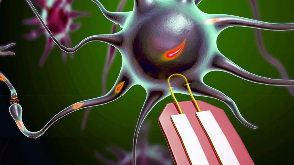
The Lieber Group at Harvard University provided this image illustrating the work,

U-shaped nanowires can record electrical chatter inside a brain or heart cell without causing any damage. The devices are 100 times smaller than their biggest competitors, which kill a cell after recording. Courtesy: University of Surrey

A July 3, 2019 University of Surrey press release (also on EurekAlert), which originated the news item, provides more details about this UK/US/China collaboration,

In a paper published by Nature Nanotechnology, scientists from Surrey’s Advanced Technology Institute (ATI) and Harvard University detail how they produced an array of the ultra-small U-shaped nanowire field-effect transistor probes for intracellular recording. This incredibly small structure was used to record, with great clarity, the inner activity of primary neurons and other electrogenic cells, and the device has the capacity for multi-channel recordings.

Dr Yunlong Zhao from the ATI at the University of Surrey said: “If our medical professionals are to continue to understand our physical condition better and help us live longer, it is important that we continue to push the boundaries of modern science in order to give them the best possible tools to do their jobs. For this to be possible, an intersection between humans and machines is inevitable.

“Our ultra-small, flexible, nanowire probes could be a very powerful tool as they can measure intracellular signals with amplitudes comparable with those measured with patch clamp techniques; with the advantage of the device being scalable, it causes less discomfort and no fatal damage to the cell (cytosol dilation). Through this work, we found clear evidence for how both size and curvature affect device internalisation and intracellular recording signal.”

Professor Charles Lieber from the Department of Chemistry and Chemical Biology at Harvard University said: “This work represents a major step towards tackling the general problem of integrating ‘synthesised’ nanoscale building blocks into chip and wafer scale arrays, and thereby allowing us to address the long-standing challenge of scalable intracellular recording.

“The beauty of science to many, ourselves included, is having such challenges to drive hypotheses and future work. In the longer term, we see these probe developments adding to our capabilities that ultimately drive advanced high-resolution brain-machine interfaces and perhaps eventually bringing cyborgs to reality.”

Professor Ravi Silva, Director of the ATI at the University of Surrey, said: “This incredibly exciting and ambitious piece of work illustrates the value of academic collaboration. Along with the possibility of upgrading the tools we use to monitor cells, this work has laid the foundations for machine and human interfaces that could improve lives across the world.”

Dr Yunlong Zhao and his team are currently working on novel energy storage devices, electrochemical probing, bioelectronic devices, sensors and 3D soft electronic systems. Undergraduate, graduate and postdoc students with backgrounds in energy storage, electrochemistry, nanofabrication, bioelectronics, tissue engineering are very welcome to contact Dr Zhao to explore the opportunities further.

Here’s a link to and a citation for the paper,

Scalable ultrasmall three-dimensional nanowire transistor probes for intracellular recording by Yunlong Zhao, Siheng Sean You, Anqi Zhang, Jae-Hyun Lee, Jinlin Huang & Charles M. Lieber. Nature Nanotechnology (2019) DOI: https://doi.org/10.1038/s41565-019-0478-y Published 01 July 2019

The link I’ve provided leads to a paywall. However, I found a freely accessible version of the paper (this may not be the final published version) here.