Monthly Archives: March 2015

Microbubbles reform into nanoparticles after bursting

It seems researchers at the Toronto-based (Canada), Princess Margaret Cancer Centre, have developed a new theranostic tool made of microbubbles used for imaging that are then burst into nanoparticles delivering therapeutics. From a March 30, 2015 news item on phys.org,

Biomedical researchers led by Dr. Gang Zheng at Princess Margaret Cancer Centre have successfully converted microbubble technology already used in diagnostic imaging into nanoparticles that stay trapped in tumours to potentially deliver targeted, therapeutic payloads.

The discovery, published online today [March 30, 2015] in Nature Nanotechnology, details how Dr. Zheng and his research team created a new type of microbubble using a compound called porphyrin – a naturally occurring pigment in nature that harvests light.

A March 30, 2015 University Health Network news release on EurekAlert, which originated the news item, describes the laboratory research on mice,

In the lab in pre-clinical experiments, the team used low-frequency ultrasound to burst the porphyrin containing bubbles and observed that they fragmented into nanoparticles. Most importantly, the nanoparticles stayed within the tumour and could be tracked using imaging.

“Our work provides the first evidence that the microbubble reforms into nanoparticles after bursting and that it also retains its intrinsic imaging properties. We have identified a new mechanism for the delivery of nanoparticles to tumours, potentially overcoming one of the biggest translational challenges of cancer nanotechnology. In addition, we have demonstrated that imaging can be used to validate and track the delivery mechanism,” says Dr. Zheng, Senior Scientist at the Princess Margaret and also Professor of Medical Biophysics at the University of Toronto.

Conventional microbubbles, on the other hand, lose all intrinsic imaging and therapeutic properties once they burst, he says, in a blink-of-an-eye process that takes only a minute or so after bubbles are infused into the bloodstream.

“So for clinicians, harnessing microbubble to nanoparticle conversion may be a powerful new tool that enhances drug delivery to tumours, prolongs tumour visualization and enables them to treat cancerous tumours with greater precision.”

For the past decade, Dr. Zheng’s research focus has been on finding novel ways to use heat, light and sound to advance multi-modality imaging and create unique, organic nanoparticle delivery platforms capable of transporting cancer therapeutics directly to tumours.

Interesting development, although I suspect there are many challenges yet to be met such as ensuring the microbubbles consistently arrive at their intended destination in sufficient mass to be effective both for imaging purposes and, later, as nanoparticles for drug delivery purposes.

Here’s a link to and citation for the paper,

In situ conversion of porphyrin microbubbles to nanoparticles for multimodality imaging by Elizabeth Huynh, Ben Y. C. Leung, Brandon L. Helfield, Mojdeh Shakiba, Julie-Anne Gandier, Cheng S. Jin, Emma R. Master, Brian C. Wilson, David E. Goertz, & Gang Zheng. Nature Nanotechnology (2015) doi:10.1038/nnano.2015.25 Published online 30 March 2015

This paper is behind a paywall but a free preview is available via ReadCube Access.

This is one of those times where I’m including the funding agencies and the ‘About’ portions of the news release,

The research published today was funded by the Canadian Institutes of Health Research (CIHR) Frederick Banting and Charles Best Canada Graduate Scholarship, the Emerging Team Grant on Regenerative Medicine and Nanomedicine co-funded by the CIHR and the Canadian Space Agency, the Natural Sciences and Engineering Research Council of Canada, the Ontario Institute for Cancer Research, the International Collaborative R&D Project of the Ministry of Knowledge Economy, South Korea, the Joey and Toby Tanenbaum/Brazilian Ball Chair in Prostate Cancer Research, the Canada Foundation for Innovation and The Princess Margaret Cancer Foundation.

About Princess Margaret Cancer Centre, University Health Network

The Princess Margaret Cancer Centre has achieved an international reputation as a global leader in the fight against cancer and delivering personalized cancer medicine. The Princess Margaret, one of the top five international cancer research centres, is a member of the University Health Network, which also includes Toronto General Hospital, Toronto Western Hospital and Toronto Rehabilitation Institute. All are research hospitals affiliated with the University of Toronto. For more information, go to http://www.theprincessmargaret.ca or http://www.uhn.ca .

I was not expecting to see South Korea or Brazil mentioned in the funding. Generally, when multiple countries are funding research, their own research institutions are also involved. As for the Princess Margaret Cancer Centre being one of the top five such centres internationally, I wonder how these rankings are determined.

Filtration membranes with twice as much ability to remove unwanted materials from water

A March 26, 2015 news item on Nanowerk offers information about a new method for removing pollutants from water and some insight into the situation regarding bisphenol A (BPA) in Europe,

New types of membrane adsorbers remove unwanted particles from water and also, at the same time, dissolved substances such as the hormonally active bis-phenol A or toxic lead. To do this, researchers at the Fraunhofer Institute for Interfacial Engineering and Biotechnology IGB imbed selective adsorber particles in filtration membranes.

It was not until January 2015 that the European Food Safety Authority (EFSA) lowered the threshold value for bisphenol A in packaging. The hormonally active bulk chemical is among other things a basic material for polycarbonate from which, for example, CDs, plastic tableware or spectacles glasses are manufactured. Due to its chemical structure, bisphenol A is not completely degraded in the biological stages of treatment plants and is discharged into rivers and lakes by the purification facility.

Activated carbon or adsorber materials are already used to remove chemicals, anti-biotics [sic] or heavy metals from waste or process water. However, a disadvantage of these highly porous materials is the long contact time that the pollutants require to diffuse into the pores. So that as many of the harmful substances as possible are captured even in a shorter time, the treatment plants use larger quantities of adsorbers in correspondingly large treatment basins. However, activated carbon can only be regenerated with a high energy input, resulting for the most part in the need to dispose of large quantities of material contaminated with pollutants.

Also, membrane filtration with nanofiltration or reverse osmosis membranes, which can remove the contaminating substances, is not yet cost-effective for the removal of dissolved molecules from high-volume flows such as process or wastewater. Membranes filter the water through their pores when a pressure is built up on one side of the membrane, thus holding back larger molecules and solid particles. But the smaller the membrane pores are, the higher the pressure – and therefore the more energy – that is required to separate the substances from water.

A March 24, 2015 Fraunhofer Institute for Interfacial Engineering and Biotechnology (IGB) press release, which originated the news item, goes on to describe their team’s new approach,

Researchers at the Fraunhofer Institute for Interfacial Engineering and Biotechnology IGB in Stuttgart have opted for a new approach that combines the advantages of both methods. When manufacturing the membranes they add small, polymeric adsorber particles. The resulting membrane adsorbers can – in addition to their filtration function – adsorptively bind substances dissolved in water. “We make use of the porous structure of the membrane located underneath the separation layer. The pores have a highly specific surface so that as many particles as possible can be imbedded, and they also provide optimum accessibility,” says Dr. Thomas Schiestel, Head of the “Inorganic Interfaces and Membranes” working group at the Fraunhofer IGB.

“Unlike conventional adsorbers, our membrane adsorbers transport the pollutants convectively. This means that, with the water flowing rapidly through the membrane pores, a contact time lasting only a few seconds is sufficient to adsorb pollutants on the particle surface,” says the scientist. Up to 40 percent of the weight of the membrane adsorbers is accounted for by the particles, so their binding capacity is correspondingly high. At the same time the membrane adsorbers can be operated at low pressures. As the membranes can be packed very tightly, very large volumes of water can be treated even with small devices.

Functional adsorber particles

The researchers manufacture the adsorber particles in a one-step, cost-efficient process. In this patented process monomeric components are polymerized with the help of a crosslinking agent to generate 50 to 500 nanometer polymer globules. “Depending on which substances are to be removed from the water, we select the most suitable one from a variety of monomers with differing functional groups,” Schiestel explains. The spectrum here ranges from pyridine, which tends to be hydrophobic, by way of cationic ammonium compounds and includes anionic phosphonates.

Selective removal of pollutants and metals

The researchers were able to show in various tests that the membrane adsorbers remove pollutants very selectively by means of the particles, which are customized for the particular contaminant in question. For example, membrane adsorbers with pyridine groups bind the hydrophobic bisphenol A especially well, whereas those with amino groups adsorb the negatively charged salt of the antibiotic penicillin G.

“The various adsorber particles can even be combined in one membrane. In this way we can remove several micropollutants simultaneously with just one membrane adsorber,” says Schiestel, pointing out a further advantage. Equipped with different functional groups, the membrane adsorbers can also remove toxic heavy metals such as lead or arsenic from the water. Phosphonate membrane adsorbers, for example, adsorb more than 5 grams of lead per square meter of membrane surface area – 40 percent more than a commercially available membrane adsorber.

Cost-effective and regenerable

So that the membrane adsorbers can be used several times, the adsorbed pollutants have to be detached once again from the particles in the membrane. “Membrane adsorbers for bisphenol A can be fully regenerated by a shift of the pH value,” Schiestel explains. The concentrated pollutants can then be disposed off cost-effectively or broken down using suitable oxidative processes.

The regenerability of the membrane adsorbers also makes possible a further application: reutilization of the separated molecules. This additionally makes the technology attractive for recovering valuable precious metals or rare earth metals.

The Fraunhofer IGB is presenting the membrane adsorbers and other innovative technologies for water purification at the “Wasser Berlin International” Trade Fair and Congress from 24th to 27th March 2015 in Berlin. The IGB is in Hall 2.2, Stand 422.

While it’s too late to attend the presentation, here are two links and citations to papers concerning the work,

Removal of micropollutants from water by nanocomposite membrane adsorbers by Klaus Niedergall, Monika Bach, Thomas Hirth, Günter E.M. Tovar, and Thomas Schiestel. Separation and Purification Technology, Volume 131, 27 June 2014 DOI: 10.1016/j.seppur.2014.04.032

Nanostructured Composite Adsorber Membranes for the Reduction of Trace Substances in Water: The Example of Bisphenol A by Klaus Niedergall, Monika Bach, Thomas Schiestel, and Günter E.M. Tovar. Ind. Eng. Chem. Res., 2013, 52 (39), pp 14011–14018 DOI: 10.1021/ie303264r Publication Date (Web): May 16, 2013

Copyright © 2013 American Chemical Society

Both articles are behind a paywall.

Entangling thousands of atoms

Quantum entanglement as an idea seems extraordinary to me like something from of the fevered imagination made possible only with certain kinds of hallucinogens. I suppose you could call theoretical physicists who’ve conceptualized entanglement a different breed as they don’t seem to need chemical assistance for their flights of fancy, which turn out to be reality. Researchers at MIT (Massachusetts Institute of Technology) and the University of Belgrade (Serbia) have entangled thousands of atoms with a single photon according to a March 26, 2015 news item on Nanotechnology Now,

Physicists from MIT and the University of Belgrade have developed a new technique that can successfully entangle 3,000 atoms using only a single photon. The results, published today in the journal Nature, represent the largest number of particles that have ever been mutually entangled experimentally.

The researchers say the technique provides a realistic method to generate large ensembles of entangled atoms, which are key components for realizing more-precise atomic clocks.

“You can make the argument that a single photon cannot possibly change the state of 3,000 atoms, but this one photon does — it builds up correlations that you didn’t have before,” says Vladan Vuletic, the Lester Wolfe Professor in MIT’s Department of Physics, and the paper’s senior author. “We have basically opened up a new class of entangled states we can make, but there are many more new classes to be explored.”

A March 26, 2015 MIT news release by Jennifer Chu (also on EurekAlert but dated March 25, 2015), which originated the news item, describes entanglement with particular attention to how it relates to atomic timekeeping,

Entanglement is a curious phenomenon: As the theory goes, two or more particles may be correlated in such a way that any change to one will simultaneously change the other, no matter how far apart they may be. For instance, if one atom in an entangled pair were somehow made to spin clockwise, the other atom would instantly be known to spin counterclockwise, even though the two may be physically separated by thousands of miles.

The phenomenon of entanglement, which physicist Albert Einstein once famously dismissed as “spooky action at a distance,” is described not by the laws of classical physics, but by quantum mechanics, which explains the interactions of particles at the nanoscale. At such minuscule scales, particles such as atoms are known to behave differently from matter at the macroscale.

Scientists have been searching for ways to entangle not just pairs, but large numbers of atoms; such ensembles could be the basis for powerful quantum computers and more-precise atomic clocks. The latter is a motivation for Vuletic’s group.

Today’s best atomic clocks are based on the natural oscillations within a cloud of trapped atoms. As the atoms oscillate, they act as a pendulum, keeping steady time. A laser beam within the clock, directed through the cloud of atoms, can detect the atoms’ vibrations, which ultimately determine the length of a single second.

“Today’s clocks are really amazing,” Vuletic says. “They would be less than a minute off if they ran since the Big Bang — that’s the stability of the best clocks that exist today. We’re hoping to get even further.”

The accuracy of atomic clocks improves as more and more atoms oscillate in a cloud. Conventional atomic clocks’ precision is proportional to the square root of the number of atoms: For example, a clock with nine times more atoms would only be three times as accurate. If these same atoms were entangled, a clock’s precision could be directly proportional to the number of atoms — in this case, nine times as accurate. The larger the number of entangled particles, then, the better an atomic clock’s timekeeping.

It seems weak lasers make big entanglements possible (from the news release),

Scientists have so far been able to entangle large groups of atoms, although most attempts have only generated entanglement between pairs in a group. Only one team has successfully entangled 100 atoms — the largest mutual entanglement to date, and only a small fraction of the whole atomic ensemble.

Now Vuletic and his colleagues have successfully created a mutual entanglement among 3,000 atoms, virtually all the atoms in the ensemble, using very weak laser light — down to pulses containing a single photon. The weaker the light, the better, Vuletic says, as it is less likely to disrupt the cloud. “The system remains in a relatively clean quantum state,” he says.

The researchers first cooled a cloud of atoms, then trapped them in a laser trap, and sent a weak laser pulse through the cloud. They then set up a detector to look for a particular photon within the beam. Vuletic reasoned that if a photon has passed through the atom cloud without event, its polarization, or direction of oscillation, would remain the same. If, however, a photon has interacted with the atoms, its polarization rotates just slightly — a sign that it was affected by quantum “noise” in the ensemble of spinning atoms, with the noise being the difference in the number of atoms spinning clockwise and counterclockwise.

“Every now and then, we observe an outgoing photon whose electric field oscillates in a direction perpendicular to that of the incoming photons,” Vuletic says. “When we detect such a photon, we know that must have been caused by the atomic ensemble, and surprisingly enough, that detection generates a very strongly entangled state of the atoms.”

Vuletic and his colleagues are currently using the single-photon detection technique to build a state-of-the-art atomic clock that they hope will overcome what’s known as the “standard quantum limit” — a limit to how accurate measurements can be in quantum systems. Vuletic says the group’s current setup may be a step toward developing even more complex entangled states.

“This particular state can improve atomic clocks by a factor of two,” Vuletic says. “We’re striving toward making even more complicated states that can go further.”

This research was supported in part by the National Science Foundation, the Defense Advanced Research Projects Agency, and the Air Force Office of Scientific Research.

Here’s a link to and a citation for the paper,

Entanglement with negative Wigner function of almost 3,000 atoms heralded by one photon by Robert McConnell, Hao Zhang, Jiazhong Hu, Senka Ćuk & Vladan Vuletić. Nature 519 439–442 (26 March 2015) doi:10.1038/nature14293 Published online 25 March 2015

This article is behind a paywall but there is a free preview via ReadCube Access.

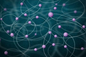
This image illustrates the entanglement of a large number of atoms. The atoms, shown in purple, are shown mutually entangled with one another. Image: Christine Daniloff/MIT and Jose-Luis Olivares/MIT

This image illustrates the entanglement of a large number of atoms. The atoms, shown in purple, are shown mutually entangled with one another.
Image: Christine Daniloff/MIT and Jose-Luis Olivares/MIT

Graphene light bulb to hit UK stores later in 2015

I gather people at the University of Manchester are quite happy about the graphene light bulb which their spin-off (or spin-out) company, Graphene Lighting PLC, is due to deliver to the market sometime later in 2015. From a March 30, 2015 news item by Nancy Owano on phys.org (Note: A link has been removed),

The BBC reported on Saturday [March 28, 2015] that a graphene bulb is set for shops, to go on sale this year. UK developers said their graphene bulb will be the first commercially viable consumer product using the super-strong carbon; bulb was developed by a Canadian-financed company, Graphene Lighting, one of whose directors is Prof Colin Bailey at the University of Manchester. [emphasis mine]

I have not been able to track down the Canadian connection mentioned (*never in any detail) in some of the stories. A March 30, 2015 University of Manchester press release makes no mention of Canada or any other country in its announcement (Note: Links have been removed),

A graphene lightbulb with lower energy emissions, longer lifetime and lower manufacturing costs has been launched thanks to a University of Manchester research and innovation partnership.

Graphene Lighting PLC is a spin-out based on a strategic partnership with the National Graphene Institute (NGI) at The University of Manchester to create graphene applications.

The UK-registered company will produce the lightbulb, which is expected to perform significantly better and last longer than traditional LED bulbs.

It is expected that the graphene lightbulbs will be on the shelves in a matter of months, at a competitive cost.

The University of Manchester has a stake in Graphene Lighting PLC to ensure that the University benefits from commercial applications coming out of the NGI.

The graphene lightbulb is believed to be the first commercial application of graphene to emerge from the UK, and is the first application from the £61m NGI, which only opened last week.

Graphene was isolated at The University of Manchester in 2004 by Sir Andre Geim and Sir Kostya Novoselov, earning them the Nobel prize for Physics in 2010. The University is the home of graphene, with more than 200 researchers and an unrivalled breadth of graphene and 2D material research projects.

The NGI will see academic and commercial partners working side by side on graphene applications of the future. It is funded by £38m from the Engineering and Physical Sciences Research Council (EPSRC) and £23m from the European Regional Development Fund (ERDF).

There are currently more than 35 companies partnering with the NGI. In 2017, the University will open the Graphene Engineering Innovation Centre (GEIC), which will accelerate the process of bringing products to market.

Professor Colin Bailey, Deputy President and Deputy Vice-Chancellor of The University of Manchester said: “This lightbulb shows that graphene products are becoming a reality, just a little more than a decade after it was first isolated – a very short time in scientific terms.

“This is just the start. Our partners are looking at a range of exciting applications, all of which started right here in Manchester. It is very exciting that the NGI has launched its first product despite barely opening its doors yet.”

James Baker, Graphene Business Director, added: “The graphene lightbulb is proof of how partnering with the NGI can deliver real-life products which could be used by millions of people.

“This shows how The University of Manchester is leading the way not only in world-class graphene research but in commercialisation as well.”

Chancellor George Osborne and Sir Kostya Novoselov with the graphene lightbulb Courtesy: University of Manchester

Chancellor George Osborne and Sir Kostya Novoselov with the graphene lightbulb Courtesy: University of Manchester

This graphene light bulb announcement comes on the heels of the university’s official opening of its National Graphene Institute mentioned here in a March 26, 2015 post.

Getting back to graphene and light bulbs, Judy Lin in a March 30, 2015 post on LEDinside.com offers some details such as proposed pricing and more,

These new bulbs will be priced at GBP 15 (US $22.23) each.

The dimmable bulb incorporates a filament-shaped LED coated in graphene, which was designed by Manchester University, where the strong carbon material was first discovered.

$22 seems like an expensive light bulb but my opinion could change depending on how long it lasts. ‘Longer lasting’ (and other variants of the term) seen in the news stories and press release are not meaningful to me. Perhaps someone could specify how many hours and under what conditions?

* ‘but’ removed as it was unnecessary, April 3, 2015.

ETA April 3, 2105: Dexter Johnson has provided a thought-provoking commentary about this graphene light bulb in an April 2, 2015 post on his Nanoclast blog (on the IEEE [Institute for Electrical and Electronics Engineers] website), Note: Links have been removed,

The big story this week in graphene, after taking into account the discovery of “grapene,” [Dexter’s April Fool’s Day joke posting] has to be the furor that has surrounded news that a graphene-coated light bulb was to be the “first commercially viable consumer product” using graphene.

Since the product is not expected to be on store shelves until next year, “commercially viable” is both a good hedge and somewhat short on meaning. The list of companies with a commercially viable graphene-based product is substantial, graphene-based conductive inks and graphene-based lithium-ion anodes come immediately to mind. Even that list neglects products that are already commercially available, never mind “viable”, like Head’s graphene-based tennis racquets.

Dexter goes on to ask more pointed questions and shares the answers he got from Daniel Cochlin, the graphene communications and marketing manager at the University of Manchester. I confess I got caught up in the hype. It’s always good to have someone bringing things back down to earth. Thank you Dexter!

And the bacteria shall save us—nanobiobots

A March 24, 2015 University of Illinois at Chicago news release (also on EurekAlert) describes the NERD, a Nano-Electro-Robotic Device which employs bacteria and graphene quantum dots,

As nanotechnology makes possible a world of machines too tiny to see, researchers are finding ways to combine living organisms with nonliving machinery to solve a variety of problems.

Like other first-generation bio-robots, the new nanobot engineered at the University of Illinois at Chicago [UIC] is a far cry from Robocop. It’s a robotic germ.

UIC researchers created an electromechanical device–a humidity sensor–on a bacterial spore. They call it NERD, for Nano-Electro-Robotic Device. …

“We’ve taken a spore from a bacteria, and put graphene quantum dots on its surface–and then attached two electrodes on either side of the spore,” said Vikas Berry, UIC associate professor of chemical engineering and principal investigator on the study.

“Then we change the humidity around the spore,” he said.

When the humidity drops, the spore shrinks as water is pushed out. As it shrinks, the quantum dots come closer together, increasing their conductivity, as measured by the electrodes.

“We get a very clean response–a very sharp change the moment we change humidity,” Berry said. The response was 10 times faster, he said, than a sensor made with the most advanced man-made water-absorbing polymers.

There was also better sensitivity in extreme low-pressure, low-humidity situations.

“We can go all the way down to a vacuum and see a response,” said Berry, which is important in applications where humidity must be kept low, for example, to prevent corrosion or food spoilage. “It’s also important in space applications, where any change in humidity could signal a leak,” he said.

Currently available sensors increase in sensitivity as humidity rises, Berry said. NERD’s sensitivity is actually higher at low humidity.

“This is a fascinating device,” Berry said. “Here we have a biological entity. We’ve made the sensor on the surface of these spores, with the spore a very active complement to this device. The biological complement is actually working towards responding to stimuli and providing information.”

Interesting, yes? Here’s a link to and a citation for the research paper,

Graphene Quantum Dots Interfaced with Single Bacterial Spore for Bio-Electromechanical Devices: A Graphene Cytobot by T. S. Sreeprasad, Phong Nguyen, Ahmed Alshogeathri, Luke Hibbeler, Fabian Martinez, Nolan McNeil, & Vikas Berry. Scientific Reports 5, Article number: 9138 doi:10.1038/srep09138 Published 16 March 2015

This paper is open access.

Dexter Johnson provides more context for this research in a March 26, 2015 post on his Nanoclast blog (on the IEEE [institute of Electrical and Electronics Engineers]) where he notes,

Recently, James Tours’ group at Rice University, who were the first to develop GQCs [graphene quantum dots] in 2013, created an improved way to manufacture them that promised to open them up to a new range of applications in optics.

Dexter’s insights make for worthwhile reading.

CelluForce celebrates a new investor but gives no details about research or applications

The most one can gather from the news item/press release is that CelluForce is researching applications in the oil and gas sector and that they’re very happy to receive money although there’s no indication as to how much. From a March 26, 2015 news item on Azonano,

CelluForce is pleased to announce an investment into the company by Schlumberger, the world’s leading supplier of technology, integrated project management and information solutions for the global oil and gas industry.

CelluForce’s March 25, 2015 press release does go on but there are no more details to be had,

This investment furthers the collaboration between CelluForce and Schlumberger to explore the use of CelluForce’s wood-derived nano-crystalline cellulose (CelluForce NCCTM) to enhance the productivity of oil and gas wells.

“We are very proud to be expanding our partnership with Schlumberger, the world’s leading oil and gas service company”, stated René Goguen, Acting President of CelluForce. “We have always believed that NCC applications hold promise extending far beyond the forest sector, and we see this investment from an international company as respected as Schlumberger as confirmation of this belief.”

NCC is a fundamental building block of trees that can be extracted from the forest biomass and has unique properties that offer a wide range of potential applications. Measured in units as small as nanometres, these tiny structures have strength properties comparable to steel and will have uses in a variety of industrial sectors.

The first small-scale NCC pilot plant was built and began operation in 2006 at FPInnovations’ laboratory in Montréal, Québec. Supported in part by Natural Resources Canada and the Ministère de l’Énergie et des Ressources naturelles du Québec, the pilot plant operation led to a scalable NCC production process and placed Canada in the pole position of the global race towards commercial NCC manufacture. Based on the success of the small-scale pilot plant, CelluForce, a joint venture of Domtar and FPInnovations, was created which led to the construction of a demonstration plant at Domtar’s mill in Windsor, Québec, having a production capacity of 1000 kg of NCC per day.

This announcement follows the recent announcement by the Honourable Greg Rickford, Minister of Natural Resources, of a $4.0 million contribution by Sustainable Development Technology Canada (SDTC) to optimize the extraction process of NCC from dry wood pulp and develop applications for its use in the oil and gas sector.

The $4M Canadian federal government investment was mentioned in my Feb. 19, 2015 post (scroll down about 40% of the way).

I get the feeling CelluForce is trying to recover from a setback and I wonder if it has anything to do with their production facility’s stockpile of NCC (aka, CNC or cellulose nanocrystals), first mentioned here in an Oct. 3, 2013 post. There was much fanfare about producing NCC/CNC but there was and is no substantive demand for the material in Canada or anywhere else globally.

Canada has three facilities that produce CNC (CelluForce being the largest) and there are production facilities in other countries. To date, there is no major application for CNC but given its properties, there is substantive research into how it could be commercialized. My Nov. 25, 2014 post covers a recent US report about commercializing nanocellulosic materials, including CNC.

I hope that CelluForce is able to overcome whatever problems it seems to be experiencing. Certainly, investments such as Schlumberger’s hint at the possibility. I wish the management team good luck.

Frogs: monitoring them, finding new species, and research about the golden ones in Panama

I have three frog-oriented items and while they’re not strictly speaking in my usual range of topics, given this blog’s name and the fact I haven’t posted a frog piece in quite a while, it seems this is a good moment to address that lack.

Monitoring frogs and amphibians at Trent University (Ontario, Canada)

From a March 23, 2015 Trent University news release,

With the decline of amphibian populations around the world, a team of researchers led by Trent University’s Dr. Dennis Murray will seek to establish environmental DNA (eDNA) monitoring of amphibian occupancy and aquatic ecosystem risk assessment with the help of a significant grant of over $596,000 from the Natural Sciences and Engineering Research Council of Canada (NSERC).

Awarded to Professor Murray, a Canada research chair in integrative wildlife conservation, bioinformatics, and ecological modelling and professor at Trent University along with colleagues Dr. Craig Brunetti of the Biology department, and Dr. Chris Kyle of the Forensic Science program, and partners at Laurentian University, University of Toronto, McGill University, Ontario Ministry of Natural Resources and Forestry and Environment Canada, the grant will support the development of tools that will promote a cleaner aquatic environment.

The project will use amphibian DNA found in natural breeding habitats to determine the presence and abundance of amphibians as well as their pathogens. This new technology capitalizes on Trent University’s expertise and infrastructure in the areas of wildlife DNA and water quality.

“We’re honoured to have received the grant to help us drive the project forward,” said Prof. Murray. “Our plan is to place Canada, and Trent, in a leadership position with respect to aquatic wildlife monitoring and amphibian conservation.”

Amphibian populations are declining worldwide, yet in Canada, amphibian numbers are not monitored closely, meaning changes in their distribution or abundance may be unnoticeable. Amphibian monitoring in Canada is conducted by citizen scientists who record frog breeding calls when visiting bodies of water during the spring. However, the lack of formalized amphibian surveys leaves Canada in a vulnerable position regarding the status of its diverse amphibian community.

Prof. Murray believes that the protocols developed from this project could revolutionize how amphibian populations are monitored in Canada and in turn lead to new insights regarding the population trends for several amphibian species across the country.

Here’s more about NSERC and Trent University from the news release,

About NSERC

NSERC is a federal agency that helps make Canada a country of discoverers and innovators. The agency supports almost 30,000 post-secondary students and postdoctoral fellows in their advanced studies. NSERC promotes discovery by funding approximately 12,000 professors every year and fosters innovation by encouraging over 2,400 Canadian companies to participate and invest in post-secondary research projects.

The NSERC Strategic Project Grants aim to increase research and training in areas that could strongly influence Canada’s economy, society or environment in the next 10 years in four target areas: environmental science and technologies; information and communications technologies; manufacturing; and natural resources and energy.

About Trent University

One of Canada’s top universities, Trent University was founded on the ideal of interactive learning that’s personal, purposeful and transformative. Consistently recognized nationally for leadership in teaching, research and student satisfaction, Trent attracts excellent students from across the country and around the world. Here, undergraduate and graduate students connect and collaborate with faculty, staff and their peers through diverse communities that span residential colleges, classrooms, disciplines, hands-on research, co-curricular and community-based activities. Across all disciplines, Trent brings critical, integrative thinking to life every day. As the University celebrates its 50th anniversary in 2014/15, Trent’s unique approach to personal development through supportive, collaborative community engagement is in more demand than ever. Students lead the way by co-creating experiences rooted in dialogue, diverse perspectives and collaboration. In a learning environment that builds life-long passion for inclusion, leadership and social change, Trent’s students, alumni, faculty and staff are engaged global citizens who are catalysts in developing sustainable solutions to complex issues. Trent’s Peterborough campus boasts award-winning architecture in a breathtaking natural setting on the banks of the Otonabee River, just 90 minutes from downtown Toronto, while Trent University Durham delivers a distinct mix of programming in the GTA.

Trent University’s expertise in water quality could be traced to its proximity to Canada’s Experimental Lakes Area (ELA), a much beleaguered research environment due to federal political imperatives. You can read more about the area and the politics in this Wikipedia entry. BTW, I am delighted to learn that it still exists under the auspices of the International Institute for Sustainable Development (IISD),

Taking this post into nanotechnology territory while mentioning the ELA, Trent University published a Dec. 8, 2014 news release about research into silver nanoparticles,

For several years, Trent University’s Dr. Chris Metcalfe and Dr. Maggie Xenopoulos have dedicated countless hours to the study of aquatic contaminants and the threat they pose to our environment.

Now, through the efforts of the International Institute for Sustainable Development (IISD), their research is reaching a wider audience thanks to a new video (Note: A link has been removed).

The video is one of a five-part series being released by the IISD that looks into environmental issues in Canada. The video entitled “Distilling Science at the Experimental Lakes Area: Nanosilver” and featuring Professors Metcalfe and Xenopoulos profiles their research around nanomaterials at the Experimental Lakes Area.

Prof. Xenopolous’ involvement in the project falls in line with other environmental issues she has tackled. In the past, her research has examined how human activities – including climate change, eutrophication and land use – affect ecosystem structure and function in lakes and rivers. She has also taken an interest in how land use affects the material exported and processed in aquatic ecosystems.

Prof. Metcalfe’s ongoing research on the fate and distribution of pharmaceutical and personal care products in the environment has generated considerable attention both nationally and internationally.

Together, their research into nanomaterials is getting some attention. Nanomaterials are submicroscopic particles whose physical and chemical properties make them useful for a variety of everyday applications. They can be found in certain pieces of clothing, home appliances, paint, and kitchenware. Initial laboratory research conducted at Trent University showed that nanosilver could strongly affect aquatic organisms at the bottom of the food chain, such as bacteria, algae and zooplankton.

To further examine these effects in a real ecosystem, a team of researchers from Trent University, Fisheries and Oceans Canada and Environment Canada has been conducting studies at undisclosed lakes in northwestern Ontario. The Lake Ecosystem Nanosilver (LENS) project has been monitoring changes in the lakes’ ecosystem that occur after the addition of nanosilver.

“In our particular case, we will be able to study and understand the effects of only nanosilver because that is the only variable that is going to change,” says Prof. Xenopoulos. “It’s really the only place in the world where we can do that.”

The knowledge gained from the study will help policy-makers make decisions about whether nanomaterials can be a threat to aquatic ecosystems and whether regulatory action is required to control their release into the environment.

You can find the 13 mins. video here: https://www.youtube.com/watch?v=_nJai_B4YH0#action=share

Shapeshifting frogs, a new species in Ecuador

Caption: This image shows skin texture variation in one individual frog (Pristimantis mutabilis) from Reserva Las Gralarias. Note how skin texture shifts from highly tubercular to almost smooth; also note the relative size of the tubercles on the eyelid, lower lip, dorsum and limbs. Credit: Zoological Journal of the Linnean Society

Caption: This image shows skin texture variation in one individual frog (Pristimantis mutabilis) from Reserva Las Gralarias. Note how skin texture shifts from highly tubercular to almost smooth; also note the relative size of the tubercles on the eyelid, lower lip, dorsum and limbs.
Credit: Zoological Journal of the Linnean Society

Here’s more about the shapeshifting and how the scientists figured out what the frogs were doing (from a March 23, 2015 Case Western Research University news release on EurekAlert; Note: A link has been removed),

A frog in Ecuador’s western Andean cloud forest changes skin texture in minutes, appearing to mimic the texture it sits on.

Originally discovered by a Case Western Reserve University PhD student and her husband, a projects manager at Cleveland Metroparks’ Natural Resources Division, the amphibian is believed to be the first known to have this shape-shifting capability.

But the new species, called Pristimantis mutabilis, or mutable rainfrog, has company. Colleagues working with the couple recently found that a known relative of the frog shares the same texture-changing quality–but it was never reported before.

The frogs are found at Reserva Las Gralarias, a nature reserve originally created to protect endangered birds in the Parish of Mindo, in north-central Ecuador.

The researchers, Katherine and Tim Krynak, and colleagues from Universidad Indoamérica and Tropical Herping (Ecuador) co-authored a manuscript describing the new animal and skin texture plasticity in the Zoological Journal of the Linnean Society this week. They believe their findings have broad implications for how species are and have been identified. The process may now require photographs and longer observations in the field to ensure the one species is not mistakenly perceived as two because at least two species of rain frogs can change their appearance.

Katherine Krynak believes the ability to change skin texture to reflect its surroundings may enable P. mutabilis to help camouflage itself from birds and other predators.

The Krynaks originally spotted the small, spiny frog, nearly the width of a marble, sitting on a moss-covered leaf about a yard off the ground on a misty July night in 2009. The Krynaks had never seen this animal before, though Tim had surveyed animals on annual trips to Las Gralarias since 2001, and Katherine since 2005.

They captured the little frog and tucked it into a cup with a lid before resuming their nightly search for wildlife. They nicknamed it “punk rocker” because of the thorn-like spines covering its body.

The next day, Katherine Krynak pulled the frog from the cup and set it on a smooth white sheet of plastic for Tim to photograph. It wasn’t “punk “–it was smooth-skinned. They assumed that, much to her dismay, she must have picked up the wrong frog.

“I then put the frog back in the cup and added some moss,” she said. “The spines came back… we simply couldn’t believe our eyes, our frog changed skin texture!

“I put the frog back on the smooth white background. Its skin became smooth.”

“The spines and coloration help them blend into mossy habitats, making it hard for us to see them,” she said. “But whether the texture really helps them elude predators still needs to be tested.”

During the next three years, a team of fellow biologists studied the frogs. They found the animals shift skin texture in a little more than three minutes.

Juan M. Guayasamin, from Universidad Tecnológica Indoamérica, Ecuador, the manuscript’s first author, performed morphological and genetic analyses showing that P. mutabilis was a unique and undescribed species. Carl R. Hutter, from the University of Kansas, studied the frog’s calls, finding three songs the species uses, which differentiate them from relatives. The fifth author of the paper, Jamie Culebras, assisted with fieldwork and was able to locate a second population of the species. Culebras is a member of Tropical Herping, an organization committed to discovering, and studying reptiles and amphibians.

Guayasamin and Hutter discovered that Prismantis sobetes, a relative with similar markings but about twice the size of P. mutabilis, has the same trait when they placed a spiny specimen on a sheet and watched its skin turn smooth. P. sobetes is the only relative that has been tested so far.

Because the appearance of animals has long been one of the keys to identifying them as a certain species, the researchers believe their find challenges the system, particularly for species identified by one or just a few preserved specimens. With those, there was and is no way to know if the appearance is changeable.

The Krynaks, who helped form Las Gralarias Foundation to support the conservation efforts of the reserve, plan to return to continue surveying for mutable rain frogs and to work with fellow researchers to further document their behaviors, lifecycle and texture shifting, and estimate their population, all in effort to improve our knowledge and subsequent ability to conserve this paradigm shifting species.

Further, they hope to discern whether more relatives have the ability to shift skin texture and if that trait comes from a common ancestor. If P. mutabilis and P. sobetes are the only species within this branch of Pristimantis frogs to have this capability, they hope to learn whether they retained it from an ancestor while relatives did not, or whether the trait evolved independently in each species.

Golden frog of Panama and its skin microbiome

Caption: Researchers studied microbial communities on the skin of Panamanian golden frogs to learn more about amphibian disease resistance. Panamanian golden frogs live only in captivity. Continued studies may help restore them back to the wild. Credit: B. Gratwicke/Smithsonian Conservation Biology Institute

Caption: Researchers studied microbial communities on the skin of Panamanian golden frogs to learn more about amphibian disease resistance. Panamanian golden frogs live only in captivity. Continued studies may help restore them back to the wild.
Credit: B. Gratwicke/Smithsonian Conservation Biology Institute

Among many of the pressures on frog populations, there’s a lethal fungus which has affected some 200 species of frogs. A March 23, 2015 news item on ScienceDaily describes some recent research into the bacterial communities present on frog skin,

A team of scientists including Virginia Tech researchers is one step closer to understanding how bacteria on a frog’s skin affects its likelihood of contracting disease.

A frog-killing fungus known as Batrachochytrium dendrobatidis, or Bd, has already led to the decline of more than 200 amphibian species including the now extinct-in-the-wild Panamanian golden frog.

In a recent study, the research team attempted to apply beneficial bacteria found on the skin of various Bd-resistant wild Panamanian frog species to Panamanian golden frogs in captivity, to see if this would stimulate a defense against the disease.

A March 23, 2015 Virginia Tech University news release on EurekAlert, which originated the news item, provides a twist and a turn in the story (Note: Links have been removed),

They found that while the treatment with beneficial bacteria was not successful due to its inability to stick to the skin, there were some frogs that survived exposure to the fungus.

These survivors actually had unique bacterial communities on their skin before the experiments started.

The next step is to explore these new bacterial communities.

“We were disappointed that the treatment didn’t work, but glad to have discovered new information about the relationship between these symbiotic microbial communities and amphibian disease resistance,” said Lisa Belden, an associate professor of biological sciences in the College of Science, a Fralin Life Science Institute affiliate, and a faculty member with the new Global Change Center at Virginia Tech. “Every bit of information gets us closer to getting these frogs back into nature.”

Studying the microbial communities of Panamanian golden frogs was the dissertation focus of Belden’s former graduate student Matthew Becker, who graduated with a Ph.D. in biological sciences from Virginia Tech in 2014 and is now a fellow at the Smithsonian Conservation Biology Institute.

“Anything that can help us predict resistance to this disease is very useful because the ultimate goal of this research is to establish healthy populations of golden frogs in their native habitat,” Becker told Smithsonian Science News. “I think identifying alternative probiotic treatment methods that optimize dosages and exposure times will be key for moving forward with the use of probiotics to mitigate chytridiomycosis.”

Here’s a link to and a citation for the paper,

Composition of symbiotic bacteria predicts survival in Panamanian golden frogs infected with a lethal fungus by Matthew H. Becker , Jenifer B. Walke , Shawna Cikanek , Anna E. Savage , Nichole Mattheus , Celina N. Santiago , Kevin P. C. Minbiole , Reid N. Harris , Lisa K. Belden , Brian Gratwicke. April 2015 Volume: 282 Issue: 1805 DOI: 10.1098/rspb.2014.2881 Published 18 March 2015

This is an open access paper.

For anyone curious about the article in the Smithsonian mentioned in the news release, you can find it here.
 

Tune in, turn on, and drop out—LSD and psychedelic talk at Vancouver’s (Canada) Café Scientifique on March 31, 2015

There seems to be a lot of interest in psychedelics these days and not least here in Vancouver. Next Tuesday, March 31, 2015 Cafe Scientifique, held in the back room of The Railway Club (2nd floor of 579 Dunsmuir St. [at Seymour St.], will be hosting a talk on LSD (from the March 16, 2015 announcement,

Our speaker for the evening will be Dr. Michael Hughesa Research Associate in the Department of Medical Genetics at UBC (University of British Columbia) …

Psychedelic Medicine: The History & Science of LSD in the Clinic

Ergot is a fungus that grows on rye and other grains that has been blamed (rightly or wrongly) for episodes of mass hysteria throughout history. Lysergic acid diethylamide (LSD) was first synthesized from ergot in 1938 by a Swiss chemist named Albert Hoffman, who, at the height of World War II, also discovered (somewhat mysteriously) its psychedelic properties. LSD soon came to the attention of the U.S. Army who quickly proceeds to buy up all the supply – primarily to keep it out of the hands of its enemies. Throughout the Cold War, elements in U.S. defense and security agencies engage in experiments by secretly slipping LSD to citizens with dangerous (and sometimes comical) consequences with the goal of perfecting brainwashing and mind control. Canadian scientists at McGill participated in some of these studies, thinking they could use LSD to cure psychoses. These unethical and largely unscientific experiments were akin to psychological torture. Meanwhile, the public discovered the recreational benefits of LSD and the hippie movement adopted the drug as a symbol and vehicle to enlightenment. Largely for this reason, in the early ‘70s LSD was classified as a Schedule-1 drug in the U.S. restricted legal access stopped most research and hopes of the clinical benefits of LSD was abandoned and all but forgotten. Recently, scientists, mostly working outside of the U.S. and Canada, have rediscovered LSD’s efficacy for the treatment of psychiatric disorders including post-traumatic stress syndrome (PTSD) and existential fear in terminally ill patients. Are we ready for a new wave of ethical human research to (re)-discover the clinical benefits of LSD? Take a journey through the strange history of LSD research and learn about its potential applications in medicine. What a long, strange trip it’s been.

Hughes works as a team member in the Hematopoietic Cell Development laboratory at the University of British Columbia’s (UBC) Biomedical Research Centre.

Last week on March 18, 2015, The UBC Neuroscience Graduate Student Association hosted a screening of Neurons to Nirvana: Understanding Psychedelic Medicines at the Pacific Cinematheque theatre in Vancouver (Note: Links have been removed),

A thought-provoking and visually-stunning documentary that explores the potential of five powerful psychedelic substances (LSD, psilocybin, MDMA, ayahuasca, and cannabis) as psychotherapeutic medicines. Despite the potential promise shown by such drugs in research conducted in the 1950s, the increasingly restrictive anti-drug policies of successive governments effectively shut down further enquiry. As one of the many world-renowned researchers, writers, psychologists, and scientists interviewed in the film says: “The government does not allow this research to take place, and then says there’s no research to support it. It’s beyond hypocrisy.” The film is a cogent call to put irrational, fear-based beliefs aside in order to allow clinical, evidence-based research into psychedelics in areas such as addictions, PTSD, anxiety, depression, and end-of-life care.

– – – – – – – – – – – – – – – – – –

Post-screening discussion with co-director Oliver Hockenhull and Mark Haden.

A teacher and essayist as well as a filmmaker, Oliver Hockenhull has presented at numerous universities in Canada, the US, and Europe. He has blended the documentary, essay, and experimental genres in such previous works as Aldous Huxley: The Gravity of Light (1996), Building Heaven, Remembering Earth (1999), and Evo (2002).

Mark Haden worked for Vancouver Coastal Health Addiction Services for 28 years and is now an Adjunct Professor at the UBC School of Population and Public Health. He is a pivotal voice in the drug policy reform movement, providing viable models for reforming drug education and regulating markets for currently illegal substances. Mark is also the Chair of the Board of MAPS Canada (Multidisciplinary Association for Psychedelic Studies).

Moderated by Dr. Harry Karlinsky, Clinical Professor, Department of Psychiatry, University of British Columbia.

Perhaps popular demand will lead to another showing. In the meantime, there’s Hughes’ talk and if his description is indicative it should be fascinating.

For anyone who did not recognize it,  ‘tune in, turn on, and drop out’, is a phrase that Timothy Leary, the high priest of psychedelics, psychologist, and former lecturer at Harvard University popularized during the 1960s and 70s. According to the ‘tune in, turn on, and drop out‘ entry in Wikipedia, the phrase was given to Leary by Canadian media theorist, Marshall McLuhan.

ETA March 27, 2015 at 1610 PDT: I just received a newsletter from Canada’s National Film Board where the feature item is this,

All About Acid: Hofmann’s Potion

Open your mind with this powerful feature documentary that retraces the history of LSD, a substance first used to treat addiction and mental illness that became the self-understanding tool of a generation.

For more on Hofmann’s Potion, read Meet the Lab Coat-Clad Granddaddies of LSD on the NFB/ blog.

Watch Now

* ‘tun’ changed to ‘turn’ (sigh) March 27, 2015 at 1615 PDT

What is a buckybomb?

I gather buckybombs have something to do with cancer treatments. From a March 18, 2015 news item on ScienceDaily,

In 1996, a trio of scientists won the Nobel Prize for Chemistry for their discovery of Buckminsterfullerene — soccer-ball-shaped spheres of 60 joined carbon atoms that exhibit special physical properties.

Now, 20 years later, scientists have figured out how to turn them into Buckybombs.

These nanoscale explosives show potential for use in fighting cancer, with the hope that they could one day target and eliminate cancer at the cellular level — triggering tiny explosions that kill cancer cells with minimal impact on surrounding tissue.

“Future applications would probably use other types of carbon structures — such as carbon nanotubes, but we started with Bucky-balls because they’re very stable, and a lot is known about them,” said Oleg V. Prezhdo, professor of chemistry at the USC [University of Southern California] Dornsife College of Letters, Arts and Sciences and corresponding author of a paper on the new explosives that was published in The Journal of Physical Chemistry on February 24 [2015].

A March 19, 2015 USC news release by Robert Perkins, which despite its publication date originated the news item, describes current cancer treatments with carbon nanotubes and this new technique with fullerenes,

Carbon nanotubes, close relatives of Bucky-balls, are used already to treat cancer. They can be accumulated in cancer cells and heated up by a laser, which penetrates through surrounding tissues without affecting them and directly targets carbon nanotubes. Modifying carbon nanotubes the same way as the Buckybombs will make the cancer treatment more efficient — reducing the amount of treatment needed, Prezhdo said.

To build the miniature explosives, Prezhdo and his colleagues attached 12 nitrous oxide molecules to a single Bucky-ball and then heated it. Within picoseconds, the Bucky-ball disintegrated — increasing temperature by thousands of degrees in a controlled explosion.

The source of the explosion’s power is the breaking of powerful carbon bonds, which snap apart to bond with oxygen from the nitrous oxide, resulting in the creation of carbon dioxide, Prezhdo said.

I’m glad this technique would make treatment more effective but I do pause at the thought of having exploding buckyballs in my body or, for that matter, anyone else’s.

The research was highlighted earlier this month in a March 5, 2015 article by Lisa Zynga for phys.org,

The buckybomb combines the unique properties of two classes of materials: carbon structures and energetic nanomaterials. Carbon materials such as C60 can be chemically modified fairly easily to change their properties. Meanwhile, NO2 groups are known to contribute to detonation and combustion processes because they are a major source of oxygen. So, the scientists wondered what would happen if NO2 groups were attached to C60 molecules: would the whole thing explode? And how?

The simulations answered these questions by revealing the explosion in step-by-step detail. Starting with an intact buckybomb (technically called dodecanitrofullerene, or C60(NO2)12), the researchers raised the simulated temperature to 1000 K (700 °C). Within a picosecond (10-12 second), the NO2 groups begin to isomerize, rearranging their atoms and forming new groups with some of the carbon atoms from the C60. As a few more picoseconds pass, the C60 structure loses some of its electrons, which interferes with the bonds that hold it together, and, in a flash, the large molecule disintegrates into many tiny pieces of diatomic carbon (C2). What’s left is a mixture of gases including CO2, NO2, and N2, as well as C2.

I encourage you to read Zynga’s article in whole as she provides more scientific detail and she notes that this discovery could have applications for the military and for industry.

Here’s a link to and a citation for the researchers’ paper,

Buckybomb: Reactive Molecular Dynamics Simulation by Vitaly V. Chaban, Eudes Eterno Fileti, and Oleg V. Prezhdo. J. Phys. Chem. Lett., 2015, 6 (5), pp 913–917 DOI: 10.1021/acs.jpclett.5b00120 Publication Date (Web): February 24, 2015

Copyright © 2015 American Chemical Society

This paper is behind a paywall.

Université de Montréal (Canada) and nanobots breech blood-brain barrier to deliver drugs to the brain

In the spirit of full disclosure, the March 25, 2014 news item on ScienceDaily describing the research about breeching the blood-brain barrier uses the term nanorobotic agents rather than nanobots, a term which makes my headline a lot catchier although less accurate. Getting back to the research,

Magnetic nanoparticles can open the blood-brain barrier and deliver molecules directly to the brain, say researchers from the University of Montreal, Polytechnique Montréal, and CHU Sainte-Justine. This barrier runs inside almost all vessels in the brain and protects it from elements circulating in the blood that may be toxic to the brain. The research is important as currently 98% of therapeutic molecules are also unable to cross the blood-brain barrier.

“The barrier is temporary [sic] opened at a desired location for approximately 2 hours by a small elevation of the temperature generated by the nanoparticles when exposed to a radio-frequency field,” explained first author and co-inventor Seyed Nasrollah Tabatabaei. “Our tests revealed that this technique is not associated with any inflammation of the brain. This new result could lead to a breakthrough in the way nanoparticles are used in the treatment and diagnosis of brain diseases,” explained the co-investigator, Hélène Girouard. “At the present time, surgery is the only way to treat patients with brain disorders. Moreover, while surgeons are able to operate to remove certain kinds of tumors, some disorders are located in the brain stem, amongst nerves, making surgery impossible,” added collaborator and senior author Anne-Sophie Carret.

A March 25, 2015 University of Montreal news release (also on EurekAlert), which originated the news item, notes that the technique was tested or rats or mice (murine model) and explains how the technology breeches the blood-brain barrier,

Although the technology was developed using murine models and has not yet been tested in humans, the researchers are confident that future research will enable its use in people. “Building on earlier findings and drawing on the global effort of an interdisciplinary team of researchers, this technology proposes a modern version of the vision described almost 40 years ago in the movie Fantastic Voyage, where a miniature submarine navigated in the vascular network to reach a specific region of the brain,” said principal investigator Sylvain Martel. In earlier research, Martel and his team had managed to manipulate the movement of nanoparticles through the body using the magnetic forces generated by magnetic resonance imaging (MRI) machines.

To open the blood-brain barrier, the magnetic nanoparticles are sent to the surface of the blood-brain barrier at a desired location in the brain. Although it was not the technique used in this study, the placement could be achieved by using the MRI technology described above. Then, the researchers generated a radio-frequency field. The nanoparticles reacted to the radio-frequency field by dissipating heat thereby creating a mechanical stress on the barrier. This allows a temporary and localized opening of the barrier for diffusion of therapeutics into the brain.

The technique is unique in many ways. “The result is quite significant since we showed in previous experiments that the same nanoparticles can also be used to navigate therapeutic agents in the vascular network using a clinical MRI scanner,” Martel remarked. “Linking the navigation capability with these new results would allow therapeutics to be delivered directly to a specific site of the brain, potentially improving significantly the efficacy of the treatment while avoiding systemic circulation of toxic agents that affect healthy tissues and organs,” Carret added. “While other techniques have been developed for delivering drugs to the blood-brain barrier, they either open it too wide, exposing the brain to great risks, or they are not precise enough, leading to scattering of the drugs and possible unwanted side effect,” Martel said.

Although there are many hurdles to overcome before the technology can be used to treat humans, the research team is optimistic. “Although our current results are only proof of concept, we are on the way to achieving our goal of developing a local drug delivery mechanism that will be able to treat oncologic, psychiatric, neurological and neurodegenerative disorders, amongst others,” Carret concluded.

Here’s a link to and a citation for the paper,

Remote control of the permeability of the blood–brain barrier by magnetic heating of nanoparticles: A proof of concept for brain drug delivery by Seyed Nasrollah Tabatabaei, Hélène Girouard, Anne-Sophie Carret, and Sylvain Martel.Journal of Controlled Release, Volume 206, 28 May 2015, Pages 49–57,  DOI: 10.1016/j.jconrel.2015.02.027  Available online 25 February 2015

This paper is behind a paywall.

For anyone unfamiliar with French, University of Montreal is Université de Montréal.