Tag Archives: nanoparticles

Ethical nanobiotechnology

This paper on ethics (aside: I have a few comments after the news release and citation) comes from the US Pacific Northwest National Laboratory (PNNL) according to a July 12, 2023 news item on phys.org,

Prosthetics moved by thoughts. Targeted treatments for aggressive brain cancer. Soldiers with enhanced vision or bionic ears. These powerful technologies sound like science fiction, but they’re becoming possible thanks to nanoparticles.

“In medicine and other biological settings, nanotechnology is amazing and helpful, but it could be harmful if used improperly,” said Pacific Northwest National Laboratory (PNNL) chemist Ashley Bradley, part of a team of researchers who conducted a comprehensive survey of nanobiotechnology applications and policies.

Their research, available in Health Security, works to sum up the very large, active field of nanotechnology in biology applications, draw attention to regulatory gaps, and offer areas for further consideration.

A July 12, 2023 PNNL news release (also on EurekAlert), which originated the news item, delves further into the topic, Note: A link has been removed,

“In our research, we learned there aren’t many global regulations yet,” said Bradley. “And we need to create a common set of rules to figure out the ethical boundaries.”

Nanoparticles, big differences

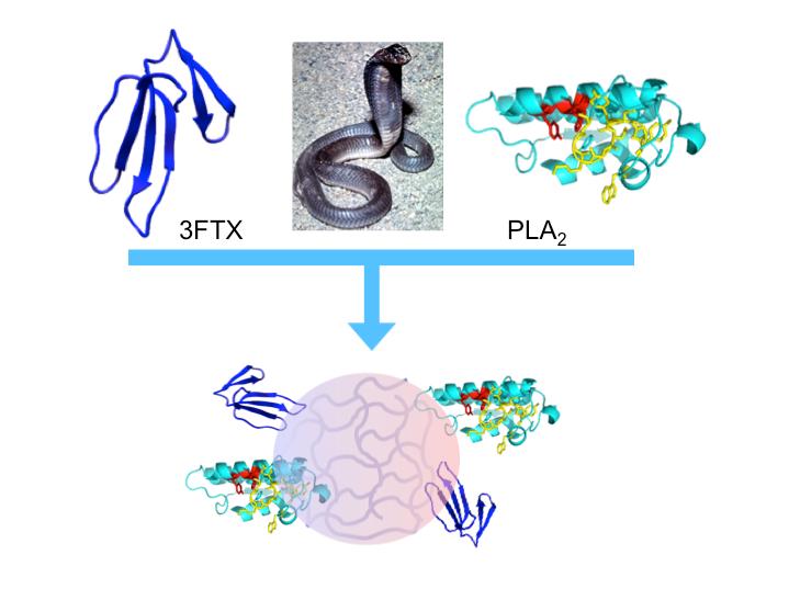
Nanoparticles are clusters of molecules with different properties than large amounts of the same substances. In medicine and other biology applications, these properties allow nanoparticles to act as the packaging that delivers treatments through cell walls and the difficult to cross blood-brain barrier.

“You can think of the nanoparticles a little bit like the plastic around shredded cheese,” said PNNL chemist Kristin Omberg. “It makes it possible to get something perishable directly where you want it, but afterwards you’ve got to deal with a whole lot of substance where it wasn’t before.”

Unfortunately, dealing with nanoparticles in new places isn’t straightforward. Carbon is pencil lead, nano carbon conducts electricity. The same material may have different properties at the nanoscale, but most countries still regulate it the same as bulk material, if the material is regulated at all.

For example, zinc oxide, a material that was stable and unreactive as a pigment in white paint, is now accumulating in oceans when used as nanoparticles in sunscreen, warranting a call to create alternative reef-safe sunscreens. And although fats and lipids aren’t regulated, the researchers suggest which agencies could weigh in on regulations were fats to become after-treatment byproducts.

The article also inventories national and international agencies, organizations, and governing bodies with an interest in understanding how nanoparticles break down or react in a living organism and the environmental life cycle of a nanoparticle. Because nanobiotechnology spans materials science, biology, medicine, environmental science, and tech, these disparate research and regulatory disciplines must come together, often for the first time—to fully understand the impact on humans and the environment.

Dual use: Good for us, bad for us

Like other quickly growing fields, there’s a time lag between the promise of new advances and the possibilities of unintended uses.

“There were so many more applications than we thought there were,” said Bradley, who collected exciting nanobio examples such as Alzheimer’s treatment, permanent contact lenses, organ replacement, and enhanced muscle recovery, among others.

The article also highlights concerns about crossing the blood-brain barrier, thought-initiated control of computers, and nano-enabled DNA editing where the researchers suggest more caution, questioning, and attention could be warranted. This attention spans everything from deep fundamental research and regulations all the way to what Omberg called “the equivalent of tattoo removal” if home-DNA splicing attempts go south.

The researchers draw parallels to more established fields such as synthetic bio and pharmacology, which offer lessons to be learned from current concerns such as the unintended consequences of fentanyl and opioids. They believe these fields also offer examples of innovative coordination between science and ethics, such as synthetic bio’s IGEM [The International Genetically Engineered Machine competition]—student competition, to think about not just how to create, but also to shape the use and control of new technologies.

Omberg said unusually enthusiastic early reviewers of the article contributed even more potential uses and concerns, demonstrating that experts in many fields recognize ethical nanobiotechnology is an issue to get in front of. “This is a train that’s going. It will be sad if 10 years from now, we haven’t figured how to talk about it.”

Funding for the team’s research was supported by PNNL’s Biorisk Beyond the List National Security Directorate Objective.

Here’s a link to and a citation for the paper,

The Promise of Emergent Nanobiotechnologies for In Vivo Applications and Implications for Safety and Security by Anne M. Arnold, Ashley M. Bradley, Karen L. Taylor, Zachary C. Kennedy, and Kristin M. Omberg. Health Security.Oct 2022.408-423.Published in Volume: 20 Issue 5: October 17, 2022 DOI: https://doi.org/10.1089/hs.2022.0014 Published Online:17 Oct 2022

This paper is open access.

You can find out more about IGEM (The International Genetically Engineered Machine competition) here.

Comments (brief)

It seems a little odd that the news release (“Prosthetics moved by thoughts …”) and the paper both reference neurotechnology without ever mentioning it by name. Here’s the reference from the paper, Note: Links have been removed,

Nanoparticles May Be Developed to Facilitate Cognitive Enhancements

The development and implementation of NPs that enhance cognitive function has yet to be realized. However, recent advances on the micro- and macro-level with neural–machine interfacing provide the building blocks necessary to develop this technology on the nanoscale. A noninvasive brain–computer interface to control a robotic arm was developed by teams at 2 universities.157 A US-based company, Neuralink, [emphasis mine] is at the forefront of implementing implantable, intracortical microelectrodes that provide an interface between the human brain and technology.158,159 Utilization of intracortical microelectrodes may ultimately provide thought-initiated access and control of computers and mobile devices, and possibly expand cognitive function by accessing underutilized areas of the brain.158

Neuralink (founded by Elon Musk) is controversial for its animal testing practices. You can find out more in Björn Ólafsson’s May 30, 2023 article for Sentient Media.

The focus on nanoparticles as the key factor in the various technologies and applications mentioned seems narrow but necessary given the breadth of topics covered in the paper as the authors themselves note in the paper’s abstract,

… In this article, while not comprehensive, we attempt to illustrate the breadth and promise of bionanotechnology developments, and how they may present future safety and security challenges. Specifically, we address current advancements to streamline the development of engineered NPs for in vivo applications and provide discussion on nano–bio interactions, NP in vivo delivery, nanoenhancement of human performance, nanomedicine, and the impacts of NPs on human health and the environment.

They have a good overview of the history and discussions about nanotechnology risks and regulation. It’s international in scope with a heavy emphasis on US efforts, as one would expect.

For anyone who’s interested in the neurotechnology end of things, I’ve got a July 17, 2023 commentary “Unveiling the Neurotechnology Landscape: Scientific Advancements, Innovations and Major Trends—a UNESCO report.” The report was launched July 13, 2023 during UNESCO’s Global dialogue on the ethics of neurotechnology (see my July 7, 2023 posting about the then upcoming dialogue for links to more UNESCO information). Both the July 17 and July 7, 2023 postings included additional information about Neuralink.

Could synergistic action of engineered nanoparticles have a health impact?

Synergistic action can be difficult to study especially when you’re looking at nanoparticles which could be naturally occurring and/or engineered. I believe this study is focused on engineered nanoparticles (ENPs) and I think it’s the first one I’ve seen that examines synergistic action of any kind. So, bravo to the scientists for tackling a very ambitious project.

An October 1, 2020 news item on phys.org describes this work from Denmark,

Nanoparticles are used in a wide range of products and manufacturing processes because the properties of a material can change dramatically when the material comes in nano-form.

They can be used, for example, to purify wastewater and to transport medicine around the body. They are also added to, for example, socks, pillows, mattresses, phone covers and refrigerators to supply the items with an antibacterial surface.

Much research has been done on how nanoparticles affect humans and the environment and a number of studies have shown that nanoparticles can disrupt or damage our cells.

This is confirmed by a new study that has also looked at how cells react when exposed to more than one kind of nano particle at the same time.

An October 1, 2020 University of Southern Denmark press release (also on EurekAlert) by Birgitte Svennevig, which originated the news item, provides more insight into the research,

The lead author of the study is Barbara Korzeniowska from the Department of Biochemistry and Molecular Biology at SDU. The head of research is Professor Frank Kjeldsen from the same department.

His research into metal nanoparticles is supported by a European Research Grant of DKK 14 million.

“Throughout a lifetime, we are exposed to many different kinds of nano-particles, and we should investigate how the combination of different nano-particles affects us and also whether an accumulation through life can harm us,” says Barbara Korzeniowska.

She herself became interested in the subject when her little daughter one day was going in the bathtub and got a rubber duck as a toy.

– It turned out that it had been treated with nano-silver, probably to keep it free of bacteria, but small children put their toys in their mouths, and she could thus ingest nano-silver. That is highly worrying when research shows that nano-silver can damage human cells, she says.

In her new study, she looked at nano-silver and nano-platinum. She has investigated their individual effect and whether exposure of both types of nanoparticles results in a synergy effect in two types of brain cells.

– There are almost no studies of the synergy effect of nano particles, so it is important to get started with these studies, she says.

She chose nano-silver because it is already known to be able to damage cells and nano-platinum, because nano-platinum is considered to be so-called bio-inert; i.e. has a minimal interaction with human tissue.

The nanoparticles were tested on two types of brain cells: astrocytes and endothelial cells. Astrocytes are supporter cells in the central nervous system, which i.a. helps to supply the nervous system with nutrients and repair damage to the brain. Endothelial cells sit on the inside of the blood vessels and transport substances from the bloodstream to the brain.

When the endothelial cells were exposed to nano-platinum, nothing happened. When exposed to nano-silver, their ability to divide deteriorated. When exposed to both nano-silver and nano-platinum, the effect was amplified, and they died in large numbers. Furthermore, their defense mechanisms decreased, and they had difficulty communicating with each other.

– So even though nano-platinum alone does not do harm, something drastic happens when they are combined with a different kind of nano-particle, says Frank Kjeldsen.

The astrocytes were more hardy and reacted “only” with impaired ability to divide when exposed to both types of nano-particles.

An earlier study, conducted by Frank Kjeldsen, has shown a dramatic synergy effect of silver nanoparticles and cadmium ions, which are found naturally all around us on Earth.

In that study, 72 % of the cells died (in this study it was intestinal cells) as they were exposed to both nano-silver and cadmium ions. When they were only exposed to nano-silver, 25% died. When exposed to cadmium ions only, 12% died.

We are involuntarily exposed

– Little is known about how large concentrations of nano-particles are used in industrial products. We also do not know what size particles they use – size also has an effect on whether they can enter a cell, says Barbara Korzeniowska and continues:

– But we know that a lot of people are involuntarily exposed to nano-particles, and that there can be lifelong exposure.

There are virtually no restrictions on adding nanoparticles to products. In the EU, however, manufacturers must have an approval if they want to use nanoparticles in products with antibacterial properties. In Denmark, they must also declare nano-content in such products on the label.

Here’s a link to and a citation for the paper,

The Cytotoxicity of Metal Nanoparticles Depends on Their Synergistic Interactions by Barbara Korzeniowska, Micaella P. Fonseca, Vladimir Gorshkov, Lilian Skytte, Kaare L. Rasmussen, Henrik D. Schrøder, Frank Kjeldsen. Particle Volume 37, Issue 8, August 2020,. 2000135 DOI: https://doi.org/10.1002/ppsc.202000135 First published: 06 July 2020

This paper is behind a paywall.

Snakebite? Roll out the nanoparticles

An October 4, 2018 news item on Nanowerk highlights some recent research into treating snakebites (Note: A link has been removed),

Venomous snakebites affect 2.5 million people, and annually cause more than 100,000 deaths and leave 400,000 individuals with permanent physical and psychological trauma each year.

Researchers reporting in PLOS Neglected Tropical Diseases (“Engineered nanoparticles bind elapid snake venom toxins and inhibit venom-induced dermonecrosis”) have now described a new approach to treating snake bites [sic], using nanoparticles to bind to venom toxins and prevent the spread of venom through the body.

Caption: “Synthetic polymer nanoparticles bind elapid snake venom toxins and inhibit venom-induced dermonecrosis.” Credit: Shea, et al. CC BY 4.0: Redistribution allowed with credit

An October 4, 2018 PLOS news release on EurekAlert, which originated the news item, expands on the theme,

The standard treatment for snakebites is the intravenous administration of IgG immune molecules that recognize venoms. However, such antivenom therapies must be administered quickly–and by trained healthcare workers– to be effective and are highly specific to particular venoms. There is an ongoing need for a snakebite treatment which can be used in a rural setting and works against the bites of diverse venomous snakes.

In the new work, Kenneth Shea, of the University of California, Irvine, and colleagues engineered nanoparticles that bind to and sequester an array of phospholipases A2 (PLA2) and three-finger toxin (3FTX) molecules found in Elapidae snake venoms. The Elapidae family is a large family of venomous snakes that includes cobras, kraits, tiger snakes, sea snakes, coral snakes and mambas, among other species. The researchers tested the ability of the nanoparticles to block Naja nigricollis (black-necked spitting cobra) venom in mice that received varying doses of the nanoparticles, injected into the skin. Envenomings by this snake in sub-Saharan Africa inflict serious cutaneous necrosis that may leave permanent tissue damage in the victims.

In experiments on isolated cells, the nanoparticles were found to sequester a wide range of Elapidae PLA and 3FTX venoms. Moreover, with collaborator José María Gutiérrez from the Instituto Clodomiro Picado (Universidad de Costa Rica), experiments with mice demonstrated that injections of the nanoparticles at the site of venom injection significantly mitigated the typical necrotic effects–including blistering and ulcers– of the spitting cobra venom. The nanoparticles administered to mice that had not received venom did not have an effect on skin and did not induce systemic toxicity.

“The stable, low-cost nanoparticles have the potential to be administered subcutaneously immediately after the bite at the site of envenoming by this spitting cobra to halt or reduce the extent of local damage and mitigate the systemic distribution of toxins post-envenoming,” the researchers say.

Here’s a link to and a citation for the paper,

Engineered nanoparticles bind elapid snake venom toxins and inhibit venom-induced dermonecrosis by Jeffrey O’Brien, Shih-Hui Lee, José María Gutiérrez, Kenneth J. Shea. PLOS Neglected Tropical Diseases 12(10): e0006736 DOI: https://doi.org/10.1371/journal.pntd.0006736 Published: October 4, 2018

This paper is open access. By the way, PLOS stands for Public Library of Science.

Therapeutic nanoparticles for agricultural crops

Nanoscale drug delivery systems developed by the biomedical community may prove useful to farmers. The Canadian Broadcasting Corporation (CBC) featured the story in a May 26, 2018 online news item (with audio file; Note: A link has been removed),

Thanks to a fortuitous conversation between an Israeli chemical engineer who works on medical nanotechnology and his farmer friend, there’s a new way to deliver nourishment to nutrient-starved crops.

Avi Schroeder, the chemical engineer and cancer researcher from Technion — Israel Institute of Technology asked his friend what are the major problems facing agriculture today. “He said, ‘You know Avi, one of the major issues we’re facing is that in some of the crops we try to grow, we actually have a lack of nutrients. And then we end up not growing those crops even though they’re very valuable or very important crops.'”

This problem is only going to become more acute in many regions of the world as global population approaches eight billion people.

“Feeding them with healthy food and nutritious food is becoming a major limiting factor. And … the land we can actually grow crops on are also becoming smaller and smaller in every country because people need to build houses too. So what we want is to get actually more crops per hectare.”

The way farmers currently deliver nutrients to malnourished agricultural crops is very inefficient. Much of what is added to the leaves of the plant is wasted. Most of it washes away or isn’t taken up by the plants.

If plants don’t get the nutrients they need, their leaves start to yellow, their growth becomes stunted and they don’t produce as much food as nutrient-rich crops.

“We work primarily in the field of medicine,” says Schroeder. “What we do many times is we’ll load minuscule doses of medicine into nanoparticles — we’ll inject them into the patient. And those nanoparticles will actually be able to detect the disease site inside the body. That sounded very, very similar to the problem the farmers were actually facing — how do you get a medicine into a crop or a nutrient into a crop and get it to the right region within the crop where it’s actually necessary.”

The nanoparticles Schroeder developed are tiny packages that can deliver nutrients — any nutrients — that are placed inside.

A June 6, 2018 news item on Nanowerk offers a few more details,

An innovative technology developed at the Technion [Israel Institute of Technology] could lead to significant increases in agricultural yields. Using a nanometric transport platform on plants that was previously utilized for targeted drug delivery, researchers increased the penetration rate of nutrients into the plants, from 1% to approximately 33%.

A May 27,2018 Technion press release, which originated the news item, fleshes out the details,

The technology exploits nanoscale delivery platforms which until now were used to transport drugs to specific targets in the patient’s body. The work was published in Scientific Reports and will be presented in Nature Press.

The use of the nanotechnology for targeted drug delivery has been the focus of research activity conducted at the Laboratory for Targeted Drug Delivery and Personalized Medicine Technologies at the Wolfson Faculty of Chemical Engineering. The present research repurposes this technology for agricultural use; and is being pursued by laboratory director Prof. Avi Schroeder and graduate student Avishai Karny.

“The constant growth in the world population demands more efficient agricultural technologies, which will produce greater supplies of healthier foods and reduce environmental damage,” said Prof. Schroeder. “The present work provides a new means of delivering essential nutrients without harming the environment.”

The researchers loaded the nutrients into liposomes which are small spheres generated in the laboratory, comprised of a fatty outer layer enveloping the required nutrients. The particles are stable in the plant’s aqueous environment and can penetrate the cells. In addition, the Technion researchers can ‘program’ them to disintegrate and release the load at precisely the location and time of interest, namely, in the roots and leaves. Disintegration occurs in acidic environments or in response to an external signal, such as light waves or heat. The molecules comprising the particles are derived from soy plants and are therefore approved and safe for consumption by both humans and animals.

In the present experiment, the researchers used 100-nanometer liposomes to deliver the nutrients iron and magnesium into both young and adult tomato crops. They demonstrated that the liposomes, which were sprayed in the form of a solution onto the leaves, penetrated the leaves and reached other leaves and roots. Only when reaching the root cells did they disintegrate and release the nutrients. As said, the technology greatly increased the nutrient penetration rate.

In addition to demonstrating the effectivity of this approach as compared to the standard spray method, the researchers also assessed the regulatory limitations associated with the spread of volatile particles.

”Our engineered liposomes are only stable within a short spraying range of up to 2 meters,” explained Prof. Schroeder. “If they travel in the air beyond that distance, they break down into safe materials (phospholipids). We hope that the success of this study will expand the research and development of similar agricultural products, to increase the yield and quality of food crops.”

This is an illustration of the work,

Each liposome (light blue bubble) was loaded with iron and magnesium particles. The liposomes sprayed on the leaves, penetrated and then spread throughout the various parts of the plant and released their load within the cells. Courtesy: Technion

Here’s a link to and a citation for the paper,

Therapeutic nanoparticles penetrate leaves and deliver nutrients to agricultural crops by Avishai Karny, Assaf Zinger, Ashima Kajal, Janna Shainsky-Roitman, & Avi Schroeder. Scientific Reportsvolume 8, Article number: 7589 (2018) DOI: https://doi.org/10.1038/s41598-018-25197-y Published 17 May 2018

This paper is open access.

Injectable bandages for internal bleeding and hydrogel for the brain

This injectable bandage could be a gamechanger (as they say) if it can be taken beyond the ‘in vitro’ (i.e., petri dish) testing stage. A May 22, 2018 news item on Nanowerk makes the announcement (Note: A link has been removed),

While several products are available to quickly seal surface wounds, rapidly stopping fatal internal bleeding has proven more difficult. Now researchers from the Department of Biomedical Engineering at Texas A&M University are developing an injectable hydrogel bandage that could save lives in emergencies such as penetrating shrapnel wounds on the battlefield (Acta Biomaterialia, “Nanoengineered injectable hydrogels for wound healing application”).

A May 22, 2018 US National Institute of Biomedical Engineering and Bioengiineering news release, which originated the news item, provides more detail (Note: Links have been removed),

The researchers combined a hydrogel base (a water-swollen polymer) and nanoparticles that interact with the body’s natural blood-clotting mechanism. “The hydrogel expands to rapidly fill puncture wounds and stop blood loss,” explained Akhilesh Gaharwar, Ph.D., assistant professor and senior investigator on the work. “The surface of the nanoparticles attracts blood platelets that become activated and start the natural clotting cascade of the body.”

Enhanced clotting when the nanoparticles were added to the hydrogel was confirmed by standard laboratory blood clotting tests. Clotting time was reduced from eight minutes to six minutes when the hydrogel was introduced into the mixture. When nanoparticles were added, clotting time was significantly reduced, to less than three minutes.

In addition to the rapid clotting mechanism of the hydrogel composite, the engineers took advantage of special properties of the nanoparticle component. They found they could use the electric charge of the nanoparticles to add growth factors that efficiently adhered to the particles. “Stopping fatal bleeding rapidly was the goal of our work,” said Gaharwar. “However, we found that we could attach growth factors to the nanoparticles. This was an added bonus because the growth factors act to begin the body’s natural wound healing process—the next step needed after bleeding has stopped.”

The researchers were able to attach vascular endothelial growth factor (VEGF) to the nanoparticles. They tested the hydrogel/nanoparticle/VEGF combination in a cell culture test that mimics the wound healing process. The test uses a petri dish with a layer of endothelial cells on the surface that create a solid skin-like sheet. The sheet is then scratched down the center creating a rip or hole in the sheet that resembles a wound.

When the hydrogel containing VEGF bound to the nanoparticles was added to the damaged endothelial cell wound, the cells were induced to grow back and fill-in the scratched region—essentially mimicking the healing of a wound.

“Our laboratory experiments have verified the effectiveness of the hydrogel for initiating both blood clotting and wound healing,” said Gaharwar. “We are anxious to begin tests in animals with the hope of testing and eventual use in humans where we believe our formulation has great potential to have a significant impact on saving lives in critical situations.”

The work was funded by grant EB023454 from the National Institute of Biomedical Imaging and Bioengineering (NIBIB), and the National Science Foundation. The results were reported in the February issue of the journal Acta Biomaterialia.

The paper was published back in April 2018 and there was an April 2, 2018 Texas A&M University news release on EurekAlert making the announcement (and providing a few unique details),

A penetrating injury from shrapnel is a serious obstacle in overcoming battlefield wounds that can ultimately lead to death.Given the high mortality rates due to hemorrhaging, there is an unmet need to quickly self-administer materials that prevent fatality due to excessive blood loss.

With a gelling agent commonly used in preparing pastries, researchers from the Inspired Nanomaterials and Tissue Engineering Laboratory have successfully fabricated an injectable bandage to stop bleeding and promote wound healing.

In a recent article “Nanoengineered Injectable Hydrogels for Wound Healing Application” published in Acta Biomaterialia, Dr. Akhilesh K. Gaharwar, assistant professor in the Department of Biomedical Engineering at Texas A&M University, uses kappa-carrageenan and nanosilicates to form injectable hydrogels to promote hemostasis (the process to stop bleeding) and facilitate wound healing via a controlled release of therapeutics.

“Injectable hydrogels are promising materials for achieving hemostasis in case of internal injuries and bleeding, as these biomaterials can be introduced into a wound site using minimally invasive approaches,” said Gaharwar. “An ideal injectable bandage should solidify after injection in the wound area and promote a natural clotting cascade. In addition, the injectable bandage should initiate wound healing response after achieving hemostasis.”

The study uses a commonly used thickening agent known as kappa-carrageenan, obtained from seaweed, to design injectable hydrogels. Hydrogels are a 3-D water swollen polymer network, similar to Jell-O, simulating the structure of human tissues.

When kappa-carrageenan is mixed with clay-based nanoparticles, injectable gelatin is obtained. The charged characteristics of clay-based nanoparticles provide hemostatic ability to the hydrogels. Specifically, plasma protein and platelets form blood adsorption on the gel surface and trigger a blood clotting cascade.

“Interestingly, we also found that these injectable bandages can show a prolonged release of therapeutics that can be used to heal the wound” said Giriraj Lokhande, a graduate student in Gaharwar’s lab and first author of the paper. “The negative surface charge of nanoparticles enabled electrostatic interactions with therapeutics thus resulting in the slow release of therapeutics.”

Nanoparticles that promote blood clotting and wound healing (red discs), attached to the wound-filling hydrogel component (black) form a nanocomposite hydrogel. The gel is designed to be self-administered to stop bleeding and begin wound-healing in emergency situations. Credit: Lokhande, et al. 1

Here’s a link to and a citation for the paper,

Nanoengineered injectable hydrogels for wound healing application by Giriraj Lokhande, James K. Carrow, Teena Thakur, Janet R. Xavier, Madasamy Parani, Kayla J. Bayless, Akhilesh K. Gaharwar. Acta Biomaterialia Volume 70, 1 April 2018, Pages 35-47
https://doi.org/10.1016/j.actbio.2018.01.045

This paper is behind a paywall.

Hydrogel and the brain

It’s been an interesting week for hydrogels. On May 21, 2018 there was a news item on ScienceDaily about a bioengineered hydrogel which stimulated brain tissue growth after a stroke (mouse model),

In a first-of-its-kind finding, a new stroke-healing gel helped regrow neurons and blood vessels in mice with stroke-damaged brains, UCLA researchers report in the May 21 issue of Nature Materials.

“We tested this in laboratory mice to determine if it would repair the brain in a model of stroke, and lead to recovery,” said Dr. S. Thomas Carmichael, Professor and Chair of neurology at UCLA. “This study indicated that new brain tissue can be regenerated in what was previously just an inactive brain scar after stroke.”

The brain has a limited capacity for recovery after stroke and other diseases. Unlike some other organs in the body, such as the liver or skin, the brain does not regenerate new connections, blood vessels or new tissue structures. Tissue that dies in the brain from stroke is absorbed, leaving a cavity, devoid of blood vessels, neurons or axons, the thin nerve fibers that project from neurons.

After 16 weeks, stroke cavities in mice contained regenerated brain tissue, including new neural networks — a result that had not been seen before. The mice with new neurons showed improved motor behavior, though the exact mechanism wasn’t clear.

Remarkable stuff.

Quantum dots derived from tea leaves inhibit growth of lung cancer cells

A May 21, 2018 news item on phys.org announces some intriguing work borne of a UK-India research collaboration,

Nanoparticles derived from tea leaves inhibit the growth of lung cancer cells, destroying up to 80% of them, new research by a joint Swansea University and Indian team has shown.

The team made the discovery while they were testing out a new method of producing a type of nanoparticle called quantum dots. These are tiny particles which measure less than 10 nanometres. A human hair is 40,000 nanometres thick.

A May 21, 2018 Swansea University (UK) press release (also on EurekAlert but dated May 20, 2018), which originated the news item, fills in the details,

Although nanoparticles are already used in healthcare, quantum dots have only recently attracted researchers’ attention.  Already they are showing promise for use in different applications, from computers and solar cells to tumour imaging and treating cancer.

600 x 292

Picture: Size comparison of quantum dots with football and with human hair, in nanometers.

Quantum dots can be made chemically, but this is complicated and expensive and has toxic side effects.  The Swansea-led research team were therefore exploring a non-toxic plant-based alternative method of producing the dots, using tea leaf extract.

Tea leaves contain a wide variety of compounds, including polyphenols, amino acids, vitamins and antioxidants.   The researchers mixed tea leaf extract with cadmium sulphate (CdSO4) and sodium sulphide (Na2S) and allowed the solution to incubate, a process which causes quantum dots to form.   They then applied the dots to lung cancer cells.

The researchers found: 

  • Tea leaves are a simpler, cheaper and less toxic method of producing quantum dots, compared with using chemicals, confirming the results of other research in the field.
  • Quantum dots produced from tea leaves inhibit the growth of lung cancer cellsThey penetrated into the nanopores of the cancer cells and destroyed up to 80% of them.  This was a brand new finding, and came as a surprise to the team.

The research, published in “Applied Nano Materials”, is a collaborative venture between Swansea University experts and colleagues from two Indian universities.

600 x 281

Picture: microscope images of A549 lung cancer cells:  left, untreated; right, treated with quantum dots

Dr Sudhagar Pitchaimuthu of Swansea University, lead researcher on the project, and a Ser Cymru-II Rising Star Fellow, said:

“Our research confirmed previous evidence that tea leaf extract can be a non-toxic alternative to making quantum dots using chemicals.

The real surprise, however, was that the dots actively inhibited the growth of the lung cancer cells.  We hadn’t been expecting this.

The CdS quantum dots derived from tea leaf extract showed exceptional fluorescence emission in cancer cell bioimaging compared to conventional CdS nanoparticles.

Quantum dots are therefore a very promising avenue to explore for developing new cancer treatments.

They also have other possible applications, for example in anti-microbial paint used in operating theatres, or in sun creams.”

Dr Pitchaimuthu outlined the next steps for research:

“Building on this exciting discovery, the next step is to scale up our operation, hopefully with the help of other collaborators.   We want to investigate the role of tea leaf extract in cancer cell imaging, and the interface between quantum dots and the cancer cell.

We would like to set up a “quantum dot factory” which will allow us to explore more fully the ways in which they can be used.”

Here’s a link to and a citation for the paper,

Green-Synthesis-Derived CdS Quantum Dots Using Tea Leaf Extract: Antimicrobial, Bioimaging, and Therapeutic Applications in Lung Cancer Cells by Kavitha Shivaji, Suganya Mani, Ponnusamy Ponmurugan, Catherine Suenne De Castro, Matthew Lloyd Davies, Mythili Gnanamangai Balasubramanian, and Sudhagar Pitchaimuthu. ACS Appl. Nano Mater., 2018, 1 (4), pp 1683–1693 DOI: 10.1021/acsanm.8b00147 Publication Date (Web): March 9, 2018

Copyright © 2018 American Chemical Society

This paper is behind a paywall.

German scientists battle tough mucus

A December 15, 2017 news item on ScienceDaily highlights cystic fibrosis research being done in Germany,

Around one in 3,300 children in Germany is born with Mucoviscidosis [cystic fibrosis; CF]. A characteristic of this illness is that one channel albumen on the cell surface is disturbed by mutations. Thus, the amount of water of different secretions in the body is reduced which creates a tough mucus. As a consequence, inner organs malfunction. Moreover, the mucus blocks the airways. Thus, the self regulatory function of the lung is disturbed, the mucus is colonized by bacteria and chronic infections follow. The lung is so significantly damaged that patients often die or need to have a lung transplant. The average life expectancy of a patient today is around 40 years. This is due to medical progress. Permanent treatment with inhaled antibiotics play a considerable part in this. The treatment can’t avoid the colonization by bacteria completely but it can keep it in check for a longer period of time. However, the bacteria defend themselves with a development of resistance and with the growth of so-called biofilms underneath the layer of mucus, which mostly block off the bacteria in the lower rows like a protective shield.

A complex way to the Pathogens

Scientists of the Friedrich Schiller University Jena, Germany succeeded in developing a much more efficient method to treat the airway infections which are often lethal. Crucial are nanoparticles that transport the antibiotics more efficiently to their destination….

A December 15, 2017 Friedrich Schiller University Jena press release (also on EurekAlert), which originated the news item, expands on the theme,

“Typically, the drugs are applied by inhalation in the body. Then they make a complicated way through the body to the pathogens and many of them don’t make it to their destination,” states Prof. Dr Dagmar Fischer of the chair for Pharmaceutical Technology at the University of Jena, who supervised the project together with her colleague Prof. Dr Mathias Pletz, a pulmonologist and infectious diseases physician, from the Center for Infectious Diseases and Infection Control at the Jena University Hospital. The project was supported by the Deutsche Forschungsgemeinschaft. First of all, the active particles need to have a certain size to be able to reach the deeper airways and not to bounce off somewhere else before. Ultimately, they have to penetrate the thick layer of mucus on the airways as well as the lower layers of the bacteria biofilm.

Nanoparticles travel more efficiently

To overcome the strong defense, the researchers encapsulated the active agents, like the antibiotic Tobramycin, in a polyester polymer. Thus, they created a nanoparticle which they then tested in the laboratory where they beforehand had simulated the present lung situation, in a static as well as in a dynamic state, i. e. with simulated flow movements. Therefore Pletz’s research group had developed new test systems, which are able to mimick the situation of the chronically infected CF-lung. The scientists discovered that their nanoparticle travels more easily through the sponge-like net of the mucus layer and is finally able to kill off the pathogens without any problems. Moreover, an additionally applied coating of polyethylenglycol makes it nearly invisible for the immune system. “All materials of a nanocarrier are biocompatible, biodegradable, nontoxic and therefore not dangerous for humans,” the researcher informs.

However, the Jena scientists don’t know yet exactly why their nanoparticle fights the bacteria so much more efficiently. But they want to finally get clarification in the year ahead. “We have two assumptions: Either the much more efficient transport method advances significantly larger amounts of active ingredients to the center of infection, or the nanoparticle circumvents a defense mechanism, which the bacterium has developed against the antibiotic,” the Jena Pharmacist Fischer explains. “This would mean, that we succeeded in giving back its impact to an antibiotic, which had already lost it through a development of resistance of the bacteria.”

“More specifically, we assume that bacteria from the lower layers of the biofilm transform into dormant persisters and hardly absorb any substances from outside. In this stadium, they are tolerant to most antibiotics, which only kill off self-dividing bacteria. The nanoparticles transport the antibiotics more or less against their will to the inner part of the cell, where they can unfold their impact,” Mathias Pletz adds.

Additionally, the Jena research team had to prepare the nanoparticles for the inhalation. Because at 200 nanometers the particle is too small to get into the deeper airways. “The breathing system filters out particles that are too big as well as those which are too small,” Dagmar Fischer explains. “So, we are left with a preferred window of between one and five micrometers.” The Jena researchers also have promising ideas for resolving this problem.

Coating of Nanoparticles enhances the impact of Antibiotics against Biofilms

The scientists from Jena are at this point already convinced to have found a very promising method to fight respiratory infections of patients with mucoviscidosis. Thus they may be able to contribute to a higher life expectancy of those affected. “We were able to show that the nanoparticle coating improves the impact of the antibiotics against biofilm by a factor of 1,000,” the pulmonologist and infectious diseases physician is happy to say.

It’s exciting news and I wish the researchers great success. Perhaps, one day, they will publish a paper about their work.

Europe’s cathedrals get a ‘lift’ with nanoparticles

That headline is a teensy bit laboured but I couldn’t resist the levels of wordplay available to me. They’re working on a cathedral close to the leaning Tower of Pisa in this video about the latest in stone preservation in Europe.

*ETA August 7, 2019: Video reinserted today.*

I have covered the topic of preserving stone monuments before (most recently in my Oct. 21, 2014 posting). The action in this field seems to be taking place mostly in Europe, specifically Italy, although other countries are also quite involved.

Finally, getting to the European Commission’s latest stone monument preservation project, Nano-Cathedral, a Sept. 26, 2017 news item on Nanowerk announces the latest developments,

Just a few meters from Pisa’s famous Leaning Tower, restorers are defying scorching temperatures to bring back shine to the city’s Cathedral.

Ordinary restoration techniques like laser are being used on much of the stonework that dates back to the 11th century. But a brand new technique is also being used: a new material made of innovative nanoparticles. The aim is to consolidate the inner structure of the stones. It’s being applied mainly on marble.

A March 7, 2017 item on the Euro News website, which originated the Nanowerk news item, provides more detail,

“Marble has very low porosity, which means we have to use nanometric particles in order to go deep inside the stone, to ensure that the treatment is both efficient while still allowing the stone to breathe,” explains Roberto Cela, civil engineer at Opera Della Primaziale Pisana.

The material developed by the European research team includes calcium carbonate, which is a mix of calcium oxide, water and carbon dioxide.

The nano-particles penetrate the stone cementing its decaying structure.

“It is important that these particles have the same chemical nature as the stones that are being treated, so that the physical and mechanical processes that occur over time don’t lead to the break-up of the stones,” says Dario Paolucci, chemist at the University of Pisa.

Vienna’s St Stephen’s is another of the five cathedrals where the new restoration materials are being tested.

The first challenge for researchers is to determine the mechanical characteristics of the cathedral’s stones. Since there are few original samples to work on, they had to figure out a way of “ageing” samples of stones of similar nature to those originally used.

“We tried different things: we tried freeze storage, we tried salts and acids, and we decided to go for thermal ageing,” explains Matea Ban, material scientist at the University of Technology in Vienna. “So what happens is that we heat the stone at certain temperatures. Minerals inside then expand in certain directions, and when they expand they build up stresses to neighbouring minerals and then they crack, and we need those cracks in order to consolidate them.”

Consolidating materials were then applied on a variety of limestones, sandstones and marble – a selection of the different types of stones that were used to build cathedrals around Europe.

What researchers are looking for are very specific properties.

“First of all, the consolidating material has to be well absorbed by the stone,” says petrologist Johannes Weber of the University of Applied Arts in Vienna. “Then, as it evaporates, it has to settle properly within the stone structure. It should not shrink too much. All materials shrink when drying, including consolidating materials. They should adhere to the particles of the stone but shouldn’t completely obstruct its pores.”

Further tests are underway in cathedrals across Europe in the hope of better protecting our invaluable cultural heritage.

There’s a bit more detail about Nano-Cathedral on the Opera della Primaziale Pisana (O₽A) website (from their Nano-Cathedral project page),

With the meeting of June 3 this year the Nano Cathedral project kicked off, supported by the European Union within the nanotechnology field applied to Horizon 2020 cultural heritage with a fund of about 6.5 million euro.

A total of six monumental buildings will be for three years under the eyes and hands of petrographers, geologists, chemists and restorers of the institutes belonging to the Consortium: five cathedrals have been selected to represent the cultural diversity within Europe from the perspective of developing shared values and transnational identity, and a contemporary monumental building entirely clad in Carrara marble, the Opera House of Oslo.

Purpose: the testing of nanomaterials for the conservation of marble and the outer surfaces of our ‘cathedrals’.
The field of investigation to check degradation, testing new consolidating and protective products is the Cathedral of Pisa together with the Cathedrals of Cologne, Vienna, Ghent and Vitoria.
For the selection of case studies we have crosschecked requirements for their historical and architectural value but also for the different types of construction materials – marble, limestone and sandstone – as well as the relocation of six monumental buildings according to European climates.

The Cathedral of Pisa is the most southern, fully positioned in Mediterranean climate, therefore subject to degradation and very different from those which the weather conditions of the Scandinavian peninsula recorded; all the intermediate climate phases are modulated through Ghent, Vitoria, Cologne and Vienna.

At the conclusion of the three-year project, once the analysis in situ and in the laboratory are completed and all the experiments are tested on each different identified portion in each monumental building, an intervention protocol will be defined in detail in order to identify the mineralogical and petrographic characteristics of stone materials and of their degradation, the assessment of the causes and mechanisms of associated alteration, including interactions with factors of environmental pollution. Then we will be able to identify the most appropriate method of restoration and testing of nanotechnology products for the consolidation and protection of different stone materials.

In 2018 we hope to have new materials to protect and safeguard the ‘skin’ of our historic buildings and monuments for a long time.

Back to my headline and the second piece of wordplay, ‘lift’ as in ‘skin lift’ in that last sentence.

I realize this is a bit off topic but it’s worth taking a look at ORA’s home page,

Gabriele D’Annunzio effectively condenses the wonder and admiration that catch whoever visits the Duomo Square of Pisa.

The Opera della Primaziale Pisana (O₽A) is a non-profit organisation which was established in order to oversee the first works for the construction of the monuments in the Piazza del Duomo, subject to its own charter which includes the protection, promotion and enhancement of its heritage, in order to pass the religious and artistic meaning onto future generations.

«L’Ardea roteò nel cielo di Cristo, sul prato dei Miracoli.»
Gabriele d’Annunzio in Forse che sì forse che no (1910)

If you go to the home page, you can buy tickets to visit the monuments surrounding the square and there are other notices including one for a competition (it’s too late to apply but the details are interesting) to construct four stained glass windows for the Pisa cathedral.

Nanoparticles from tattoo inks circulate through your body

English: Tattoo of Hand of Fatima,. Model: Casini. Date: 4 July 2017, 18:13:41. Source : Own work. Author: Stephencdickson.

For those who like their news in video format, there’s this Canadian Broadcasting Corporation (CBC) news item broadcast on Sep. 11, 2017 (after the commercials),

For those who like text and more detail, scientists at the European Synchrotron Radiation Facility (ESRF) have produced a study of the (at the nanoparticle scale) inks in tattoos. From a Sept. 12, 2017 news item on phys.org,

The elements that make up the ink in tattoos travel inside the body in micro and nanoparticle forms and reach the lymph nodes, according to a study published in Scientific Reports on 12 September [2017] by scientists from Germany and the ESRF, the European Synchrotron, Grenoble (France). It is the first time researchers have found analytical evidence of the transport of organic and inorganic pigments and toxic element impurities as well as in depth characterization of the pigments ex vivo in tattooed tissues. Two ESRF beamlines were crucial in this breakthrough.

A Sept. 12, 2017 ESRF press release (also on EurkeAlert), which originated the news item, explains further,

The reality is that little is known about the potential impurities in the colour mixture applied to the skin. Most tattoo inks contain organic pigments, but also include preservatives and contaminants like nickel, chromium, manganese or cobalt. Besides carbon black, the second most common ingredient used in tattoo inks is titanium dioxide (TiO2), a white pigment usually applied to create certain shades when mixed with colorants. Delayed healing, along with skin elevation and itching, are often associated with white tattoos, and by consequence with the use of TiO2. TiO2 is also commonly used in food additives, sun screens and paints. Scientists from the ESRF, the German Federal Institute for Risk Assessment, Ludwig-Maximilians University, and the Physikalisch-Technische Bundesanstalt have managed to get a very clear picture on the location of titanium dioxide once it gets in the tissue. This work was done on the ESRF beamlines ID21 and ID16B.

drawing tattookinetics.jpg

Translocation of tattoo particles from skin to lymph nodes. Upon injection of tattoo inks, particles can be either passively transported via blood and lymph fluids or phagocytized by immune cells and subsequently deposited in regional lymph nodes. After healing, particles are present in the dermis and in the sinusoids of the draining lymph nodes. Credits: C. Seim.

The hazards that potentially derive from tattoos were, until now, only investigated by chemical analysis of the inks and their degradation products in vitro. “We already knew that pigments from tattoos would travel to the lymph nodes because of visual evidence: the lymph nodes become tinted with the colour of the tattoo. It is the response of the body to clean the site of entrance of the tattoo. What we didn’t know is that they do it in a nano form, which implies that they may not have the same behaviour as the particles at a micro level. And that is the problem: we don’t know how nanoparticles react”, explains Bernhard Hesse, one of the two first authors of the study (together with Ines Schreiver, from the German Federal Institute for Risk Assessment) and ESRF visiting scientist.

titaniumdistribution.jpg

Particle mapping and size distribution of different tattoo pigment elements.  a, d) Ti and the Br containing pigment phthalocyanine green 36 are located next to each other. b, e) Log scale mappings of Ti, Br and Fe in the same areas as displayed in a) and d) reveal primary particle sizes of different pigment species. c, f) Magnifications of the indicated areas in b) and e), respectively. Credits: C. Seim.

X-ray fluorescence measurements on ID21 allowed the team to locate titanium dioxide at the micro and nano range in the skin and the lymphatic environment. They found a broad range of particles with up to several micrometres in size in human skin, but only smaller (nano) particles transported to the lymph nodes. This can lead to the chronic enlargement of the lymph nodes and lifelong exposure. Scientists also used the technique of Fourier transform infrared spectroscopy to assess biomolecular changes in the tissues in the proximity of the tattoo particles.

ESRF16_Tattoo-1low.jpg

Ines Schreiver doing experiments on ID16B with Julie Villanova. Credits: B. Hesse.

Altogether the scientists report strong evidence for both migration and long-term deposition of toxic elements and tattoo pigments as well as for conformational alterations of biomolecules that are sometimes linked to cutaneous adversities upon tattooing.

Then next step for the team is to inspect further samples of patients with adverse effects in their tattoos in order to find links with chemical and structural properties of the pigments used to create these tattoos.

Here’s a link to and  a citation for the paper,

Synchrotron-based ν-XRF mapping and μ-FTIR microscopy enable to look into the fate and effects of tattoo pigments in human skin by Ines Schreiver, Bernhard Hesse, Christian Seim, Hiram Castillo-Michel, Julie Villanova, Peter Laux, Nadine Dreiack, Randolf Penning, Remi Tucoulou, Marine Cotte, & Andreas Luch. Scientific Reports 7, Article number: 11395 (2017) doi:10.1038/s41598-017-11721-z Published online: 12 September 2017

This paper is open access.

Measurably fewer nanoparticles in São Paulo’s (Brazil) air after ethanol use

An Aug. 28, 2017 news item on Nanotechnology Now features news about nanoparticles and the environment in São Paulo, Brazil,

When ethanol prices at the pump rise for whatever reason, it becomes economically advantageous for drivers of dual-fuel vehicles to fill up with gasoline. However, the health of the entire population pays a high price: substitution of gasoline for ethanol leads to a 30% increase in the atmospheric concentration of ultrafine particulate matter, which consists of particles with a diameter of less than 50 nanometers (nm).

An Aug. 23, 2017 Fundação de Amparo à Pesquisa do Estado de São Paulo (The São Paulo Research Foundation [FAPESP]) press release, which originated the news item, explains further,

The phenomenon was detected in São Paulo City, Brazil, in a study supported by FAPESP and published in July 2017 in Nature Communications.

“These polluting nanoparticles are so tiny that they behave like gas molecules. When inhaled, they can penetrate the respiratory system’s defensive barriers and reach the pulmonary alveoli, so that potentially toxic substances enter the bloodstream and may increase the incidence of respiratory and cardiovascular problems,” said Paulo Artaxo, Full Professor at the University of São Paulo’s Physics Institute (IF-USP) and a co-author of the study.

Levels of ultrafine particulate matter in the atmosphere are neither monitored nor regulated by environmental agencies not only in Brazil but practically anywhere in the world, according to Artaxo. The São Paulo State Environmental Corporation (CETESB), for example, routinely monitors only solid particles with diameters of 10,000 nm (PM10) and 2,500 nm (PM2.5) – as well as other gaseous pollutants such as ozone (O3), carbon monoxide (CO) and nitrogen dioxide (NO2).

“Between 75% and 80% of the mass of the nanoparticles we measured in this study corresponds to organic compounds emitted by motor vehicles – carbon in different chemical forms. What these compounds are exactly and how they affect health are questions that require further research,” Artaxo said.

He added that a consensus is forming in the United States and Europe based on recent research indicating that these emissions are a potential health hazard and should be regulated. Several US states, such as California, have laws requiring a 20%-30% ethanol blend in gasoline, which also helps reduce emissions of ultrafine particulate matter.

Methodology

The data analyzed in the study were collected during the period of January-May 2011, when ethanol prices fluctuated sharply compared with gasoline prices, owing to macroeconomic factors such as variations in the international price of sugar (Brazilian ethanol is made from sugarcane).

Collection was performed at the top of a ten-story building belonging to IF-USP in the western part of São Paulo City. According to Artaxo, the site was chosen because it is relatively distant from the main traffic thoroughfares so that the aerosols there are “older” in the sense that they have already interacted with other substances present in the atmosphere.

“Generally speaking, the pollution we inhale every day at home or at work isn’t what comes out of vehicular exhaust pipes but particles already processed in the atmosphere,” he explained. “For this reason, we chose a site that isn’t directly impacted by primary vehicle emissions.”

The study was conducted during Joel Ferreira de Brito’s postdoctoral research, which Artaxo supervised. The model used to analyze the data was developed by Brazilian economist Alberto Salvo, a professor at the National University of Singapore and first author of the article. Franz Geiger, a chemist at Northwestern University in the US, also collaborated.

“We adapted a sophisticated statistical model originally developed for economic analysis and used here for the first time to analyze the chemistry of atmospheric nanoparticles,” Artaxo said. “The main strength of this tool is that it can work with a large number of variables, such as the presence or absence of rainfall, wind direction, traffic intensity, and levels of ozone, carbon monoxide and other pollutants.”

Analyses were performed before, during and after a sharp fluctuation in ethanol prices leading consumers to switch motor fuels in São Paulo City. While no significant changes were detected in levels of inhalable fine particulate matter (PM2.5 and PM10), the study proved in a real, day-to-day situation that choosing ethanol reduces emissions of ultrafine particles. To date, this phenomenon had only been observed in the laboratory.

“These results reinforce the need for public policies to encourage the use of biofuels, as they clearly show that the public lose in health what they save at the pump when opting for gasoline,” Artaxo said.

In São Paulo, a city with 7 million motor vehicles and the largest urban fleet of flexible-fuel cars, it would be feasible to run all buses on biofuel. “We have the technology for this in Brazil – and at a competitive price,” he said.

The fact that the city’s bus fleet still depends on diesel, Artaxo warned, creates an even worse health hazard in the shape of emissions of black carbon, one of the main components of soot and a pollutant that contributes to global warming. Alongside electricity generation, the transportation sector is the largest emitter of pollutants produced by the burning of fossil fuels.

For Artaxo, incentives for electric, hybrid or biofuel vehicles are vital to reduce greenhouse gas emissions. “By incentivizing biofuels, we could solve several problems at once,” he said. “We could combat climate change, reduce harm to health and foster advances in automotive technology by offering a stimulus for auto makers to develop more economical and efficient cars fueled by ethanol.”

Here’s a link to and a citation for the paper,

Reduced ultrafine particle levels in São Paulo’s atmosphere during shifts from gasoline to ethanol use by Alberto Salvo, Joel Brito, Paulo Artaxo, & Franz M. Geiger. Nature Communications 8, Article number: 77 (2017) doi:10.1038/s41467-017-00041-5 Published online: 18 July 2017

This paper is open access.