Tag Archives: bacteria

Biobattery with a 100-year shelf life

According to an April 18, 2023 news item on ScienceDaily this long-lasting (100 years potentially) biobattery runs on bacteria,

A tiny biobattery that could still work after 100 years has been developed by researchers at Binghamton University, State University of New York.

Last fall [2022], Binghamton University Professor Seokheun “Sean” Choi and his Bioelectronics and Microsystems Laboratory published their research into an ingestible biobattery activated by the Ph factor of the human intestine.

Now, he and PhD student Maryam Rezaie have taken what they learned and incorporated it into new ideas for use outside the body.

A new study in the journal Small, which covers nanotechnology, shares the results from using spore-forming bacteria similar to the previous ingestible version to create a device that potentially would still work after 100 years.

An April 12, 2023 Binghamton University news release (also on EurekAlert but published April 18, 2023) by Chris Kocher, which originated the news item, highlights the researcher’s perspective on this work,

“The overall objective is to develop a microbial fuel cell that can be stored for a relatively long period without degradation of biocatalytic activity and also can be rapidly activated by absorbing moisture from the air,” said Choi, a faculty member in the Department of Electrical and Computer Engineering at the Thomas J. Watson College of Engineering and Applied Science.

“We wanted to make these biobatteries for portable, storable and on-demand power generation capabilities,” Choi said. “The problem is, how can we provide the long-term storage of bacteria until used? And if that is possible, then how would you provide on-demand battery activation for rapid and easy power generation? And how would you improve the power?”

The dime-sized fuel cell was sealed with a piece of Kapton tape, a material that can withstand temperatures from -500 to 750 degrees Fahrenheit. When the tape was removed and moisture allowed in, the bacteria mixed with a chemical germinant that encouraged the microbes to produce spores. The energy from that reaction produced enough to power an LED, a digital thermometer or a small clock.

Heat activation of the bacterial spores cut the time to full power from 1 hour to 20 minutes, and increasing the humidity led to higher electrical output. After a week of storage at room temperature, there was only a 2% drop in power generation.

The study is funded by the [US] Office of Naval Research, and it’s easy to imagine the military applications for a power source that could be deployed on the battlefield or in remote locations. However, there would be plenty of civilian uses for such a fuel cell, too.

While these are all good results, Choi knows that a fuel cell like this needs to power up more quickly and produce more voltage to become a viable alternative to traditional batteries.

“I think this is a good start,” he said. “Hopefully, we can make a commercial product using these ideas.”

Here’s a link to and a citation for the paper,

Moisture-Enabled Germination of Heat-Activated Bacillus Endospores for Rapid and Practical Bioelectricity Generation: Toward Portable, Storable Bacteria-Powered Biobatteries by Maryam Rezaie, Seokheun Choi. Small, Online Version of Record before inclusion in an issue 2301135 DOI: https://doi.org/10.1002/smll.202301135 First published online: 18 March 2023

This paper is behind a paywall.

Living photovoltaics with carbon nanotubes (CNTs)?

A September 12, 2022 news item on phys.org has an interesting lede,

“We put nanotubes inside of bacteria,” says Professor Ardemis Boghossian at EPFL’s School of Basic Sciences. “That doesn’t sound very exciting on the surface, but it’s actually a big deal. Researchers have been putting nanotubes in mammalian cells that use mechanisms like endocytosis, that are specific to those kinds of cells. Bacteria, on the other hand, don’t have these mechanisms and face additional challenges in getting particles through their tough exterior. Despite these barriers, we’ve managed to do it, and this has very exciting implications in terms of applications.”

A September 16, 2022 Ecole Polytechnique Fédérale de Lausanne (EPFL) press release (also on EurekAlert but published September 12, 2022), which originated the news item, goes on to describe this work in the field of ‘nanobionics,

Boghossian’s research focuses on interfacing artificial nanomaterials with biological constructs, including living cells. The resulting “nanobionic” technologies combine the advantages of both the living and non-living worlds. For years, her group has worked on the nanomaterial applications of single-walled carbon nanotubes (SWCNTs), tubes of carbon atoms with fascinating mechanical and optical properties.

These properties make SWCNTs [single-walled carbon nanotubes] ideal for many novel applications in the field of nanobiotechnology. For example, SWCNTs have been placed inside mammalian cells to monitor their metabolisms using near-infrared imaging. The insertion of SWCNTs in mammalian cells has also led to new technologies for delivering therapeutic drugs to their intracellular targets, while in plant cells they have been used for genome editing. SWCNTs have also been implanted in living mice to demonstrate their ability to image biological tissue deep inside the body.

Fluorescent nanotubes in bacteria: A first

In an article published in Nature Nanotechnology, Boghossian’s group with their international colleagues were able to “convince” bacteria to spontaneously take up SWCNTs by “decorating” them with positively charged proteins that are attracted by the negative charge of the bacteria’s outer membrane. The two types of bacteria explored in the study, Synechocystis and Nostoc, belong to the Cyanobacteria phylum, an enormous group of bacteria that get their energy through photosynthesis – like plants. They are also “Gram-negative”, which means that their cell wall is thin, and they have an additional outer membrane that “Gram-positive” bacteria lack.

The researchers observed that the cyanobacteria internalized SWCNTs through a passive, length-dependent and selective process. This process allowed the SWCNTs to spontaneously penetrate the cell walls of both the unicellular Synechocystis and the long, snake-like, multicellular Nostoc.

Following this success, the team wanted to see if the nanotubes can be used to image cyanobacteria – as is the case with mammalian cells. “We built a first-of-its-kind custom setup that allowed us to image the special near-infrared fluorescence we get from our nanotubes inside the bacteria,” says Boghossian.

Alessandra Antonucci, a former PhD student at Boghossian’s lab adds: “When the nanotubes are inside the bacteria, you could very clearly see them, even though the bacteria emit their own light. This is because the wavelengths of the nanotubes are far in the red, the near-infrared. You get a very clear and stable signal from the nanotubes that you can’t get from any other nanoparticle sensor. We’re excited because we can now use the nanotubes to see what is going on inside of cells that have been difficult to image using more traditional particles or proteins. The nanotubes give off a light that no natural living material gives off, not at these wavelengths, and that makes the nanotubes really stand out in these cells.”

“Inherited nanobionics”

The scientists were able to track the growth and division of the cells by monitoring the bacteria in real-time. Their findings revealed that the SWCNTs were being shared by the daughter cells of the dividing microbe.  “When the bacteria divide, the daughter cells inherent the nanotubes along with the properties of the nanotubes,” says Boghossian. “We call this ‘inherited nanobionics.’ It’s like having an artificial limb that gives you capabilities beyond what you can achieve naturally. And now imagine that your children can inherit its properties from you when they are born. Not only did we impart the bacteria with this artificial behavior, but this behavior is also inherited by their descendants. It’s our first demonstration of inherited nanobionics.”

Living photovoltaics

“Another interesting aspect is when we put the nanotubes inside the bacteria, the bacteria show a significant enhancement in the electricity it produces when it is illuminated by light,” says Melania Reggente, a postdoc with Boghossian’s group. “And our lab is now working towards the idea of using these nanobionic bacteria in a living photovoltaic.”

“Living” photovoltaics are biological energy-producing devices that use photosynthetic microorganisms. Although still in the early stages of development, these devices represent a real solution to our ongoing energy crisis and efforts against climate change.

“There’s a dirty secret in photovoltaic community,” says Boghossian. “It is green energy, but the carbon footprint is really high; a lot of CO2 is released just to make most standard photovoltaics. But what’s nice about photosynthesis is not only does it harness solar energy, but it also has a negative carbon footprint. Instead of releasing CO2, it absorbs it. So it solves two problems at once: solar energy conversion and CO2 sequestration. And these solar cells are alive. You do not need a factory to build each individual bacterial cell; these bacteria are self-replicating. They automatically take up CO2 to produce more of themselves.  This is a material scientist’s dream.”

Boghossian envisions a living photovoltaic device based on cyanobacteria that have automated control over electricity production that does not rely on the addition of foreign particles. “In terms of implementation, the bottleneck now is the cost and environmental effects of putting nanotubes inside of cyanobacteria on a large scale.”

With an eye towards large-scale implementation, Boghossian and her team are looking to synthetic biology for answers: “Our lab is now working towards bioengineering cyanobacteria that can produce electricity without the need for nanoparticle additives. Advancements in synthetic biology allow us to reprogram these cells to behave in totally artificial ways. We can engineer them so that producing electricity is literally in their DNA.”

Other contributors

University of Freiburg
Swiss Center for Electronics and Microtechnology
University of Salento
Sapienza University of Rome

Here’s a link to and a citation for the paper,

Carbon nanotube uptake in cyanobacteria for near-infrared imaging and enhanced bioelectricity generation in living photovoltaics by Alessandra Antonucci, Melania Reggente, Charlotte Roullier, Alice J. Gillen, Nils Schuergers, Vitalijs Zubkovs, Benjamin P. Lambert, Mohammed Mouhib, Elisabetta Carata, Luciana Dini & Ardemis A. Boghossian. Nature Nanotechnology (2022) DOI: https://doi.org/10.1038/s41565-022-01198-x Published: 12 September 2022

This paper is behind a paywall.

Cyborg soil?

Edith Hammer, lecturer (Biology) at Lund University (Sweden) has written a July 22, 2021 essay for The Conversation (h/t July 23, 2021 news item on phys.org) that has everything.: mystery, cyborgs, unexpected denizens, and a phenomenon explored for the first time (Note: Links have been removed),

Dig a teaspoon into your nearest clump of soil, and what you’ll emerge with will contain more microorganisms than there are people on Earth. We know this from lab studies that analyse samples of earth scooped from the microbial wild to determine which forms of microscopic life exist in the world beneath our feet.

The problem is, such studies can’t actually tell us how this subterranean kingdom of fungi, flagellates and amoebae operates in the ground. Because they entail the removal of soil from its environment, these studies destroy the delicate structures of mud, water and air in which the soil microbes reside.

This prompted my lab to develop a way to spy on these underground workers, who are indispensable in their role as organic matter recycling agents, without disturbing their micro-habitats.

Our study revealed the dark, dank cities in which soil microbes reside [emphasis mine]. We found labyrinths of tiny highways, skyscrapers, bridges and rivers which are navigated by microorganisms to find food, or to avoid becoming someone’s next meal. This new window into what’s happening underground could help us better appreciate and preserve Earth’s increasingly damaged soils.

Here’s how the soil scientists probed the secrets buried in soil (Note: A link has been removed),

In our study, we developed a new kind of “cyborg soil”, which is half natural and half artificial. It consists of microengineered chips that we either buried in the wild, or surrounded with soil in the lab for enough time for the microbial cities to emerge within the mud.

The chips literally act like windows to the underground. A transparent patch in the otherwise opaque soil, the chip is cut to mimic the pore structures of actual soil, which are often strange and counter-intuitive at the scale that microbes experience them.

Different physical laws become dominant at the micro scale compared to what we’re acquainted to in our macro world. Water clings to surfaces, and resting bacteria get pushed around by the movement of water molecules. Air bubbles form insurmountable barriers for many microorganisms, due to the surface tension of the water around them.

Here’s some of the what they found,

When we excavated our first chips, we were met with the full variety of single-celled organisms, nematodes, tiny arthropods and species of bacteria that exist in our soils. Fungal hyphae, which burrow like plant roots underground, had quickly grown into the depths of our cyborg soil pores, creating a direct living connection between the real soil and our chips.

This meant we could study a phenomenon known only from lab studies: the “fungal highways” along which bacteria “hitchhike” to disperse through soil. Bacteria usually disperse through water, so by making some of our chips air-filled we could watch how bacteria smuggle themselves into new pores by following the groping arms of fungal hyphae.

Unexpectedly, we also found a high number of protists – enigmatic single-celled organisms which are neither animal, plant or fungus – in the spaces around hyphae. Clearly they too hitch a ride on the fungal highway – a so-far completely unexplored phenomenon.

The essay has a number of embedded videos and images illustrating a fascinating world in a ‘teaspoon of soil’.

Here’s a link to and a citation for the study by the researchers at Lund University,

Microfluidic chips provide visual access to in situ soil ecology by Paola Micaela Mafla-Endara, Carlos Arellano-Caicedo, Kristin Aleklett, Milda Pucetaite, Pelle Ohlsson & Edith C. Hammer. Communications Biology volume 4, Article number: 889 (2021) DOI: https://doi.org/10.1038/s42003-021-02379-5 Published: 20 July 2021

This paper is open access.

CRISPR technology is like a pair of scissors and a dimmer switch?

The ‘pair of scissors’ analogy is probably the most well known of the attempts to describe how the CRISPR (clustered regularly interspaced short palindromic repeats)-Cas9 gene editing system works. It seems a new analogy is about to be added according to a January 19 2021 news item on ScienceDaily (Note: This October 30, 2019 posting features more CRISPR analogies),

In a series of experiments with laboratory-cultured bacteria, Johns Hopkins scientists have found evidence that there is a second role for the widely used gene-cutting system CRISPR-Cas9 — as a genetic dimmer switch for CRISPR-Cas9 genes. Its role of dialing down or dimming CRISPR-Cas9 activity may help scientists develop new ways to genetically engineer cells for research purposes.

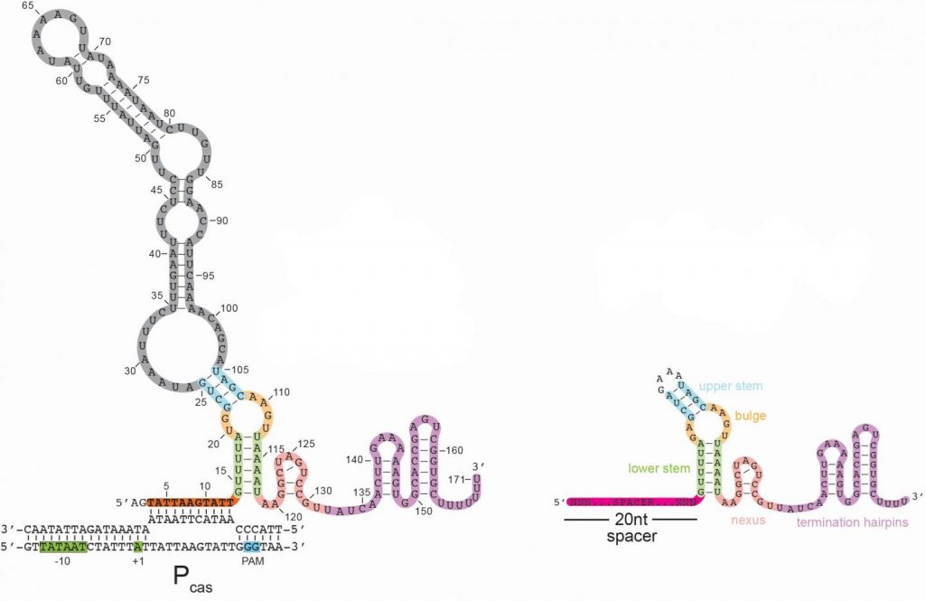
Here’s an image illustrating the long form of the tracrRNA or ‘dimmer switch’ alongside the more commonly used short form,

Caption: Left – a schematic of the long form of the tracrRNA used by the CRISPR-Cas9 system in bacteria; Right – the standard guide RNA used by many scientists as part of the gene-cutting CRISPR-Cas9 system. Credit: Joshua Modell, Rachael Workman and Johns Hopkins Medicine

A January 19 ,2021 Johns Hopkins Medicine news release (also on EurekAlert), which originated the news item, explains about CRISPR and what the acronym stands for, as well as, giving more details about the discovery,

First identified in the genome of gut bacteria in 1987, CRISPR-Cas9 is a naturally occurring but unusual group of genes with a potential for cutting DNA sequences in other types of cells that was realized 25 years later. Its value in genetic engineering — programmable gene alteration in living cells, including human cells — was rapidly appreciated, and its widespread use as a genome “editor” in thousands of laboratories worldwide was recognized in the awarding of the Nobel Prize in Chemistry last year to its American and French co-developers.

CRISPR stands for clustered, regularly interspaced short palindromic repeats. Cas9, which refers to CRISPR-associated protein 9, is the name of the enzyme that makes the DNA slice. Bacteria naturally use CRISPR-Cas9 to cut viral or other potentially harmful DNA and disable the threat, says Joshua Modell, Ph.D., assistant professor of molecular biology and genetics at the Johns Hopkins University School of Medicine. In this role, Modell says, “CRISPR is not only an immune system, it’s an adaptive immune system — one that can remember threats it has previously encountered by holding onto a short piece of their DNA, which is akin to a mug shot.” These mug shots are then copied into “guide RNAs” that tell Cas9 what to cut.

Scientists have long worked to unravel the precise steps of CRISPR-Cas9’s mechanism and how its activity in bacteria is dialed up or down. Looking for genes that ignite or inhibit the CRISPR-Cas9 gene-cutting system for the common, strep-throat causing bacterium Streptococcus pyogenes, the Johns Hopkins scientists found a clue regarding how that aspect of the system works.

Specifically, the scientists found a gene in the CRISPR-Cas9 system that, when deactivated, led to a dramatic increase in the activity of the system in bacteria. The product of this gene appeared to re-program Cas9 to act as a brake, rather than as a “scissor,” to dial down the CRISPR system.

“From an immunity perspective, bacteria need to ramp up CRISPR-Cas9 activity to identify and rid the cell of threats, but they also need to dial it down to avoid autoimmunity — when the immune system mistakenly attacks components of the bacteria themselves,” says graduate student Rachael Workman, a bacteriologist working in Modell’s laboratory.

To further nail down the particulars of the “brake,” the team’s next step was to better understand the product of the deactivated gene (tracrRNA). RNA is a genetic cousin to DNA and is vital to carrying out DNA “instructions” for making proteins. TracrRNAs belong to a unique family of RNAs that do not make proteins. Instead, they act as a kind of scaffold that allows the Cas9 enzyme to carry the guide RNA that contains the mug shot and cut matching DNA sequences in invading viruses.

TracrRNA comes in two sizes: long and short. Most of the modern gene-cutting CRISPR-Cas9 tools use the short form. However, the research team found that the deactivated gene product was the long form of tracrRNA, the function of which has been entirely unknown.

The long and short forms of tracrRNA are similar in structure and have in common the ability to bind to Cas9. The short form tracrRNA also binds to the guide RNA. However, the long form tracrRNA doesn’t need to bind to the guide RNA, because it contains a segment that mimics the guide RNA. “Essentially, long form tracrRNAs have combined the function of the short form tracrRNA and guide RNA,” says Modell.

In addition, the researchers found that while guide RNAs normally seek out viral DNA sequences, long form tracrRNAs target the CRISPR-Cas9 system itself. The long form tracrRNA tends to sit on DNA, rather than cut it. When this happens in a particular area of a gene, it prevents that gene from expressing, — or becoming functional.

To confirm this, the researchers used genetic engineering to alter the length of a certain region in long form tracrRNA to make the tracrRNA appear more like a guide RNA. They found that with the altered long form tracrRNA, Cas9 once again behaved more like a scissor.

Other experiments showed that in lab-grown bacteria with a plentiful amount of long form tracrRNA, levels of all CRISPR-related genes were very low. When the long form tracrRNA was removed from bacteria, however, expression of CRISPR-Cas9 genes increased a hundredfold.

Bacterial cells lacking the long form tracrRNA were cultured in the laboratory for three days and compared with similarly cultured cells containing the long form tracrRNA. By the end of the experiment, bacteria without the long form tracrRNA had completely died off, suggesting that long form tracrRNA normally protects cells from the sickness and death that happen when CRISPR-Cas9 activity is very high.

“We started to get the idea that the long form was repressing but not eliminating its own CRISPR-related activity,” says Workman.

To see if the long form tracrRNA could be re-programmed to repress other bacterial genes, the research team altered the long form tracrRNA’s spacer region to let it sit on a gene that produces green fluorescence. Bacteria with this mutated version of long form tracrRNA glowed less green than bacteria containing the normal long form tracrRNA, suggesting that the long form tracrRNA can be genetically engineered to dial down other bacterial genes.

Another research team, from Emory University, found that in the parasitic bacteria Francisella novicida, Cas9 behaves as a dimmer switch for a gene outside the CRISPR-Cas9 region. The CRISPR-Cas9 system in the Johns Hopkins study is more widely used by scientists as a gene-cutting tool, and the Johns Hopkins team’s findings provide evidence that the dimmer action controls the CRISPR-Cas9 system in addition to other genes.

The researchers also found the genetic components of long form tracrRNA in about 40% of the Streptococcus group of bacteria. Further study of bacterial strains that don’t have the long form tracrRNA, says Workman, will potentially reveal whether their CRISPR-Cas9 systems are intact, and other ways that bacteria may dial back the CRISPR-Cas9 system.

The dimmer capability that the experiments uncovered, says Modell, offers opportunities to design new or better CRISPR-Cas9 tools aimed at regulating gene activity for research purposes. “In a gene editing scenario, a researcher may want to cut a specific gene, in addition to using the long form tracrRNA to inhibit gene activity,” he says.

Here’s a link to and a citation for the paper,

A natural single-guide RNA repurposes Cas9 to autoregulate CRISPR-Cas expression by Rachael E. Workman, Teja Pammi, Binh T.K. Nguyen, Leonardo W. Graeff, Erika Smith, Suzanne M. Sebald, Marie J. Stoltzfus, Chad W. Euler, Joshua W. Modell. Cell DOI:https://doi.org/10.1016/j.cell.2020.12.017 Published Online:J anuary 08, 2021

This paper is behind a paywall.

Bacteria and graphene oxide as a basis for producing computers

A July 10, 2019 news item on ScienceDaily announces a more environmentally friendly way to produce graphene leading to more environmentally friendly devices such as computers,

In order to create new and more efficient computers, medical devices, and other advanced technologies, researchers are turning to nanomaterials: materials manipulated on the scale of atoms or molecules that exhibit unique properties.

Graphene — a flake of carbon as thin as a single later of atoms — is a revolutionary nanomaterial due to its ability to easily conduct electricity, as well as its extraordinary mechanical strength and flexibility. However, a major hurdle in adopting it for everyday applications is producing graphene at a large scale, while still retaining its amazing properties.

In a paper published in the journal ChemOpen, Anne S. Meyer, an associate professor of biology at the University of Rochester [New York state, US], and her colleagues at Delft University of Technology in the Netherlands, describe a way to overcome this barrier. The researchers outline their method to produce graphene materials using a novel technique: mixing oxidized graphite with bacteria. Their method is a more cost-efficient, time-saving, and environmentally friendly way of producing graphene materials versus those produced chemically, and could lead to the creation of innovative computer technologies and medical equipment.

A July 10, 2019 University of Rochester news release (also on EurekAlert), which originated the news item, provides details as to how this new technique for extracting graphene differs from the technique currently used,

Graphene is extracted from graphite, the material found in an ordinary pencil. At exactly one atom thick, graphene is the thinnest–yet strongest–two-dimensional material known to researchers. Scientists from the University of Manchester in the United Kingdom were awarded the 2010 Nobel Prize in Physics for their discovery of graphene; however, their method of using sticky tape to make graphene yielded only small amounts of the material.

“For real applications you need large amounts,” Meyer says. “Producing these bulk amounts is challenging and typically results in graphene that is thicker and less pure. This is where our work came in.”

In order to produce larger quantities of graphene materials, Meyer and her colleagues started with a vial of graphite. They exfoliated the graphite–shedding the layers of material–to produce graphene oxide (GO), which they then mixed with the bacteria Shewanella. They let the beaker of bacteria and precursor materials sit overnight, during which time the bacteria reduced the GO to a graphene material.

“Graphene oxide is easy to produce, but it is not very conductive due to all of the oxygen groups in it,” Meyer says. “The bacteria remove most of the oxygen groups, which turns it into a conductive material.”

While the bacterially-produced graphene material created in Meyer’s lab is conductive, it is also thinner and more stable than graphene produced chemically. It can additionally be stored for longer periods of time, making it well suited for a variety of applications, including field-effect transistor (FET) biosensors and conducting ink. FET biosensors are devices that detect biological molecules and could be used to perform, for example, real-time glucose monitoring for diabetics.

“When biological molecules bind to the device, they change the conductance of the surface, sending a signal that the molecule is present,” Meyer says. “To make a good FET biosensor you want a material that is highly conductive but can also be modified to bind to specific molecules.” Graphene oxide that has been reduced is an ideal material because it is lightweight and very conductive, but it typically retains a small number of oxygen groups that can be used to bind to the molecules of interest.

The bacterially produced graphene material could also be the basis for conductive inks, which could, in turn, be used to make faster and more efficient computer keyboards, circuit boards, or small wires such as those used to defrost car windshields. Using conductive inks is an “easier, more economical way to produce electrical circuits, compared to traditional techniques,” Meyer says. Conductive inks could also be used to produce electrical circuits on top of nontraditional materials like fabric or paper.

“Our bacterially produced graphene material will lead to far better suitability for product development,” Meyer says. “We were even able to develop a technique of ‘bacterial lithography’ to create graphene materials that were only conductive on one side, which can lead to the development of new, advanced nanocomposite materials.”

Here’s a link to and a citation for the paper,

Creation of Conductive Graphene Materials by Bacterial Reduction Using Shewanella Oneidensis by Benjamin A. E. Lehner, Vera A. E. C. Janssen, Dr. Ewa M. Spiesz, Dominik Benz, Dr. Stan J. J. Brouns, Dr. Anne S. Meyer, Prof. Dr. Herre S. J. van der Zant. ChemistryOpen Volume 8, Issue 7 July 2019 Pages 888-895 DOI: https://doi.org/10.1002/open.201900186
First published: 04 July 2019

As you would expect given the journal’s title, this paper is open access.

Cleaning water with bacteria

There seems to be much interest in bacteria as collaborators as opposed to the old ‘enemy that must be destoyed’ concept. The latest collaborative effort was announced in a January 19,2019 news item on Nanowerk,

More than one in 10 people in the world lack basic drinking water access, and by 2025, half of the world’s population will be living in water-stressed areas, which is why access to clean water is one of the National Academy of Engineering’s Grand Challenges. Engineers at Washington University in St. Louis [WUSTL] have designed a novel membrane technology that purifies water while preventing biofouling, or buildup of bacteria and other harmful microorganisms that reduce the flow of water.

And they used bacteria to build such filtering membranes.

A January 17, 2019 WUSTL news release by Beth Miller, which originated the news item, provides more detail,

Srikanth Singamaneni, professor of mechanical engineering & materials science, and Young-Shin Jun, professor of energy, environmental & chemical engineering, and their teams blended their expertise to develop an ultrafiltration membrane using graphene oxide and bacterial nanocellulose that they found to be highly efficient, long-lasting and environmentally friendly. If their technique were to be scaled up to a large size, it could benefit many developing countries where clean water is scarce.


Biofouling accounts for nearly half of all membrane fouling and is highly challenging to eradicate completely. Singamaneni and Jun have been tackling this challenge together for nearly five years. They previously developed other membranes using gold nanostars, but wanted to design one that used less expensive materials.

Their new membrane begins with feeding Gluconacetobacter hansenii bacteria a sugary substance so that they form cellulose nanofibers when in water. The team then incorporated graphene oxide (GO) flakes into the bacterial nanocellulose while it was growing, essentially trapping GO in the membrane to make it stable and durable.

After GO is incorporated, the membrane is treated with base solution to kill Gluconacetobacter. During this process, the oxygen groups of GO are eliminated, making it reduced GO.  When the team shone sunlight onto the membrane, the reduced GO flakes immediately generated heat, which is dissipated into the surrounding water and bacteria nanocellulose.

Ironically, the membrane created from bacteria also can kill bacteria.
“If you want to purify water with microorganisms in it, the reduced graphene oxide in the membrane can absorb the sunlight, heat the membrane and kill the bacteria,” Singamaneni said.

Singamaneni and Jun and their team exposed the membrane to E. coli bacteria, then shone light on the membrane’s surface. After being irradiated with light for just 3 minutes, the E. coli bacteria died. The team determined that the membrane quickly heated to above the 70 degrees Celsius required to deteriorate the cell walls of E. coli bacteria.

While the bacteria are killed, the researchers had a pristine membrane with a high quality of nanocellulose fibers that was able to filter water twice as fast as commercially available ultrafiltration membranes under a high operating pressure.

When they did the same experiment on a membrane made from bacterial nanocellulose without the reduced GO, the E. coli bacteria stayed alive.

“This is like 3-D printing with microorganisms,” Jun said. “We can add whatever we like to the bacteria nanocellulose during its growth. We looked at it under different pH conditions similar to what we encounter in the environment, and these membranes are much more stable compared to membranes prepared by vacuum filtration or spin-coating of graphene oxide.”

While Singamaneni and Jun acknowledge that implementing this process in conventional reverse osmosis systems is taxing, they propose a spiral-wound module system, similar to a roll of towels. It could be equipped with LEDs or a type of nanogenerator that harnesses mechanical energy from the fluid flow to produce light and heat, which would reduce the overall cost.

Here’s a link to and a citation for the paper,

Photothermally Active Reduced Graphene Oxide/Bacterial Nanocellulose Composites as Biofouling-Resistant Ultrafiltration Membranes by Qisheng Jiang, Deoukchen Ghim, Sisi Cao, Sirimuvva Tadepalli, Keng-Ku Liu, Hyuna Kwon, Jingyi Luan, Yujia Min, Young-Shin Jun, and Srikanth Singamaneni. Environ. Sci. Technol., 2019, 53 (1), pp 412–421 DOI: 10.1021/acs.est.8b02772 Publication Date (Web): September 14, print Jan. 2, 2019.

Copyright © 2018 American Chemical Society

This paper is behind a paywall.

Food nanoparticles and their effect on intestinal flora (i.e., your gut microbiome)

This work from Germany is largely speculative. The scientists seem to be interested in exploring how engineered nanoparticles and naturally occurring nanoparticles in food affect your gut. From a January 29, 2019 news item on ScienceDaily,

The intestinal microbiome is not only key for food processing but an accepted codeterminant for various diseases. Researchers led by the University Medical Center of Johannes Gutenberg University Mainz (JGU) identified effects of nanoparticles on intestinal microorganisms. The ultra-small particles adhere to intestinal microorganisms, thereby affecting their life cycle as well as cross talk with the host. One of the researchers’ observations was that nanoparticles’ binding inhibits the infection with Helicobacter pylori, a pathogen implicated in gastric cancer. The findings will stimulate further epidemiological studies and pave the way for the development of potential ‘probiotic’ nanoparticles for food. The discoveries were published in Science of Food.

A January 29, 2019 Johannes Gutenberg University Mainz (JGU) press release (also on EurekAlert), which originated the news item, provides more detail,

Due to their minute size, nanoparticles have unique characteristics and capabilities, such as adhering to microstructures. Nanotechnology is as an important driver of innovation for both consumer industry and medicine. In medicine, the focus is on improving diagnostics and therapeutics, while industry addresses mainly product optimization. Hence, synthetic nanoparticles are already used as additives to improve the characteristics of food. But how can we use nanotechnology more efficiently and safely in food? And are there unknown effects of nanoparticles, which need to be further exploited?

Nutrition strongly influences the diversity and composition of our microbiome. ‘Microbiome’ describes all colonizing microorganisms present in a human being, in particular, all the bacteria in the gut. In other words, your microbiome includes your intestinal flora as well as the microorganisms that colonize your skin, mouth, and nasal cavity.

Scientists and clinicians are interested in microbiomes because of their positive or negative effects on the host. These include modulation of our immune system, metabolism, vascular aging, cerebral functioning, and our hormonal system. The composition of the microbiome seems to play an important role for the development of various disorders, such as cardiovascular diseases, cancer, allergies, obesity, and even mental disorders. “Hence, nutrition and its containing nanoparticulates may affect the microbiome-host balance, finally influencing human health. In order to reduce potential risks and, ideally, promote health, the impact of dietary nanoparticles needs to be understood,” emphasized Professor David J. McClements from the Department of Food Science at the University of Massachusetts in Amherst, USA.

“Prior to our studies, nobody really looked whether and how nano-additives directly influence the gastrointestinal flora,” commented Professor Roland Stauber of the Department of Otolaryngology, Head, and Neck Surgery at the Mainz University Medical Center. “Hence, we studied at a wide range of technical nanoparticles with clearly defined properties in order to mimic what happens to currently used or potential future nanosized food additives. By simulating the journey of particles through the different environments of the digestive tract in the laboratory, we found that the all tested nanomaterials were indeed able to bind to bacteria.” explained Stauber.

The scientists discovered that these binding processes can have different outcomes. On the one hand, nanoparticle-bound microorganisms were less efficiently recognized by the immune system, which may lead to increased inflammatory responses. On the other hand, ‘nano-food’ showed beneficial effects. In cell culture models, silica nanoparticles inhibited the infectivity of Helicobacter pylori, which is considered to be one of the main agents involved in gastric cancer.

‘It was puzzling that we were able to also isolate naturally occurring nanoparticles from food, like beer, which showed similar effects. Nanoparticles in our daily food are not just those added deliberately but can also be generated naturally during preparation. Nanoparticulates are already omnipresent,” concluded Stauber.

The insights of the study will allow to derive strategies for developing and utilizing synthetic or natural nanoparticles to modulate the microbiome as beneficial ingredients in functional foods. “The challenge is to identify nanoparticles that fit the desired purpose, perhaps even as probiotic food supplements in the future. Challenge accepted,” emphasized Stauber and his team.

Here’s a link to and a citation for the paper,

Nanosized food additives impact beneficial and pathogenic bacteria in the human gut: a simulated gastrointestinal study by Svenja Siemer, Angelina Hahlbrock, Cecilia Vallet, David Julian McClements, Jan Balszuweit, Jens Voskuhl, Dominic Docter, Silja Wessler, Shirley K. Knauer, Dana Westmeier, & Roland H. Stauber. npj Science of Foodvolume 2, Article number: 22 (2018) DOI: https://doi.org/10.1038/s41538-018-0030-8 Published 04 December 2018

This paper is open access.

An artificial enzyme uses light to kill bacteria

An April 4, 2018 news item on ScienceDaily announces a light-based approach to killing bacteria,

Researchers from RMIT University [Australia] have developed a new artificial enzyme that uses light to kill bacteria.

The artificial enzymes could one day be used in the fight against infections, and to keep high-risk public spaces like hospitals free of bacteria like E. coli and Golden Staph.

E. coli can cause dysentery and gastroenteritis, while Golden Staph is the major cause of hospital-acquired secondary infections and chronic wound infections.

Made from tiny nanorods — 1000 times smaller than the thickness of the human hair — the “NanoZymes” use visible light to create highly reactive oxygen species that rapidly break down and kill bacteria.

Lead researcher, Professor Vipul Bansal who is an Australian Future Fellow and Director of RMIT’s Sir Ian Potter NanoBioSensing Facility, said the new NanoZymes offer a major cutting edge over nature’s ability to kill bacteria.

Dead bacteria made beautiful,

Caption: A 3-D rendering of dead bacteria after it has come into contact with the NanoZymes.
Credit: Dr. Chaitali Dekiwadia/ RMIT Microscopy and Microanalysis Facility

An April 5, 2018 RMIT University press release (also on EurekAlert but dated April 4, 2018), which originated the news item, expands on the theme,

“For a number of years we have been attempting to develop artificial enzymes that can fight bacteria, while also offering opportunities to control bacterial infections using external ‘triggers’ and ‘stimuli’,” Bansal said. “Now we have finally cracked it.

“Our NanoZymes are artificial enzymes that combine light with moisture to cause a biochemical reaction that produces OH radicals and breaks down bacteria. Nature’s antibacterial activity does not respond to external triggers such as light.

“We have shown that when shined upon with a flash of white light, the activity of our NanoZymes increases by over 20 times, forming holes in bacterial cells and killing them efficiently.

“This next generation of nanomaterials are likely to offer new opportunities in bacteria free surfaces and controlling spread of infections in public hospitals.”

The NanoZymes work in a solution that mimics the fluid in a wound. This solution could be sprayed onto surfaces.

The NanoZymes are also produced as powders to mix with paints, ceramics and other consumer products. This could mean bacteria-free walls and surfaces in hospitals.

Public toilets — places with high levels of bacteria, and in particular E. coli — are also a prime location for the NanoZymes, and the researchers believe their new technology may even have the potential to create self-cleaning toilet bowls.

While the NanoZymes currently use visible light from torches or similar light sources, in the future they could be activated by sunlight.

The researchers have shown that the NanoZymes work in a lab environment. The team is now evaluating the long-term performance of the NanoZymes in consumer products.

“The next step will be to validate the bacteria killing and wound healing ability of these NanoZymes outside of the lab,” Bansal said.

“This NanoZyme technology has huge potential, and we are seeking interest from appropriate industries for joint product development.”

Here’s a link to and a citation for the paper,

Visible-Light-Triggered Reactive-Oxygen-Species-Mediated Antibacterial Activity of Peroxidase-Mimic CuO Nanorods by Md. Nurul Karim, Mandeep Singh, Pabudi Weerathunge, Pengju Bian, Rongkun Zheng, Chaitali Dekiwadia, Taimur Ahmed, Sumeet Walia, Enrico Della Gaspera, Sanjay Singh, Rajesh Ramanathan, and Vipul Bansal. ACS Appl. Nano Mater., Article ASAP DOI: 10.1021/acsanm.8b00153 Publication Date (Web): March 6, 2018

Copyright © 2018 American Chemical Society

This paper is open access.

Immune to CRISPR?

I guess if you’re going to use bacteria as part of your gene editing technology (CRISPR [clustered regularly interspaced short palindromic repeats]/Cas9) then, you might half expect the body’s immune system may have developed some defenses. A Jan. 9, 2018 article by Sarah Zhang for The Atlantic provides some insight into what the new research suggests (Note: Links have been removed),

2018 is supposed to be the year of CRISPR in humans. The first U.S. and European clinical trials that test the gene-editing tool’s ability to treat diseases—such as sickle-cell anemia, beta thalassemia, and a type of inherited blindness—are slated to begin this year.

But the year has begun on a cautionary note. On Friday [January 5, 2018], Stanford researchers posted a preprint (which has not been peer reviewed) to the website biorXiv highlighting a potential obstacle to using CRISPR in humans: Many of us may already be immune to it. That’s because CRISPR actually comes from bacteria that often live on or infect humans, and we have built up immunity to the proteins from these bacteria over our lives.

Not all CRISPR therapies in humans will be doomed. “We don’t think this is the end of the story. This is the start of the story,” says Porteus [Matthew Porteus, a pediatrician and stem-cell researcher at Stanford]. There are likely ways around the problem of immunity to CRISPR proteins, and many of the early clinical trials appear to be designed around this problem.

Porteus and his colleagues focused on two versions of Cas9, the bacterial protein mostly commonly used in CRISPR gene editing. One comes from Staphylococcus aureus, which often harmlessly lives on skin but can sometimes causes staph infections, and another from Streptococcus pyogenes, which causes strep throat but can also become “flesh-eating bacteria” when it spreads to other parts of the body. So yeah, you want your immune system to be on guard against these bacteria.

The human immune system has a couple different ways of recognizing foreign proteins, and the team tested for both. First, they looked to see if people have molecules in their blood called antibodies that can specifically bind to Cas9. Among 34 people they tested, 79 percent had antibodies against the staph Cas9 and 65 percent against the strep Cas9.

The Stanford team only tested for preexisting immunity against Cas9, but anytime you inject a large bacterial protein into the human body, it can provoke an immune response. After all, that’s how the immune system learns to fight off bacteria it’s never seen before. (Preexisting immunity can make the response faster and more robust, though.)

The danger of the immune system turning on a patient’s body hangs over a lot of research into correcting genes. In the late 1990s and 2000s, research into gene therapy was derailed by the death of 18-year-old Jesse Gelsinger, who died from an immune reaction to the virus used to deliver the corrected gene. This is the worst-case scenario that the CRISPR world hopes to avoid.

Here’s a link to and a citation for the preprint,

Identification of Pre-Existing Adaptive Immunity to Cas9 Proteins in Humans by Carsten Trevor Charlesworth, Priyanka S Deshpande, Daniel P Dever, Beruh Dejene, Natalia Gomez-Ospina, Sruthi Mantri, Mara Pavel-Dinu, Joab Camarena, Kenneth I Weinberg, Matthew H Porteus. bioRxiv posted January 5, 2018 doi: https://doi.org/10.1101/243345

This article is a preprint and has not been peer-reviewed …

This preprint (not yet published paper) is open access and open for feedback.

Meanwhile, the year of CRISPR takes off (from a January 10, 2018 American Chemical Society news release on EurekAlert),

This year could be a defining one for CRISPR, the gene editing technique, which has been hailed as an important breakthrough in laboratory research. That’s because the first company-sponsored clinical studies will be conducted to see if it can help treat diseases in humans, according to an article in Chemical & Engineering News (C&EN), the weekly newsmagazine of the American Chemical Society.

C&EN Assistant Editor Ryan Cross reports that a big push is coming from industry, specifically from three companies that are each partly founded by one of the three inventors of the method. They are zeroing in on the blood diseases called sickle-cell anemia and β-thalassemia, mostly because their precise cause is known. In these diseases, hemoglobin doesn’t function properly, leading to severe health issues in some people. Crispr Therapeutics and Intellia Therapeutics plan to test the technique to boost levels of an alternative version of healthy hemoglobin. Editas Medicine, however, will also use CRISPR to correct mutations in the faulty hemoglobin gene. Labs led by university researchers are also joining the mix, starting or continuing clinical trials with the approach in 2018.

Because CRISPR is being used to cut a cell’s DNA and insert a new sequence, concerns have been raised about the potential for accidents. A cut in the wrong place could mean introducing a new mutation that could be benign — or cancerous. But according to proponents of the method, researchers are conducting extensive computer predictions and in vitro tests to help avoid this outcome.

The January 8, 2018 Chemical and Engineering News (C&EN) open access article by Ryan Cross is here.

Finally, if you are interested in how this affects research as it’s being developed, there’s University of British Columbia researcher Rosie Redfield’s January 16, 2018 posting on RRResearch blog,

Thursday’s [January 11, 2018] post described the hypothesis that bacteria might use gene transfer agent particles to inoculate other cells in the population with fragments of phage DNA, and outlined an experiment to test this.  Now I’m realizing that I need to know a lot more about the kind of immunity I should expect to see if this GTA-as-vaccine hypothesis is correct.

That should give you some idea of what I meant by “research as it’s being developed.” Redfield’s blog is not for the mildly interested.

Redfield is well-known internationally as being one of the first to refute research which suggested the existence of an ‘arsenic bacterium’ (see my Dec. 8, 2010 posting: My apologies for arsenic blooper. She’s first mentioned in the second excerpt, second paragraph.) The affair was known online as #arseniclife. There’s a May 27, 2011 essay by Carl Zimmer on Slate titled: The Discovery of Arsenic-Based Twitter: How #arseniclife changed science.

German scientists battle tough mucus

A December 15, 2017 news item on ScienceDaily highlights cystic fibrosis research being done in Germany,

Around one in 3,300 children in Germany is born with Mucoviscidosis [cystic fibrosis; CF]. A characteristic of this illness is that one channel albumen on the cell surface is disturbed by mutations. Thus, the amount of water of different secretions in the body is reduced which creates a tough mucus. As a consequence, inner organs malfunction. Moreover, the mucus blocks the airways. Thus, the self regulatory function of the lung is disturbed, the mucus is colonized by bacteria and chronic infections follow. The lung is so significantly damaged that patients often die or need to have a lung transplant. The average life expectancy of a patient today is around 40 years. This is due to medical progress. Permanent treatment with inhaled antibiotics play a considerable part in this. The treatment can’t avoid the colonization by bacteria completely but it can keep it in check for a longer period of time. However, the bacteria defend themselves with a development of resistance and with the growth of so-called biofilms underneath the layer of mucus, which mostly block off the bacteria in the lower rows like a protective shield.

A complex way to the Pathogens

Scientists of the Friedrich Schiller University Jena, Germany succeeded in developing a much more efficient method to treat the airway infections which are often lethal. Crucial are nanoparticles that transport the antibiotics more efficiently to their destination….

A December 15, 2017 Friedrich Schiller University Jena press release (also on EurekAlert), which originated the news item, expands on the theme,

“Typically, the drugs are applied by inhalation in the body. Then they make a complicated way through the body to the pathogens and many of them don’t make it to their destination,” states Prof. Dr Dagmar Fischer of the chair for Pharmaceutical Technology at the University of Jena, who supervised the project together with her colleague Prof. Dr Mathias Pletz, a pulmonologist and infectious diseases physician, from the Center for Infectious Diseases and Infection Control at the Jena University Hospital. The project was supported by the Deutsche Forschungsgemeinschaft. First of all, the active particles need to have a certain size to be able to reach the deeper airways and not to bounce off somewhere else before. Ultimately, they have to penetrate the thick layer of mucus on the airways as well as the lower layers of the bacteria biofilm.

Nanoparticles travel more efficiently

To overcome the strong defense, the researchers encapsulated the active agents, like the antibiotic Tobramycin, in a polyester polymer. Thus, they created a nanoparticle which they then tested in the laboratory where they beforehand had simulated the present lung situation, in a static as well as in a dynamic state, i. e. with simulated flow movements. Therefore Pletz’s research group had developed new test systems, which are able to mimick the situation of the chronically infected CF-lung. The scientists discovered that their nanoparticle travels more easily through the sponge-like net of the mucus layer and is finally able to kill off the pathogens without any problems. Moreover, an additionally applied coating of polyethylenglycol makes it nearly invisible for the immune system. “All materials of a nanocarrier are biocompatible, biodegradable, nontoxic and therefore not dangerous for humans,” the researcher informs.

However, the Jena scientists don’t know yet exactly why their nanoparticle fights the bacteria so much more efficiently. But they want to finally get clarification in the year ahead. “We have two assumptions: Either the much more efficient transport method advances significantly larger amounts of active ingredients to the center of infection, or the nanoparticle circumvents a defense mechanism, which the bacterium has developed against the antibiotic,” the Jena Pharmacist Fischer explains. “This would mean, that we succeeded in giving back its impact to an antibiotic, which had already lost it through a development of resistance of the bacteria.”

“More specifically, we assume that bacteria from the lower layers of the biofilm transform into dormant persisters and hardly absorb any substances from outside. In this stadium, they are tolerant to most antibiotics, which only kill off self-dividing bacteria. The nanoparticles transport the antibiotics more or less against their will to the inner part of the cell, where they can unfold their impact,” Mathias Pletz adds.

Additionally, the Jena research team had to prepare the nanoparticles for the inhalation. Because at 200 nanometers the particle is too small to get into the deeper airways. “The breathing system filters out particles that are too big as well as those which are too small,” Dagmar Fischer explains. “So, we are left with a preferred window of between one and five micrometers.” The Jena researchers also have promising ideas for resolving this problem.

Coating of Nanoparticles enhances the impact of Antibiotics against Biofilms

The scientists from Jena are at this point already convinced to have found a very promising method to fight respiratory infections of patients with mucoviscidosis. Thus they may be able to contribute to a higher life expectancy of those affected. “We were able to show that the nanoparticle coating improves the impact of the antibiotics against biofilm by a factor of 1,000,” the pulmonologist and infectious diseases physician is happy to say.

It’s exciting news and I wish the researchers great success. Perhaps, one day, they will publish a paper about their work.