Tag Archives: ribonucleic acid (RNA)

Visualization of RNA structures at near-atomic resolution enabled by nanotechnology

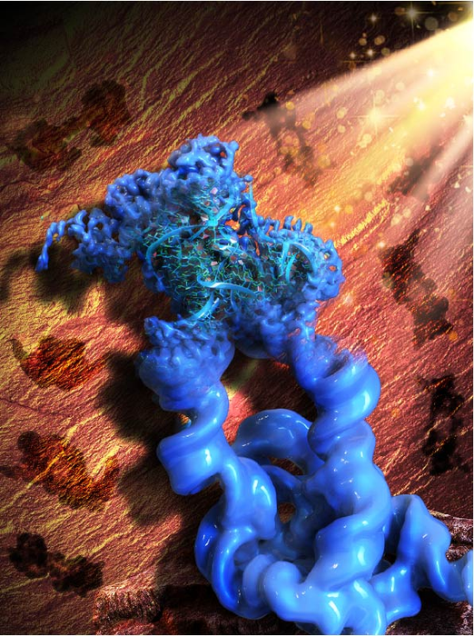
The illustration that accompanies the research is both fascinating and baffling as its caption,

Caption: This illustration is inspired by the Paleolithic rock painting in the Lascaux cave, signifying the acronym of our method, ROCK. Figuratively, the patterns of the rock art in the background (brown) are the 2D projections of the engineered dimeric construct of the Tetrahymena group I intron, while the main object in the front (blue) is the reconstructed 3D cryo-EM map of the dimer, with one monomer in focus and refined to the high resolution that allowed the collaborators to build an atomic model of the RNA. Credit: Wyss Institute at Harvard University

This May 2, 2022 news item on ScienceDaily announces the research into RNA molecules made possible by ROCK (the technology being illustrated in the above),

We live in a world made and run by RNA [ribonucleic acid], the equally important sibling of the genetic molecule DNA. In fact, evolutionary biologists hypothesize that RNA existed and self-replicated even before the appearance of DNA and the proteins encoded by it. Fast forward to modern day humans: science has revealed that less than 3% of the human genome is transcribed into messenger RNA (mRNA) molecules that in turn are translated into proteins. In contrast, 82% of it is transcribed into RNA molecules with other functions many of which still remain enigmatic.

To understand what an individual RNA molecule does, its 3D structure needs to be deciphered at the level of its constituent atoms and molecular bonds. Researchers have routinely studied DNA and protein molecules by turning them into regularly packed crystals that can be examined with an X-ray beam (X-ray crystallography) or radio waves (nuclear magnetic resonance). However, these techniques cannot be applied to RNA molecules with nearly the same effectiveness because their molecular composition and structural flexibility prevent them from easily forming crystals.

Now, a research collaboration led by Wyss Core Faculty member Peng Yin, Ph.D. at the Wyss Institute for Biologically Inspired Engineering at Harvard University, and Maofu Liao, Ph.D. at Harvard Medical School (HMS), has reported a fundamentally new approach to the structural investigation of RNA molecules. ROCK, as it is called, uses an RNA nanotechnological technique that allows it to assemble multiple identical RNA molecules into a highly organized structure, which significantly reduces the flexibility of individual RNA molecules and multiplies their molecular weight. Applied to well-known model RNAs with different sizes and functions as benchmarks, the team showed that their method enables the structural analysis of the contained RNA subunits with a technique known as cryo-electron microscopy (cryo-EM). Their advance is reported in Nature Methods.

A May 2, 2022 Wyss Institute for Biologically Inspired Engineering at Harvard University news release (also on EurekAlert) by Benjamin Boettner, which originated the news item, delves further into the imaging technology, Note: Links have been removed,

“ROCK is breaking the current limits of RNA structural investigations and enables 3D structures of RNA molecules to be unlocked that are difficult or impossible to access with existing methods, and at near-atomic resolution,” said Yin, who together with Liao led the study. “We expect this advance to invigorate many areas of fundamental research and drug development, including the burgeoning field of RNA therapeutics.” Yin also is a leader of the Wyss Institute’s Molecular Robotics Initiative and Professor in the Department of Systems Biology at HMS.

Gaining control over RNA

Yin’s team at the Wyss Institute has pioneered various approaches that enable DNA and RNA molecules to self-assemble into large structures based on different principles and requirements, including DNA bricks and DNA origami. They hypothesized that such strategies could also be used to assemble naturally occurring RNA molecules into highly ordered circular complexes in which their freedom to flex and move is highly restricted by specifically linking them together. Many RNAs fold in complex yet predictable ways, with small segments base-pairing with each other. The result often is a stabilized “core” and “stem-loops” bulging out into the periphery. 

“In our approach we install ‘kissing loops’ that link different peripheral stem-loops belonging to two copies of an identical RNA in a way that allows a overall stabilized ring to be formed, containing multiple copies of the RNA of interest,” said Di Liu, Ph.D., one of two first-authors and a Postdoctoral Fellow in Yin’s group. “We speculated that these higher-order rings could be analyzed with high resolution by cryo-EM, which had been applied to RNA molecules with first success.”

Picturing stabilized RNA

In cryo-EM, many single particles are flash-frozen at cryogenic temperatures to prevent any further movements, and then visualized with an electron microscope and the help of computational algorithms that compare the various aspects of a particle’s 2D surface projections and reconstruct its 3D architecture. Peng and Liu teamed up with Liao and his former graduate student François Thélot, Ph.D., the other co-first author of the study. Liao with his group has made important contributions to the rapidly advancing cryo-EM field and the experimental and computational analysis of single particles formed by specific proteins.

“Cryo-EM has great advantages over traditional methods in seeing high-resolution details of biological molecules including proteins, DNAs and RNAs, but the small size and moving tendency of most RNAs prevent successful determination of RNA structures. Our novel method of assembling RNA multimers solves these two problems at the same time, by increasing the size of RNA and reducing its movement,” said Liao, who also is Associate Professor of Cell Biology at HMS. “Our approach has opened the door to rapid structure determination of many RNAs by cryo-EM.” The integration of RNA nanotechnology and cryo-EM approaches led the team to name their method “RNA oligomerization-enabled cryo-EM via installing kissing loops” (ROCK).

To provide proof-of-principle for ROCK, the team focused on a large intron RNA from Tetrahymena, a single-celled organism, and a small intron RNA from Azoarcus, a nitrogen-fixing bacterium, as well as the so-called FMN riboswitch. Intron RNAs are non-coding RNA sequences scattered throughout the sequences of freshly-transcribed RNAs and have to be “spliced” out in order for the mature RNA to be generated. The FMN riboswitch is found in bacterial RNAs involved in the biosynthesis of flavin metabolites derived from vitamin B2. Upon binding one of them, flavin mononucleotide (FMN), it switches its 3D conformation and suppresses the synthesis of its mother RNA.  

“The assembly of the Tetrahymena group I intron into a ring-like structure made the samples more homogenous, and enabled the use of computational tools leveraging the symmetry of the assembled structure. While our dataset is relatively modest in size, ROCK’s innate advantages allowed us to resolve the structure at an unprecedented resolution,” said Thélot. “The RNA’s core is resolved at 2.85 Å [one Ångström is one ten-billions (US) of a meter and the preferred metric used by structural biologists], revealing detailed features of the nucleotide bases and sugar backbone. I don’t think we could have gotten there without ROCK – or at least not without considerably more resources.” 

Cryo-EM also is able to capture molecules in different states if they, for example, change their 3D conformation as part of their function. Applying ROCK to the Azoarcus intron RNA and the FMN riboswitch, the team managed to identify the different conformations that the Azoarcus intron transitions through during its self-splicing process, and to reveal the relative conformational rigidity of the ligand-binding site of the FMN riboswitch.

“This study by Peng Yin and his collaborators elegantly shows how RNA nanotechnology can work as an accelerator to advance other disciplines. Being able to visualize and understand the structures of many naturally occurring RNA molecules could have tremendous impact on our understanding of many biological and pathological processes across different cell types, tissues, and organisms, and even enable new drug development approaches,” said Wyss Founding Director Donald Ingber, M.D., Ph.D., who is also the Judah Folkman Professor of Vascular Biology at Harvard Medical School and Boston Children’s Hospital, and Professor of Bioengineering at the Harvard John A. Paulson School of Engineering and Applied Sciences.

The study was also authored by Joseph Piccirilli, Ph.D., an expert in RNA chemistry and biochemistry and Professor at The University of Chicago. It was supported by the National Science Foundation (NSF; grant# CMMI-1333215, CCMI-1344915, and CBET-1729397), Air Force Office of Scientific Research (AFOSR; grant MURI FATE, #FA9550-15-1-0514), National Institutes of Health (NIH; grant# 5DP1GM133052, R01GM122797, and R01GM102489), and the Wyss Institute’s Molecular Robotics Initiative.

Here’s a link to and a citation for the paper,

Sub-3-Å cryo-EM structure of RNA enabled by engineered homomeric self-assembly by Di Liu, François A. Thélot, Joseph A. Piccirilli, Maofu Liao & Peng Yin. Nature Methods (2022) DOI: https://doi.org/10.1038/s41592-022-01455-w Published: 02 May 2022

This paper is behind a paywall.

mRNA, COVID-19 vaccines, treating genetic diseases before birth, and the scientist who started it all

This post was going to be about new research into fetal therapeutics and mRNA.But, since I’ve been very intrigued by the therapeutic agent, mRNA, which has been a big part of the COVID-19 vaccine story; this seemed like a good opportunity to dive a little more deeply into that topic at the same time.

It’s called messenger ribonucleic acid (mRNA) and until seeing this video I had only the foggiest idea of how it works, which is troubling since at least two COVID-19 vaccines are based on this ‘new’ technology. From a November 10, 2020 article by Damian Garde for STAT,

Garde’s article offers detail about mRNA along with fascinating insight into how science and entreneurship works.

mRNA—it’s in the details, plus, the loneliness of pioneer researchers, a demotion, and squabbles

Garde’s November 10, 2020 article provides some explanation about how mRNA vaccines work and it takes a look at what can happen to pioneering scientists (Note: A link has been removed),

For decades, scientists have dreamed about the seemingly endless possibilities of custom-made messenger RNA, or mRNA.

Researchers understood its role as a recipe book for the body’s trillions of cells, but their efforts to expand the menu have come in fits and starts. The concept: By making precise tweaks to synthetic mRNA and injecting people with it, any cell in the body could be transformed into an on-demand drug factory. [emphasis mine]

But turning scientific promise into medical reality has been more difficult than many assumed. Although relatively easy and quick to produce compared to traditional vaccine-making, no mRNA vaccine or drug has ever won approval [until 2021].

Whether mRNA vaccines succeed or not, their path from a gleam in a scientist’s eye to the brink of government approval has been a tale of personal perseverance, eureka moments in the lab, soaring expectations — and an unprecedented flow of cash into the biotech industry.

Before messenger RNA was a multibillion-dollar idea, it was a scientific backwater. And for the Hungarian-born scientist behind a key mRNA discovery, it was a career dead-end.

Katalin Karikó spent the 1990s collecting rejections. Her work, attempting to harness the power of mRNA to fight disease, was too far-fetched for government grants, corporate funding, and even support from her own colleagues.

It all made sense on paper. In the natural world, the body relies on millions of tiny proteins to keep itself alive and healthy, and it uses mRNA to tell cells which proteins to make. If you could design your own mRNA, you could, in theory, hijack that process and create any protein you might desire — antibodies to vaccinate against infection, enzymes to reverse a rare disease, or growth agents to mend damaged heart tissue.

In 1990, researchers at the University of Wisconsin managed to make it work in mice. Karikó wanted to go further.

The problem, she knew, was that synthetic RNA was notoriously vulnerable to the body’s natural defenses, meaning it would likely be destroyed before reaching its target cells. And, worse, the resulting biological havoc might stir up an immune response that could make the therapy a health risk for some patients.

It was a real obstacle, and still may be, but Karikó was convinced it was one she could work around. Few shared her confidence.

“Every night I was working: grant, grant, grant,” Karikó remembered, referring to her efforts to obtain funding. “And it came back always no, no, no.”

By 1995, after six years on the faculty at the University of Pennsylvania, Karikó got demoted. She had been on the path to full professorship, but with no money coming in to support her work on mRNA, her bosses saw no point in pressing on.

She was back to the lower rungs of the scientific academy.

“Usually, at that point, people just say goodbye and leave because it’s so horrible,” Karikó said.

There’s no opportune time for demotion, but 1995 had already been uncommonly difficult. Karikó had recently endured a cancer scare, and her husband was stuck in Hungary sorting out a visa issue. Now the work to which she’d devoted countless hours was slipping through her fingers.

“I thought of going somewhere else, or doing something else,” Karikó said. “I also thought maybe I’m not good enough, not smart enough. I tried to imagine: Everything is here, and I just have to do better experiments.”

In time, those better experiments came together. After a decade of trial and error, Karikó and her longtime collaborator at Penn — Drew Weissman, an immunologist with a medical degree and Ph.D. from Boston University — discovered a remedy for mRNA’s Achilles’ heel.

The stumbling block, as Karikó’s many grant rejections pointed out, was that injecting synthetic mRNA typically led to that vexing immune response; the body sensed a chemical intruder, and went to war. The solution, Karikó and Weissman discovered, was the biological equivalent of swapping out a tire.

Every strand of mRNA is made up of four molecular building blocks called nucleosides. But in its altered, synthetic form, one of those building blocks, like a misaligned wheel on a car, was throwing everything off by signaling the immune system. So Karikó and Weissman simply subbed it out for a slightly tweaked version, creating a hybrid mRNA that could sneak its way into cells without alerting the body’s defenses.

“That was a key discovery,” said Norbert Pardi, an assistant professor of medicine at Penn and frequent collaborator. “Karikó and Weissman figured out that if you incorporate modified nucleosides into mRNA, you can kill two birds with one stone.”

That discovery, described in a series of scientific papers starting in 2005, largely flew under the radar at first, said Weissman, but it offered absolution to the mRNA researchers who had kept the faith during the technology’s lean years. And it was the starter pistol for the vaccine sprint to come.

Entrepreneurs rush in

Garde’s November 10, 2020 article shifts focus from Karikó, Weissman, and specifics about mRNA to the beginnings of what might be called an entrepreneurial gold rush although it starts sedately,

Derrick Rossi [emphasis mine], a native of Toronto who rooted for the Maple Leafs and sported a soul patch, was a 39-year-old postdoctoral fellow in stem cell biology at Stanford University in 2005 when he read the first paper. Not only did he recognize it as groundbreaking, he now says Karikó and Weissman deserve the Nobel Prize in chemistry.

“If anyone asks me whom to vote for some day down the line, I would put them front and center,” he said. “That fundamental discovery is going to go into medicines that help the world.”

But Rossi didn’t have vaccines on his mind when he set out to build on their findings in 2007 as a new assistant professor at Harvard Medical School running his own lab.

He wondered whether modified messenger RNA might hold the key to obtaining something else researchers desperately wanted: a new source of embryonic stem cells [emphasis mine].

In a feat of biological alchemy, embryonic stem cells can turn into any type of cell in the body. That gives them the potential to treat a dizzying array of conditions, from Parkinson’s disease to spinal cord injuries.

But using those cells for research had created an ethical firestorm because they are harvested from discarded embryos.

Rossi thought he might be able to sidestep the controversy. He would use modified messenger molecules to reprogram adult cells so that they acted like embryonic stem cells.

He asked a postdoctoral fellow in his lab to explore the idea. In 2009, after more than a year of work, the postdoc waved Rossi over to a microscope. Rossi peered through the lens and saw something extraordinary: a plate full of the very cells he had hoped to create.

Rossi excitedly informed his colleague Timothy Springer, another professor at Harvard Medical School and a biotech entrepreneur. Recognizing the commercial potential, Springer contacted Robert Langer, the prolific inventor and biomedical engineering professor at the Massachusetts Institute of Technology.

On a May afternoon in 2010, Rossi and Springer visited Langer at his laboratory in Cambridge. What happened at the two-hour meeting and in the days that followed has become the stuff of legend — and an ego-bruising squabble.

Langer is a towering figure in biotechnology and an expert on drug-delivery technology. At least 400 drug and medical device companies have licensed his patents. His office walls display many of his 250 major awards, including the Charles Stark Draper Prize, considered the equivalent of the Nobel Prize for engineers.

As he listened to Rossi describe his use of modified mRNA, Langer recalled, he realized the young professor had discovered something far bigger than a novel way to create stem cells. Cloaking mRNA so it could slip into cells to produce proteins had a staggering number of applications, Langer thought, and might even save millions of lives.

“I think you can do a lot better than that,” Langer recalled telling Rossi, referring to stem cells. “I think you could make new drugs, new vaccines — everything.”

Within several months, Rossi, Langer, Afeyan [Noubar Afeyan, venture capitalist, founded and runs Flagship Ventures], and another physician-researcher at Harvard formed the firm Moderna — a new word combining modified and RNA.

Springer was the first investor to pledge money, Rossi said. In a 2012 Moderna news release, Afeyan said the firm’s “promise rivals that of the earliest biotechnology companies over 30 years ago — adding an entirely new drug category to the pharmaceutical arsenal.”

But although Moderna has made each of the founders hundreds of millions of dollars — even before the company had produced a single product — Rossi’s account is marked by bitterness. In interviews with the [Boston] Globe in October [2020], he accused Langer and Afeyan of propagating a condescending myth that he didn’t understand his discovery’s full potential until they pointed it out to him.

Garde goes on to explain how BioNTech came into the mRNA picture and contrasts the two companies’ approaches to biotechnology as a business. It seems BioNTech has not cashed in the same way as has Moderna. (For some insight into who’s making money from COVID-19 check out Giacomo Tognini’s December 23, 2020 article (Meet The 50 Doctors, Scientists And Healthcare Entrepreneurs Who Became Pandemic Billionaires In 2020) for Forbes.)

Garde ends his November 10, 2020 article on a mildly cautionary note,

“You have all these odd clinical and pathological changes caused by this novel bat coronavirus [emphasis mine], and you’re about to meet it with all of these vaccines with which you have no experience,” said Paul Offit, an infectious disease expert at Children’s Hospital of Philadelphia and an authority on vaccines.

What happened to Katalin Karikó?

Matthew Rosza’s January 25, 2021 article about Karikó and her pioneering work features an answer to my question and some advice,

“I want young people to feel — if my example, because I was demoted, rejected, terminated, I was even subject for deportation one point — [that] if they just pursue their thing, my example helps them to wear rejection as a badge,” Karikó, who today is a senior vice president at BioNTech RNA Pharmaceuticals, told Salon last month when discussing her story. “‘Okay, well, I was rejected. I know. Katalin was rejected and still [succeeded] at the end.’ So if it helps them, then it helps them.”

Despite her demotion, Karikó continued with her work and, along with a fellow immunologist named Dr. Drew Weissman, penned a series of influential articles starting in 2005. These articles argued that mRNA vaccines would not be neutralized by the human immune system as long as there were specific modifications to nucleosides, a compound commonly found in RNA.

By 2013, Karikó’s work had sufficiently impressed experts that she left the University of Pennsylvania for BioNTech RNA Pharmaceuticals.

Karikó tells Salon that the experience taught her one important lesson: In life there will be people who, for various reasons, will try to hold you back, and you can’t let them get you down.

“People that are in power, they can help you or block you,” Karikó told Salon. “And sometimes people select to make your life miserable. And now they cannot be happy with me because now they know that, ‘Oh, you know, we had the confrontation and…’ But I don’t spend too much time on these things.”

Before moving onto the genetic research which prompted this posting, I have an answer to the following questions:

Could an mRNA vaccine affect your DNA (deoxyribonucleic acid) and how do mRNA vaccines differ from the traditional ones?

No, DNA is not affected by the COVID-19 mRNA vaccines, according to a January 5, 2021 article by Jason Murdock for Newsweek,

The type of vaccines used against COVID-19 do not interact with or alter human genetic code, also known as DNA, scientists say.

In traditional vaccines, a piece of a virus, known as an “antigen,” would be injected into the body to force the immune system to make antibodies to fight off future infection. But mRNA-based methods do not use a live virus, and cannot give someone COVID.

Instead, mRNA vaccines give cells the instructions to make a “spike” protein also found on the surface of the virus that causes COVID. The body kickstarts its immune response by creating the antibodies needed to combat those specific virus proteins.

Once the spike protein is created, the cell breaks down the instructions provided by the mRNA molecule, leaving the human immune system prepared to combat infection. The mRNA vaccines are not a medicine—nor a cure—but a preventative measure.

Gavi, a vaccine alliance partnered with the World Health Organization (WHO), has said that mRNA instructions will become degraded in approximately 72 hours.

It says mRNA strands are “chemical intermediaries” between DNA in our chromosomes and the “cellular machinery that produces the proteins we need to function.”

But crucially, while mRNA vaccines will give the human body the blueprints on how to assemble proteins, the alliance said in a fact-sheet last month that “mRNA isn’t the same as DNA, and it can’t combine with our DNA to change our genetic code.”

It explained: “Some viruses like HIV can integrate their genetic material into the DNA of their hosts, but this isn’t true of all viruses… mRNA vaccines don’t carry these enzymes, so there is no risk of the genetic material they contain altering our DNA.”

The [US] Centers for Disease Control and Prevention (CDC) says on its website that mRNA vaccines that are rolling out don’t “interact with our DNA in any way,” and “mRNA never enters the nucleus of the cell, which is where our DNA (genetic material) is kept.”

Therapeutic fetal mRNA treatment

Rossi’s work on mRNA and embryonic stem cells bears a relationship of sorts to this work focusing on prebirth therapeutics. (From a January 13, 2021 news item on Nanowerk), Note: A link has been removed,

Researchers at Children’s Hospital of Philadelphia and the School of Engineering and Applied Science at the University of Pennsylvania have identified ionizable lipid nanoparticles that could be used to deliver mRNA as part of fetal therapy.

The proof-of-concept study, published in Science Advances (“Ionizable Lipid Nanoparticles for In Utero mRNA Delivery”), engineered and screened a number of lipid nanoparticle formulations for targeting mouse fetal organs and has laid the groundwork for testing potential therapies to treat genetic diseases before birth.

A January 13, 2021 Children’s Hospital of Philadelphia (CHOP) news release (also on EurekAlert), which originated the news item, delves further into the research,

“This is an important first step in identifying nonviral mediated approaches for delivering cutting-edge therapies before birth,” said co-senior author William H. Peranteau, MD, an attending surgeon in the Division of General, Thoracic and Fetal Surgery and the Adzick-McCausland Distinguished Chair in Fetal and Pediatric Surgery at CHOP. “These lipid nanoparticles may provide a platform for in utero mRNA delivery, which would be used in therapies like fetal protein replacement and gene editing.”

Recent advances in DNA sequencing technology and prenatal diagnostics have made it possible to diagnose many genetic diseases before birth. Some of these diseases are treated by protein or enzyme replacement therapies after birth, but by then, some of the damaging effects of the disease have taken hold. Thus, applying therapies while the patient is still in the womb has the potential to be more effective for some conditions. The small fetal size allows for maximal therapeutic dosing, and the immature fetal immune system may be more tolerant of replacement therapy.

Of the potential vehicles for introducing therapeutic protein replacement, mRNA is distinct from other nucleic acids, such as DNA, because it does not need to enter the nucleus and can use the body’s own machinery to produce the desired proteins. Currently, the common methods of nucleic acid delivery include viral vectors and nonviral approaches. Although viral vectors may be well-suited to gene therapy, they come with the potential risk of unwanted integration of the transgene or parts of the viral vector in the recipient genome. Thus, there is an important need to develop safe and effective nonviral nucleic acid delivery technologies to treat prenatal diseases.

In order to identify potential nonviral delivery systems for therapeutic mRNA, the researchers engineered a library of lipid nanoparticles, small particles less than 100 nanometers in size that effectively enter cells in mouse fetal recipients. Each lipid nanoparticle formulation was used to encapsulate mRNA, which was administered to mouse fetuses. The researchers found that several of the lipid nanoparticles enabled functional mRNA delivery to fetal livers and that some of those lipid nanoparticles also delivered mRNA to the fetal lungs and intestines. They also assessed the lipid nanoparticles for toxicity and found them to be as safe or safer than existing formulations.

Having identified the lipid nanoparticles that were able to accumulate within fetal livers, lungs, and intestines with the highest efficiency and safety, the researchers also tested therapeutic potential of those designs by using them to deliver erythropoietin (EPO) mRNA, as the EPO protein is easily trackable. They found that EPO mRNA delivery to liver cells in mouse fetuses resulted in elevated levels of EPO protein in the fetal circulation, providing a model for protein replacement therapy via the liver using these lipid nanoparticles.

“A central challenge in the field of gene therapy is the delivery of nucleic acids to target cells and tissues, without causing side effects in healthy tissue. This is difficult to achieve in adult animals and humans, which have been studied extensively. Much less is known in terms of what is required to achieve in utero nucleic acid delivery,” said Mitchell. “We are very excited by the initial results of our lipid nanoparticle technology to deliver mRNA in utero in safe and effective manner, which could open new avenues for lipid nanoparticles and mRNA therapeutics to treat diseases before birth.”

Here’s a link to and a citation for the paper,

Ionizable lipid nanoparticles for in utero mRNA delivery by Rachel S. Riley, Meghana V. Kashyap, Margaret M. Billingsley, Brandon White, Mohamad-Gabriel Alameh, Sourav K. Bose, Philip W. Zoltick, Hiaying Li, Rui Zhang, Andrew Y. Cheng, Drew Weissman, William H. Peranteau, Michael J. Mitchell. Science Advances 13 Jan 2021: Vol. 7, no. 3, eaba1028 DOI: 10.1126/sciadv.aba1028

This paper appears to be open access. BTW, I noticed Drew Weissman’s name as one of the paper’s authors and remembered him as one of the first to recognize Karikó’s pioneering work. I imagine that when he co-authored papers with Karikó he was risking his reputation.

Funny how a despised field of research has sparked a ‘gold rush’ for research and for riches, yes?.
CRISPR technology is like a pair of scissors and a dimmer switch?

The ‘pair of scissors’ analogy is probably the most well known of the attempts to describe how the CRISPR (clustered regularly interspaced short palindromic repeats)-Cas9 gene editing system works. It seems a new analogy is about to be added according to a January 19 2021 news item on ScienceDaily (Note: This October 30, 2019 posting features more CRISPR analogies),

In a series of experiments with laboratory-cultured bacteria, Johns Hopkins scientists have found evidence that there is a second role for the widely used gene-cutting system CRISPR-Cas9 — as a genetic dimmer switch for CRISPR-Cas9 genes. Its role of dialing down or dimming CRISPR-Cas9 activity may help scientists develop new ways to genetically engineer cells for research purposes.

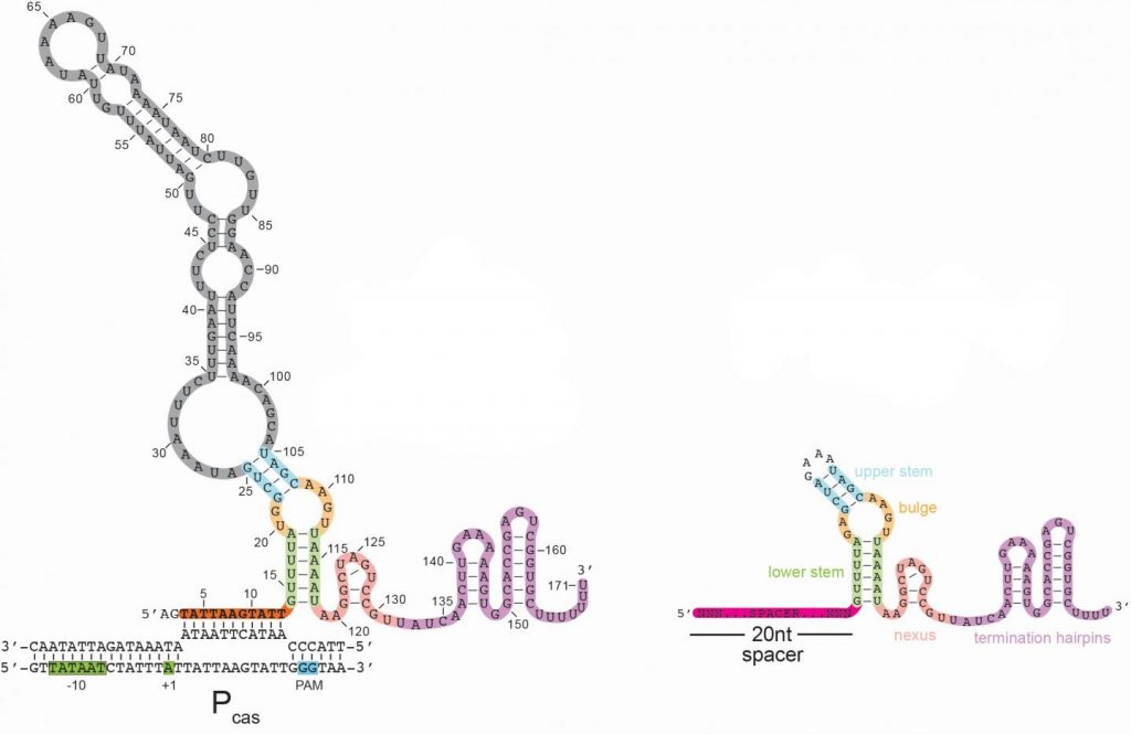
Here’s an image illustrating the long form of the tracrRNA or ‘dimmer switch’ alongside the more commonly used short form,

Caption: Left – a schematic of the long form of the tracrRNA used by the CRISPR-Cas9 system in bacteria; Right – the standard guide RNA used by many scientists as part of the gene-cutting CRISPR-Cas9 system. Credit: Joshua Modell, Rachael Workman and Johns Hopkins Medicine

A January 19 ,2021 Johns Hopkins Medicine news release (also on EurekAlert), which originated the news item, explains about CRISPR and what the acronym stands for, as well as, giving more details about the discovery,

First identified in the genome of gut bacteria in 1987, CRISPR-Cas9 is a naturally occurring but unusual group of genes with a potential for cutting DNA sequences in other types of cells that was realized 25 years later. Its value in genetic engineering — programmable gene alteration in living cells, including human cells — was rapidly appreciated, and its widespread use as a genome “editor” in thousands of laboratories worldwide was recognized in the awarding of the Nobel Prize in Chemistry last year to its American and French co-developers.

CRISPR stands for clustered, regularly interspaced short palindromic repeats. Cas9, which refers to CRISPR-associated protein 9, is the name of the enzyme that makes the DNA slice. Bacteria naturally use CRISPR-Cas9 to cut viral or other potentially harmful DNA and disable the threat, says Joshua Modell, Ph.D., assistant professor of molecular biology and genetics at the Johns Hopkins University School of Medicine. In this role, Modell says, “CRISPR is not only an immune system, it’s an adaptive immune system — one that can remember threats it has previously encountered by holding onto a short piece of their DNA, which is akin to a mug shot.” These mug shots are then copied into “guide RNAs” that tell Cas9 what to cut.

Scientists have long worked to unravel the precise steps of CRISPR-Cas9’s mechanism and how its activity in bacteria is dialed up or down. Looking for genes that ignite or inhibit the CRISPR-Cas9 gene-cutting system for the common, strep-throat causing bacterium Streptococcus pyogenes, the Johns Hopkins scientists found a clue regarding how that aspect of the system works.

Specifically, the scientists found a gene in the CRISPR-Cas9 system that, when deactivated, led to a dramatic increase in the activity of the system in bacteria. The product of this gene appeared to re-program Cas9 to act as a brake, rather than as a “scissor,” to dial down the CRISPR system.

“From an immunity perspective, bacteria need to ramp up CRISPR-Cas9 activity to identify and rid the cell of threats, but they also need to dial it down to avoid autoimmunity — when the immune system mistakenly attacks components of the bacteria themselves,” says graduate student Rachael Workman, a bacteriologist working in Modell’s laboratory.

To further nail down the particulars of the “brake,” the team’s next step was to better understand the product of the deactivated gene (tracrRNA). RNA is a genetic cousin to DNA and is vital to carrying out DNA “instructions” for making proteins. TracrRNAs belong to a unique family of RNAs that do not make proteins. Instead, they act as a kind of scaffold that allows the Cas9 enzyme to carry the guide RNA that contains the mug shot and cut matching DNA sequences in invading viruses.

TracrRNA comes in two sizes: long and short. Most of the modern gene-cutting CRISPR-Cas9 tools use the short form. However, the research team found that the deactivated gene product was the long form of tracrRNA, the function of which has been entirely unknown.

The long and short forms of tracrRNA are similar in structure and have in common the ability to bind to Cas9. The short form tracrRNA also binds to the guide RNA. However, the long form tracrRNA doesn’t need to bind to the guide RNA, because it contains a segment that mimics the guide RNA. “Essentially, long form tracrRNAs have combined the function of the short form tracrRNA and guide RNA,” says Modell.

In addition, the researchers found that while guide RNAs normally seek out viral DNA sequences, long form tracrRNAs target the CRISPR-Cas9 system itself. The long form tracrRNA tends to sit on DNA, rather than cut it. When this happens in a particular area of a gene, it prevents that gene from expressing, — or becoming functional.

To confirm this, the researchers used genetic engineering to alter the length of a certain region in long form tracrRNA to make the tracrRNA appear more like a guide RNA. They found that with the altered long form tracrRNA, Cas9 once again behaved more like a scissor.

Other experiments showed that in lab-grown bacteria with a plentiful amount of long form tracrRNA, levels of all CRISPR-related genes were very low. When the long form tracrRNA was removed from bacteria, however, expression of CRISPR-Cas9 genes increased a hundredfold.

Bacterial cells lacking the long form tracrRNA were cultured in the laboratory for three days and compared with similarly cultured cells containing the long form tracrRNA. By the end of the experiment, bacteria without the long form tracrRNA had completely died off, suggesting that long form tracrRNA normally protects cells from the sickness and death that happen when CRISPR-Cas9 activity is very high.

“We started to get the idea that the long form was repressing but not eliminating its own CRISPR-related activity,” says Workman.

To see if the long form tracrRNA could be re-programmed to repress other bacterial genes, the research team altered the long form tracrRNA’s spacer region to let it sit on a gene that produces green fluorescence. Bacteria with this mutated version of long form tracrRNA glowed less green than bacteria containing the normal long form tracrRNA, suggesting that the long form tracrRNA can be genetically engineered to dial down other bacterial genes.

Another research team, from Emory University, found that in the parasitic bacteria Francisella novicida, Cas9 behaves as a dimmer switch for a gene outside the CRISPR-Cas9 region. The CRISPR-Cas9 system in the Johns Hopkins study is more widely used by scientists as a gene-cutting tool, and the Johns Hopkins team’s findings provide evidence that the dimmer action controls the CRISPR-Cas9 system in addition to other genes.

The researchers also found the genetic components of long form tracrRNA in about 40% of the Streptococcus group of bacteria. Further study of bacterial strains that don’t have the long form tracrRNA, says Workman, will potentially reveal whether their CRISPR-Cas9 systems are intact, and other ways that bacteria may dial back the CRISPR-Cas9 system.

The dimmer capability that the experiments uncovered, says Modell, offers opportunities to design new or better CRISPR-Cas9 tools aimed at regulating gene activity for research purposes. “In a gene editing scenario, a researcher may want to cut a specific gene, in addition to using the long form tracrRNA to inhibit gene activity,” he says.

Here’s a link to and a citation for the paper,

A natural single-guide RNA repurposes Cas9 to autoregulate CRISPR-Cas expression by Rachael E. Workman, Teja Pammi, Binh T.K. Nguyen, Leonardo W. Graeff, Erika Smith, Suzanne M. Sebald, Marie J. Stoltzfus, Chad W. Euler, Joshua W. Modell. Cell DOI:https://doi.org/10.1016/j.cell.2020.12.017 Published Online:J anuary 08, 2021

This paper is behind a paywall.

A Vancouver (Canada) connection to the Pfizer COVID-19 vaccine

Canada’s NanoMedicines Innovation Network (NMIN) must have been excited over the COVID-19 vaccine news (Pfizer Nov. 9, 2020 news release) since it’s a Canadian company (Acuitas Therapeutics) that is providing the means of delivering the vaccine once it enters the body.

Here’s the company’s president and CEO [chief executive officer], Dr. Thomas Madden explaining his company’s delivery system (from Acuitas’ news and events webpage),

For anyone who might find a textual description about the vaccine helpful, I have a Nov. 9, 2020 article by Adele Peters for Fast Company,

… a handful of small biotech companies began scrambling to develop vaccines using an as-yet-unproven technology platform that relies on something called messenger RNA [ribonucleic acid], usually shortened to mRNA …

Like other vaccines, mRNA vaccines work by training the immune system to recognize a threat like a virus and begin producing antibodies to protect itself. But while traditional vaccines often use inactivated doses of the organisms that cause disease, mRNA vaccines are designed to make the body produce those proteins itself. Messenger RNA—a molecule that contains instructions for cells to make DNA—is injected into cells. In the case of COVID-19, mRNA vaccines provide instructions for cells to start producing the “spike” protein of the new coronavirus, the protein that helps the virus get into cells. On its own, the spike protein isn’t harmful. But it triggers the immune system to begin a defensive response. As Bill Gates, who has supported companies like Moderna and BioNTech through the Gates Foundation, has described it, “you essentially turn your body into its own manufacturing unit.”

Amy Judd’s Nov. 9, 2020 article for Global news online explains (or you can just take another look at the video to refresh your memory) how the Acuitas technology fits into the vaccine picture,

Vancouver-based Acuitas Therapeutics, a biotechnology company, is playing a key role through a technology known as lipid nanoparticles, which deliver messenger RNA into cells.

“The technology we provide to our partners is lipid nanoparticles and BioNTech and Pfizer are developing a vaccine that’s using a messenger RNA that tells our cells how to make a protein that’s actually found in the COVID-19 virus,” Dr. Thomas Madden, president and CEO of Acuitas Therapeutics, told Global News Monday [Nov. 9, 2020].

“But the messenger RNA can’t work by itself, it needs a delivery technology to protect this after it’s administered and then to carry it into the cells where it can be expressed and give rise to an immune response.”

Madden said they like to think of the lipid nanoparticles as protective wrapping around a fragile glass ornament [emphasis mine] being shipped to your house online. That protective wrapping would then make sure the ornament made it to your house, through your front door, then unwrap itself and leave in your hallway, ready for you to come and grab it when you came home.

Acuitas Therapeutics employs 29 people and Madden said he believes everyone is feeling very proud of their work.

“Not many people are aware of the history of this technology and the fact that it originated in Vancouver,” he added.

“Dr. Pieter Cullis was one of the key scientists who brought together a team to develop this technology many, many years ago. UBC and Vancouver and companies associated with those scientists have been at the global centre of this technology for many years now.

“I think we’ve been looking for a light at the end of the tunnel for quite some time. I think everybody has been hoping that a vaccine would be able to provide the protection we need to move out of our current situation and I think this is now a confirmation that this hope wasn’t misplaced.”

Nanomedicine in Vancouver

For anyone who’s curious about the Canadian nanomedicine scene, you can find out more about it on Canada’s NanoMedicines Innovation Network (NMIN) website. They recently held a virtual event (Vancouver Nanomedicine Day) on Sept. 17, 2020 (see my Sept. 11, 2020 posting for details), which featured a presentation about Aquitas’ technology.

Happily, the organizers have posted videos for most of the sessions. Dr. Ying Tam of Acuitas made this presentation (about 22 mins. running time) “A Novel Vaccine Approach Using Messenger RNA‐Lipid Nanoparticles: Preclinical and Clinical Perspectives.” If you’re interested in that video or any of the others go to the NanoMedicines Innovation Network’s Nanomedicine Day 2020 webpage.

Acuitas Therapeutics can be found here.