Monthly Archives: October 2014

Simon Fraser University – SCFC861Nanotechnology, The Next Big Idea: course Week 2

Yesterday (Oct. 30, 2014) I taught week 2 of a course called, Nanotechnology: The Next Big Idea for Simon Fraser University’s (SFU) Continuing Studies programme. At the end of this post you will find a link to my Week Two PowerPoint slides and notes of a sort.

For those who may be mildly curious, here’s a description of what was covered in the second week (from SFU’s course description webpage),

Week 2: Not as New as You Might Think

The thinking that underlies nanotechnology can be traced to ancient Greek philosophers while accidental nanotechnology-enabled objects can even be traced to ancient Rome. Many of the field’s luminaries contest claims that one of physicist Richard Feynman’s 1959 lectures is the basis for contemporary nanotechnology.

Here’s the week 2 slide deck:

Week2_history

Here are my ‘notes’ for yesterday’s class consisting largely of brief heads designed to remind me of the content to be found by clicking the link directly after the head.

Week2_Not as new_history

Happy Reading and don’t forget to turn back the clock Saturday night (Nov. 1, 2014) should you live in Canada or the US.

Toughening up your electronics: kevlar with a tungsten fibre coating

An upcoming presentation at the 61st annual AVS Conference (Nov. 9 – 14, 2014) features a fibre made of tungsten that when added to kevlar offers the possibility of ‘tough’ electronics. From an Oct. 31, 2014 news item on Nanowerk (Note: A link has been removed),

A group of North Carolina State University researchers is exploring novel ways to apply semiconductor industry processes to unique substrates, such as textiles and fabrics, to “weave together” multifunctional materials with distinct capabilities.

During the AVS 61st International Symposium & Exhibition, being held November 9-14, 2014, in Baltimore, Maryland, the researchers will describe how they were able to “weave” high-strength, highly conductive yarns made of tungsten metal on Kevlar — aka body armor material — by using atomic layer deposition (ALD), a process commonly used for producing memory and logic devices.

An Oct. 28, 2014 AVS: Science & Technology of Materials, Interfaces, and Processing news release on Newswire, which originated the news item provides more details about this multifunctional material and a good description of atomic layer deposition (ALD),

“As a substrate, Kevlar was intriguing to us because it’s capable of withstanding the relatively high temperature (220°C) required by the ALD deposition process,” explains Sarah Atanasov, a Ph.D. candidate in the Biomolecular Engineering Department at North Carolina State University. “Kevlar doesn’t begin to degrade until it reaches nearly 400°C.”

The group selected ALD as a process because it allows them to deposit highly conformal films on nonplanar surfaces with nanometer-thickness precision. “This ensures that the entire surface of the yarn — made of nearly 600 fibers, each 12 microns in diameter — is evenly coated,” said Atanasov.

How does the ALD process work? It’s actually a cyclical process, which begins by exposing the substrate’s surface to one gas-phase chemical, in this case tungsten hexafluoride (WF6), followed by removal of any unreacted material. This is chased with surface exposure to a second gas-phase chemical, silane (SiH4), after which any unreacted material is once again removed.

By the end of the ALD cycle, the two chemicals have reacted to produce tungsten. “This is a self-limited process, meaning that a single atomic layer is deposited during each cycle — in this case ~5.5 Angstroms per cycle,” Atanasov said. “The process can be cycled through a number of times to achieve any specifically desired thickness. As a bonus, ALD occurs in the gas phase, so it doesn’t require any solution processing and is considered to be a more sustainable deposition technique.”

While weaving together multiple fabrics to combine multiple capabilities certainly isn’t new, characteristics such as high strength, high conductivity, and flexibility are frequently regarded as being mutually exclusive — so concessions are often made to get the most important one.

The work by Atanasov and colleagues shows, however, that ALD of tungsten on Kevlar yields yarns that are highly flexible and highly conductive, around 2,000 S/cm (“Siemens per centimeter,” a common unit used for conductivity). The yards are also within 90 percent of their original prior-to-coating tensile strength.

“Introducing well-established processes from one area into a completely new field can lead to some very interesting and useful results,” Atanasov noted.

The group’s tungsten-on-Kevlar yarns are expected to find applications in multifunctional protective electronics materials for electromagnetic shielding and communications, as well as erosion-resistant antistatic fabrics for space and automated technologies.

Presentation #MS+PS+TF-ThA4, “Multifunctional Fabrics via Tungsten ALD on Kevlar,” authored by Sarah Atanasov, B. Kalanyan and G.N. Parsons, will be at 3:20 p.m. ET on Thursday, Nov. 13, 2014.

Atanasov recently published a paper about another kevlar project where she worked to enhance its ‘stab resistance’ with a titanium dioxide/aluminum mixture as Anisha Ratan notes in her Sept. 12, 2014 article (Oxide armour offers Kevlar better stab resistance)  (excerpt from Ratan’s article for the Royal Society; Note: Links have been removed),

Scientists in the US have synthesised an ultrathin inorganic bilayer coating for Kevlar that could improve its stab resistance by 30% and prove invaluable for military and first-responders requiring multi-threat protection clothes.

Developed in 1965 by Stephanie Kwolek at DuPont, poly(p-phenylene terephthalamide) (PPTA), or Kevlar, is a para-aramid synthetic fiber deriving its strength from interchain hydrogen bonding. It finds use in flexible energy and electronic systems, but is most commonly associated with bullet-proof body armour.

However, despite its anti-ballistic properties, it offers limited cut and stab protection. In a bid to overcome this drawback, Sarah Atanasov, from Gregory Parsons’ group at North Carolina State University, and colleagues, have developed a TiO2/Al2O3 bilayer that significantly enhances the cut resistance of Kevlar fibers. The coating is added to Kevlar by atomic layer deposition, a low temperature technique with nanoscale precision.

Unfortunately the team’s research paper is no longer open access but you can find a link to it from Ratan’s article.

Nanozen: protecting us from nanoparticles (maybe)

Friday, Oct. 24, 2014 the Vancouver Sun (Canada) featured a local nanotechnology company, Nanozen in an article by ‘digital life’ writer, Gillian Shaw. Unfortunately, the article is misleading. Before noting the issues, it should be said that most reporters don’t have much time to prepare stories and are often asked to write on topics that are new or relatively unknown to them. It is a stressful position to be in especially when one is reliant on the interviewee’s expertise and agenda. As for the interviewee, sometimes scientists get excited and enthused and don’t speak with their usual caution.

The article starts off in an unexceptionable manner,

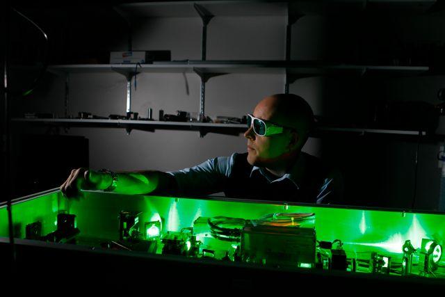
Vancouver startup Nanozen is a creating real-time, wearable particle sensor for use in mines, mills and other industrial locations where dust and other particles can lead to dangerous explosions and debilitating respiratory diseases.

The company founder and, presumably, lead researcher Winnie Chu is described as a former professor of environmental health at the University of British Columbia who has devoted herself to developing a new means of monitoring particles, in particular nanoparticles. Chu is quoted as saying this,

“The current technology is not sufficient to protect workers or the community when concentrations exceed the acceptable level,” she said.

It seems ominous and is made more so with this,

Chu said more than 90 per cent of the firefighters who responded to the 9/11 disaster developed lung disease, having walked into a site full of small and very damaging particles in the air.

“Those nanoparticles go deep into your lungs and cause inflammation and other problems,” Chu said.

It seems odd to mention this particular disaster. The lung issues for the firefighters, first responders and people living close to the site of World Trade Centers collapse are due to a complex mix of materials in the air. Most of the research I can find focuses on micrsoscale particles such as the work from the University of California at Davis’s Delta Group (Detection and Evaluation of the Long-Range Transport of Aerosols). From the Group’s World Trade Center webpage,

The fuming World Trade Center debris pile was a chemical factory that exhaled pollutants in particularly dangerous forms that could penetrate deep into the lungs of workers at Ground Zero, says a new study by UC Davis air-quality experts.

You can find the group’s presentation (-Presentation download (WTC aersols ACS 2003.ppt; 7,500kb)) to an American Chemical Society meeting in 2003 along more details such as this on their webpage,

The conditions would have been “brutal” for people working at Ground Zero without respirators and slightly less so for those working or living in immediately adjacent buildings, said the study’s lead author, Thomas Cahill, a UC Davis professor emeritus of physics and atmospheric science and research professor in engineering.

“Now that we have a model of how the debris pile worked, it gives us a much better idea of what the people working on and near the pile were actually breathing,” Cahill said. “Our first report was based on particles that we collected one mile away. This report gives a reasonable estimate of what type of pollutants were actually present at Ground Zero.

“The debris pile acted like a chemical factory. It cooked together the components of the buildings and their contents, including enormous numbers of computers, and gave off gases of toxic metals, acids and organics for at least six weeks.”

The materials found by this group were not at the nanoscale. In fact, the focus was then and subsequently on materials such as glass shards, asbestos, and metallic aerosols at the microscale, all of which can cause well documented health problems. No doubt effective monitoring would have been helpful It seems the critical issue in the early stages of the disaster was access to a respirator. Also, effective monitoring at later stages which did not seem to have happened would have been a good idea.

A 2004 (?) New York Magazine article by Jennifer Senior titled ‘Fallout‘ had this to say about the air content,

Here, today, is what we know about the dust and air at ground zero: It contained glass shards, pulverized concrete, and many carcinogens, including hundreds of thousands of pounds of asbestos, tens of thousands of pounds of lead, mercury, cadmium, dioxins, PCBs, and polycyclic aromatic hydrocarbons, or PAHs. It also contained benzene. According to a study done by the U.S. Geological Survey, the dust was so caustic in places that its pH exceeded that of ammonia. Thomas Cahill, a scientist who analyzed the plumes from a rooftop one mile away, says that the levels of acids, insoluble particles, high-temperature organic materials, and metals were in most cases higher in very fine particles (which can slip deep into the lungs) than anyplace ever recorded on earth, including the oil fires of Kuwait.

The article describes at some length the problems for first responders and for those who later moved back into their homes nearby the disaster site under the impression the air was clean.

Getting back to the nanoscale, there were carbon nanotubes (CNTs) present as this 2009 research paper, Case Report: Lung Disease in World Trade Center Responders Exposed to Dust and Smoke: Carbon Nanotubes Found in the Lungs of World Trade Center Patients and Dust Samples, noted in relation to a sample of seven patients,

It may well be the most frequent injury pattern in exposed patients with severe respiratory impairment. b) Interstitial disease was present in four cases (Patients A, B, C, and E), characterized by a generally bronchiolocentric pattern of interstitial inflammation and fibrosis of variable severity. The lungs of these patients contained large amounts of silicates, and three of them showed nanotubes.

CNT of commercial origin, common now, would not have been present in substantial numbers in the WTC complex before the disaster in 2001. However, the high temperatures generated during the WTC disaster as a result of the combustion of fuel in the presence of carbon and metals would have been sufficient to locally generate large numbers of CNT. This scenario could have caused the generation of CNT that we have noted in the dust samples and in the lung biopsy specimens.

Given that CNTs are more common now, it would suggest that a monitor for nanoscale materials such as Chu’s proposed equipment could be an excellent idea. Unfortunately, it’s not clear what Chu is trying to achieve as she appears to make a blunder in the article,

Chu said environmental agencies require testing to distinguish between particles equal to or less than 10 microns and smaller particles 2.5 microns or less.

“When we inhale we inhale both size particles but they go into different parts of the lung,” said Chu, who said research shows the smaller the particle the higher the toxicity. [emphasis mine] The monitor she has developed can detect particles as small as one micron and even less.

The word ‘nanoparticle’ is often used generically to include, CNTs, quantum dots, silver nanoparticles, etc. as Chu seems to be doing throughout the article. The only nanomaterial/nanoparticle that researchers agree unequivocally cause lung problems are long carbon nanotubes which resemble asbestos fibres. This is precisely the opposite of Chu’s statement.

For validation, you can conduct your own search or you can check Swiss toxicologist Harald Krug’s (mentioned in my Nanosafety research: a quality control issue posting of Oct. 30, 2014) statement that most health and safety research of nanomaterials and the resultant conclusions are problematic. But he too is unequivocal with regard long carbon nanotubes (from Krug’s study, Nanosafety Research—Are We on the Right Track?).

Comparison of instillation and inhalation experiments: instillation studies have to be carried out with relatively high local doses and, thus, more often meet overload conditions than inhalation studies. Transient inflammatory effects have been observed frequently in both types of lung exposure, irrespective of the type of ENMs used for the experiment. This finding suggests an unspecific particle effect; moreover, the biological response seems to be comparable to a scenario involving exposure to fine dust. Prominent exceptions are long and rigid carbon nanotube (CNT) bundles, which induce a severe tissue reaction (chronic inflammation) that may ultimately result in tumor formation. Overall, the evaluated studies showed no indication of a “nanospecific” effect in the lung. [from the Summary section; 2nd bulleted point]

You can find the Nanozen website here but there doesn’t appear to be any information on the site yet. These search terms ‘about’, ‘team’, ‘technology’, and ‘product’ yielded no results on website as of Oct. 30, 2014 at 1000 hours PDT.

Nanosafety research: a quality control issue

Toxicologist Dr. Harald Krug has published a review of several thousand studies on nanomaterials safety exposing problematic research methodologies and conclusions. From an Oct. 29, 2014 news item on Nanowerk (Note: A link has been removed),

Empa [Swiss Federal Laboratories for Materials Science and Technology] toxicologist Harald Krug has lambasted his colleagues in the journal Angewandte Chemie (“Nanosafety Research—Are We on the Right Track?”). He evaluated several thousand studies on the risks associated with nanoparticles and discovered no end of shortcomings: poorly prepared experiments and results that don’t carry any clout. Instead of merely leveling criticism, however, Empa is also developing new standards for such experiments within an international network.

An Oct. 29, 2014 Empa press release (also on EurekAlert), which originated the news item, describes the new enthusiasm for research into nanomaterials and safety,

Researching the safety of nanoparticles is all the rage. Thousands of scientists worldwide are conducting research on the topic, examining the question of whether titanium dioxide nanoparticles from sun creams can get through the skin and into the body, whether carbon nanotubes from electronic products are as hazardous for the lungs as asbestos used to be or whether nanoparticles in food can get into the blood via the intestinal flora, for instance. Public interest is great, research funds are flowing – and the number of scientific projects is skyrocketing: between 1980 and 2010, a total of 5,000 projects were published, followed by another 5,000 in just the last three years. However, the amount of new knowledge has only increased marginally. After all, according to Krug the majority of the projects are poorly executed and all but useless for risk assessments.

The press release goes on to describe various pathways into the body and problems with research methodologies,

How do nanoparticles get into the body?

Artificial nanoparticles measuring between one and 100 nanometers in size can theoretically enter the body in three ways: through the skin, via the lungs and via the digestive tract. Almost every study concludes that healthy, undamaged skin is an effective protective barrier against nanoparticles. When it comes to the route through the stomach and gut, however, the research community is at odds. But upon closer inspection the value of many alarmist reports is dubious – such as when nanoparticles made of soluble substances like zinc oxide or silver are being studied. Although the particles disintegrate and the ions drifting into the body are cytotoxic, this effect has nothing to do with the topic of nanoparticles but is merely linked to the toxicity of the (dissolved) substance and the ingested dose.

Laboratory animals die in vain – drastic overdoses and other errors

Krug also discovered that some researchers maltreat their laboratory animals with absurdly high amounts of nanoparticles. Chinese scientists, for instance, fed mice five grams of titanium oxide per kilogram of body weight, without detecting any effects. By way of comparison: half the amount of kitchen salt would already have killed the animals. A sloppy job is also being made of things in the study of lung exposure to nanoparticles: inhalation experiments are expensive and complex because a defined number of particles has to be swirled around in the air. Although it is easier to place the particles directly in the animal’s windpipe (“instillation”), some researchers overdo it to such an extent that the animals suffocate on the sheer mass of nanoparticles.

While others might well make do without animal testing and conduct in vitro experiments on cells, here, too, cell cultures are covered by layers of nanoparticles that are 500 nanometers thick, causing them to die from a lack of nutrients and oxygen alone – not from a real nano-effect. And even the most meticulous experiment is worthless if the particles used have not been characterized rigorously beforehand. Some researchers simply skip this preparatory work and use the particles “straight out of the box”. Such experiments are irreproducible, warns Krug.

As noted in the news item, the scientists at Empa have devised a solution to some to of the problems (from the press release),

The solution: inter-laboratory tests with standard materials
Empa is thus collaborating with research groups like EPFL’s Powder Technology Laboratory, with industrial partners and with Switzerland’s Federal Office of Public Health (FOPH) to find a solution to the problem: on 9 October the “NanoScreen” programme, one of the “CCMX Materials Challenges”, got underway, which is expected to yield a set of pre-validated methods for lab experiments over the next few years. It involves using test materials that have a closely defined particle size distribution, possess well-documented biological and chemical properties and can be altered in certain parameters – such as surface charge. “Thanks to these methods and test substances, international labs will be able to compare, verify and, if need be, improve their experiments,” explains Peter Wick, Head of Empa’s laboratory for Materials-Biology Interactions.

Instead of the all-too-familiar “fumbling around in the dark”, this would provide an opportunity for internationally coordinated research strategies to not only clarify the potential risks of new nanoparticles in retrospect but even be able to predict them. The Swiss scientists therefore coordinate their research activities with the National Institute of Standards and Technology (NIST) in the US, the European Commission’s Joint Research Center (JRC) and the Korean Institute of Standards and Science (KRISS).

Bravo! and thank you Dr. Krug and Empa for confirming something I’ve suspected due to hints from more informed commentators. Unfortunately my ignorance. about research protocols has not permitted me to undertake a better analysis of the research. ,

Here’s a link to and a citation for the paper,

Nanosafety Research—Are We on the Right Track? by Prof. Dr. Harald F. Krug. Angewandte Chemie International Edition DOI: 10.1002/anie.201403367 Article first published online: 10 OCT 2014

This is an open access paper.

Nanodiamond alternative to organic fluorophores to view inside living human cells

No sooner is a Nobel prize (2014) awarded for nanoscopy which makes use of fluorescence to observe processes in living cells than there is an announcement about a new technique that avoids fluorescence and its attendant shortcomings. From an Oct. 27, 2014 news item on Nanowerk (Note: A link has been removed),

Nanodiamonds are providing scientists with new possibilities for accurate measurements of processes inside living cells with potential to improve drug delivery and cancer therapeutics.

Published in Nature Nanotechnology (“Coherent anti-Stokes Raman scattering microscopy of single nanodiamonds”), researchers from Cardiff University have unveiled a new method for viewing nanodiamonds inside human living cells for purposes of biomedical research.

An Oct. 27, 2014 Cardiff University (Wales) news release, which originated the news item, explains why the use of nanodiamonds is superior to the use of organic flurophores,

Nanodiamonds are very small particles (a thousand times smaller than human hair) and because of their low toxicity they can be used as a carrier to transport drugs inside cells. They also show huge promise as an alternative to the organic fluorophores usually used by scientists to visualise processes inside cells and tissues.

A major limitation of organic fluorophores is that they have the tendency to degrade and bleach over time under light illumination. This makes it difficult to use them for accurate measurements of cellular processes. Moreover, the bleaching and chemical degradation can often be toxic and significantly perturb or even kill cells.

There is a growing consensus among scientists that nanodiamonds are one of the best inorganic material alternatives for use in biomedical research, because of their compatibility with human cells, and due to their stable structural and chemical properties.

Previous attempts by other research teams to visualise nanodiamonds under powerful light microscopes have run into the obstacle that the diamond material per se is transparent to visible light. Locating the nanodiamonds under a microscope had relied on tiny defects in the crystal lattice, which fluoresce under light illumination.

Production of the defects proved both costly and difficult to realise in a controlled way. Furthermore, the fluorescence light emitted by these defects, and in turn the image gleaned from the microscopic exploration of these flawed nanodiamonds, is sometimes also unstable.

In their latest paper, researchers from Cardiff University’s Schools of Biosciences and Physics showed that non-fluorescing nanodiamonds (diamonds without defects) can be imaged optically and far more stably via the interaction between the illuminating light and the vibrating chemical bonds in the diamond lattice structure which results in scattered light at a different colour.

The paper describes how two laser beams beating at a specific frequency are used to drive chemical bonds to vibrate in sync. One of these beams is then used to probe this vibration and generate a light, called coherent anti-Stokes Raman scattering (CARS).

By focusing these laser beams onto the nanodiamond, a high-resolution CARS image is generated. Using an in-house built microscope, the research team was able to measure the intensity of the CARS light on a series of single nanodiamonds of different sizes.

The nanodiamond size was accurately measured by means of electron microscopy and other quantitative optical contrast methods developed within the researcher’s lab. In this way, they were able to quantify the relationship between the CARS light intensity and the nanoparticle size.

Consequently, the calibrated CARS signal enabled the team to analyse the size and number of nanodiamonds that had been delivered into living cells, with a level of accuracy hitherto not achieved by other methods.

Professor Paola Borri from the School of Biosciences, who led the study, said: “This new imaging modality opens the exciting prospect of following complex cellular trafficking pathways quantitatively with important applications in drug delivery. The next step for us will be to push the technique to detect nanodiamonds of even smaller sizes than what we have shown so far and to demonstrate a specific application in drug delivery.”

Here’s a link to and a citation for the paper,

Coherent anti-Stokes Raman scattering microscopy of single nanodiamonds by Iestyn Pope, Lukas Payne, George Zoriniants, Evan Thomas, Oliver Williams, Peter Watson, Wolfgang Langbein, & Paola Borri. Nature Nanotechnology (2014) doi:10.1038/nnano.2014.210 Published online 12 October 2014

The paper is behind a paywall but there is a free preview with ReadCube Access.

For anyone who’d like to read more about fluorescence and its use in nanoscopy there’s my Oct. 8, 2014 posting about the 2014 Nobel Prize in Chemistry and in my Oct. 27, 2014 posting about a specific use for determining how bipolar disorder may affect the brain.

Friendlier (halogen-free) lithium-ion batteries

An Oct. 24, 2014 news item on ScienceDaily mentions a greener type of lithium-ion battery from a theoretical (keep reading till you reach the first paragraph of the university news release) perspective,

Physics researchers at Virginia Commonwealth University have discovered that most of the electrolytes used in lithium-ion batteries — commonly found in consumer electronic devices — are superhalogens, and that the vast majority of these electrolytes contain toxic halogens.

At the same time, the researchers also found that the electrolytes in lithium-ion batteries (also known as Li-ion batteries) could be replaced with halogen-free electrolytes that are both nontoxic and environmentally friendly.

“The significance [of our findings] is that one can have a safer battery without compromising its performance,” said lead author Puru Jena, Ph.D., distinguished professor in the Department of Physics of the College of Humanities and Sciences. “The implication of our research is that similar strategies can also be used to design cathode materials in Li-ion batteries.”

An Oct. 24, 2014 Virginia Commonwealth University news release by Brian McNeill (also on EurekAlert), which originated the news item, describes the researchers’ hopes and the inspiration for this work,

“We hope that our theoretical prediction will stimulate experimentalists to synthesize halogen-free salts which will then lead manufacturers to use such salts in commercial applications,” he said.

The researchers also found that the procedure outlined for Li-ion batteries is equally valid for other metal-ion batteries, such as sodium-ion or magnesium-ion batteries.

Jena became interested in the topic several months ago when he saw a flyer on Li-ion batteries that mentioned the need for halogen-free electrolytes.

“I had not done any work on Li-ion batteries at the time, but I was curious to see what the current electrolytes are,” he said. “I found that the negative ions that make up the electrolytes are large and complex in nature and they contain one less electron than what is needed for electronic shell closure.”

Jena had already been working for more than five years on superhalogens, a class of molecules that mimic the chemistry of halogens but have electron affinities that are much larger than that of the halogen atoms.

“I knew of many superhalogen molecules that do not contain a single halogen atom,” he said. “My immediate thought was first to see if the anionic components of the current electrolytes are indeed superhalogens. And, if so, do the halogen-free superhalogens that we knew serve the purpose as halogen-free electrolytes? Our research proved that to be the case.”

Here’s a link to and a citation for the paper,

Superhalogens as Building Blocks of Halogen-Free Electrolytes in Lithium-Ion Batteries by Dr. Santanab Giri, Swayamprabha Behera and Prof. Puru Jena. Angewandte Chemie, DOI: 10.1002/ange.201408648 Article first published online: 14 OCT 2014

© 2014 WILEY-VCH Verlag GmbH & Co. KGaA, Weinheim

This paper is behind a paywall.

Faster, cheaper, and just as good—nanoscale device for measuring cancer drug methotrexate

Lots of cancer drugs can be toxic if the dosage is too high for individual metabolisms, which can vary greatly in their ability to break drugs down. The University of Montréal (Université de Montréal) has announced a device that could help greatly in making the technology to determine toxicity in the bloodstream faster and cheaper according to an Oct. 27, 2014 news item on Nanowerk,

In less than a minute, a miniature device developed at the University of Montreal can measure a patient’s blood for methotrexate, a commonly used but potentially toxic cancer drug. Just as accurate and ten times less expensive than equipment currently used in hospitals, this nanoscale device has an optical system that can rapidly gauge the optimal dose of methotrexate a patient needs, while minimizing the drug’s adverse effects. The research was led by Jean-François Masson and Joelle Pelletier of the university’s Department of Chemistry.

An Oct. 27, 2014 University of Montréal news release, which originated the news item, provides more specifics about the cancer drug being monitored and the research that led to the new device,

Methotrexate has been used for many years to treat certain cancers, among other diseases, because of its ability to block the enzyme dihydrofolate reductase (DHFR). This enzyme is active in the synthesis of DNA precursors and thus promotes the proliferation of cancer cells. “While effective, methotrexate is also highly toxic and can damage the healthy cells of patients, hence the importance of closely monitoring the drug’s concentration in the serum of treated individuals to adjust the dosage,” Masson explained.

Until now, monitoring has been done in hospitals with a device using fluorescent bioassays to measure light polarization produced by a drug sample. “The operation of the current device is based on a cumbersome, expensive platform that requires experienced personnel because of the many samples that need to be manipulated,” Masson said.

Six years ago, Joelle Pelletier, a specialist of the DHFR enzyme, and Jean-François Masson, an expert in biomedical instrument design, investigated how to simplify the measurement of methotrexate concentration in patients.

Gold nanoparticles on the surface of the receptacle change the colour of the light detected by the instrument. The detected colour reflects the exact concentration of the drug in the blood sample. In the course of their research, they developed and manufactured a miniaturized device that works by surface plasmon resonance. Roughly, it measures the concentration of serum (or blood) methotrexate through gold nanoparticles on the surface of a receptacle. In “competing” with methotrexate to block the enzyme, the gold nanoparticles change the colour of the light detected by the instrument. And the colour of the light detected reflects the exact concentration of the drug in the blood sample.

The accuracy of the measurements taken by the new device were compared with those produced by equipment used at the Maisonneuve-Rosemont Hospital in Montreal. “Testing was conclusive: not only were the measurements as accurate, but our device took less than 60 seconds to produce results, compared to 30 minutes for current devices,” Masson said. Moreover, the comparative tests were performed by laboratory technicians who were not experienced with surface plasmon resonance and did not encounter major difficulties in operating the new equipment or obtaining the same conclusive results as Masson and his research team.

In addition to producing results in real time, the device designed by Masson is small and portable and requires little manipulation of samples. “In the near future, we can foresee the device in doctors’ offices or even at the bedside, where patients would receive individualized and optimal doses while minimizing the risk of complications,” Masson said. Another benefit, and a considerable one: “While traditional equipment requires an investment of around $100,000, the new mobile device would likely cost ten times less, around $10,000.”

For those who prefer to read the material in French here’s a link to ‘le 27 Octobre 2014 communiqué de nouvelles‘.

Here’s a prototype of the device,

Les nanoparticules d’or situées à la surface de la languette réceptrice modifient la couleur de la lumière détectée par l’instrument. La couleur captée reflète la concentration exacte du médicament contenu dans l’échantillon sanguin. Courtesy  Université de Montréal

Les nanoparticules d’or situées à la surface de la languette réceptrice modifient la couleur de la lumière détectée par l’instrument. La couleur captée reflète la concentration exacte du médicament contenu dans l’échantillon sanguin. Courtesy Université de Montréal

There is no indication as to when this might come to market, in English  or in French.

Bipolar disorder at the nanoscale

In all the talk generated by the various brain projects (BRAIN initiative [US], The Human Brain Project [European Union], Brain Canada), there’s remarkably little discussion about mental illness. So, this news is a little unusual.

Using super-high resolution technique scientists at Northwestern University (Chicago, Illinois, US) believe they’ve made a discovery which explains how bipolar disorder affects the brain according to an Oct. 22, 2014 Northwestern University news release (also on EurekAlert and ScienceDaily) by Erin White,

Scientists used a new super-resolution imaging method — the same method recognized with the 2014 Nobel Prize in chemistry — to peer deep into brain tissue from mice with bipolar-like behaviors. In the synapses (where communication between brain cells occurs), they discovered tiny “nanodomain” structures with concentrated levels of ANK3 — the gene most strongly associated with bipolar disorder risk. ANK3 is coding for the protein ankyrin-G.

“We knew that ankyrin-G played an important role in bipolar disease, but we didn’t know how,” said Northwestern Medicine scientist Peter Penzes, corresponding author of the paper. “Through this imaging method we found the gene formed in nanodomain structures in the synapses, and we determined that these structures control or regulate the behavior of synapses.”

Penzes is a professor in physiology and psychiatry and behavioral sciences at Northwestern University Feinberg School of Medicine. The results were published Oct. 22 in the journal Neuron.

High-profile cases, including actress Catherine Zeta-Jones and politician Jesse Jackson, Jr., have brought attention to bipolar disorder. The illness causes unusual shifts in mood, energy, activity levels and the ability to carry out day-to-day tasks. About 3 percent of Americans experience bipolar disorder symptoms, and there is no cure.

Recent large-scale human genetic studies have shown that genes can contribute to disease risk along with stress and other environmental factors. However, how these risk genes affect the brain is not known.

This is the first time any psychiatric risk gene has been analyzed at such a detailed level of resolution. As explained in the paper, Penzes used the Nikon Structured Illumination Super-resolution Microscope to study a mouse model of bipolar disorder. The microscope realizes resolution of up to 115 nanometers. To put that size in perspective, a nanometer is one-tenth of a micron, and there are 25,400 microns in one inch. Very few of these microscopes exist worldwide.

“There is important information about genes and diseases that can only been seen at this level of resolution,” Penzes said. “We provide a neurobiological explanation of the function of the leading risk gene, and this might provide insight into the abnormalities in bipolar disorder.”

The biological framework presented in this paper could be used in human studies of bipolar disorder in the future, with the goal of developing therapeutic approaches to target these genes.

Here’s a link to and a citation for the paper,

Psychiatric Risk Factor ANK3/Ankyrin-G Nanodomains Regulate the Structure and Function of Glutamatergic Synapses by Katharine R. Smith, Katherine J. Kopeikina, Jessica M. Fawcett-Patel, Katherine Leaderbrand, Ruoqi Gao, Britta Schürmann, Kristoffer Myczek, Jelena Radulovic, Geoffrey T. Swanson, and Peter Penzes. Neuron, Volume 84, Issue 2, p399–415, 22 October 2014 DOI: http://dx.doi.org/10.1016/j.neuron.2014.10.010

This paper is behind a paywall.

You can find more about super-high resolution and nanoscopy in my Oct. 8, 2014 post about the 2014 Nobel Chemistry prize winners.