Tag Archives: blood

CRISPR-Cas12a as a new diagnostic tool

Similar to Cas9, Cas12a is has an added feature as noted in this February 15, 2018 news item on ScienceDaily,

Utilizing an unsuspected activity of the CRISPR-Cas12a protein, researchers created a simple diagnostic system called DETECTR to analyze cells, blood, saliva, urine and stool to detect genetic mutations, cancer and antibiotic resistance and also diagnose bacterial and viral infections. The scientists discovered that when Cas12a binds its double-stranded DNA target, it indiscriminately chews up all single-stranded DNA. They then created reporter molecules attached to single-stranded DNA to signal when Cas12a finds its target.

A February 15, 2018 University of California at Berkeley (UC Berkeley) news release by Robert Sanders and which originated the news item, provides more detail and history,

CRISPR-Cas12a, one of the DNA-cutting proteins revolutionizing biology today, has an unexpected side effect that makes it an ideal enzyme for simple, rapid and accurate disease diagnostics.

blood in test tube

(iStock)

Cas12a, discovered in 2015 and originally called Cpf1, is like the well-known Cas9 protein that UC Berkeley’s Jennifer Doudna and colleague Emmanuelle Charpentier turned into a powerful gene-editing tool in 2012.

CRISPR-Cas9 has supercharged biological research in a mere six years, speeding up exploration of the causes of disease and sparking many potential new therapies. Cas12a was a major addition to the gene-cutting toolbox, able to cut double-stranded DNA at places that Cas9 can’t, and, because it leaves ragged edges, perhaps easier to use when inserting a new gene at the DNA cut.

But co-first authors Janice Chen, Enbo Ma and Lucas Harrington in Doudna’s lab discovered that when Cas12a binds and cuts a targeted double-stranded DNA sequence, it unexpectedly unleashes indiscriminate cutting of all single-stranded DNA in a test tube.

Most of the DNA in a cell is in the form of a double-stranded helix, so this is not necessarily a problem for gene-editing applications. But it does allow researchers to use a single-stranded “reporter” molecule with the CRISPR-Cas12a protein, which produces an unambiguous fluorescent signal when Cas12a has found its target.

“We continue to be fascinated by the functions of bacterial CRISPR systems and how mechanistic understanding leads to opportunities for new technologies,” said Doudna, a professor of molecular and cell biology and of chemistry and a Howard Hughes Medical Institute investigator.

DETECTR diagnostics

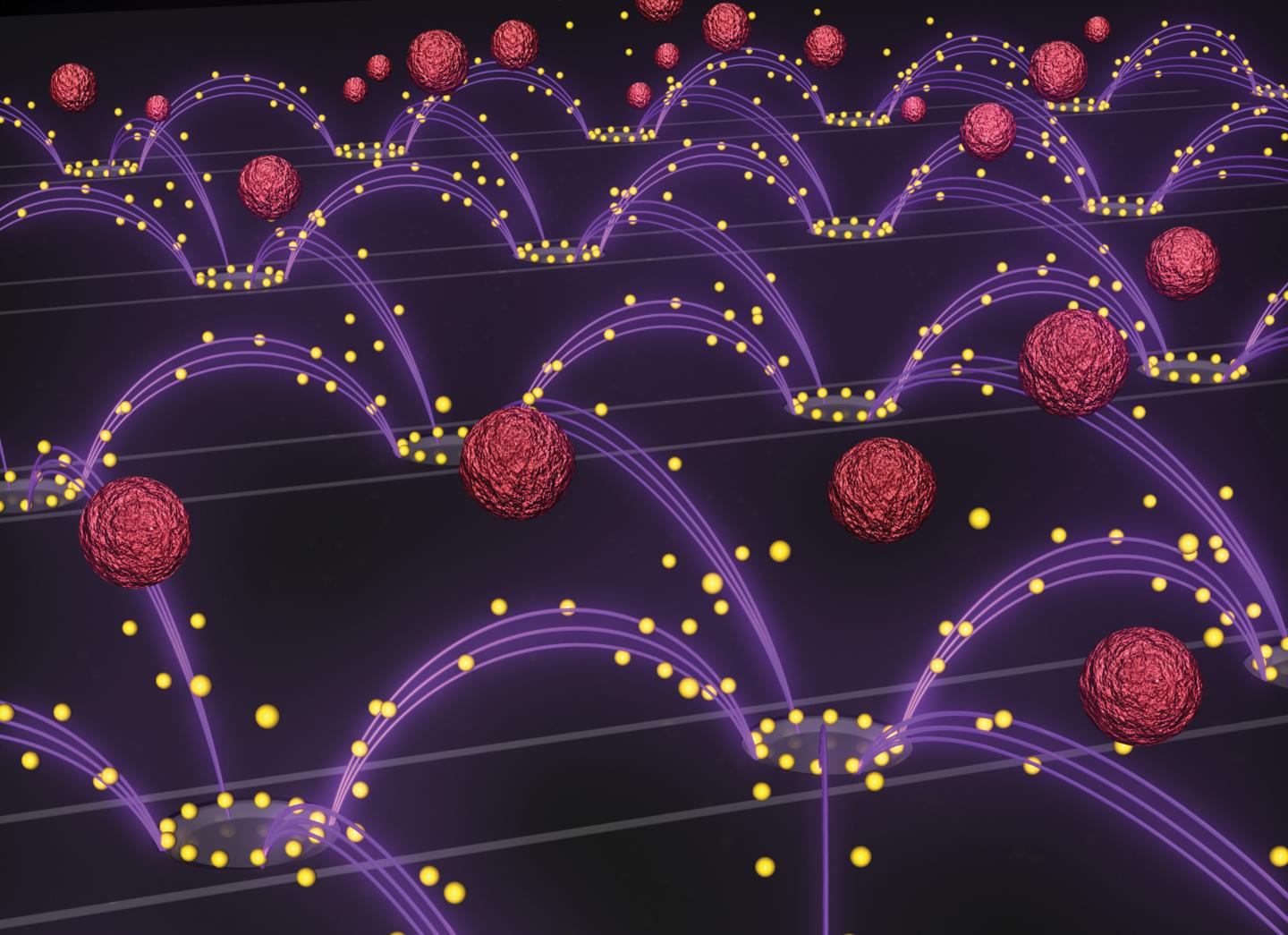
The new DETECTR system based on CRISPR-Cas12a can analyze cells, blood, saliva, urine and stool to detect genetic mutations, cancer and antibiotic resistance as well as diagnose bacterial and viral infections. Target DNA is amplified by RPA to make it easier for Cas12a to find it and bind, unleashing indiscriminate cutting of single-stranded DNA, including DNA attached to a fluorescent marker (gold star) that tells researchers that Cas12a has found its target.

The UC Berkeley researchers, along with their colleagues at UC San Francisco, will publish their findings Feb. 15 [2018] via the journal Science’s fast-track service, First Release.

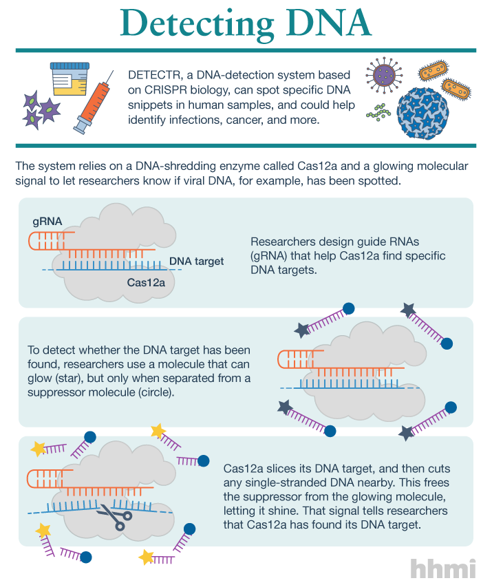
The researchers developed a diagnostic system they dubbed the DNA Endonuclease Targeted CRISPR Trans Reporter, or DETECTR, for quick and easy point-of-care detection of even small amounts of DNA in clinical samples. It involves adding all reagents in a single reaction: CRISPR-Cas12a and its RNA targeting sequence (guide RNA), fluorescent reporter molecule and an isothermal amplification system called recombinase polymerase amplification (RPA), which is similar to polymerase chain reaction (PCR). When warmed to body temperature, RPA rapidly multiplies the number of copies of the target DNA, boosting the chances Cas12a will find one of them, bind and unleash single-strand DNA cutting, resulting in a fluorescent readout.

The UC Berkeley researchers tested this strategy using patient samples containing human papilloma virus (HPV), in collaboration with Joel Palefsky’s lab at UC San Francisco. Using DETECTR, they were able to demonstrate accurate detection of the “high-risk” HPV types 16 and 18 in samples infected with many different HPV types.

“This protein works as a robust tool to detect DNA from a variety of sources,” Chen said. “We want to push the limits of the technology, which is potentially applicable in any point-of-care diagnostic situation where there is a DNA component, including cancer and infectious disease.”

The indiscriminate cutting of all single-stranded DNA, which the researchers discovered holds true for all related Cas12 molecules, but not Cas9, may have unwanted effects in genome editing applications, but more research is needed on this topic, Chen said. During the transcription of genes, for example, the cell briefly creates single strands of DNA that could accidentally be cut by Cas12a.

The activity of the Cas12 proteins is similar to that of another family of CRISPR enzymes, Cas13a, which chew up RNA after binding to a target RNA sequence. Various teams, including Doudna’s, are developing diagnostic tests using Cas13a that could, for example, detect the RNA genome of HIV.

infographic about DETECTR system

(Infographic by the Howard Hughes Medical Institute)

These new tools have been repurposed from their original role in microbes where they serve as adaptive immune systems to fend off viral infections. In these bacteria, Cas proteins store records of past infections and use these “memories” to identify harmful DNA during infections. Cas12a, the protein used in this study, then cuts the invading DNA, saving the bacteria from being taken over by the virus.

The chance discovery of Cas12a’s unusual behavior highlights the importance of basic research, Chen said, since it came from a basic curiosity about the mechanism Cas12a uses to cleave double-stranded DNA.

“It’s cool that, by going after the question of the cleavage mechanism of this protein, we uncovered what we think is a very powerful technology useful in an array of applications,” Chen said.

Here’s a link to and a citation for the paper,

CRISPR-Cas12a target binding unleashes indiscriminate single-stranded DNase activity by Janice S. Chen, Enbo Ma, Lucas B. Harrington, Maria Da Costa, Xinran Tian, Joel M. Palefsky, Jennifer A. Doudna. Science 15 Feb 2018: eaar6245 DOI: 10.1126/science.aar6245

This paper is behind a paywall.

Monster science (a book announcement and interview)

Helaine Becker has launched a new children’s science book incorporating monsters with science. The title, unsurprisingly, is: ‘Monster Science’. Here’s more about the book from Helaine’s Oct. 14, 2016 post on Sci/Why where she shares two reviews,

“From Frankenstein’s creation to Nessie, Becker uses the creatures of our scariest stories as a springboard for an introduction to the scientific understandings that might make such creatures possible—or impossible. In addition to man-made monsters and legendary sea creatures, she covers vampires, zombies, werewolves, and wild, humanlike creatures like Bigfoot. Chapter by chapter, she provides references from literature, film, and popular culture, including a bit of science, a bit of history, and a plentiful helping of humor. She includes numerous monster facts, suggests weapons of defense, and concludes each section with a test-yourself quiz. Science topics covered range widely: electricity, genetic engineering, “demonic diseases,” the nature of our blood and the circulatory system, the possibility of immortality, animal classification, evolution, cannibalism, optical illusions, heredity, hoaxes, and the very real profession of cryptozoology, or the search for hitherto unidentified creatures. … Kirkus

Then, there’s this one,

A highlight of this work is its exploration of the often symbiotic relationship between culture and science; figures such as Shelley, John Polidori (The Vampyre), and filmmaker George Romero (Night of the Living Dead) merged cultural fascination with scientific development to create truly inspiring works and further public interest in science… School Library Journal

Interview with Helaine Becker

Not to be confused with ‘Interview with a vampire’, this one is not novel-length and includes a scoop about an upcoming book in 2017,

Were you surprised by anything when you were researching and/or witting the book?

I learned so much while writing Monster Science – it’s one of the reasons I enjoy writing nonfiction, especially for kids. I always turn up fascinating stuff. I was surprised to learn that werewolves were rounded up and burned at the stake, just like witches, during the period of the Inquisition. Werewolves, it turns out, were thought to be witches – usually male ones – who could shape shift.

My fave fact of all is that vampires would still have to eat their vegetables.

Did you have to leave any monsters/pop culture references/science out of the book? And, why?

Children’s books have very tight space constraints, but my research is comprehensive and complete. That means we have to pick and choose what stays in. It’s gotta be the very best! I work closely with my editors on this, and sometimes we have, shall we say, “heated” discussions.” For Monster Science, I was particularly sorry to see the fascinating back story of the mad scientist trope end up with a stake in its heart.

Did you have a favourite monster before you started? If so, has your favourite changed? Or if you didn’t have one before writing the book, have you since developed a favourite monster?

I’ve had an uneasy relationship with vampires from the age of about 7, after watching an episode of Gilligan’s Island. It featured a “humorous” dream sequence with Gilligan as the vampire. I failed to see the humor at that tender age, and was terrified out of my socks. And anyone remember the original Dark Shadows? Barnabus Collins? Yeah. That show should have never been on in the afternoon. I slept with the blankies up to my ears until my mid-thirties. (Who am I kidding? I still do!)

Are you hoping to tie this book into the Frankenstein bicentennial celebrations?

Illustrated children’s books have very long time lines from concept to finished book. I wrote Monster Science several years ago, before I had any notion of Frankenstein bicentennials. But now that we’ve arrived at this auspicious date, I’m excited! I’d love to participate in some way. I will put on my zzz zzzz zzzt thinking cap.

Where can your fans come to a reading or some other event?

I do dozens of school visits and festival events every year. Some of them might be focused on a specific book, like Monster Science, but most usually feature discussions around several of my titles. This holiday season, for example, I will be doing events around my latest picture book, a very Canadian Christmas-themed title called Deck the Halls. It’s the third in a very popular series. Anyone can drop in to the Sherway Gardens branch of Indigo Book Store [in Toronto] at noon on Sunday, Dec. 4 [2016], to take part in that.

I’ll be doing many events in association with the Forest of Reading, one of North America’s largest children’s choice award programs this spring. More than 250,000 children participate! I am honored to have two science-related books nominated this year, Worms for Breakfast: How to Feed a Zoo (Owlkids) and Everything: Space (National Geographic Kids). I will also be the keynote at the Killaloe Literary Festival in beautiful northern Ontario at the end of May. Best place to look for my latest book and schedule info is my blog, http://helainebecker.blogspot.ca/.

Is there anything you’d like to add?

For insiders only: Coming soon! Look for my upcoming picture book biography of William Playfair, the Victorian era scoundrel who single-handedly invented the field of infographics. It’s called Lines, Bars and Circles and will be published by Kids Can Press early in 2017.

Thank you, Helaine! (I usually don’t get funny interviews. It makes for a good change of pace.)

Getting back to ‘Monster Science’, you can purchase the book here.

Biohackers (also known as bodyhackers or grinders) become more common?

Stephen Melendez’s June 11, 2016 story about biohackers/bodyhackers/grinders for Fast Company sports a striking image in the banner, an x-ray of a pair hands featuring some mysterious additions to the webbing between thumbs and forefingers (Note: Links have been removed),

Tim Shank can guarantee he’ll never leave home without his keys. Why? His house keys are located inside his body.

Shank, the president of the Minneapolis futurist group TwinCities+, has a chip installed in his hand that can communicate electronically with his front door and tell it to unlock itself. His wife has one, too.

In fact, Shank has several chips in his hand, including a near field communication (NFC) chip like the ones used in Apple Pay and similar systems, which stores a virtual business card with contact information for TwinCities+. “[For] people with Android phones, I can just tap their phone with my hand, right over the chip, and it will send that information to their phone,” he says. In the past, he’s also used a chip to store a bitcoin wallet.

Shank is one of a growing number of “biohackers” who implant hardware ranging from microchips to magnets inside their bodies.

Certainly the practice seems considerably more developed since the first time it was mentioned here in a May 27, 2010 posting about a researcher who’d implanted a chip into his body which he then contaminated with a computer virus. In the comments, you’ll find Amal Grafstraa who’s mentioned in the Melendez article at some length, from the Melendez article (Note: Links have been removed),

Some biohackers use their implants in experimental art projects. Others who have disabilities or medical conditions use them to improve their quality of life, while still others use the chips to extend the limits of human perception. …

Experts sometimes caution that the long-term health risks of the practice are still unknown. But many biohackers claim that, if done right, implants can be no more dangerous than getting a piercing or tattoo. In fact, professional body piercers are frequently the ones tasked with installing these implants, given that they possess the training and sterilization equipment necessary to break people’s skin safely.

“When you talk about things like risk, things like putting it in your body, the reality is the risk of having one of these installed is extremely low—it’s even lower than an ear piercing,” claims Amal Graafstra, the founder of Dangerous Things, a biohacking supply company.

Graafstra, who is also the author of the book RFID Toys, says he first had an RFID chip installed in his hand in 2005, which allowed him to unlock doors without a key. When the maker movement took off a few years later, and as more hackers began to explore what they could put inside their bodies, he founded Dangerous Things with the aim of ensuring these procedures were done safely.

“I decided maybe it’s time to wrap a business model around this and make sure that the things people are trying to put in their bodies are safe,” he says. The company works with a network of trained body piercers and offers online manuals and videos for piercers looking to get up to speed on the biohacking movement.

At present, these chips are capable of verifying users’ identities and opening doors. And according to Graafstra, a next-generation chip will have enough on-board cryptographic power to potentially work with credit card terminals securely.

“The technology is there—we can definitely talk to payment terminals with it—but we don’t have the agreements in place with banks [and companies like] MasterCard to make that happen,” he says.

Paying for goods with an implantable chip might sound unusual for consumers and risky for banks, but Graafstra thinks the practice will one day become commonplace. He points to a survey released by Visa last year that found that 25% of Australians are “at least slightly interested” in paying for purchases through a chip implanted in their bodies.

Melendez’s article is fascinating and well worth reading in its entirety. It’s not all keys and commerce as this next and last excerpt shows,

Other implantable technology has more of an aesthetic focus: Pittsburgh biohacking company Grindhouse Wetware offers a below-the-skin, star-shaped array of LED lights called Northstar. While the product was inspired by the on-board lamps of a device called Circadia that Grindhouse founder Tim Cannon implanted to send his body temperature to a smartphone, the commercially available Northstar features only the lights and is designed to resemble natural bioluminescence.

“This particular device is mainly aesthetic,” says Grindhouse spokesman Ryan O’Shea. “It can backlight tattoos or be used in any kind of interpretive dance, or artists can use it in various ways.”

The lights activate in the presence of a magnetic field—one that is often provided by magnets already implanted in the same user’s fingertips. Which brings up another increasingly common piece of bio-hardware: magnetic finger implants. ….

There are other objects that can be implanted in bodies. In one case, an artist, Wafaa Bilal had a camera implanted into the back of his head for a 3rd eye. I mentioned the Iraqi artist in my April 13, 2011 posting titled: Blood, memristors, cyborgs plus brain-controlled computers, prosthetics, and art (scroll down about 75% of the way). Bilal was unable to find a doctor who would perform the procedure so he went to a body-piercing studio. Unfortunately, the posting chronicles his infection and subsequent removal of the camera (h/t Feb. 11, 2011 BBC [British Broadcasting Corporation] news online article).

Observations

It’s been a while since I’ve written about bodyhacking and I’d almost forgotten about the practice relegating it to the category of “one of those trendy ideas that get left behind as interest shifts.” My own interest had shifted more firmly to neuroprosthetics (the integration of prostheses into the nervous system).

I had coined a tag for bodyhacking and neuroprostheses: machine/flesh which covers both those topics and more (e.g. cyborgs) as we continue to integrate machines into our bodies.

Final note

I was reminded of Wafaa Bilal recently when checking out a local arts magazine, Preview: the gallery guide, June/July/August 2016 issue. His work (the 168:01show) is being shown in Calgary, Alberta, Canada at the Esker Foundation from May 27 to August 28, 2016,

168:01 is a major solo exhibition of new and recent work by Iraqi-born, New York-based artist Wafaa Bilal, renowned for his online performances and technologically driven encounters that speak to the impact of international politics on individual lives.

In 168:01, Bilal takes the Bayt al-Hikma, or House of Wisdom, as a starting point for a sculptural installation of a library. The Bayt al-Hikma was a major academic center during the Islamic Golden Age where Muslim, Jewish, and Christian scholars studied the humanities and science. By the middle of the Ninth Century, the House of Wisdom had accumulated the largest library in the world. Four centuries later, a Mongol siege laid waste to all the libraries of Baghdad along with the House of Wisdom. According to some accounts, the library was thrown into the Tigris River to create a bridge of books for the Mongol army to cross. The pages bled ink into the river for seven days – or 168 hours, after which the books were drained of knowledge. Today, the Bayt al-Hikma represents one of the most well-known examples of historic cultural loss as a casualty of wartime.

For this exhibition, Bilal has constructed a makeshift library filled with empty white books. The white books symbolize the priceless cultural heritage destroyed at Bayt al-Hikma as well as the libraries, archives, and museums whose systematic decimation by occupying forces continues to ravage his homeland. Throughout the duration of the exhibition, the white books will slowly be replaced with visitor donations from a wishlist compiled by The College of Fine Arts at the University of Baghdad, whose library was looted and destroyed in 2003. At the end of each week a volunteer unpacks the accumulated shipments, catalogues each new book by hand, and places the books on the shelves. At the end of the exhibition, all the donated books will be sent to the University of Baghdad to help rebuild their library. This exchange symbolizes the power of individuals to rectify violence inflicted on cultural spaces that are meant to preserve and store knowledge for future generations.

In conjunction with the library, Bilal presents a powerful suite of photographs titled The Ashes Series that brings the viewer closer to images of violence and war in the Middle East. In an effort to foster empathy and humanize the onslaught of violent images that inundate Western media during wartime, Bilal has reconstructed journalistic images of the destruction caused by the Iraq War. He writes, “Reconstructing the destructed spaces is a way to exist in them, to share them with an audience, and to provide a layer of distance, as the original photographs are too violent and run the risk of alienating the viewer. It represents an attempt to make sense of the destruction and to preserve the moment of serenity after the dust has settled, to give the ephemeral moment extended life in a mix of beauty and violence.” In the photograph Al-Mutanabbi Street from The Ashes Series, the viewer encounters dilapidated historic and modern buildings on a street covered with layers upon layers of rubble and fragments of torn books. Bilal’s images emanate a slowness that deepens engagement between the viewer and the image, thereby inviting them to share the burden of obliterated societies and reimagine a world built on the values of peace and hope.

The House of Wisdom has been mentioned here a few times perhaps most comprehensively and in the context of the then recent opening of the King Abdullah University for Science and Technology (KAUST; located in Saudi Arabia) in this Sept. 24, 2009 posting (scroll down about 45% of the way).

Anyone interested in hacking their own body?


I expect you can find out more Amal Grafstraa’s website.

Clearing nanoparticles from blood using electric fields

With all the excitement about using nanoparticles to deliver medication (drugs), there hasn’t been much mention of removing these nanoparticles once they’ve served their purpose. Apparently, there is a new technique which makes removal much easier.

 Caption An artist's representation of the nanoparticle removal chip developed by researchers in Professor Michael Heller's lab at the UC San Diego Jacobs School of Engineering. An oscillating electric field (purple arcs) separates drug-delivery nanoparticles (yellow spheres) from blood (red spheres) and pulls them towards rings surrounding the chip's electrodes. The image is featured as the inside cover of the Oct. 14 issue of the journal Small. Credit: Stuart Ibsen and Steven Ibsen.

Caption: An artist’s representation of the nanoparticle removal chip developed by researchers in Professor Michael Heller’s lab at the UC San Diego Jacobs School of Engineering. An oscillating electric field (purple arcs) separates drug-delivery nanoparticles (yellow spheres) from blood (red spheres) and pulls them towards rings surrounding the chip’s electrodes. The image is featured as the inside cover of the Oct. 14 issue of the journal Small. Credit: Stuart Ibsen and Steven Ibsen.

Engineers at the University of California at San Diego (UCSD) provide a description of the new technology and the problems with current techniques for removing nanoparticles in a Nov. 20, 2015 UCSD news release (also on EurekAlert but dated Nov. 23, 2015),

Engineers at the University of California, San Diego developed a new technology that uses an oscillating electric field to easily and quickly isolate drug-delivery nanoparticles from blood. The technology could serve as a general tool to separate and recover nanoparticles from other complex fluids for medical, environmental, and industrial applications.

Nanoparticles, which are generally one thousand times smaller than the width of a human hair, are difficult to separate from plasma, the liquid component of blood, due to their small size and low density. Traditional methods to remove nanoparticles from plasma samples typically involve diluting the plasma, adding a high concentration sugar solution to the plasma and spinning it in a centrifuge, or attaching a targeting agent to the surface of the nanoparticles. These methods either alter the normal behavior of the nanoparticles or cannot be applied to some of the most common nanoparticle types.

“This is the first example of isolating a wide range of nanoparticles out of plasma with a minimum amount of manipulation,” said Stuart Ibsen, a postdoctoral fellow in the Department of NanoEngineering at UC San Diego and first author of the study published October in the journal Small. “We’ve designed a very versatile technique that can be used to recover nanoparticles in a lot of different processes.”

This new nanoparticle separation technology will enable researchers — particularly those who design and study drug-delivery nanoparticles for disease therapies — to better monitor what happens to nanoparticles circulating in a patient’s bloodstream. One of the questions that researchers face is how blood proteins bind to the surfaces of drug-delivery nanoparticles and make them less effective. Researchers could also use this technology in the clinic to determine if the blood chemistry of a particular patient is compatible with the surfaces of certain drug-delivery nanoparticles.

“We were interested in a fast and easy way to take these nanoparticles out of plasma so we could find out what’s going on at their surfaces and redesign them to work more effectively in blood,” said Michael Heller, a nanoengineering professor at the UC San Diego Jacobs School of Engineering and senior author of the study.

The device used to isolate the drug-delivery nanoparticles was a dime-sized electric chip manufactured by La Jolla-based Biological Dynamics, which licensed the original technology from UC San Diego. The chip contains hundreds of tiny electrodes that generate a rapidly oscillating electric field that selectively pulls the nanoparticles out of a plasma sample. Researchers inserted a drop of plasma spiked with nanoparticles into the electric chip and demonstrated nanoparticle recovery within 7 minutes. The technology worked on different types of drug-delivery nanoparticles that are typically studied in various labs.

The breakthrough in the technology relies on designing a chip that can work in the high salt concentration of blood plasma. The chip’s ability to pull the nanoparticles out of plasma is based on differences in the material properties between the nanoparticles and plasma components. When the chip’s electrodes apply an oscillating electric field, the positive and negative charges inside the nanoparticles reorient themselves at a different speed than the charges in the surrounding plasma. This momentary imbalance in the charges creates an attractive force between the nanoparticles and the electrodes. As the electric field oscillates, the nanoparticles are continually pulled towards the electrodes, leaving the rest of the plasma behind. Also, the electric field is designed to oscillate at just the right frequency: 15,000 times per second.

“It’s amazing that this method works without any modifications to the plasma samples or to the nanoparticles,” said Ibsen.

Here’s a link to and a citation for the paper,

Recovery of Drug Delivery Nanoparticles from Human Plasma Using an Electrokinetic Platform Technology by Stuart Ibsen, Avery Sonnenberg, Carolyn Schutt, Rajesh Mukthavaram, Yasan Yeh, Inanc Ortac, Sareh Manouchehri, Santosh Kesari, Sadik Esener, and Michael J. Heller. Small Volume 11, Issue 38, pages 5088–5096, October 14, 2015 DOI: 10.1002/smll.201500892 Article first published online: 14 AUG 2015

© 2015 WILEY-VCH Verlag GmbH & Co. KGaA, Weinheim

That’s quite a gap between the publication date and promotion of the study. Presumably this is the second time around for the promotion efforts. In any event, the paper is behind a paywall.

Observing nanoparticle therapeutics interact with blood in real time

Sadly, there are no images showing nanoparticle therapeutics interacting with blood or anything else for that matter to illustrate this story but perhaps the insights offered should suffice. From Sept. 15, 2015 news item on Nanowerk,

Researchers at the National University of Singapore (NUS) have developed a technique to observe, in real time, how individual blood components interact and modify advanced nanoparticle therapeutics. The method, developed by an interdisciplinary team consisting clinician-scientist Assistant Professor Chester Lee Drum of the Department of Medicine at the NUS Yong Loo Lin School of Medicine, Professor T. Venky Venkatesan, Director of NUS Nanoscience and Nanotechnology Institute, and Assistant Professor James Kah of the Department of Biomedical Engineering at the NUS Faculty of Engineering, helps guide the design of future nanoparticles to interact in concert with human blood components, thus avoiding unwanted side effects.

A Sept. 15, 2015 NUS press release, which originated the news item, describes the research in more specific detail,

With their small size and multiple functionalities, nanoparticles have attracted intense attention as both diagnostic and drug delivery systems. However, within minutes of being delivered into the bloodstream, nanoparticles are covered with a shell of serum proteins, also known as a protein ‘corona’.

“The binding of serum proteins can profoundly change the behaviour of nanoparticles, at times leading to rapid clearance by the body and a diminished clinical outcome,” said Asst Prof Kah.

Existing methods such as mass spectroscopy and diffusional radius estimation, although useful for studying important nanoparticle parameters, are unable to provide detailed, real-time binding kinetics.

Novel method to understand nano-bio interactions

The NUS team, together with external collaborator Professor Bo Liedberg from the Nanyang Technological University, showed highly reproducible kinetics for the binding between gold nanoparticles and the four most common serum proteins: human serum albumin, fibrinogen, apolipoprotein A-1, and polyclonal IgG.

“What was remarkable about this project was the initiative taken by Abhijeet Patra, my graduate student from NUS Graduate School for Integrative Sciences and Engineering, in conceptualising the problem, and bringing together the various teams in NUS and beyond to make this a successful programme,” said Prof Venkatesan. “The key development is the use of a new technique using surface plasmon resonance (SPR) technology to measure the protein corona formed when common proteins in the bloodstream bind to nanoparticles,” he added.

The researchers first immobilised the gold nanoparticles to the surface of a SPR sensor chip with a linker molecule. The chip was specially modified with an alginate polymer layer which both provided a negative charge and active sites for ligand immobilisation, and prevented non-specific binding. Using a 6 x 6 microfluidic channel array, they studied up to 36 nanoparticle-protein interactions in a single experiment, running test samples alongside experimental controls.

“Reproducibility and reliability have been a bottleneck in the studies of protein coronas,” said Mr Abhijeet Patra. “The quality and reliability of the data depends most importantly upon the design of good control experiments. Our multiplexed SPR setup was therefore key to ensuring the reliability of our data.”

Testing different concentrations of each of the four proteins, the team found that apolipoprotein A-1 had the highest binding affinity for the gold nanoparticle surface, with an association constant almost 100 times that of the lowest affinity protein, polyclonal IgG.

“Our results show that the rate of association, rather than dissociation, is the main determinant of binding with the tested blood components,” said Asst Prof Drum.

The multiplex SPR system was also used to study the effect of modification with polyethylene (PEG), a synthetic polymer commonly used in nanoparticle formulations to prevent protein accumulation. The researchers found that shorter PEG chains (2-10 kilodaltons) are about three to four times more effective than longer PEG chains (20-30 kilodaltons) at preventing corona formation.

“The modular nature of our protocol allows us to study any nanoparticle which can be chemically tethered to the sensing surface,” explained Asst Prof Drum. “Using our technique, we can quickly evaluate a series of nanoparticle-based drug formulations before conducting in vivo studies, thereby resulting in savings in time and money and a reduction of in vivo testing,” he added.

The researchers plan to use the technology to quantitatively study protein corona formation for a variety of nanoparticle formulations, and rationally design nanomedicines for applications in cardiovascular diseases and cancer.

Here’s a link to and a citation for the paper,

Component-Specific Analysis of Plasma Protein Corona Formation on Gold Nanoparticles Using Multiplexed Surface Plasmon Resonance by Abhijeet Patra, Tao Ding, Gokce Engudar, Yi Wang, Michal Marcin Dykas, Bo Liedberg, James Chen Yong Kah, Thirumalai Venkatesan, and Chester Lee Drum. Small  DOI: 10.1002/smll.201501603 Article first published online: 10 SEP 2015

© 2015 WILEY-VCH Verlag GmbH & Co. KGaA, Weinheim

This paper is behind a paywall.

Pain in your blood—converting blood cells to neurons at McMaster University (Canada)

Having once spent several months doing a literature search on pain and morphine, I have a particular interest in this breakthrough from McMaster University (Canada) announced in a May 21, 2015 news item on ScienceDaily,

Scientists at McMaster University have discovered how to make adult sensory neurons from human patients simply by having them roll up their sleeve and providing a blood sample.

Specifically, stem cell scientists at McMaster can now directly convert adult human blood cells to both central nervous system (brain and spinal cord) neurons as well as neurons in the peripheral nervous system (rest of the body) that are responsible for pain, temperature and itch perception. This means that how a person’s nervous system cells react and respond to stimuli, can be determined from his blood.

A May 21, 2015 McMaster University news release on EurekAlert, which originated the news item, describes why this will make a difference for pain management,

Currently, scientists and physicians have a limited understanding of the complex issue of pain and how to treat it. The peripheral nervous system is made up of different types of nerves – some are mechanical (feel pressure) and others detect temperature (heat). In extreme conditions, pain or numbness is perceived by the brain using signals sent by these peripheral nerves.

“The problem is that unlike blood, a skin sample or even a tissue biopsy, you can’t take a piece of a patient’s neural system. It runs like complex wiring throughout the body and portions cannot be sampled for study,” said Bhatia [Mick Bhatia, director of the McMaster Stem Cell and Cancer Research Institute and much more].

“Now we can take easy to obtain blood samples, and make the main cell types of neurological systems – the central nervous system and the peripheral nervous system – in a dish that is specialized for each patient,” said Bhatia. “Nobody has ever done this with adult blood. Ever.

“We can actually take a patient’s blood sample, as routinely performed in a doctor’s office, and with it we can produce one million sensory neurons, that make up the peripheral nerves in short order with this new approach. We can also make central nervous system cells, as the blood to neural conversion technology we developed creates neural stem cells during the process of conversion.”

His team’s revolutionary, patented direct conversion technology has “broad and immediate applications,” said Bhatia, adding that it allows researchers to start asking questions about understanding disease and improving treatments such as: Why is it that certain people feel pain versus numbness? Is this something genetic? Can the neuropathy that diabetic patients experience be mimicked in a dish?

It also paves the way for the discovery of new pain drugs that don’t just numb the perception of pain. Bhatia said non-specific opioids used for decades are still being used today.

“If I was a patient and I was feeling pain or experiencing neuropathy, the prized pain drug for me would target the peripheral nervous system neurons, but do nothing to the central nervous system, thus avoiding non-addictive drug side effects,” said Bhatia.

“You don’t want to feel sleepy or unaware, you just want your pain to go away. But, up until now, no one’s had the ability and required technology to actually test different drugs to find something that targets the peripheral nervous system and not the central nervous system in a patient specific, or personalized manner.”

Bhatia’s team successfully tested their process using fresh blood, but also cryopreserved (frozen) blood. Since blood samples are taken and frozen with many clinical trials, this allows them “almost a bit of a time machine” to go back and explore questions around pain or neuropathy to run tests on neurons created from blood samples of patients taken in past clinical trials where responses and outcomes have already been recorded”.

In the future, the process may have prognostic potential, explained Bhatia, in that one might be able to look at a patient with Type 2 Diabetes and predict whether they will experience neuropathy by running tests in the lab using their own neural cells derived from their blood sample.

“This bench to bedside research is very exciting and will have a major impact on the management of neurological diseases, particularly neuropathic pain,” said Akbar Panju, medical director of the Michael G. DeGroote Institute for Pain Research and Care, a clinician and professor of medicine.

“This research will help us understand the response of cells to different drugs and different stimulation responses, and allow us to provide individualized or personalized medical therapy for patients suffering with neuropathic pain.”

Here’s a link to and a citation for the paper,

Single Transcription Factor Conversion of Human Blood Fate to NPCs with CNS and PNS Developmental Capacity by Jong-Hee Lee, Ryan R. Mitchell, Jamie D. McNicol, Zoya Shapovalova, Sarah Laronde, Borko Tanasijevic, Chloe Milsom, Fanny Casado, Aline Fiebig-Comyn, Tony J. Collins, Karun K. Singh, and Mickie Bhatia.
Publication stage: In Press Corrected Proof Open Access DOI: http://dx.doi.org/10.1016/j.celrep.2015.04.056 Open access funded by the Author(s)

This is an open access paper. h/t Speaking Up For Science May 21, 2015 item

Multi-walled carbon nanotubes and blood clotting

There’s been a lot of interest in using carbon nanotubes (CNTs) for biomedical applications such as drug delivery. New research from Trinity College Dublin (TCD) suggests that multi-walled carbon nanotubes (MWCNTs) may have some limitations when applied to biomedical uses. From a Jan. 20, 2014 news item on Nanowerk (Note: A link has been removed),

Scientists in the School of Pharmacy and Pharmaceutical Sciences in Trinity College Dublin, have made an important discovery about the safety issues of using carbon nanotubes as biomaterials which come into contact with blood. The significance of their findings is reflected in their paper being published as the feature story and front page cover of the international, peer-reviewed journal Nanomedicine (“Blood biocompatibility of surface-bound multi-walled carbon nanotubes”).

A Jan. 19, 2015 TCD press release, which originated the news item, offers a good description of the issues around blood clotting and the research problem (nonfunctionalized CNTs and blood compartibility) the scientists were addressing (Note: Links have been removed),

When blood comes into contact with foreign surfaces the blood’s platelets are activated which in turn leads to blood clots being formed. This can be catastrophic in clinical settings where extracorporeal circulation technologies are used such as during heart-lung bypass, in which the blood is circulated in PVC tubing outside the body. More than one million cardiothoracic surgeries are performed each year and while new circulation surfaces that prevent platelet activation are urgently needed, effective technologies have remained elusive.

One hope has been that carbon nanotubes, which are enormously important as potentially useful biomedical materials, might provide a solution to this challenge and this led the scientists from the School of Pharmacy and Pharmaceutical Sciences in collaboration with Trinity’s School of Chemistry and with colleagues from UCD and the University of Michigan in Ann Arbour to test the blood biocompatibility of carbon nanotubes. They found that the carbon nanotubes did actually stimulate blood platelet activation, subsequently leading to serious and devastating blood clotting. The findings have implications for the design of medical devices which contain nanoparticles and which are used in conjunction with flowing blood.

Speaking about their findings, Professor Marek Radomski, Chair of Pharmacology, Trinity and the paper’s senior author said: “Our results bear significance for the design of blood-facing medical devices, surface-functionalised with nanoparticles or containing surface-shedding nanoparticles. We feel that the risk/benefit ratio with particular attention to blood compatibility should be carefully evaluated during the development of such devices. Furthermore, it is clear that non-functionalised carbon nanotubes both soluble and surface-bound are not blood-compatible”.

The press release also quotes a TCD graduate,

Speaking about the significance of these findings for Nanomedicine research, the paper’s first author Dr Alan Gaffney, a Trinity PhD graduate who is now Assistant Professor of Anaesthesiology in Columbia University Medical Centre, New York said: “When new and exciting technologies with enormous potential benefits for medicine are being studied, there is often a bias towards the publication of positive findings. [emphasis mine] The ultimate successful and safe application of nanotechnology in medicine requires a complete understanding of the negative as well as positive effects so that un-intended side effects can be prevented. Our study is an important contribution to the field of nanomedicine and nanotoxicology research and will help to ensure that nanomaterials that come in contact with blood are thoroughly tested for their interaction with blood platelets before they are used in patients.”

Point well taken Dr. Gaffney. Too often there’s an almost euphoric quality to the nanomedicine discussion where nanoscale treatments are described as if they are perfectly benign in advance of any real testing. For example, I wrote about surgical nanobots being used in a human clinical trial in a Jan. 7, 2015 post which features a video of the researcher ‘selling’ his idea. The enthusiasm is laudable and necessary (researchers work for years trying to develop new treatments) but as Gaffney notes there needs to be some counter-ballast and recognition of the ‘positive bias’ issue.

Getting back to the TCD research, here’s a link to and a citation for the paper (or counter-ballast),

Blood biocompatibility of surface-bound multi-walled carbon nanotubes by Alan M. Gaffney, MD, PhD, Maria J. Santos-Martinez, MD, Amro Satti, Terry C. Major, Kieran J. Wynne, Yurii K. Gun’ko, PhD, Gail M. Annich, Giuliano Elia, Marek W. Radomski, MD. January 2015 Volume 11, Issue 1, Pages 39–46 DOI: http://dx.doi.org/10.1016/j.nano.2014.07.005 Published Online: July 26, 2014

This paper is open access.

Faster, cheaper, and just as good—nanoscale device for measuring cancer drug methotrexate

Lots of cancer drugs can be toxic if the dosage is too high for individual metabolisms, which can vary greatly in their ability to break drugs down. The University of Montréal (Université de Montréal) has announced a device that could help greatly in making the technology to determine toxicity in the bloodstream faster and cheaper according to an Oct. 27, 2014 news item on Nanowerk,

In less than a minute, a miniature device developed at the University of Montreal can measure a patient’s blood for methotrexate, a commonly used but potentially toxic cancer drug. Just as accurate and ten times less expensive than equipment currently used in hospitals, this nanoscale device has an optical system that can rapidly gauge the optimal dose of methotrexate a patient needs, while minimizing the drug’s adverse effects. The research was led by Jean-François Masson and Joelle Pelletier of the university’s Department of Chemistry.

An Oct. 27, 2014 University of Montréal news release, which originated the news item, provides more specifics about the cancer drug being monitored and the research that led to the new device,

Methotrexate has been used for many years to treat certain cancers, among other diseases, because of its ability to block the enzyme dihydrofolate reductase (DHFR). This enzyme is active in the synthesis of DNA precursors and thus promotes the proliferation of cancer cells. “While effective, methotrexate is also highly toxic and can damage the healthy cells of patients, hence the importance of closely monitoring the drug’s concentration in the serum of treated individuals to adjust the dosage,” Masson explained.

Until now, monitoring has been done in hospitals with a device using fluorescent bioassays to measure light polarization produced by a drug sample. “The operation of the current device is based on a cumbersome, expensive platform that requires experienced personnel because of the many samples that need to be manipulated,” Masson said.

Six years ago, Joelle Pelletier, a specialist of the DHFR enzyme, and Jean-François Masson, an expert in biomedical instrument design, investigated how to simplify the measurement of methotrexate concentration in patients.

Gold nanoparticles on the surface of the receptacle change the colour of the light detected by the instrument. The detected colour reflects the exact concentration of the drug in the blood sample. In the course of their research, they developed and manufactured a miniaturized device that works by surface plasmon resonance. Roughly, it measures the concentration of serum (or blood) methotrexate through gold nanoparticles on the surface of a receptacle. In “competing” with methotrexate to block the enzyme, the gold nanoparticles change the colour of the light detected by the instrument. And the colour of the light detected reflects the exact concentration of the drug in the blood sample.

The accuracy of the measurements taken by the new device were compared with those produced by equipment used at the Maisonneuve-Rosemont Hospital in Montreal. “Testing was conclusive: not only were the measurements as accurate, but our device took less than 60 seconds to produce results, compared to 30 minutes for current devices,” Masson said. Moreover, the comparative tests were performed by laboratory technicians who were not experienced with surface plasmon resonance and did not encounter major difficulties in operating the new equipment or obtaining the same conclusive results as Masson and his research team.

In addition to producing results in real time, the device designed by Masson is small and portable and requires little manipulation of samples. “In the near future, we can foresee the device in doctors’ offices or even at the bedside, where patients would receive individualized and optimal doses while minimizing the risk of complications,” Masson said. Another benefit, and a considerable one: “While traditional equipment requires an investment of around $100,000, the new mobile device would likely cost ten times less, around $10,000.”

For those who prefer to read the material in French here’s a link to ‘le 27 Octobre 2014 communiqué de nouvelles‘.

Here’s a prototype of the device,

Les nanoparticules d’or situées à la surface de la languette réceptrice modifient la couleur de la lumière détectée par l’instrument. La couleur captée reflète la concentration exacte du médicament contenu dans l’échantillon sanguin. Courtesy  Université de Montréal

Les nanoparticules d’or situées à la surface de la languette réceptrice modifient la couleur de la lumière détectée par l’instrument. La couleur captée reflète la concentration exacte du médicament contenu dans l’échantillon sanguin. Courtesy Université de Montréal

There is no indication as to when this might come to market, in English  or in French.

Batteryfree cardiac pacemaker

This particular energy-havesting pacemaker has been tested ‘in vivo’ or, as some like to say, ‘on animal models’. From an Aug. 31, 2014 European Society of Cardiology news release (also on EurekAlert),

A new batteryless cardiac pacemaker based on an automatic wristwatch and powered by heart motion was presented at ESC Congress 2014 today by Adrian Zurbuchen from Switzerland. The prototype device does not require battery replacement.

Mr Zurbuchen, a PhD candidate in the Cardiovascular Engineering Group at ARTORG, University of Bern, Switzerland, said: “Batteries are a limiting factor in today’s medical implants. Once they reach a critically low energy level, physicians see themselves forced to replace a correctly functioning medical device in a surgical intervention. This is an unpleasant scenario which increases costs and the risk of complications for patients.”

Four years ago Professor Rolf Vogel, a cardiologist and engineer at the University of Bern, had the idea of using an automatic wristwatch mechanism to harvest the energy of heart motion. Mr Zurbuchen said: “The heart seems to be a very promising energy source because its contractions are repetitive and present for 24 hours a day, 7 days a week. Furthermore the automatic clockwork, invented in the year 1777, has a good reputation as a reliable technology to scavenge energy from motion.”

The researchers’ first prototype is based on a commercially available automatic wristwatch. All unnecessary parts were removed to reduce weight and size. In addition, they developed a custom-made housing with eyelets that allows suturing the device directly onto the myocardium (photo 1).

The prototype works the same way it would on a person’s wrist. When it is exposed to an external acceleration, the eccentric mass of the clockwork starts rotating. This rotation progressively winds a mechanical spring. After the spring is fully charged it unwinds and thereby spins an electrical micro-generator.

To test the prototype, the researchers developed an electronic circuit to transform and store the signal into a small buffer capacity. They then connected the system to a custom-made cardiac pacemaker (photo 2). The system worked in three steps. First, the harvesting prototype acquired energy from the heart. Second, the energy was temporarily stored in the buffer capacity. And finally, the buffered energy was used by the pacemaker to apply minute stimuli to the heart.

The researchers successfully tested the system in in vivo experiments with domestic pigs. The newly developed system allowed them for the first time to perform batteryless overdrive-pacing at 130 beats per minute.

Mr Zurbuchen said: “We have shown that it is possible to pace the heart using the power of its own motion. The next step in our prototype is to integrate both the electronic circuit for energy storage and the custom-made pacemaker directly into the harvesting device. This will eliminate the need for leads.”

He concluded: “Our new pacemaker tackles the two major disadvantages of today’s pacemakers. First, pacemaker leads are prone to fracture and can pose an imminent threat to the patient. And second, the lifetime of a pacemaker battery is limited. Our energy harvesting system is located directly on the heart and has the potential to avoid both disadvantages by providing the world with a batteryless and leadless pacemaker.”

This project seems the furthest along with regard to its prospects for replacing batteries in pacemakers (with leadlessness being a definite plus) but there are other projects such as Korea’s Professor Keon Jae Lee of KAIST and Professor Boyoung Joung, M.D. at Severance Hospital of Yonsei University who are working on a piezoelectric nanogenerator according to a June 26, 2014 article by Colin Jeffrey for Gizmodo.com,

… Unfortunately, the battery technology used to power these devices [cardiac pacemakers] has not kept pace and the batteries need to be replaced on average every seven years, which requires further surgery. To address this problem, a group of researchers from Korea Advanced Institute of Science and Technology (KAIST) has developed a cardiac pacemaker that is powered semi-permanently by harnessing energy from the body’s own muscles.

The research team, headed by Professor Keon Jae Lee of KAIST and Professor Boyoung Joung, M.D. at Severance Hospital of Yonsei University, has created a flexible piezoelectric nanogenerator that has been used to directly stimulate the heart of a live rat using electrical energy produced from small body movements of the animal.

… the team created their new high-performance flexible nanogenerator from a thin film semiconductor material. In this case, lead magnesium niobate-lead titanate (PMN-PT) was used rather than the graphene oxide and carbon nanotubes of previous versions. As a result, the new device was able to harvest up to 8.2 V and 0.22 mA of electrical energy as a result of small flexing motions of the nanogenerator. The resultant voltage and current generated in this way were of sufficient levels to stimulate the rat’s heart directly.

I gather this project too was tested on animal models, in this case, rats.

Gaining some attention at roughly the same time as the Korean researchers, a French team’s work with a ‘living battery’ is mentioned in a June 17, 2014 news item on the Open Knowledge website,

Philippe Cinquin, Serge Cosnier and their team at Joseph Fourier University in France have invented a ‘living battery.’ The device – a fuel cell and conductive wires modified with reactive enzymes – has the power to tap into the body’s endless supply of glucose and convert simple sugar, which constitutes the energy source of living cells, into electricity.

Visions of implantable biofuel cells that use the body’s natural energy sources to power pacemakers or artificial hearts have been around since the 1960s, but the French team’s innovations represents the closest anyone has ever come to harnessing this energy.

The French team was a finalist for the 2014 European Inventor Award. Here’s a description of how their invention works, from their 2014 European Inventor Award’s webpage,

Biofuel cells that harvest energy from glucose in the body function much like every-day batteries that conduct electricity through positive and negative terminals called anodes and cathodes and a medium conducive to electric charge known as the electrolyte. Electricity is produced via a series of electrochemical reactions between these three components. These reactions are catalysed using enzymes that react with glucose stored in the blood.

Bodily fluids, which contain glucose and oxygen, serve as the electrolyte. To create an anode, two enzymes are used. The first enzyme breaks down the sugar glucose, which is produced every time the animal or person consumes food. The second enzyme oxidises the simpler sugars to release electrons. A current then flows as the electrons are drawn to the cathode. A capacitor that is hooked up to the biofuel cell stores the electric charge produced.

I wish all the researchers good luck as they race towards a new means of powering pacemakers, deep brain stimulators, and other implantable devices that now rely on batteries which need to be changed thus forcing the patient to undergo major surgery.

Self-powered batteries for pacemakers, etc. have been mentioned here before:

April 3, 2009 posting

July 12, 2010 posting

March 8, 2013 posting

Nanodiamonds detect the iron in your blood

Too little iron in the blood can lead to anemia and too much can signal problems with the immune system; German researchers have devised a promising new technique for detecting the amount of iron in the blood according to an Oct. 2, 2013 news item on ScienceDaily,

Lack of iron — caused by malnutrition — can lead to anemia while an increased level of iron may signal the presence of an acute inflammatory response. Therefore, the blood iron level is an important medical diagnostic agent. Researchers at Ulm University [Germany], led by experimental physicist Fedor Jelezko, theoretical physicist Martin Plenio and chemist Tanja Weil, have developed a novel biosensor for determination of iron content that is based on nanodiamonds.

Here’s an image of microscopic diamonds before they’ve been ground down to the nanoscale,

(Photo: Fedor Jelezko): Microscope picture of small diamonds, 100 microns in diameter. Specific lattice defects do not only impart colour on the diamonds but also provide the basis for the magnetic field sensor. In their experiments the team at Ulm ground down these diamonds to a size of 20 nanometers (as a comparison, a human hair has a diameter of 70 microns and is therefore 3000 times thicker than the nanodiamonds).

(Photo: Fedor Jelezko): Microscope picture of small diamonds, 100 microns in diameter. Specific lattice defects do not only impart colour on the diamonds but also provide the basis for the magnetic field sensor. In their experiments the team at Ulm ground down these diamonds to a size of 20 nanometers (as a comparison, a human hair has a diameter of 70 microns and is therefore 3000 times thicker than the nanodiamonds).

The Oct. 2, 2013 University of Ulm news release (on the Alpha Galileo Foundation website,) which originated the news item, describes the problem the scientists were addressing and their solution,

“Standard blood tests do not capture — as one might expect — free iron ions in the blood, because free iron is toxic and is therefore hardly detectable in blood,” explains Professor Tanja Weil, director of the Institute for Organic Chemistry III, University of Ulm. These methods are based on certain proteins instead that are responsible for the storage and transport of iron. One of these proteins is Ferritin that can contain up to 4,500 magnetic iron ions. Most standard tests are based on immunological techniques and estimate the iron concentration indirectly based on different markers. Results from different tests may however lead to inconsistent results in some clinical situations.

The Ulm scientists have developed a completely new approach to detect Ferritin. This required a combination of several new ideas. First, each ferritin-bound iron atom generates a magnetic field but as there are only 4,500 of them, the total magnetic field they generate is very small indeed and therefore hard to measure. This indeed, posed the second challenge for the team: to develop a method that is sufficiently sensitive to detect such weak magnetic fields. This they achieved by making use of a completely new, innovative technology based on tiny artificial diamonds of nanometer size. Crucially these diamonds are not perfect —colorless and transparent — but contain lattice defects which are optically active and thus provide the color of diamonds.

“These color centers allow us to measure the orientation of electron spins in external fields and thus measure their strength” explains Professor Fedor Jelezko, director of the Ulm Institute of Quantum Optics. Thirdly, the team had to find a way to adsorb ferritin on the surface of the diamond. “This we achieved with the help of electrostatic interactions between the tiny diamond particles and ferritin proteins,” adds Weil. Finally, “Theoretical modeling was essential to ensure that the signal measured is in fact consistent with the presence of ferritin and thus to validate the method,” states Martin Plenio, director of the Institute for Theoretical Physics. Future plans of the Ulm team include the precise determination of the number of ferritin proteins and the average iron load of individual proteins.

As the news release notes, this research is part of a larger project,

The demonstration of this innovative method, reported in Nano Letters [journal], represents a first step towards the goals of their recently awarded BioQ Synergy Grant. [10.3 million Euro which the scientists were awarded last December 2012 by the European Research Council] The focus of this project is the exploration of quantum properties in biology and the creation of self-organized diamond structures.

“Diamond sensors can thus be applied in biology and medicine,” say the Ulm scientists. But their new invention has its limits “. Whether the children have actually eaten their spinach cannot be detected with the diamond sensor, that’s still the prerogative of parents “, confesses quantum physicist Plenio

Here’s a link to and a citation for the paper,

Detection of a Few Metallo-Protein Molecules Using Color Centers in Nanodiamonds by A. Ermakova, G. Pramanik, J.-M. Cai, G. Algara-Siller,  U. Kaiser, T. Weil, Y.-K. Tzeng, H. C. Chang, L. P. McGuinness, M. B. Plenio, B. Naydenov, and F. Jelezko. Nano Lett., 2013, 13 (7), pp 3305–3309 DOI: 10.1021/nl4015233 Publication Date (Web): June 5, 2013
Copyright © 2013 American Chemical Society

This paper is behind a paywall.