Tag Archives: ribonucleic acid

The CRISPR ((clustered regularly interspaced short palindromic repeats)-CAS9 gene-editing technique may cause new genetic damage kerfuffle

Setting the stage

Not unexpectedly, CRISPR-Cas9  or clustered regularly interspaced short palindromic repeats-CRISPR-associated protein 9 can be dangerous as these scientists note in a July 16, 2018 news item on phys.org,

Scientists at the Wellcome Sanger Institute have discovered that CRISPR/Cas9 gene editing can cause greater genetic damage in cells than was previously thought. These results create safety implications for gene therapies using CRISPR/Cas9 in the future as the unexpected damage could lead to dangerous changes in some cells.

Reported today (16 July 2018) in the journal Nature Biotechnology, the study also revealed that standard tests for detecting DNA changes miss finding this genetic damage, and that caution and specific testing will be required for any potential gene therapies.

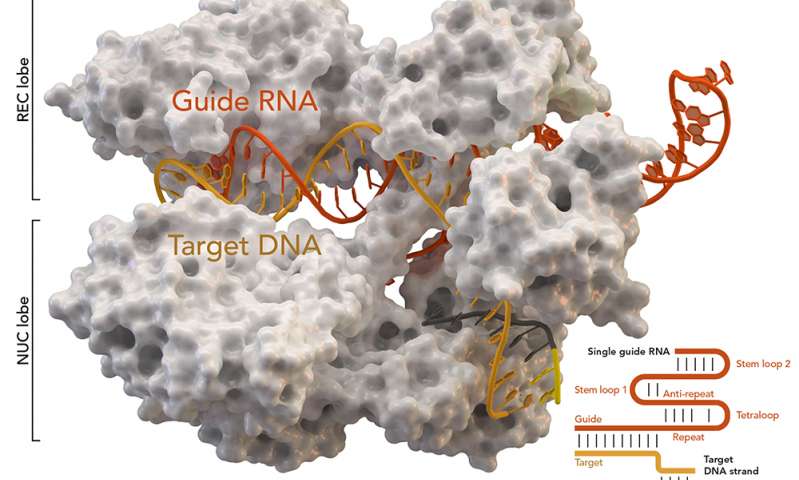
This CRISPR-Cas9 image reminds me of popcorn,

CRISPR-associated protein Cas9 (white) from Staphylococcus aureus based on Protein Database ID 5AXW. Credit: Thomas Splettstoesser (Wikipedia, CC BY-SA 4.0)[ downloaded from https://phys.org/news/2018-07-genome-crisprcas9-gene-higher-thought.html#jCp]

A July 16, 2018 Wellcome Sanger Institute press release (also on EurekAlert), which originated the news item, offers a little more explanation,

CRISPR/Cas9 is one of the newest genome editing tools. It can alter sections of DNA in cells by cutting at specific points and introducing changes at that location. Already extensively used in scientific research, CRISPR/Cas9 has also been seen as a promising way to create potential genome editing treatments for diseases such as HIV, cancer or sickle cell disease. Such therapeutics could inactivate a disease-causing gene, or correct a genetic mutation. However, any potential treatments would have to prove that they were safe.

Previous research had not shown many unforeseen mutations from CRISPR/Cas9 in the DNA at the genome editing target site. To investigate this further the researchers carried out a full systematic study in both mouse and human cells and discovered that CRISPR/Cas9 frequently caused extensive mutations, but at a greater distance from the target site.

The researchers found many of the cells had large genetic rearrangements such as DNA deletions and insertions. These could lead to important genes being switched on or off, which could have major implications for CRISPR/Cas9 use in therapies. In addition, some of these changes were too far away from the target site to be seen with standard genotyping methods.

Prof Allan Bradley, corresponding author on the study from the Wellcome Sanger Institute, said: “This is the first systematic assessment of unexpected events resulting from CRISPR/Cas9 editing in therapeutically relevant cells, and we found that changes in the DNA have been seriously underestimated before now. It is important that anyone thinking of using this technology for gene therapy proceeds with caution, and looks very carefully to check for possible harmful effects.”

Michael Kosicki, the first author from the Wellcome Sanger Institute, said: “My initial experiment used CRISPR/Cas9 as a tool to study gene activity, however it became clear that something unexpected was happening. Once we realised the extent of the genetic rearrangements we studied it systematically, looking at different genes and different therapeutically relevant cell lines, and showed that the CRISPR/Cas9 effects held true.”

The work has implications for how CRISPR/Cas9 is used therapeutically and is likely to re-spark researchers’ interest in finding alternatives to the standard CRISPR/Cas9 method for gene editing.

Prof Maria Jasin, an independent researcher from Memorial Slone Kettering Cancer Centre, New York, who was not involved in the study said: “This study is the first to assess the repertoire of genomic damage arising at a CRISPR/Cas9 cleavage site. While it is not known if genomic sites in other cell lines will be affected in the same way, this study shows that further research and specific testing is needed before CRISPR/Cas9 is used clinically.”

For anyone who’d like to better understand the terms gene editing and CRISPR-Cas9, the Wellcome Sanger Institute provides these explanatory webpages, What is genome editing? and What is CRISPR-Cas9?

For the more advanced, here’s a link and a citation for the paper,

Repair of double-strand breaks induced by CRISPR–Cas9 leads to large deletions and complex rearrangements by Michael Kosicki, Kärt Tomberg, & Allan Bradley. Nature Biotechnology DOI: https://doi.org/10.1038/nbt.4192 Published 16 July 2018

This paper appears to be open access.

The kerfuffle

It seems this news has affected the CRISPR market. From a July 16, 2018 article by Cale Guthrie Weissman for Fast Company,

… CRISPR could unknowingly delete or alter non-targeted genes, which could lead to myriad unintended consequences. This is especially frightening, since the technology is going to be used in human clinical trials.

Meanwhile, other scientists working with CRISPR are trying to downplay the findings, telling STAT [a life sciences and business journalism website] that there have been no reported adverse effects similar to what the study describes. The news, however, has brought about a market reaction–at least three publicly traded companies that focus on CRISPR-based therapies are in stock nosedive. Crispr Therapeutics is down by over 6%; Editas fell by over 3%; and Intellia Therapeutics dropped by over 5%. [emphasis mine]

Damage control

Gaetan Burgio (geneticist, Australian National University)  in a July 16, 2018 essay on phys.org (originating from The Conversation) suggests some calm (Note: Links have been removed),

But a new study has called into question the precision of the technique [CRISPR gene editing technology].

The hope for gene editing is that it will be able to cure and correct diseases. To date, many successes have been reported, including curing deafness in mice, and in altering cells to cure cancer.

Some 17 clinical trials in human patients are registered [emphasis mine] testing gene editing on leukaemias, brain cancers and sickle cell anaemia (where red blood cells are misshaped, causing them to die). Before implementing CRISPR technology in clinics to treat cancer or congenital disorders, we must address whether the technique is safe and accurate.

There are a few options for getting around this problem. One option is to isolate the cells we wish to edit from the body and reinject only the ones we know have been correctly edited.

For example, lymphocytes (white blood cells) that are crucial to killing cancer cells could be taken out of the body, then modified using CRISPR to heighten their cancer-killing properties. The DNA of these cells could be sequenced in detail, and only the cells accurately and specifically gene-modified would be selected and delivered back into the body to kill the cancer cells.

While this strategy is valid for cells we can isolate from the body, some cells, such as neurons and muscles, cannot be removed from the body. These types of cells might not be suitable for gene editing using Cas9 scissors.

Fortunately, researchers have discovered other forms of CRISPR systems that don’t require the DNA to be cut. Some CRISPR systems only cut the RNA, not the DNA (DNA contains genetic instructions, RNA convey the instructions on how to synthesise proteins).

As RNA [ribonucleic acid] remains in our cells only for a specific period of time before being degraded, this would allow us to control the timing and duration of the CRISPR system delivery and reverse it (so the scissors are only functional for a short period of time).

This was found to be successful for dementia in mice. Similarly, some CRISPR systems simply change the letters of the DNA, rather than cutting them. This was successful for specific mutations causing diseases such as hereditary deafness in mice.

I agree with Burgio’s conclusion (not included here) that we have a lot more to learn and I can’t help wondering why there are 17 registered human clinical trials at this point.

Nanoparticle-based delivery platform for CRISPR-Cas9 (gene-editing technology)

A February 18, 2018 King Abdullah University of Science and Technology (KAUST; Saudi Arabia) news release (also on EurekAlert but published on Feb. 20, 2018) describes a new technology for delivering CRISPR (clustered regularly interspaced short palindromic repeats)-Cas9 into cells,

A new delivery system for introducing gene-editing technology into cells could help safely and efficiently correct disease-causing mutations in patients.

The system, developed by KAUST scientists, is the first to use sponge-like ensembles of metal ions and organic molecules to coat the molecular components of the precision DNA-editing technology known as CRISPR/Cas9, allowing efficient release of the genome-editing machinery inside the cell.

“This method presents an easy and economically feasible route to improve on the delivery problems that accompany RNA-based therapeutic approaches,” says Niveen Khashab, the associate professor of chemical sciences at KAUST who led the study. “This may permit such formulations to be eventually used for treating genetic diseases effectively in the future.”

CRISPR/Cas9 has a double delivery problem: For the gene-editing technology to work like a molecular Swiss Army knife, both a large protein (the Cas9 cutting enzyme) and a highly charged RNA component (the guide RNA used for DNA targeting) must each get from the outside of the cell into the cytoplasm and finally into the nucleus, all without getting trapped in the tiny intracellular bubbles that are known as endosomes.

To solve this problem, Khashab and her lab turned to a nano-sized type of porous material known as a zeolitic imidazolate framework, which forms a cage-like structure into which other molecules can be placed. The researchers encapsulated the Cas9 protein and guide RNA in this material and then introduced the resulting nanoparticles into hamster cells.

The encapsulated CRISPR-Cas9 constructs were not toxic to the cells. And because particles in the coating material become positively charged when absorbed into endosomes, they caused these membrane-bound bubbles to burst, freeing the CRISPR-Cas9 machinery to travel to the nucleus, home to the cell’s genome. There the gene-editing technology could get to work.

Using a guide RNA designed to target a gene that caused the cells to glow green under fluorescent light, Khashab and her team showed that they could reduce the expression of this gene by 37 percent over four days with their technology. “These cage-like structures are biocompatible and can be triggered on demand, making them smart options to overcome delivery problems of genetic materials and proteins,” says the study’s first author Shahad Alsaiari, a Ph.D. student in Khashab’s lab.

The researchers’ plan to test their system in human cells and in mice, and eventually, they hope, in clinical trials.

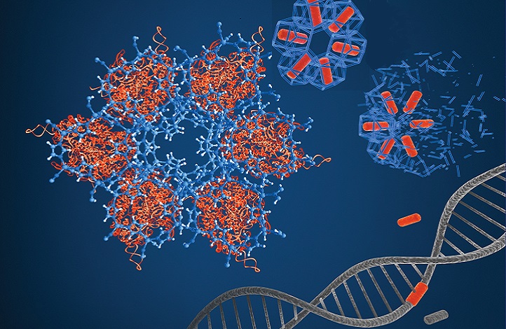
The zeolitic imidazolate framework forms a cage-like scaffold over the CRISPR/Cas9 machinery.. Reprinted (adapted) with permission from Alsaiari, S.K., Patil, S., Alyami, M., Alamoudi, K.O., Aleisa, F.A., Merzaban, J., Li M. & Khashab, N.M. Endosomal escape and delivery of CRISPR/Cas9 genome editing machinery enabled by nanoscale zeolitic imidazolate framework. Journal of the American Chemical Society 140, 143–146 (2018). © 2018 American Chemical Society; KAUST Xavier Pita and Heno Huang ][downloaded from https://discovery.kaust.edu.sa/en/article/475/a%250adelivery-platform-for-gene-editing-technology]

Here’s a link to and a citation for the paper,

Endosomal Escape and Delivery of CRISPR/Cas9 Genome Editing Machinery Enabled by Nanoscale Zeolitic Imidazolate Framework by Shahad K. Alsaiari, Sachin Patil, Mram Alyami, Kholod O. Alamoudi, Fajr A. Aleisa, Jasmeen S. Merzaban, Mo Li, and Niveen M. Khashab. J. Am. Chem. Soc., 2018, 140 (1), pp 143–146 DOI: 10.1021/jacs.7b11754 Publication Date (Web): December 22, 2017

Copyright © 2017 American Chemical Society

This paper is behind a paywall.

CRISPR-Cas12a as a new diagnostic tool

Similar to Cas9, Cas12a is has an added feature as noted in this February 15, 2018 news item on ScienceDaily,

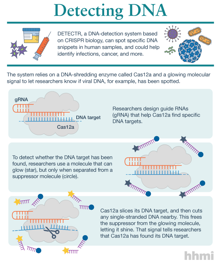
Utilizing an unsuspected activity of the CRISPR-Cas12a protein, researchers created a simple diagnostic system called DETECTR to analyze cells, blood, saliva, urine and stool to detect genetic mutations, cancer and antibiotic resistance and also diagnose bacterial and viral infections. The scientists discovered that when Cas12a binds its double-stranded DNA target, it indiscriminately chews up all single-stranded DNA. They then created reporter molecules attached to single-stranded DNA to signal when Cas12a finds its target.

A February 15, 2018 University of California at Berkeley (UC Berkeley) news release by Robert Sanders and which originated the news item, provides more detail and history,

CRISPR-Cas12a, one of the DNA-cutting proteins revolutionizing biology today, has an unexpected side effect that makes it an ideal enzyme for simple, rapid and accurate disease diagnostics.

blood in test tube

(iStock)

Cas12a, discovered in 2015 and originally called Cpf1, is like the well-known Cas9 protein that UC Berkeley’s Jennifer Doudna and colleague Emmanuelle Charpentier turned into a powerful gene-editing tool in 2012.

CRISPR-Cas9 has supercharged biological research in a mere six years, speeding up exploration of the causes of disease and sparking many potential new therapies. Cas12a was a major addition to the gene-cutting toolbox, able to cut double-stranded DNA at places that Cas9 can’t, and, because it leaves ragged edges, perhaps easier to use when inserting a new gene at the DNA cut.

But co-first authors Janice Chen, Enbo Ma and Lucas Harrington in Doudna’s lab discovered that when Cas12a binds and cuts a targeted double-stranded DNA sequence, it unexpectedly unleashes indiscriminate cutting of all single-stranded DNA in a test tube.

Most of the DNA in a cell is in the form of a double-stranded helix, so this is not necessarily a problem for gene-editing applications. But it does allow researchers to use a single-stranded “reporter” molecule with the CRISPR-Cas12a protein, which produces an unambiguous fluorescent signal when Cas12a has found its target.

“We continue to be fascinated by the functions of bacterial CRISPR systems and how mechanistic understanding leads to opportunities for new technologies,” said Doudna, a professor of molecular and cell biology and of chemistry and a Howard Hughes Medical Institute investigator.

DETECTR diagnostics

The new DETECTR system based on CRISPR-Cas12a can analyze cells, blood, saliva, urine and stool to detect genetic mutations, cancer and antibiotic resistance as well as diagnose bacterial and viral infections. Target DNA is amplified by RPA to make it easier for Cas12a to find it and bind, unleashing indiscriminate cutting of single-stranded DNA, including DNA attached to a fluorescent marker (gold star) that tells researchers that Cas12a has found its target.

The UC Berkeley researchers, along with their colleagues at UC San Francisco, will publish their findings Feb. 15 [2018] via the journal Science’s fast-track service, First Release.

The researchers developed a diagnostic system they dubbed the DNA Endonuclease Targeted CRISPR Trans Reporter, or DETECTR, for quick and easy point-of-care detection of even small amounts of DNA in clinical samples. It involves adding all reagents in a single reaction: CRISPR-Cas12a and its RNA targeting sequence (guide RNA), fluorescent reporter molecule and an isothermal amplification system called recombinase polymerase amplification (RPA), which is similar to polymerase chain reaction (PCR). When warmed to body temperature, RPA rapidly multiplies the number of copies of the target DNA, boosting the chances Cas12a will find one of them, bind and unleash single-strand DNA cutting, resulting in a fluorescent readout.

The UC Berkeley researchers tested this strategy using patient samples containing human papilloma virus (HPV), in collaboration with Joel Palefsky’s lab at UC San Francisco. Using DETECTR, they were able to demonstrate accurate detection of the “high-risk” HPV types 16 and 18 in samples infected with many different HPV types.

“This protein works as a robust tool to detect DNA from a variety of sources,” Chen said. “We want to push the limits of the technology, which is potentially applicable in any point-of-care diagnostic situation where there is a DNA component, including cancer and infectious disease.”

The indiscriminate cutting of all single-stranded DNA, which the researchers discovered holds true for all related Cas12 molecules, but not Cas9, may have unwanted effects in genome editing applications, but more research is needed on this topic, Chen said. During the transcription of genes, for example, the cell briefly creates single strands of DNA that could accidentally be cut by Cas12a.

The activity of the Cas12 proteins is similar to that of another family of CRISPR enzymes, Cas13a, which chew up RNA after binding to a target RNA sequence. Various teams, including Doudna’s, are developing diagnostic tests using Cas13a that could, for example, detect the RNA genome of HIV.

infographic about DETECTR system

(Infographic by the Howard Hughes Medical Institute)

These new tools have been repurposed from their original role in microbes where they serve as adaptive immune systems to fend off viral infections. In these bacteria, Cas proteins store records of past infections and use these “memories” to identify harmful DNA during infections. Cas12a, the protein used in this study, then cuts the invading DNA, saving the bacteria from being taken over by the virus.

The chance discovery of Cas12a’s unusual behavior highlights the importance of basic research, Chen said, since it came from a basic curiosity about the mechanism Cas12a uses to cleave double-stranded DNA.

“It’s cool that, by going after the question of the cleavage mechanism of this protein, we uncovered what we think is a very powerful technology useful in an array of applications,” Chen said.

Here’s a link to and a citation for the paper,

CRISPR-Cas12a target binding unleashes indiscriminate single-stranded DNase activity by Janice S. Chen, Enbo Ma, Lucas B. Harrington, Maria Da Costa, Xinran Tian, Joel M. Palefsky, Jennifer A. Doudna. Science 15 Feb 2018: eaar6245 DOI: 10.1126/science.aar6245

This paper is behind a paywall.

New nanomapping technology: CRISPR-CAS9 as a programmable nanoparticle

A November 21, 2017 news item on Nanowerk describes a rather extraordinary (to me, anyway) approach to using CRRISP ( Clustered Regularly Interspaced Short Palindromic Repeats)-CAS9 (Note: A link has been removed),

A team of scientists led by Virginia Commonwealth University physicist Jason Reed, Ph.D., have developed new nanomapping technology that could transform the way disease-causing genetic mutations are diagnosed and discovered. Described in a study published today [November 21, 2017] in the journal Nature Communications (“DNA nanomapping using CRISPR-Cas9 as a programmable nanoparticle”), this novel approach uses high-speed atomic force microscopy (AFM) combined with a CRISPR-based chemical barcoding technique to map DNA nearly as accurately as DNA sequencing while processing large sections of the genome at a much faster rate. What’s more–the technology can be powered by parts found in your run-of-the-mill DVD player.

A November 21, 2017 Virginia Commonwealth University news release by John Wallace, which originated the news item, provides more detail,

The human genome is made up of billions of DNA base pairs. Unraveled, it stretches to a length of nearly six feet long. When cells divide, they must make a copy of their DNA for the new cell. However, sometimes various sections of the DNA are copied incorrectly or pasted together at the wrong location, leading to genetic mutations that cause diseases such as cancer. DNA sequencing is so precise that it can analyze individual base pairs of DNA. But in order to analyze large sections of the genome to find genetic mutations, technicians must determine millions of tiny sequences and then piece them together with computer software. In contrast, biomedical imaging techniques such as fluorescence in situ hybridization, known as FISH, can only analyze DNA at a resolution of several hundred thousand base pairs.

Reed’s new high-speed AFM method can map DNA to a resolution of tens of base pairs while creating images up to a million base pairs in size. And it does it using a fraction of the amount of specimen required for DNA sequencing.

“DNA sequencing is a powerful tool, but it is still quite expensive and has several technological and functional limitations that make it difficult to map large areas of the genome efficiently and accurately,” said Reed, principal investigator on the study. Reed is a member of the Cancer Molecular Genetics research program at VCU Massey Cancer Center and an associate professor in the Department of Physics in the College of Humanities and Sciences.

“Our approach bridges the gap between DNA sequencing and other physical mapping techniques that lack resolution,” he said. “It can be used as a stand-alone method or it can complement DNA sequencing by reducing complexity and error when piecing together the small bits of genome analyzed during the sequencing process.”

IBM scientists made headlines in 1989 when they developed AFM technology and used a related technique to rearrange molecules at the atomic level to spell out “IBM.” AFM achieves this level of detail by using a microscopic stylus — similar to a needle on a record player — that barely makes contact with the surface of the material being studied. The interaction between the stylus and the molecules creates the image. However, traditional AFM is too slow for medical applications and so it is primarily used by engineers in materials science.

“Our device works in the same fashion as AFM but we move the sample past the stylus at a much greater velocity and use optical instruments to detect the interaction between the stylus and the molecules. We can achieve the same level of detail as traditional AFM but can process material more than a thousand times faster,” said Reed, whose team proved the technology can be mainstreamed by using optical equipment found in DVD players. “High-speed AFM is ideally suited for some medical applications as it can process materials quickly and provide hundreds of times more resolution than comparable imaging methods.”

Increasing the speed of AFM was just one hurdle Reed and his colleagues had to overcome. In order to actually identify genetic mutations in DNA, they had to develop a way to place markers or labels on the surface of the DNA molecules so they could recognize patterns and irregularities. An ingenious chemical barcoding solution was developed using a form of CRISPR technology.

CRISPR has made a lot of headlines recently in regard to gene editing. CRISPR is an enzyme that scientists have been able to “program” using targeting RNA in order to cut DNA at precise locations that the cell then repairs on its own. Reed’s team altered the chemical reaction conditions of the CRISPR enzyme so that it only sticks to the DNA and does not actually cut it.

“Because the CRISPR enzyme is a protein that’s physically bigger than the DNA molecule, it’s perfect for this barcoding application,” Reed said. “We were amazed to discover this method is nearly 90 percent efficient at bonding to the DNA molecules. And because it’s easy to see the CRISPR proteins, you can spot genetic mutations among the patterns in DNA.”

To demonstrate the technique’s effectiveness, the researchers mapped genetic translocations present in lymph node biopsies of lymphoma patients. Translocations occur when one section of the DNA gets copied and pasted to the wrong place in the genome. They are especially prevalent in blood cancers such as lymphoma but occur in other cancers as well.

While there are many potential uses for this technology, Reed and his team are focusing on medical applications. They are currently developing software based on existing algorithms that can analyze patterns in sections of DNA up to and over a million base pairs in size. Once completed, it would not be hard to imagine this shoebox-sized instrument in pathology labs assisting in the diagnosis and treatment of diseases linked to genetic mutations.

Here’s a link to and a citation for the paper,

DNA nanomapping using CRISPR-Cas9 as a programmable nanoparticle by Andrey Mikheikin, Anita Olsen, Kevin Leslie, Freddie Russell-Pavier, Andrew Yacoot, Loren Picco, Oliver Payton, Amir Toor, Alden Chesney, James K. Gimzewski, Bud Mishra, & Jason Reed. Nature Communications 8, Article number: 1665 (2017) doi:10.1038/s41467-017-01891-9 Published online: 21 November 2017

This paper is open access.

Viewing RNA (ribonucleic acid) more closely at the nanoscale with expansion microscopy (EXM) and off-the-shelf parts

A close cousin to DNA (deoxyribonucleic acid), RNA (ribonucleic acid) is a communicator according to a July 4, 2016 news item on ScienceDaily describing how a team at the Massachusetts Institute of Technology (MIT) managed to image RNA more precisely,

Cells contain thousands of messenger RNA molecules, which carry copies of DNA’s genetic instructions to the rest of the cell. MIT engineers have now developed a way to visualize these molecules in higher resolution than previously possible in intact tissues, allowing researchers to precisely map the location of RNA throughout cells.

Key to the new technique is expanding the tissue before imaging it. By making the sample physically larger, it can be imaged with very high resolution using ordinary microscopes commonly found in research labs.

“Now we can image RNA with great spatial precision, thanks to the expansion process, and we also can do it more easily in large intact tissues,” says Ed Boyden, an associate professor of biological engineering and brain and cognitive sciences at MIT, a member of MIT’s Media Lab and McGovern Institute for Brain Research, and the senior author of a paper describing the technique in the July 4, 2016 issue of Nature Methods.

A July 4, 2016 MIT news release (also on EurekAlert), which originated the news item, explains why scientists want a better look at RNA and how the MIT team accomplished the task,

Studying the distribution of RNA inside cells could help scientists learn more about how cells control their gene expression and could also allow them to investigate diseases thought to be caused by failure of RNA to move to the correct location.

Boyden and colleagues first described the underlying technique, known as expansion microscopy (ExM), last year, when they used it to image proteins inside large samples of brain tissue. In a paper appearing in Nature Biotechnology on July 4, the MIT team has now presented a new version of the technology that employs off-the-shelf chemicals, making it easier for researchers to use.

MIT graduate students Fei Chen and Asmamaw Wassie are the lead authors of the Nature Methods paper, and Chen and graduate student Paul Tillberg are the lead authors of the Nature Biotechnology paper.

A simpler process

The original expansion microscopy technique is based on embedding tissue samples in a polymer that swells when water is added. This tissue enlargement allows researchers to obtain images with a resolution of around 70 nanometers, which was previously possible only with very specialized and expensive microscopes. However, that method posed some challenges because it requires generating a complicated chemical tag consisting of an antibody that targets a specific protein, linked to both a fluorescent dye and a chemical anchor that attaches the whole complex to a highly absorbent polymer known as polyacrylate. Once the targets are labeled, the researchers break down the proteins that hold the tissue sample together, allowing it to expand uniformly as the polyacrylate gel swells.

In their new studies, to eliminate the need for custom-designed labels, the researchers used a different molecule to anchor the targets to the gel before digestion. This molecule, which the researchers dubbed AcX, is commercially available and therefore makes the process much simpler.

AcX can be modified to anchor either proteins or RNA to the gel. In the Nature Biotechnology study, the researchers used it to anchor proteins, and they also showed that the technique works on tissue that has been previously labeled with either fluorescent antibodies or proteins such as green fluorescent protein (GFP).

“This lets you use completely off-the-shelf parts, which means that it can integrate very easily into existing workflows,” Tillberg says. “We think that it’s going to lower the barrier significantly for people to use the technique compared to the original ExM.”

Using this approach, it takes about an hour to scan a piece of tissue 500 by 500 by 200 microns, using a light sheet fluorescence microscope. The researchers showed that this technique works for many types of tissues, including brain, pancreas, lung, and spleen.

Imaging RNA

In the Nature Methods paper, the researchers used the same kind of anchoring molecule but modified it to target RNA instead. All of the RNAs in the sample are anchored to the gel, so they stay in their original locations throughout the digestion and expansion process.

After the tissue is expanded, the researchers label specific RNA molecules using a process known as fluorescence in situ hybridization (FISH), which was originally developed in the early 1980s and is widely used. This allows researchers to visualize the location of specific RNA molecules at high resolution, in three dimensions, in large tissue samples.

This enhanced spatial precision could allow scientists to explore many questions about how RNA contributes to cellular function. For example, a longstanding question in neuroscience is how neurons rapidly change the strength of their connections to store new memories or skills. One hypothesis is that RNA molecules encoding proteins necessary for plasticity are stored in cell compartments close to the synapses, poised to be translated into proteins when needed.

With the new system, it should be possible to determine exactly which RNA molecules are located near the synapses, waiting to be translated.

“People have found hundreds of these locally translated RNAs, but it’s hard to know where exactly they are and what they’re doing,” Chen says. “This technique would be useful to study that.”

Boyden’s lab is also interested in using this technology to trace the connections between neurons and to classify different subtypes of neurons based on which genes they are expressing.

There’s a brief (30 secs.), silent video illustrating the work (something about a ‘Brainbow Hippocampus’) made available by MIT,


Here’s a link to and a citation for the paper,

Nanoscale imaging of RNA with expansion microscopy by Fei Chen, Asmamaw T Wassie, Allison J Cote, Anubhav Sinha, Shahar Alon, Shoh Asano, Evan R Daugharthy, Jae-Byum Chang, Adam Marblestone, George M Church, Arjun Raj, & Edward S Boyden.     Nature Methods (2016)  doi:10.1038/nmeth.3899 Published online 04 July 2016

This paper is behind a paywall.

Sticky-flares nanotechnology to track and observe RNA (ribonucleic acid) regulation

I like the name ‘sticky-flares’ and had hoped there was an amusing story about its origins. Ah well, perhaps I’ll have better luck next time.

This work comes out of Chad Mirkin’s lab at Northwestern University (Chicago, US) according to a July 21, 2015 news item on Azonano,

RNA [ribonucleic acid] is a fundamental ingredient in all known forms of life — so when RNA goes awry, a lot can go wrong. RNA misregulation plays a critical role in the development of many disorders, such as mental disability, autism and cancer.

A new technology — called “Sticky-flares” — developed by nanomedicine experts at Northwestern University offers the first real-time method to track and observe the dynamics of RNA distribution as it is transported inside living cells.

A July 20, 2015 Northwestern University news release by Erin Spain, which originated the news item, describes the research in a little more detail also including information about predecessor technology,

Sticky-flares have the potential to help scientists understand the complexities of RNA better than any analytical technique to date and observe and study the biological and medical significance of RNA misregulation.

Previous technologies made it possible to attain static snapshots of RNA location, but that isn’t enough to understand the complexities of RNA transport and localization within a cell. Instead of analyzing snapshots of RNA to try to understand functioning, Sticky-flares help create an experience that is more like watching live-streaming video.

“This is very exciting because much of the RNA in cells has very specific quantities and localization, and both are critical to the cell’s function, but until this development it has been very difficult, and often impossible, to probe both attributes of RNA in a live cell,” said Chad A. Mirkin, a nanomedicine expert and corresponding author of the study. “We hope that many more researchers will be able to use this platform to increase our understanding of RNA function inside cells.”

Sticky-flares are tiny spherical nucleic acid gold nanoparticle conjugates that can enter living cells and target and transfer a fluorescent reporter or “tracking device” to RNA transcripts. This fluorescent labeling can be tracked via fluorescence microscopy as it is transported throughout the cell, including the nucleus.

In the … paper, the scientists explain how they used Sticky-flares to quantify β–actin mRNA in HeLa cells (the oldest and most commonly used human cell line) as well as to follow the real-time transport of β–actin mRNA in mouse embryonic fibroblasts.

Sticky-flares are built upon another technology from Mirkin’s group called NanoFlares, which was the first genetic-based approach that is able to detect live circulating tumor cells out of the complex matrix that is human blood.

NanoFlares have been very useful for researchers that operate in the arena of quantifying gene expression. AuraSense, Inc., a biotechnology company that licensed the NanoFlare technology from Northwestern University, and EMD-Millipore, another biotech company, have commercialized NanoFlares. There are now more than 1,700 commercial forms of NanoFlares sold under the SmartFlareä name in more than 230 countries.

The Sticky-flare is designed to address limitations of SmartFlares, most notably their inability to track RNA location and enter the nucleus. The Northwestern team believes Sticky-flares are poised to become a valuable tool for researchers who desire to understand the function of RNA in live cells.

Based on the paragraph about the precursor technology’s commercial success , I gather they are excited about similar possibilities for sticky-flares.

Here’s a link to and a citation for the paper,

Quantification and real-time tracking of RNA in live cells using Sticky-flares by William E. Briley, Madison H. Bondy, Pratik S. Randeria, Torin J. Dupper, and Chad A. Mirkin. Published online before print July 20, 2015, doi: 10.1073/pnas.1510581112 PNAS July 20, 2015

This paper is behind a paywall.

A newish Tekmira results from a merger with OnCore Biopharma

A Jan. 12, 2015 news item on Azonano announces a new business entity, a combined Tekmira Pharmaceuticals (located in North Vancouver, Canada) and OnCore Biopharma (located in Pennsylvania, US),

Tekmira Pharmaceuticals Corporation, a leading developer of RNA interference (RNAi) therapeutics, and OnCore Biopharma, Inc., a biopharmaceutical company dedicated to discovering, developing and commercializing an all-oral cure for patients suffering from chronic hepatitis B virus (HBV) infection, announced today that they have agreed to merge to create a new leading global HBV company focused on developing a curative regimen for hepatitis B patients by combining multiple therapeutic approaches.

A Jan. 11, 2015 Tekmira news release, which originated the news item, provides details including how this merger will affect the work on the Tekmira ebola treatment,

This transaction is expected to bring together the companies’ broad expertise in antiviral drug development, Tekmira’s Phase 1-ready HBV RNAi therapeutic and OnCore’s multiple HBV programs, to build a robust portfolio of compounds aimed at eradicating HBV. The combined company’s most advanced products are expected to be TKM-HBV, an RNAi therapeutic designed to eliminate HBV surface antigen (HBsAg) expression, a key component of host immune suppression, which is on track to begin human clinical trials in the first quarter of 2015; and OCB-030, a second-generation cyclophilin inhibitor focused on the suppression of viral replication, as well as stimulation and reactivation of the body’s immune response, which is anticipated to enter human clinical trials in the second half of 2015. The combined company anticipates progressing additional programs toward the clinic to achieve the goal of expeditiously evaluating combination regimens.

The combined pipeline is expected to target the three pillars necessary to develop a curative regimen for HBV, including assets focused on suppressing HBV replication, reactivating and stimulating the host immune response directed at HBV and eliminating covalently closed circular DNA (cccDNA). The parties believe that, together, these three pillars are the foundation for achieving a curative regimen.

Dr. Mark J. Murray, Chief Executive Officer of Tekmira, said, “We believe that the merger between Tekmira and OnCore has the potential to transform the HBV treatment landscape by bringing together the technologies and science needed to eradicate the virus and develop a cure for this debilitating and deadly disease. Our new company has the potential to advance multiple, highly active, complementary agents into the clinic in rapid succession, and create an HBV therapeutics powerhouse, thereby potentially offering significant benefits to the global medical community working to improve the lives of HBV patients. Importantly, we also believe this transaction has the potential to create significant value for our shareholders.”

Patrick Higgins, Chief Executive Officer of OnCore, said, “Tekmira and OnCore share a vision that effective combination regimens will ultimately cure HBV, a goal now being realized for hepatitis C virus. This merger is expected to bring together the promise of TKM-HBV with our existing HBV portfolio and accelerate our timeline for combination clinical trials. It is expected to deliver both near-term catalysts and long-term value creation. We believe that the ability to rapidly and sequentially combine novel HBV therapeutics is extremely valuable. We intend to utilize our collective expertise in liver disease and a focused development program, as we did at Pharmasset, to expeditiously and efficiently meet our shared goals.”

An Industry-Leading, Multi-Functional HBV Portfolio

Through the combined portfolio, OnCore and Tekmira intend to advance a robust pipeline of assets that uniquely targets the three pillars for delivering a curative regimen for HBV, including suppressing HBV replication, reactivating and stimulating the host immune response directed at HBV and eliminating cccDNA, the stable source of HBV viral genomic material. Post-closing, the combined company’s HBV portfolio is expected to include  product assets, which can be viewed in a chart by clicking on the following  link: http://media.globenewswire.com/cache/14025/file/31117.pdf

“We intend to take a focused, iterative approach to identifying the most effective combination regimens, while applying what we learn at each stage to optimize future compounds and combinations,” said Dr. Michael Sofia, the combined company’s Chief Scientific Officer and an inventor of sofosbuvir (Sovaldi) for the treatment of hepatitis C. “We believe that the ability to combine multiple unique programs housed in the same company is a significant competitive advantage, and should provide considerable efficiency in terms of speed and ease of decision-making. Combining the OnCore and Tekmira HBV portfolios underpins our vision to accelerate the delivery of a curative HBV regimen.”

Non-HBV Programs Continuing to Move Forward

Tekmira is a global leader in the RNAi field, and has created a diverse pipeline of products in development to treat serious human diseases, such as cancer and viral infections, including Ebola. The company has also licensed its leading lipid nanoparticle (LNP) delivery technology to partners around the world.

The management teams and Boards of Directors of Tekmira and OnCore believe that there is significant value in Tekmira’s non-HBV assets and collaborations. TKM-PLK1 is currently in Phase 2 in multiple indications and TKM-Ebola is expected to enter Phase 2 in West Africa in early 2015. Tekmira also maintains an active RNAi research and development effort. The combined management team and Board of Directors plans to continue to move forward with these programs with the goal of maximizing their value.

The news release goes on to describe the deal,

Under the terms of the agreement, the transaction will be carried out by way of a merger pursuant to which OnCore will merge with a wholly-owned subsidiary of Tekmira and thereby become a wholly-owned subsidiary of Tekmira. Upon closing of the transaction the stockholders of OnCore will hold approximately fifty percent (50%) of the total number of outstanding shares of capital stock of Tekmira, calculated on a fully-diluted and as-converted basis using the treasury stock method. The terms and conditions of the transaction are more fully set forth in the Merger Agreement. The implied market value of the combined company, based on the closing price of Tekmira common shares on the NASDAQ Global Market on January 9, 2015, is approximately USD$750 million.

The merger is subject to approval of a majority of the shareholders of Tekmira present, in person or by proxy, at a special meeting of Tekmira shareholders. Completion of the transaction is also subject to customary closing conditions, including regulatory approvals.  The transaction is expected to close in the first half of 2015, shortly after completion of the Securities and Exchange Commission (SEC) review process and receipt of Tekmira shareholder approval. The Tekmira Board of Directors unanimously approved and recommends that Tekmira shareholders vote FOR the proposed transaction at a special meeting of shareholders.

Details regarding these and other terms of the transaction are set out in the Merger Agreement, which will be filed by Tekmira on the SEC website at www.sec.gov and on the Canadian securities administrator’s website at www.sedar.com.

The combined company plans to retain top executives and board members from Tekmira and OnCore. The new company’s management team will include Mark J. Murray, PhD, Chief Executive Officer; Patrick T. Higgins, President and Chief Operating Officer; Bruce Cousins, Chief Financial Officer; Michael J. Sofia, PhD, Chief Scientific Officer; Mark Kowalski, MD, PhD, Chief Medical Officer; Bryce Roberts, Chief Legal Officer; Michael J. McElhaugh, Chief Business Officer; and Michael J. Abrams, PhD, Chief Discovery Officer. William T. Symonds, PharmD, who led the clinical development of sofosbuvir for the treatment of HCV infection at Pharmasset and later Gilead Sciences, Inc., will be Chief Development Officer and lead the clinical development of the portfolio.

Vivek Ramaswamy will serve as Chairman of the combined company; Dr. Daniel Kisner MD will serve as its Vice-Chairman. The combined company will be headquartered in Vancouver, BC.

I don’t understand how a company, OnCore, which is becoming a subsidiary qualifies as an equal partner in a merger but I gather this is business speak. In any event, the truly curious can find the webcast for a conference call about the deal held on Jan. 12, 2015 at 5 am PT (8 am ET)  along with an accompanying presentation here. The webcast will be available only from January 12, 2015 at 9:00 am PT  / 12 noon ET to January 17, 2015 at 9:00 am PT  / 12 noon ET and, for access, you must register on the site.

I have written previously about Tekmira, in a Nov. 19, 2014 post regarding another of its business deals and in a Sept. 23, 2014 post about its ebola treatment.

RNA interference: a Tekmira deal and a new technique births Solstice Biologics

I have two news items concerning ribonucleic acid interference (RNAi). The first item features Tekmira Pharmaceuticals Corporation (a Canadian company located in the Vancouver area) and a licencing deal with Dicerna Pharmaceuticals (Massachusetts, US), according to a Nov. 18, 2014 news item on Azonano,

Tekmira Pharmaceuticals Corporation a leading developer of RNA interference (RNAi) therapeutics, today announces a licensing and collaboration agreement with Dicerna Pharmaceuticals, Inc. Tekmira has licensed its proprietary lipid nanoparticle (LNP) delivery technology for exclusive use in Dicerna’s primary hyperoxaluria type 1 (PH1) development program.

Under the agreement, Dicerna will pay Tekmira $2.5 million upfront and payments of $22 million in aggregate development milestones, plus a mid-single-digit royalty on future PH1 sales. This new partnership also includes a supply agreement with Tekmira providing clinical drug supply and regulatory support in the rapid advancement of the product candidate.

The agreement announced today follows the successful testing and demonstration of positive results combining Tekmira’s LNP technology with DCR-PH1 in pre-clinical animal models.

I don’t entirely understand what they mean by “pre-clinical animal models” as I’ve not noticed the term “pre-clinical” applied to animal testing before this. It’s possible they mean they’ve run tests on animals (in vivo) and are now proceeding to human clinical trials or it could mean they’ve run in silico (computer modeling) or in vitro (test tube/test slide) tests and are now proceeding to animal tests. If anyone should have some insights, please do share them with me in the comments section.

A Nov. 17, 2014 Tekmira news release, which originated the news item, describes the deal in more detail,

Dicerna will use Tekmira’s third generation LNP technology for delivery of DCR-PH1, Dicerna’s Dicer substrate RNA (DsiRNA) molecule, for the treatment of PH1, a rare, inherited liver disorder that often results in kidney failure and for which there are no approved therapies.

“This new agreement validates our leadership position in RNAi delivery with LNP technology, and it underscores the significant value we can bring to partners who leverage our technology. Our LNP technology is enabling the most advanced applications of RNAi therapeutics in the clinic, and it continues to do so. We are excited to be working with Dicerna to be able to advance a needed therapeutic for the treatment of PH1,” said Dr. Mark J. Murray, Tekmira’s President and CEO.

“As a core pillar of our business strategy, we continue to engage in partnerships where our technology improves the risk profile and accelerates the development programs of our collaborators and provides meaningful non-dilutive financing to TKMR,” added Dr. Murray.

“Dicerna is focused on realizing the full clinical potential of our proprietary pipeline of highly targeted RNAi therapies by applying proven technologies,” said Douglas Fambrough, Ph.D., Chief Executive Officer of Dicerna. “By drawing on Tekmira’s extensive and deep experience with lipid nanoparticle delivery to the liver, the agreement will streamline the development path for DCR-PH1. We look forward to initiating Phase 1 trials of DCR-PH1 in 2015, aiming to fill a high unmet medical need for patients with PH1.”

The news release also provides a high level description of the various technologies being researched and brought to market and a bit more information about the liver disorder being addressed by this research,

About RNAi

RNAi therapeutics have the potential to treat a number of human diseases by “silencing” disease-causing genes. The discoverers of RNAi, a gene silencing mechanism used by all cells, were awarded the 2006 Nobel Prize for Physiology or Medicine. RNAi trigger molecules often require delivery technology to be effective as therapeutics.

AboutTekmira’s LNP Technology

Tekmira believes its LNP technology represents the most widely adopted delivery technology for the systemic delivery of RNAi triggers. Tekmira’s LNP platform is being utilized in multiple clinical trials by Tekmira and its partners. Tekmira’s LNP technology (formerly referred to as stable nucleic acid-lipid particles, or SNALP) encapsulates RNAi triggers with high efficiency in uniform lipid nanoparticles that are effective in delivering these therapeutic compounds to disease sites. Tekmira’s LNP formulations are manufactured by a proprietary method which is robust, scalable and highly reproducible, and LNP-based products have been reviewed by multiple regulatory agencies for use in clinical trials. LNP formulations comprise several lipid components that can be adjusted to suit the specific application.

About Primary Hyperoxaluria Type 1 ( PH1)

PH1 is a rare, inherited liver disorder that often results in severe damage to the kidneys. The disease can be fatal unless the patient undergoes a liver-kidney transplant, a major surgical procedure that is often difficult to perform due to the lack of donors and the threat of organ rejection. In the event of a successful transplant, the patient must live the rest of his or her life on immunosuppressant drugs, which have substantial associated risks. Currently, there are no FDA approved treatments for PH1.

PH1 is characterized by a genetic deficiency of the liver enzyme alanine:glyoxalate-aminotransferase (AGT), which is encoded by the AGXT gene. AGT deficiency induces overproduction of oxalate by the liver, resulting in the formation of crystals of calcium oxalate in the kidneys. Oxalate crystal formation often leads to chronic and painful cases of kidney stones and subsequent fibrosis (scarring), which is known as nephrocalcinosis. Many patients progress to end-stage renal disease (ESRD) and require dialysis or transplant. Aside from having to endure frequent dialysis, PH1 patients with ESRD may experience a build-up of oxalate in the bone, skin, heart and retina, with concomitant debilitating complications. While the true prevalence of primary hyperoxaluria is unknown, it is estimated to be one to three cases per one million people.1 Fifty percent of patients with PH1 reach ESRD by their mid-30s.2

About DCR-PH1

Dicerna is developing DCR-PH1, which is in preclinical development, for the treatment of PH1. DCR-PH1 is engineered to address the pathology of PH1 by targeting and destroying the messenger RNA (mRNA) produced by HAO1, a gene implicated in the pathogenesis of PH1. HAO1 encodes glycolate oxidase, a protein involved in producing oxalate. By reducing oxalate production, this approach is designed to prevent the complications of PH1. In preclinical studies, DCR-PH1 has been shown to induce potent and long-term inhibition of HAO1 and to significantly reduce levels of urinary oxalate, while demonstrating long-term efficacy and tolerability in animal models of PH1.

About Dicerna’s Dicer Substrate Technology

Dicerna’s proprietary RNAi molecules are known as Dicer substrates, or DsiRNAs, so called because they are processed by the Dicer enzyme, which is the initiation point for RNAi in the human cell cytoplasm. Dicerna’s discovery approach is believed to maximize RNAi potency because the DsiRNAs are structured to be ideal for processing by Dicer. Dicer processing enables the preferential use of the correct RNA strand of the DsiRNA, which may increase the efficacy of the RNAi mechanism, as well as the potency of the DsiRNA molecules relative to other molecules used to induce RNAi.

You can find more information about Tekmira here and about Dicerna here. I mentioned Tekmira previously in a Sept. 28, 2014 post about Ebola and treatments.

Further south at the University of California at San Diego (UCSD), researcher and founder of Solstice Biologics, Dr Steven Dowdy has developed and patented a new technique for delivering RNAi drugs into cells according to a Nov. 18, 2014 news item on Azonano,

Small pieces of synthetic RNA trigger a RNA interference (RNAi) response that holds great therapeutic potential to treat a number of diseases, especially cancer and pandemic viruses. The problem is delivery — it is extremely difficult to get RNAi drugs inside the cells in which they are needed. To overcome this hurdle, researchers at University of California, San Diego School of Medicine have developed a way to chemically disguise RNAi drugs so that they are able to enter cells. Once inside, cellular machinery converts these disguised drug precursors — called siRNNs — into active RNAi drugs. …

A Nov. 17, 2014 UCSD news release (also on EurekAlert) by Heather Buschman, which originated the news item, describes the issues with delivering RNAi drugs to cells and the new technique,

“Many current approaches use nanoparticles to deliver RNAi drugs into cells,” said Steven F. Dowdy, PhD, professor in the Department of Cellular and Molecular Medicine and the study’s principal investigator. “While nanotechnology protects the RNAi drug, from a molecular perspective nanoparticles are huge, some 5,000 times larger than the RNAi drug itself. Think of delivering a package into your house by having an 18-wheeler truck drive it through your living room wall — that’s nanoparticles carrying standard RNAi drugs. Now think of a package being slipped through the mail slot — that’s siRNNs.”

The beauty of RNAi is that it selectively blocks production of target proteins in a cell, a finding that garnered a Nobel Prize in 2006. While this is a normal process that all cells use, researchers have taken advantage of RNAi to inhibit specific proteins that cause disease when overproduced or mutated, such as in cancer. First, researchers generate RNAi drugs with a sequence that corresponds to the gene blueprint for the disease protein and then delivers them into cells. Once inside the cell, the RNAi drug is loaded into an enzyme that specifically slices the messenger RNA encoding the target protein in half. This way, no protein is produced.

As cancer and viral genes mutate, RNAi drugs can be easily evolved to target them. This allows RNAi therapy to keep pace with the genetics of the disease — something that no other type of therapy can do. Unfortunately, due to their size and negatively charged chemical groups (phosphates) on their backbone, RNAi drugs are repelled by the cellular membrane and cannot be delivered into cells without a special delivery agent.

It took Dowdy and his team, including Bryan Meade, PhD, Khirud Gogoi, PhD, and Alexander S. Hamil, eight years to find a way to mask RNAi’s negative phosphates in such a way that gets them into cells, but is still capable of inducing an RNAi response once inside.

In the end, the team added a chemical tag called a phosphotriester group. The phosphotriester neutralizes and protects the RNA backbone — converting the ribonucleic acid (RNA) to ribonucleic neutral (RNN), and thus giving the name siRNN. The neutral (uncharged) nature of siRNNs allows them to pass into the cell much more efficiently. Once inside the cell, enzymes cleave off the neutral phosphotriester group to expose a charged RNAi drug that shuts down production of the target disease protein. siRNNs represent a transformational next-generation RNAi drug.

“siRNNs are precursor drugs, or prodrugs, with no activity. It’s like having a tool still in the box, it won’t work until you take it out,” Dowdy said. “Only when the packaging — the phosphotriester groups — is removed inside the cells do you have an active tool or RNAi drug.”

The findings held up in a mouse model, too. There, Dowdy’s team found that siRNNs were significantly more effective at blocking target protein production than typical RNAi drugs — demonstrating that once siRNNs get inside a cell they can do a better job.

“There remains a lot of work ahead to get this into the clinics. But, in theory, the therapeutic potential of siRNNs is endless,” Dowdy said. “Particularly for cancer, viral infections and genetic diseases.”

The siRNN technology forms the basis for Solstice Biologics, a biotech company in La Jolla, Calif. that is now taking the technique to the next level. Dowdy is a co-founder of Solstice Biologics and serves as a Board Director.

Here’s a link to and a citation for the research paper,

Efficient delivery of RNAi prodrugs containing reversible charge-neutralizing phosphotriester backbone modifications by Bryan R Meade, Khirud Gogoi, Alexander S Hamil, Caroline Palm-Apergi, Arjen van den Berg, Jonathan C Hagopian, Aaron D Springer, Akiko Eguchi, Apollo D Kacsinta, Connor F Dowdy, Asaf Presente, Peter Lönn, Manuel Kaulich, Naohisa Yoshioka, Edwige Gros, Xian-Shu Cui, & Steven F Dowdy. Nature Biotechnology (2014) doi:10.1038/nbt.3078 Published online 17 November 2014

This paper is behind a paywall.

I have not been able to locate a website for Solstice Biologics but did find a rather curious item about Dr. Dowdy and a shooting incident last year. From a Sept. 18, 2013 news article by Kat Robinson for thewire.sheknows.com,

A wealthy San Diego community is shaken after a man opens fire on his former neighborhood early Wednesday morning. Police say Hans Petersen, a 48-year-old man, is the prime suspect in the shooting of Steven Dowdy and Michael Fletcher.

There’s also a Nov. 8, 2013 article about the incident by Lucas Laursen for Nature magazine,

On September 18 [2013], former Traversa Therapeutics CEO Hans Petersen went on a shooting spree. One of two people wounded was molecular biologist Steven Dowdy, a professor at University of California San Diego (UCSD) School of Medicine, in La Jolla, and cofounder of Traversa, according to a San Diego police report.…

The rest of the article is behind a paywall.

Treatment for patients infected with the ebola virus (a response to crisis in West African countries)

I’ve not actively kept up with the situation in the West African countries suffering an outbreak of the ebola virus other than to note that it is ongoing. My Aug. 15, 2014 post provides a snapshot of the situation and various new treatments, including one based on tobacco, which could be helpful but appeared not to have been tested and/or deployed. There was a lot of secrecy (especially from Medicago, a Canadian company) regarding the whole matter of treatments and vaccines.

There seem to have been some new developments on the treatment side, involving yet another Canadian company, Tekmira, according to a Sept. 23, 2013 news item on Azonano,

Tekmira Pharmaceuticals Corporation, a leading developer of RNA interference (RNAi) therapeutics, today announced that the FDA [US Food and Drug Administration] has authorized Tekmira to provide TKM-Ebola for treatment under expanded access protocols to subjects with confirmed or suspected Ebola virus infections.

A Sept. 22, 2014 Tekmira news release, which originated the news item, expands on the topic of regulatory issues associated with bringing this treatment to the areas suffering the outbreak,

“Tekmira is reporting that an appropriate regulatory and clinical framework is now in place to allow the use of TKM-Ebola in patients. We have worked with the FDA and Health Canada to establish this framework and a treatment protocol allowing us to do what we can to help these patients,” said Dr. Mark J. Murray Tekmira’s President and CEO.

“We have insisted on acting responsibly in the interest of patients and our stakeholders,” added Dr. Murray. “Today we are reporting that, working closely with regulators in the United States and Canada, we have established a framework for TKM-Ebola use in multiple patients. In the US, the FDA has granted expanded access use of TKM-Ebola under our Investigational New Drug application (IND) and Health Canada has established a similar framework, both of which allow the use of our investigational therapeutic in more patients.”

“We have already responded to requests for the use of our investigational agent in several patients under emergency protocols, in an effort to help these patients, a goal we share with the FDA and Health Canada. TKM-Ebola has been administered to a number of patients and the repeat infusions have been well tolerated. However, it must be kept in mind that any uses of the product under expanded access, does not constitute controlled clinical trials. These patients may be infected with a strain of Ebola virus which has emerged subsequent to the strain that our product is directed against, and physicians treating these patients may use more than one therapeutic intervention in an effort to achieve the best outcome,” said Dr. Murray. “Our TKM-Ebola drug supplies are limited, but we will continue to help where we can, as we continue to focus on the other important objectives we have to advance therapies to meet the unmet needs of patients.”

TKM-Ebola is an investigational therapeutic, being developed under an FDA approved IND, which is currently the subject of a partial clinical hold under which the FDA has allowed the potential use of TKM-Ebola in individuals with a confirmed or suspected Ebola virus infection.

About FDA Expanded Access Program

Expanded access is the use of an investigational drug outside of a clinical trial to treat a patient, with a serious or immediately life-threatening disease or condition, who has no comparable or satisfactory alternative treatment options. FDA regulations allow access to investigational drugs for treatment purposes on a case-by-case basis for an individual patient, or for intermediate-size groups of patients with similar treatment needs who otherwise do not qualify to participate in a clinical trial. (Source: www.fda.com)

About TKM-Ebola, an Anti-Ebola Virus RNAi Therapeutic

TKM-Ebola, an anti-Ebola virus RNAi therapeutic, is being developed under a $140 million contract with the U.S. Department of Defense’s Medical Countermeasure Systems BioDefense Therapeutics (MCS-BDTX) Joint Product Management Office. Earlier preclinical studies were published in the medical journal The Lancet and demonstrated that when siRNA targeting the Ebola virus and delivered by Tekmira’s LNP [Lipid Nanoparticle] technology were used to treat previously infected non-human primates, the result was 100 percent protection from an otherwise lethal dose of Zaire Ebola virus (Geisbert et al., The Lancet, Vol. 375, May 29, 2010). In March 2014, Tekmira was granted a Fast Track designation from the U.S. Food and Drug Administration for the development of TKM-Ebola.

About Joint Project Manager Medical Countermeasure Systems (JPM-MCS)

This work is being conducted under contract with the U.S. Department of Defense Joint Project Manager Medical Countermeasure Systems (JPM-MCS). JPM-MCS, a component of the Joint Program Executive Office for Chemical and Biological Defense, aims to provide U.S. military forces and the nation with safe, effective, and innovative medical solutions to counter chemical, biological, radiological, and nuclear threats. JPM-MCS facilitates the advanced development and acquisition of medical countermeasures and systems to enhance biodefense response capability. For more information, visit www.jpeocbd.osd.mil.

About Tekmira

Tekmira Pharmaceuticals Corporation is a biopharmaceutical company focused on advancing novel RNAi therapeutics and providing its leading lipid nanoparticle (LNP) delivery technology to pharmaceutical partners. Tekmira has been working in the field of nucleic acid delivery for over a decade and has broad intellectual property covering LNPs. Further information about Tekmira can be found at www.tekmira.com. Tekmira is based in Vancouver, B.C. Canada.

Forward-Looking Statements and Information

This news release contains “forward-looking statements” or “forward-looking information” within the meaning of applicable securities laws (collectively, “forward-looking statements”). Forward-looking statements in this news release include statements about Tekmira’s strategy, future operations, clinical trials, prospects and the plans of management; an appropriate regulatory and clinical  framework for emergency use of TKM-Ebola in subjects with confirmed or suspected Ebola infections; FDA grant of expanded access use of TKM-Ebola under Tekmira’s IND; Health Canada’s establishment of a similar framework for TKM-Ebola; Tekmira’s response to requests for the use of TKM-Ebola in several patients under emergency protocols and the results thereon; the current supply of TKM-Ebola drug; the partial clinical hold on the TKM-Ebola IND by the FDA (enabling the potential use of TKM-Ebola in individuals with a confirmed or suspected Ebola virus infection); the quantum value of the contract with the JPM-MCS; and Fast Track designation from the FDA for the development of TKM-Ebola.

With respect to the forward-looking statements contained in this news release, Tekmira has made numerous assumptions regarding, among other things, the clinical framework for emergency use of TKM-Ebola. While Tekmira considers these assumptions to be reasonable, these assumptions are inherently subject to significant business, economic, competitive, market and social uncertainties and contingencies.

Additionally, there are known and unknown risk factors which could cause Tekmira’s actual results, performance or achievements to be materially different from any future results, performance or achievements expressed or implied by the forward-looking statements contained herein. Known risk factors include, among others: TKM-Ebola may not prove to be effective in the treatment of Ebola infection under the emergency use framework, or at all; any uses of TKM-Ebola under emergency INDs are not controlled trails, and TKM-Ebola may be used on Ebola strains that have diverged from the strain to which TKM-Ebola is directed, and physicians treating patients may use more than one therapeutic intervention in addition to TKM-Ebola; the current supply of TKM-Ebola is limited, and Tekmira may not be able to respond to future requests for help in the current Ebola outbreak; the FDA may not remove the partial clinical hold on the TKM-Ebola IND; the FDA may refuse to approve Tekmira’s products, or place restrictions on Tekmira’s ability to commercialize its products; anticipated pre-clinical and clinical trials may be more costly or take longer to complete than anticipated, and may never be initiated or completed, or may not generate results that warrant future development of the tested drug candidate; and Tekmira may not receive the necessary regulatory approvals for the clinical development of Tekmira’s products.

A more complete discussion of the risks and uncertainties facing Tekmira appears in Tekmira’s Annual Report on Form 10-K and Tekmira’s continuous disclosure filings, which are available at www.sedar.com or www.sec.gov. All forward-looking statements herein are qualified in their entirety by this cautionary statement, and Tekmira disclaims any obligation to revise or update any such forward-looking statements or to publicly announce the result of any revisions to any of the forward-looking statements contained herein to reflect future results, events or developments, except as required by law.

In the midst of all those ‘cover your rear end’ statements to investors, it’s easy to miss the fact that people are actually being treated and the results are promising, if not guaranteed,

Tekmira has distributed a Sept. 23, 2014 news release touting its membership in a new consortium, which suggests that in parallel with offering treatment, human clinical trials will  also be conducted,

Tekmira Pharmaceuticals Corporation (Nasdaq:TKMR) (TSX:TKM), a leading developer of RNA interference (RNAi) therapeutics, today reported that it is collaborating with an international consortium to provide an RNAi based investigational therapeutic for expedited clinical studies in West Africa.

Led by Dr. Peter Horby of the Centre for Tropical Medicine and Global Health at the University of Oxford and the International Severe Acute Respiratory and Emerging Infection Consortium (ISARIC), the consortium includes representatives from the World Health Organization (WHO), US Centers for Disease Control, Médecins Sans Frontières – Doctors without Borders (MSF), ISARIC, and Fondation Mérieux, among others.

The Wellcome Trust has announced it has awarded £3.2 million to the consortium to fund this initiative. The award will include funds for the manufacture of investigational therapeutics as well as the establishment of an operational clinical trials platform in two or more Ebola Virus Disease (EVD) treatment centers in West Africa. RNAi has been prioritized as an investigational therapeutic and may be selected for clinical trials at these centers.

The objective of the clinical trials is to assess the efficacy and safety of promising therapeutics and vaccines, reliably and safely, in patients with EVD by adopting strict protocols that comply with international standards.  It is hoped this initiative will permit the adoption of safe and effective interventions rapidly.

The genetic sequence of the Ebola virus variant responsible for the ongoing outbreak in West Africa is now available. Under this program, Tekmira will produce an RNAi based product specifically targeting the viral variant responsible for this outbreak.  The ability to rapidly and accurately match the evolving genetic sequences of emerging infectious agents is one of the powerful features of RNAi therapeutics.

“We commend the Wellcome Trust for their leadership in providing the necessary funds to launch and expedite this ground breaking initiative. We are gratified that RNAi has been prioritized as a potential investigational therapeutic to assist in the ongoing public health and humanitarian crisis in Africa,” said Dr. Murray, Tekmira’s President and CEO.

“We are an active collaborator in this consortium and through our ongoing dialogue with the WHO, NGOs and governments in various countries; we have been discussing the creation of appropriate clinical and regulatory frameworks for the potential use of investigational therapeutics in Africa. This initiative goes a long way towards achieving this aim.  Many complex decisions remain to fully implement this unique clinical trial platform.  At this time, there can be no assurances that our product will be selected by the consortium for clinical trials in Africa,” said Dr. Murray.

About Wellcome Trust

The Wellcome Trust is the largest charity in the UK. It funds innovative biomedical research, in the UK and internationally, spending over £600 million each year to support the brightest scientists with the best ideas. The Wellcome Trust supports public debate about biomedical research and its impact on health and wellbeing. For more information, visit www.wellcome.ac.uk

I’m glad they’re being careful while giving people treatment, i. e., trying to do something rather than waiting to conduct human clinical trials as has sometimes been the case in the past. This business of running the trials almost parallel to offering treatment suggests an agility not often associated with the international health care community.

ETA Sept. 23 2014 1200 hours PDT: For more information about the status of the Ebola outbreak read Tara Smith’s Sept. 22, 2014 article Slate titled, Here’s Where We Stand With Ebola; Even experienced international disaster responders are shocked at how bad it’s gotten (Note: Links have been removed).

Now, terms like “exponential spread” are being thrown around as the epidemic continues to expand more and more rapidly. Just last week, an increase of 700 new cases was reported, and the case count is now doubling in size approximately every three weeks.

A Doctors Without Borders worker in Monrovia, Liberia, named Jackson Naimah describes the situation in his home country, noting that patients are literally dying at the front door of his treatment center because it lacks patient beds and assistance; the sufferers are left to die a “horrible, undignified death” and potentially infect others as they do so: …

… Health care workers who are treating the sick are dying because they also lack basic protective equipment, or because they have been so overwhelmed by taking care of the ill and dying that they begin to make potentially fatal errors. They have gone on strike in Liberia because they are not being adequately protected or even paid for their risky service.

Fear and misinformation are as deadly as the virus itself. Eight Ebola workers were recently murdered in Guinea, in the area where the virus first came to the world’s attention in March. Liberia’s largest newspaper featured a story describing Ebola as a man-made virus being purposely unleashed upon Africans by Western pharmaceutical companies. Reports abound of doctors and other workers being chased away, sometimes violently, by fearful families. …

It’s not a pleasant read but, I think, a necessary one. For anyone who may think the panic and fear are unique to this situation, I once worked with a nurse who described being lifted by her neck after someone came through the door of a clinic demanding a vaccine and had been refused. He was in such a panic and so fearful he wasn’t going to take a ‘no’. The incident took place in Vancouver (Canada) in a ‘nice’ part of town.

ETA Sept. 24, 2014: Kelly Grant has written a Sept. 22, 2014 article for the Globe and Mail which provides more information about Tekmira, some of which contradicts the details I have here about TKM-Ebola and clinical trials in Africa although the key points remain the same. She also provides more information about the ZMapp therapy (mentioned in my Aug. 15, 2014 post) noting yet a third Canadian connection.* Canada’s National Microbiology Laboratory was somehow involved in developing ZMApp, unfortunately, Grant does not or is not able to provide more details about that involvement.

ETA Oct. 16, 2014: David Bruggeman recommends a digital journalism site Ebola Deeply for some in depth reporting in his Oct. 16, 2014 posting.

* This sentence “She also provides more information about the ZMapp therapy mentioned in my Aug. 15, 2014 post mentioning yet a third Canadian connection.” was altered for grammatical purposes on Dec. 4, 2014.
RNA (ribonucleic acid) video game

I am a great fan of  Foldit, a protein-folding game I have mentioned several times here (my first posting about Foldit was Aug. 6, 2010) and now via the Foresight Insitute’s July 16, 2012 blog posting, I have discovered an RNA video game (Note: I have removed links),

As we pointed out a few months ago, the greater complexity of folding rules for RNA compared to its chemical cousin DNA gives RNA a greater variety of compact, three-dimensional shapes and a different set of potential functions than is the case with DNA, and this gives RNA nanotechnology a different set of advantages compared to DNA nanotechnology … Proteins have even more complex folding rules and an even greater variety of structures and functions. We also noted here that online gamers playing Foldit topped scientists in redesigning a protein to achieve a novel enzymatic activity that might be especially useful in developing molecular building blocks for molecular manufacturing. Now KurzweilAI.net brings news of an online game that allows players to design RNA molecules …

Here’s more from the KurzwelAI.net June 26, 3012 posting about the new RNA game EteRNA,

EteRNA, an online game with more than 38,000 registered users, allows players to design molecules of ribonucleic acid — RNA — that have the power to build proteins or regulate genes.

EteRNA players manipulate nucleotides, the fundamental building blocks of RNA, to coax molecules into shapes specified by the game.

Those shapes represent how RNA appears in nature while it goes about its work as one of life’s most essential ingredients.

EteRNA was developed by scientists at Stanford and Carnegie Mellon universities, who use the designs created by players to decipher how real RNA works. The game is a direct descendant of Foldit — another science crowdsourcing tool disguised as entertainment — which gets players to help figure out the folding structures of proteins.

Here’s how the EteRNA folks describe this game (from the About EteRNA page),

By playing EteRNA, you will participate in creating the first large-scale library of synthetic RNA designs. Your efforts will help reveal new principles for designing RNA-based switches and nanomachines — new systems for seeking and eventually controlling living cells and disease-causing viruses. By interacting with thousands of players and learning from real experimental feedback, you will be pioneering a completely new way to do science. Join the global laboratory!

The About EteRNA webpage also offers a discussion about RNA,

RNA is often called the “Dark Matter of Biology.” While originally thought to be an unstable cousin of DNA, recent discoveries have shown that RNA can do amazing things. They play key roles in the fundamental processes of life and disease, from protein synthesis and HIV replication, to cellular control. However, the full biological and medical implications of these discoveries is still being worked out.

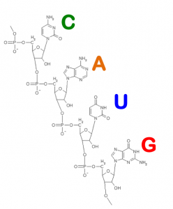
RNA is made of four nucleotides (A, C,G,and U, which stand for adenine, cytosine, guanine, and uracil). Chemically, each of these building blocks is made of atoms of carbon, oxygen, nitrogen, phosphorus, and hydrogen. When you design RNAs with EteRNA, you’re really creating a chain of these nucleotides.

RNA Nucleotides (from the About EteRNA webpage)

Scientists do not yet understand all of RNA’s roles, but we already know about a large collection of RNAs that are critical for life: (see the Thermus Thermophilus image representing following points)

  1. mRNAs are short copies of a cell’s DNA genome that gets cut up, pasted, spliced, and otherwise remixed before getting translated into proteins.
  1. rRNA forms the core machinery of an ancient machine, the ribosome. This machine synthesizes the proteins of your cells and all living cells, and is the target of most antibiotics.
  2. miRNAs (microRNAs) are short molecules (about 22-letters) that are used by all complex cells as commands for silencing genes and appear to have roles in cancer, heart disease, and other medical problems.
  3. Riboswitches are ubiquitous in bacteria. They sense all sorts of small molecules that could be food or signals from other bacteria, and turn on or off genes by changing their shapes. These are interesting targets for new antibiotics.
  4. Ribozymes are RNAs that can act as enzymes. They catalyze chemical reactions like protein synthesis and RNA splicing, and provide evidence of RNA’s dominance in a primordial stage of Life’s evolution.
  5. Retroviruses, like Hepatitis C, poliovirus, and HIV, are very large RNAs coated with proteins.
  6. And much much more… shRNA, piRNA, snRNA, and other new classes of important RNAs are being discovered every year.

Thermus Thermophilus – Large Subunit Ribosomal RNA
Source: Center for Molecular Biology (downloaded from the About EteRNA webpage)

I do wonder about the wordplay EteRNA/eternal. Are these scientists trying to tell us something?