Tag Archives: brains

Fractal brain structures and story listening

For anyone who needs to brush up on their fractals,

Caption: Zoomed in detail of the Mandelbrot set, a famous fractal, at different spatial scales of 1x, 4x, 16x, and 64x (from left to right). Credit: Image by Jeremy R. Manning.

My September 3, 2012 posting (Islands of Benoît Mandelbrot: Fractals, Chaos, and the Materiality of Thinking exhibition opening in Sept. 2012 in New York) includes an explanation of fractals. There is another explanation in the news release that follows below.

The story

A September 30, 2021 Dartmouth College news release announces work from a team of researchers using the concept of fractals as a way of understanding how the brain works (Note: Links have been removed),

Understanding how the human brain produces complex thought is daunting given its intricacy and scale. The brain contains approximately 100 billion neurons that coordinate activity through 100 trillion connections, and those connections are organized into networks that are often similar from one person to the next. A Dartmouth study has found a new way to look at brain networks using the mathematical notion of fractals, to convey communication patterns between different brain regions as people listened to a short story.The results are published in Nature Communications.

“To generate our thoughts, our brains create this amazing lightning storm of connection patterns,” said senior author Jeremy R. Manning, an assistant professor of psychological and brain sciences, and director of the Contextual Dynamics Lab at Dartmouth. “The patterns look beautiful, but they are also incredibly complicated. Our mathematical framework lets us quantify how those patterns relate at different scales, and how they change over time.”

In the field of geometry, fractals are shapes that appear similar at different scales. Within a fractal, shapes and patterns are repeated in an infinite cascade, such as spirals comprised of smaller spirals that are in turn comprised of still-smaller spirals, and so on. Dartmouth’s study shows that brain networks organize in a similar way: patterns of brain interactions are mirrored simultaneously at different scales. When people engage in complex thoughts, their networks seem to spontaneously organize into fractal-like patterns. When those thoughts are disrupted, the fractal patterns become scrambled and lose their integrity.

The researchers developed a mathematical framework that identifies similarities in network interactions at different scales or “orders.” When brain structures do not exhibit any consistent patterns of interaction, the team referred to this as a “zero-order” pattern. When individual pairs of brain structures interact, this is called a “first-order” pattern. “Second-order” patterns refer to similar patterns of interactions in different sets of brain structures, at different scales. When patterns of interaction become fractal— “first-order” or higher— the order denotes the number of times the patterns are repeated at different scales.

The study shows that when people listened to an audio recording of a 10-minute story, their brain networks spontaneously organized into fourth-order network patterns. However, this organization was disrupted when people listened to altered versions of the recording. For example, when the story’s paragraphs were randomly shuffled, preserving some but not all of the story’s meaning, people’s brain networks displayed only second-order patterns. When every word of the story was shuffled, this disrupted all but the lowest level (zero-order) patterns.

“The more finely the story was shuffled, the more the fractal structures of the network patterns were disrupted,” said first author Lucy Owen, a graduate student in psychological and brain sciences at Dartmouth. “Since the disruptions in those fractal patterns seemed directly linked with how well people could make sense of the story, this finding may provide clues about how our brain structures work together to understand what is happening in the narrative.”

The fractal network patterns were surprisingly similar across people: patterns from one group could be used to accurately estimate what part of the story another group was listening to.

The team also studied which brain structures were interacting to produce these fractal patterns. The results show that the smallest scale (first-order) interactions occurred in brain regions that process raw sounds. Second-order interactions linked these raw sounds with speech processing regions, and third-order interactions linked sound and speech areas with a network of visual processing regions. The largest-scale (fourth-order) interactions linked these auditory and visual sensory networks with brain structures that support high-level thinking. According to the researchers, when these networks organize at multiple scales, this may show how the brain processes raw sensory information into complex thought—from raw sounds, to speech, to visualization, to full-on understanding.

The researchers’ computational framework can also be applied to areas beyond neuroscience and the team has already begun using an analogous approach to explore interactions in stock prices and animal migration patterns.

Here’s a link to and a citation for the paper,

High-level cognition during story listening is reflected in high-order dynamic correlations in neural activity patterns by Lucy L. W. Owen, Thomas H. Chang & Jeremy R. Manning. Nature Communications volume 12, Article number: 5728 (2021) DOI: https://doi.org/10.1038/s41467-021-25876-x Published: 30 September 2021

This paper is open access.

Controlling neurons with light: no batteries or wires needed

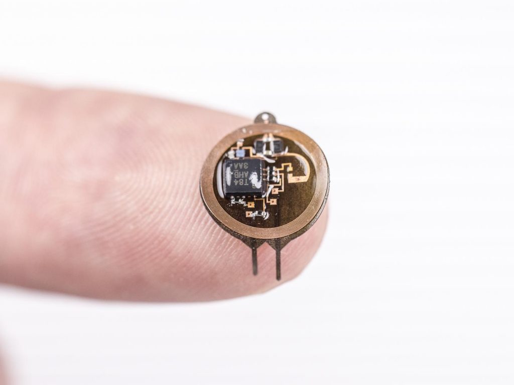
Caption: Wireless and battery-free implant with advanced control over targeted neuron groups. Credit: Philipp Gutruf

This January 2, 2019 news item on ScienceDaily describes the object seen in the above and describes the problem it’s designed to solve,

University of Arizona biomedical engineering professor Philipp Gutruf is first author on the paper Fully implantable, optoelectronic systems for battery-free, multimodal operation in neuroscience research, published in Nature Electronics.

Optogenetics is a biological technique that uses light to turn specific neuron groups in the brain on or off. For example, researchers might use optogenetic stimulation to restore movement in case of paralysis or, in the future, to turn off the areas of the brain or spine that cause pain, eliminating the need for — and the increasing dependence on — opioids and other painkillers.

“We’re making these tools to understand how different parts of the brain work,” Gutruf said. “The advantage with optogenetics is that you have cell specificity: You can target specific groups of neurons and investigate their function and relation in the context of the whole brain.”

In optogenetics, researchers load specific neurons with proteins called opsins, which convert light to electrical potentials that make up the function of a neuron. When a researcher shines light on an area of the brain, it activates only the opsin-loaded neurons.

The first iterations of optogenetics involved sending light to the brain through optical fibers, which meant that test subjects were physically tethered to a control station. Researchers went on to develop a battery-free technique using wireless electronics, which meant subjects could move freely.

But these devices still came with their own limitations — they were bulky and often attached visibly outside the skull, they didn’t allow for precise control of the light’s frequency or intensity, and they could only stimulate one area of the brain at a time.

A Dec. 21, 2018 University of Azrizona news release (published Jan. 2, 2019 on EurekAlert), which originated the news item, discusses the work in more detail,

“With this research, we went two to three steps further,” Gutruf said. “We were able to implement digital control over intensity and frequency of the light being emitted, and the devices are very miniaturized, so they can be implanted under the scalp. We can also independently stimulate multiple places in the brain of the same subject, which also wasn’t possible before.”

The ability to control the light’s intensity is critical because it allows researchers to control exactly how much of the brain the light is affecting — the brighter the light, the farther it will reach. In addition, controlling the light’s intensity means controlling the heat generated by the light sources, and avoiding the accidental activation of neurons that are activated by heat.

The wireless, battery-free implants are powered by external oscillating magnetic fields, and, despite their advanced capabilities, are not significantly larger or heavier than past versions. In addition, a new antenna design has eliminated a problem faced by past versions of optogenetic devices, in which the strength of the signal being transmitted to the device varied depending on the angle of the brain: A subject would turn its head and the signal would weaken.

“This system has two antennas in one enclosure, which we switch the signal back and forth very rapidly so we can power the implant at any orientation,” Gutruf said. “In the future, this technique could provide battery-free implants that provide uninterrupted stimulation without the need to remove or replace the device, resulting in less invasive procedures than current pacemaker or stimulation techniques.”

Devices are implanted with a simple surgical procedure similar to surgeries in which humans are fitted with neurostimulators, or “brain pacemakers.” They cause no adverse effects to subjects, and their functionality doesn’t degrade in the body over time. This could have implications for medical devices like pacemakers, which currently need to be replaced every five to 15 years.

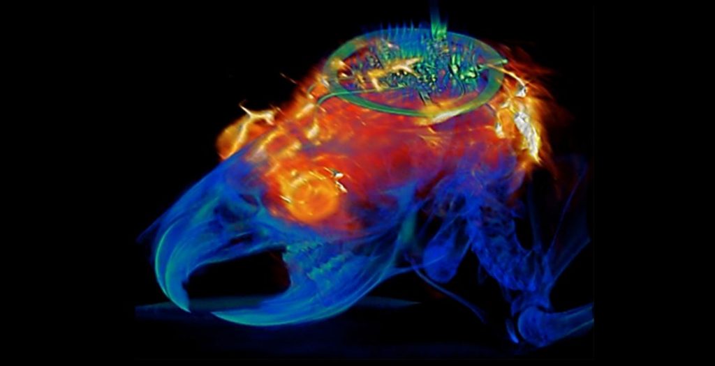
The paper also demonstrated that animals implanted with these devices can be safely imaged with computer tomography, or CT, and magnetic resonance imaging, or MRI, which allow for advanced insights into clinically relevant parameters such as the state of bone and tissue and the placement of the device.

This image of a combined MRI (magnetic resonance image) and CT (computer tomography) scan bookends, more or less, the picture of the device which headed this piece,

Combined image analysis with MRI and CT results superimposed on a 3D rendering of the animal implanted with the programmable bilateral multi µ-ILED device. Courtesy: University of Arizona

Here’s a link to and a citation for the paper,

Fully implantable optoelectronic systems for battery-free, multimodal operation in neuroscience research by Philipp Gutruf, Vaishnavi Krishnamurthi, Abraham Vázquez-Guardado, Zhaoqian Xie, Anthony Banks, Chun-Ju Su, Yeshou Xu, Chad R. Haney, Emily A. Waters, Irawati Kandela, Siddharth R. Krishnan, Tyler Ray, John P. Leshock, Yonggang Huang, Debashis Chanda, & John A. Rogers. Nature Electronics volume 1, pages652–660 (2018) DOI: https://doi.org/10.1038/s41928-018-0175-0 Published 13 December 2018

This paper is behind a paywall.

Walking again with exoskeletons and brain-controlled, non-invasive muscle stimulation enabling people to walk

I have two news bits about paraplegics and the possibility of walking. The first is from Alberta, Canada and the second is from Brazil.

Alberta

The fellow in the video is wearing a robotic exoskeleton. As you can see, it’s not perfect but it represents an extraordinary breakthrough (from an April 16, 2019 article by Sarah Lawrynuik for the Canadian Broadcasting Corporation [CBC] Radio),

On his fifteenth birthday in December 2015, Calgary’s Alex McEwan was injured in a tobogganing accident with friends and lost the ability to walk. It’s the kind of change that could destroy a person, but Alex has thrived and is learning new skills. Watch him walk onstage, with some help from a powered exoskeleton, to receive his diploma. 1:21

Sometimes events conspire to move us in a completely unexpected ways. After his accident, Alex McEwan participated in a very special study (from an August 3, 2019 article by Colin Zak for Alberta Health Services),

Researchers at Foothills Medical Centre (FMC) are the first in Canada to examine the benefits of using an exoskeleton robotic device to rehabilitate patients with spinal cord injuries (SCI) in the days and weeks following their injury.

The device, known as the Ekso Bionic Exoskeleton, consists of a metal frame that supports and stabilizes a patient’s torso, core, legs and feet. It is moved robotically by a therapist, enabling patients with a spinal cord injury to get up and walk around. Although it is controlled by remote control, the device offers varying levels of physical control by the patient, depending on the nature and extent of their injury.

Dr. Ho [ Dr. Chester Ho, Head of Physical Medicine and Rehabilitation at FMC ] says exoskeletons may potentially promote recovery and reduce complications in SCI patients by reducing loss of bone and muscle mass caused by spending so much time lying down, and also improve breathing and bowel function.

The year-long study, which begins this summer, will include between five and 10 patients selected from across Calgary [Alberta]. It aims to examine whether treatment is safe and feasible in the days and weeks after an SCI. This study will be followed by larger studies involving more patients.

Participants in the study will receive 60-minute therapy sessions with the exoskeleton device two to three times a week, for a total of 25 training hours over an eight- to 10-week period. Safety and feasibility outcomes will be monitored and tracked by the research team throughout all sessions.

Before the advent of exoskeletons, rehabilitation for patients with an SCI required them to be hoisted with a physical therapist moving their legs.

“Every step is different with this device, so patients learn from their mistakes in real time. Patients really like to use the device; it gives them hope.”

Alex, [emphasis mine] 15, sustained a spinal cord injury while tobogganing last December.

He says rehabilitation sessions with the exoskeleton have made a difference in how he feels and gives him hope for the future.

Over 2 1/2 years later, the CBC has made a radio documentary about this study and the people who took part. Lawrynuik’s April 16, 2019 article describes some highlights from the radio documentary,

Imagine waking up in a hospital bed surrounded by the beeps and whirring sounds of the machines keeping you alive. The doctor tells you that you will likely never walk again.

But then, just as you begin to process that news, a physiotherapist shows up at your bedside and says, “Hold up. I might have a special opportunity for you.”

That’s the journey taken by a number of Albertans who landed in Calgary’s Foothills Medical Centre after accidents or trauma to their spine in the last three years. Three of those people are Alex McEwan, a university student in Lethbridge; Jean Ogilvie, a 77-year-old woman living in Calgary; and Josh Pelland, a former climber turned motivational speaker in Three Hills, Alta.

All three are united by a technology called an exoskeleton, created by a company called Ekso Bionics, that allowed them to walk despite no longer being able to use their legs. 

“The first time was a bit scary actually,” Ogilvie said. “It’s like a great big skeleton that sort of clasps you in its body. [It’s] black and all sorts of straps and sensors tell you how I’m doing.”

Pelland agrees about how daunting the experience is to start.

“They just said, ‘OK, the machine is going to assist you and lift you up.’ And I was a bit like, ‘OK, this is the strangest thing ever.'”

Once the frame of the exoskeleton is strapped along the outside of the patient’s legs and up their back, starting from the seated position, it does lift them completely without the help of their own muscles.

From there they shift their upper-body weight within the machine to hit certain targets — once your body weight is shifted forward and laterally enough, a beep sounds and the exoskeleton pulls each leg forward, one at a time. 

As patients learn to use the machine, they walk with the assistance of a walker. Then, as they progress, they upgrade to forearm crutches. The entire time, they’re accompanied by the man behind the machine, Kyle McIntosh.

McIntosh is a physiotherapist and he worked with the exoskeleton both to help patients and to conduct research into the machine’s impact on rehabilitation.

After being discharged and living once again without the exoskeleton, and therefore without the ability to walk — McEwan got an idea: maybe he’d be allowed to use the robot, just one last time.

“High school wasn’t high school for me. I only really got one semester of grade 10 before I broke my spine. So that first semester was great. I enjoyed it. I played sports. I was a good student. But then it was no longer about high school anymore. It was more about adjusting to my new life.”

McIntosh and McEwan hatched the plan together and kept it a closely guarded secret. Then, on the day McEwan was set to graduate from Grade 12, he asked to be placed last on the list of students to cross the stage.

“I remember taking a first few steps and not hearing very much. Hearing people cheer because I was the kid in the wheelchair at the high school, so it makes sense. But the second they saw the canes and my first few steps, just one kid erupted: ‘Yeah!’ And then everyone went crazy.”

“I think walking across the stage — just like I got to walk into my high school on the first day of Grade 10 — was a really good closing story. The chapter of me learning to live in a wheelchair was done. And it was now my turn to go live my life. So that’s why I think it was such an important day because it gave me a lot of closure. I got to walk into the high school, I got to walk out.”

If you have the time, you might want to read Lawrynuik’s April 16, 2019 article in its entirety. It turns out that the study did much more than give a people a chance to walk again, even if just for a short time.

Anyone interested in the robotic, wearable exoskeleton used in the study can go here to EksoHealth, the company that produces the EksoGT, a bionic exoskeleton. (Lawrynuik’s article has another name for the product, i.e., Ekso Bionic Exoskeleton but all I could find was the EksoGT.)

Brazil and Walk Again

The most recent post featuring the Walk Again project is my May 20, 2014 edition which was part of a larger series on ‘Brain research, ethics, and nanotechnology’. The May 20, 2014 posting covered Walk Again’s debut at the 2014 World Cup (soccer/football) in Brazil. Unfortunately,, the lead researcher Miguel Nicolelis oversold the technology. I think people were expecting someone with paraplegia to come bounding out onto the field and give a flashy opening kick for the tournament what they saw was something a great deal more restrained.

The person was wheeled out onto the field, stood up, shuffled a bit, and nudged the ball with his foot. It represented a huge breakthrough but it wasn’t flashy.

The latest from Walk Again is in a May 14, 2019 Associação Alberto Santos Dumont para Apoio à Pesquisa press release on EurekAlert,

In another major clinical breakthrough of the Walk Again Project, a non-profit international consortium aimed at developing new neuro-rehabilitation protocols, technologies and therapies for spinal cord injury, two patients with paraplegia regained the ability to walk with minimal assistance, through the employment of a fully non-invasive brain-machine interface that does not require the use of any invasive spinal cord surgical procedure. The results of this study appeared on the May 1 [2019] issue of the journal Scientific Reports.

The two patients with paraplegia (AIS C) used their own brain activity to control the non-invasive delivery of electrical pulses to a total of 16 muscles (eight in each leg), allowing them to produce a more physiological walk than previously reported, requiring only a conventional walker and a body weight support system as assistive devices. Overall, the two patients were able to produce more than 4,500 steps using this new technology, which combines a non-invasive brain-machine interface, based on a 16-channel EEG, to control a multi-channel functional electrical stimulation system (FES), tailored to produce a much smoother gait pattern than the state of the art of this technique.

“What surprised us was that, in addition to allowing these patients to walk with little help, one of them displayed a clear motor improvement by practicing with this new approach. Patients required approximatively [sic] 25 sessions to master the training before they were able to walk using this apparatus,” said Solaiman Shokur one of the authors of the study.

The two patients that used this new rehabilitation approach had previously participated in the long-term neurorehabilitation study carried out using the Walk Again Project Neurorehabilitation (WANR) protocol. As reported in a recent publication from the same team (Shokur et al., PLoS One, Nov. 2018), all seven patients who participated in that protocol for a period of 28 months improved their clinical status, from complete paraplegia (AIS A or B, meaning no motor functions below the level of the injury, according to the ASIA classification) to partial paraplegia (AIS C, meaning partial recovery of sensory and motor function below the injury level). This significant neurological recovery included major clinical improvements in sensory discrimination (tactile, nociception, vibration, and pressure), voluntary motor control of abdomen and leg muscles, and important gains in autonomic control, such as bladder, bowel, and sexual functions.

“The last two studies published by the Walk Again Project clearly indicate that partial neurological and functional recovery can be induced in chronic spinal cord injury patients by combining multiple non-invasive technologies that are based around the concept of using a brain-machine interface to control different types of actuators, like virtual avatars, robotic walkers, or muscle stimulating devices, to allow the total involvement of patients in their own rehabilitation routine,” said Miguel Nicolelis, scientific director of the Walk Again Project and one of the authors of the study.

In a recent report by another group, one AIS C and two AIS D patients were able to walk thanks to the employment of an invasive method for spinal cord electrical stimulation, which required a spinal surgical procedure. In contrast, in the present study two AIS C patients – which originally were AIS A (see Supplemental Material below)- and a third AIS B subject, who recently achieved similar results, were able to regain a significant degree of autonomous walking without the need for such invasive treatments. Instead, these patients only received electrical stimulation patterns delivered to the skin surface of their legs, so that a total of eight muscles in each limb could be electrically stimulated in a physiologically accurate sequence. This was done in order to produce a smoother and more natural pattern of locomotion.

“Crucial for this implementation was the development of a closed-loop controller that allowed real-time correction of the patients’ walking pattern, taking into account muscle fatigue and external perturbations, in order to produce a predefined gait trajectory. Another major component of our approach was the use of a wearable haptic display to deliver tactile feedback to the patients´ forearms in order to provide them with a continuous source of proprioceptive feedback related to their walking,” said Solaiman Shokur.

To control the pattern of electrical muscle stimulation in each leg, these patients utilized an EEG-based brain-machine interface. In this setup, patients learned to alternate the generation of “stepping motor imagery” activity in their right and left motor cortices, in order to create alternated movements of their left and right legs.

According to the authors, the patients exhibited not only “less dependency on walking assistance, but also partial neurological recovery, with substantial rates of motor improvement in one of them.” The improvement in motor control in this last AIS C patient was 9 points in the lower extremity motor score (LEMS), which was comparable with that observed using invasive spinal cord stimulation.

Based on the results obtained over the past 5 years, the WAP now intends to combine all its neurorehabilitation tools into a single integrated, non-invasive platform to treat spinal cord injury patients. This platform will allow patients to begin training soon after the injury occurs. It will also allow the employment of a multi-dimensional integrated brain-machine interface capable of simultaneously controlling virtual and robotic actuators (like a lowerlimb exoskeleton), a multi-channel non-invasive electrical muscle stimulation system (like the FES used in the present study), and a novel non-invasive spinal cord stimulation approach. In this final configuration, this WAP platform will incorporate all these technologies together in order to maximize neurological and functional recovery in the shortest possible time, without the need of any invasive procedure.

According to Dr. Nicolelis, “there is no silver bullet to treat spinal cord injuries. More and more, it looks like we need to implement multiple techniques simultaneously to achieve the best neurorehabilitation results. In this context, it is also imperative to consider the occurrence of cortical plasticity as a major component in the planning of our rehabilitation approach.”

Here’s a link to and a citation for the paper,

Non-invasive, Brain-controlled Functional Electrical Stimulation for Locomotion Rehabilitation in Individuals with Paraplegia by Aurelie Selfslagh, Solaiman Shokur, Debora S. F. Campos, Ana R. C. Donati, Sabrina Almeida, Seidi Y. Yamauti, Daniel B. Coelho, Mohamed Bouri & Miguel A. L. Nicolelis. Scientific Reports volume 9, Article number: 6782 (2019) DOI: https://doi.org/10.1038/s41598-019-43041-9 Published 01 May 2019

This paper is open access.

There’s also a video for Walk Again,

Transparent graphene electrode technology and complex brain imaging

Michael Berger has written a May 24, 2018 Nanowerk Spotlight article about some of the latest research on transparent graphene electrode technology and the brain (Note: A link has been removed),

In new work, scientists from the labs of Kuzum [Duygu Kuzum, an Assistant Professor of Electrical and Computer Engineering at the University of California, San Diego {UCSD}] and Anna Devor report a transparent graphene microelectrode neural implant that eliminates light-induced artifacts to enable crosstalk-free integration of 2-photon microscopy, optogenetic stimulation, and cortical recordings in the same in vivo experiment. The new class of transparent brain implant is based on monolayer graphene. It offers a practical pathway to investigate neuronal activity over multiple spatial scales extending from single neurons to large neuronal populations.

Conventional metal-based microelectrodes cannot be used for simultaneous measurements of multiple optical and electrical parameters, which are essential for comprehensive investigation of brain function across spatio-temporal scales. Since they are opaque, they block the field of view of the microscopes and generate optical shadows impeding imaging.

More importantly, they cause light induced artifacts in electrical recordings, which can significantly interfere with neural signals. Transparent graphene electrode technology presented in this paper addresses these problems and allow seamless and crosstalk-free integration of optical and electrical sensing and manipulation technologies.

In their work, the scientists demonstrate that by careful design of key steps in the fabrication process for transparent graphene electrodes, the light-induced artifact problem can be mitigated and virtually artifact-free local field potential (LFP) recordings can be achieved within operating light intensities.

“Optical transparency of graphene enables seamless integration of imaging, optogenetic stimulation and electrical recording of brain activity in the same experiment with animal models,” Kuzum explains. “Different from conventional implants based on metal electrodes, graphene-based electrodes do not generate any electrical artifacts upon interacting with light used for imaging or optogenetics. That enables crosstalk free integration of three modalities: imaging, stimulation and recording to investigate brain activity over multiple spatial scales extending from single neurons to large populations of neurons in the same experiment.”

The team’s new fabrication process avoids any crack formation in the transfer process, resulting in a 95-100% yield for the electrode arrays. This fabrication quality is important for expanding this technology to high-density large area transparent arrays to monitor brain-scale cortical activity in large animal models or humans.

“Our technology is also well-suited for neurovascular and neurometabolic studies, providing a ‘gold standard’ neuronal correlate for optical measurements of vascular, hemodynamic, and metabolic activity,” Kuzum points out. “It will find application in multiple areas, advancing our understanding of how microscopic neural activity at the cellular scale translates into macroscopic activity of large neuron populations.”

“Combining optical techniques with electrical recordings using graphene electrodes will allow to connect the large body of neuroscience knowledge obtained from animal models to human studies mainly relying on electrophysiological recordings of brain-scale activity,” she adds.

Next steps for the team involve employing this technology to investigate coupling and information transfer between different brain regions.

This work is part of the US BRAIN (Brain Research through Advancing Innovative Neurotechnologies) initiative and there’s more than one team working with transparent graphene electrodes. John Hewitt in an Oct. 21, 2014 posting on ExtremeTech describes two other teams’ work (Note: Links have been removed),

The solution [to the problems with metal electrodes], now emerging from multiple labs throughout the universe is to build flexible, transparent electrode arrays from graphene. Two studies in the latest issue of Nature Communications, one from the University of Wisconsin-Madison and the other from Penn [University of Pennsylvania], describe how to build these devices.

The University of Wisconsin researchers are either a little bit smarter or just a little bit richer, because they published their work open access. It’s a no-brainer then that we will focus on their methods first, and also in more detail. To make the arrays, these guys first deposited the parylene (polymer) substrate on a silicon wafer, metalized it with gold, and then patterned it with an electron beam to create small contact pads. The magic was to then apply four stacked single-atom-thick graphene layers using a wet transfer technique. These layers were then protected with a silicon dioxide layer, another parylene layer, and finally molded into brain signal recording goodness with reactive ion etching.

PennTransparentelectrodeThe researchers went with four graphene layers because that provided optimal mechanical integrity and conductivity while maintaining sufficient transparency. They tested the device in opto-enhanced mice whose neurons expressed proteins that react to blue light. When they hit the neurons with a laser fired in through the implant, the protein channels opened and fired the cell beneath. The masterstroke that remained was then to successfully record the electrical signals from this firing, sit back, and wait for the Nobel prize office to call.

The Penn State group [Note: Every reearcher mentioned in the paper Hewitt linked to is from the University of Pennsylvania] in the  used a similar 16-spot electrode array (pictured above right), and proceeded — we presume — in much the same fashion. Their angle was to perform high-resolution optical imaging, in particular calcium imaging, right out through the transparent electrode arrays which simultaneously recorded in high-temporal-resolution signals. They did this in slices of the hippocampus where they could bring to bear the complex and multifarious hardware needed to perform confocal and two-photon microscopy. These latter techniques provide a boost in spatial resolution by zeroing in over narrow planes inside the specimen, and limiting the background by the requirement of two photons to generate an optical signal. We should mention that there are voltage sensitive dyes available, in addition to standard calcium dyes, which can almost record the fastest single spikes, but electrical recording still reigns supreme for speed.

What a mouse looks like with an optogenetics system plugged in

What a mouse looks like with an optogenetics system plugged in

One concern of both groups in making these kinds of simultaneous electro-optic measurements was the generation of light-induced artifacts in the electrical recordings. This potential complication, called the Becqueral photovoltaic effect, has been known to exist since it was first demonstrated back in 1839. When light hits a conventional metal electrode, a photoelectrochemical (or more simply, a photovoltaic) effect occurs. If present in these recordings, the different signals could be highly disambiguatable. The Penn researchers reported that they saw no significant artifact, while the Wisconsin researchers saw some small effects with their device. In particular, when compared with platinum electrodes put into the opposite side cortical hemisphere, the Wisconsin researchers found that the artifact from graphene was similar to that obtained from platinum electrodes.

Here’s a link to and a citation for the latest research from UCSD,

Deep 2-photon imaging and artifact-free optogenetics through transparent graphene microelectrode arrays by Martin Thunemann, Yichen Lu, Xin Liu, Kıvılcım Kılıç, Michèle Desjardins, Matthieu Vandenberghe, Sanaz Sadegh, Payam A. Saisan, Qun Cheng, Kimberly L. Weldy, Hongming Lyu, Srdjan Djurovic, Ole A. Andreassen, Anders M. Dale, Anna Devor, & Duygu Kuzum. Nature Communicationsvolume 9, Article number: 2035 (2018) doi:10.1038/s41467-018-04457-5 Published: 23 May 2018

This paper is open access.

You can find out more about the US BRAIN initiative here and if you’re curious, you can find out more about the project at UCSD here. Duygu Kuzum (now at UCSD) was at  the University of Pennsylvania in 2014 and participated in the work mentioned in Hewitt’s 2014 posting.

Injectable bandages for internal bleeding and hydrogel for the brain

This injectable bandage could be a gamechanger (as they say) if it can be taken beyond the ‘in vitro’ (i.e., petri dish) testing stage. A May 22, 2018 news item on Nanowerk makes the announcement (Note: A link has been removed),

While several products are available to quickly seal surface wounds, rapidly stopping fatal internal bleeding has proven more difficult. Now researchers from the Department of Biomedical Engineering at Texas A&M University are developing an injectable hydrogel bandage that could save lives in emergencies such as penetrating shrapnel wounds on the battlefield (Acta Biomaterialia, “Nanoengineered injectable hydrogels for wound healing application”).

A May 22, 2018 US National Institute of Biomedical Engineering and Bioengiineering news release, which originated the news item, provides more detail (Note: Links have been removed),

The researchers combined a hydrogel base (a water-swollen polymer) and nanoparticles that interact with the body’s natural blood-clotting mechanism. “The hydrogel expands to rapidly fill puncture wounds and stop blood loss,” explained Akhilesh Gaharwar, Ph.D., assistant professor and senior investigator on the work. “The surface of the nanoparticles attracts blood platelets that become activated and start the natural clotting cascade of the body.”

Enhanced clotting when the nanoparticles were added to the hydrogel was confirmed by standard laboratory blood clotting tests. Clotting time was reduced from eight minutes to six minutes when the hydrogel was introduced into the mixture. When nanoparticles were added, clotting time was significantly reduced, to less than three minutes.

In addition to the rapid clotting mechanism of the hydrogel composite, the engineers took advantage of special properties of the nanoparticle component. They found they could use the electric charge of the nanoparticles to add growth factors that efficiently adhered to the particles. “Stopping fatal bleeding rapidly was the goal of our work,” said Gaharwar. “However, we found that we could attach growth factors to the nanoparticles. This was an added bonus because the growth factors act to begin the body’s natural wound healing process—the next step needed after bleeding has stopped.”

The researchers were able to attach vascular endothelial growth factor (VEGF) to the nanoparticles. They tested the hydrogel/nanoparticle/VEGF combination in a cell culture test that mimics the wound healing process. The test uses a petri dish with a layer of endothelial cells on the surface that create a solid skin-like sheet. The sheet is then scratched down the center creating a rip or hole in the sheet that resembles a wound.

When the hydrogel containing VEGF bound to the nanoparticles was added to the damaged endothelial cell wound, the cells were induced to grow back and fill-in the scratched region—essentially mimicking the healing of a wound.

“Our laboratory experiments have verified the effectiveness of the hydrogel for initiating both blood clotting and wound healing,” said Gaharwar. “We are anxious to begin tests in animals with the hope of testing and eventual use in humans where we believe our formulation has great potential to have a significant impact on saving lives in critical situations.”

The work was funded by grant EB023454 from the National Institute of Biomedical Imaging and Bioengineering (NIBIB), and the National Science Foundation. The results were reported in the February issue of the journal Acta Biomaterialia.

The paper was published back in April 2018 and there was an April 2, 2018 Texas A&M University news release on EurekAlert making the announcement (and providing a few unique details),

A penetrating injury from shrapnel is a serious obstacle in overcoming battlefield wounds that can ultimately lead to death.Given the high mortality rates due to hemorrhaging, there is an unmet need to quickly self-administer materials that prevent fatality due to excessive blood loss.

With a gelling agent commonly used in preparing pastries, researchers from the Inspired Nanomaterials and Tissue Engineering Laboratory have successfully fabricated an injectable bandage to stop bleeding and promote wound healing.

In a recent article “Nanoengineered Injectable Hydrogels for Wound Healing Application” published in Acta Biomaterialia, Dr. Akhilesh K. Gaharwar, assistant professor in the Department of Biomedical Engineering at Texas A&M University, uses kappa-carrageenan and nanosilicates to form injectable hydrogels to promote hemostasis (the process to stop bleeding) and facilitate wound healing via a controlled release of therapeutics.

“Injectable hydrogels are promising materials for achieving hemostasis in case of internal injuries and bleeding, as these biomaterials can be introduced into a wound site using minimally invasive approaches,” said Gaharwar. “An ideal injectable bandage should solidify after injection in the wound area and promote a natural clotting cascade. In addition, the injectable bandage should initiate wound healing response after achieving hemostasis.”

The study uses a commonly used thickening agent known as kappa-carrageenan, obtained from seaweed, to design injectable hydrogels. Hydrogels are a 3-D water swollen polymer network, similar to Jell-O, simulating the structure of human tissues.

When kappa-carrageenan is mixed with clay-based nanoparticles, injectable gelatin is obtained. The charged characteristics of clay-based nanoparticles provide hemostatic ability to the hydrogels. Specifically, plasma protein and platelets form blood adsorption on the gel surface and trigger a blood clotting cascade.

“Interestingly, we also found that these injectable bandages can show a prolonged release of therapeutics that can be used to heal the wound” said Giriraj Lokhande, a graduate student in Gaharwar’s lab and first author of the paper. “The negative surface charge of nanoparticles enabled electrostatic interactions with therapeutics thus resulting in the slow release of therapeutics.”

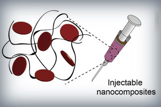
Nanoparticles that promote blood clotting and wound healing (red discs), attached to the wound-filling hydrogel component (black) form a nanocomposite hydrogel. The gel is designed to be self-administered to stop bleeding and begin wound-healing in emergency situations. Credit: Lokhande, et al. 1

Here’s a link to and a citation for the paper,

Nanoengineered injectable hydrogels for wound healing application by Giriraj Lokhande, James K. Carrow, Teena Thakur, Janet R. Xavier, Madasamy Parani, Kayla J. Bayless, Akhilesh K. Gaharwar. Acta Biomaterialia Volume 70, 1 April 2018, Pages 35-47
https://doi.org/10.1016/j.actbio.2018.01.045

This paper is behind a paywall.

Hydrogel and the brain

It’s been an interesting week for hydrogels. On May 21, 2018 there was a news item on ScienceDaily about a bioengineered hydrogel which stimulated brain tissue growth after a stroke (mouse model),

In a first-of-its-kind finding, a new stroke-healing gel helped regrow neurons and blood vessels in mice with stroke-damaged brains, UCLA researchers report in the May 21 issue of Nature Materials.

“We tested this in laboratory mice to determine if it would repair the brain in a model of stroke, and lead to recovery,” said Dr. S. Thomas Carmichael, Professor and Chair of neurology at UCLA. “This study indicated that new brain tissue can be regenerated in what was previously just an inactive brain scar after stroke.”

The brain has a limited capacity for recovery after stroke and other diseases. Unlike some other organs in the body, such as the liver or skin, the brain does not regenerate new connections, blood vessels or new tissue structures. Tissue that dies in the brain from stroke is absorbed, leaving a cavity, devoid of blood vessels, neurons or axons, the thin nerve fibers that project from neurons.

After 16 weeks, stroke cavities in mice contained regenerated brain tissue, including new neural networks — a result that had not been seen before. The mice with new neurons showed improved motor behavior, though the exact mechanism wasn’t clear.

Remarkable stuff.

Crowdsourcing brain research at Princeton University to discover 6 new neuron types

Spritely music!

There were already 1/4M registered players as of May 17, 2018 but I’m sure there’s room for more should you be inspired. A May 17, 2018 Princeton University news release (also on EurekAlert) reveals more about the game and about the neurons,

With the help of a quarter-million video game players, Princeton researchers have created and shared detailed maps of more than 1,000 neurons — and they’re just getting started.

“Working with Eyewirers around the world, we’ve made a digital museum that shows off the intricate beauty of the retina’s neural circuits,” said Sebastian Seung, the Evnin Professor in Neuroscience and a professor of computer science and the Princeton Neuroscience Institute (PNI). The related paper is publishing May 17 [2018] in the journal Cell.

Seung is unveiling the Eyewire Museum, an interactive archive of neurons available to the general public and neuroscientists around the world, including the hundreds of researchers involved in the federal Brain Research through Advancing Innovative Neurotechnologies (BRAIN) Initiative.

“This interactive viewer is a huge asset for these larger collaborations, especially among people who are not physically in the same lab,” said Amy Robinson Sterling, a crowdsourcing specialist with PNI and the executive director of Eyewire, the online gaming platform for the citizen scientists who have created this data set.

“This museum is something like a brain atlas,” said Alexander Bae, a graduate student in electrical engineering and one of four co-first authors on the paper. “Previous brain atlases didn’t have a function where you could visualize by individual cell, or a subset of cells, and interact with them. Another novelty: Not only do we have the morphology of each cell, but we also have the functional data, too.”

The neural maps were developed by Eyewirers, members of an online community of video game players who have devoted hundreds of thousands of hours to painstakingly piecing together these neural cells, using data from a mouse retina gathered in 2009.

Eyewire pairs machine learning with gamers who trace the twisting and branching paths of each neuron. Humans are better at visually identifying the patterns of neurons, so every player’s moves are recorded and checked against each other by advanced players and Eyewire staffers, as well as by software that is improving its own pattern recognition skills.

Since Eyewire’s launch in 2012, more than 265,000 people have signed onto the game, and they’ve collectively colored in more than 10 million 3-D “cubes,” resulting in the mapping of more than 3,000 neural cells, of which about a thousand are displayed in the museum.

Each cube is a tiny subset of a single cell, about 4.5 microns across, so a 10-by-10 block of cubes would be the width of a human hair. Every cell is reviewed by between 5 and 25 gamers before it is accepted into the system as complete.

“Back in the early years it took weeks to finish a single cell,” said Sterling. “Now players complete multiple neurons per day.” The Eyewire user experience stays focused on the larger mission — “For science!” is a common refrain — but it also replicates a typical gaming environment, with achievement badges, a chat feature to connect with other players and technical support, and the ability to unlock privileges with increasing skill. “Our top players are online all the time — easily 30 hours a week,” Sterling said.

Dedicated Eyewirers have also contributed in other ways, including donating the swag that gamers win during competitions and writing program extensions “to make game play more efficient and more fun,” said Sterling, including profile histories, maps of player activity, a top 100 leaderboard and ever-increasing levels of customizability.

“The community has really been the driving force behind why Eyewire has been successful,” Sterling said. “You come in, and you’re not alone. Right now, there are 43 people online. Some of them will be admins from Boston or Princeton, but most are just playing — now it’s 46.”

For science!

With 100 billion neurons linked together via trillions of connections, the brain is immeasurably complex, and neuroscientists are still assembling its “parts list,” said Nicholas Turner, a graduate student in computer science and another of the co-first authors. “If you know what parts make up the machine you’re trying to break apart, you’re set to figure out how it all works,” he said.

The researchers have started by tackling Eyewire-mapped ganglion cells from the retina of a mouse. “The retina doesn’t just sense light,” Seung said. “Neural circuits in the retina perform the first steps of visual perception.”

The retina grows from the same embryonic tissue as the brain, and while much simpler than the brain, it is still surprisingly complex, Turner said. “Hammering out these details is a really valuable effort,” he said, “showing the depth and complexity that exists in circuits that we naively believe are simple.”

The researchers’ fundamental question is identifying exactly how the retina works, said Bae. “In our case, we focus on the structural morphology of the retinal ganglion cells.”

“Why the ganglion cells of the eye?” asked Shang Mu, an associate research scholar in PNI and fellow first author. “Because they’re the connection between the retina and the brain. They’re the only cell class that go back into the brain.” Different types of ganglion cells are known to compute different types of visual features, which is one reason the museum has linked shape to functional data.

Using Eyewire-produced maps of 396 ganglion cells, the researchers in Seung’s lab successfully classified these cells more thoroughly than has ever been done before.

“The number of different cell types was a surprise,” said Mu. “Just a few years ago, people thought there were only 15 to 20 ganglion cell types, but we found more than 35 — we estimate between 35 and 50 types.”

Of those, six appear to be novel, in that the researchers could not find any matching descriptions in a literature search.

A brief scroll through the digital museum reveals just how remarkably flat the neurons are — nearly all of the branching takes place along a two-dimensional plane. Seung’s team discovered that different cells grow along different planes, with some reaching high above the nucleus before branching out, while others spread out close to the nucleus. Their resulting diagrams resemble a rainforest, with ground cover, an understory, a canopy and an emergent layer overtopping the rest.

All of these are subdivisions of the inner plexiform layer, one of the five previously recognized layers of the retina. The researchers also identified a “density conservation principle” that they used to distinguish types of neurons.

One of the biggest surprises of the research project has been the extraordinary richness of the original sample, said Seung. “There’s a little sliver of a mouse retina, and almost 10 years later, we’re still learning things from it.”

Of course, it’s a mouse’s brain that you’ll be examining and while there are differences between a mouse brain and a human brain, mouse brains still provide valuable data as they did in the case of some groundbreaking research published in October 2017. James Hamblin wrote about it in an Oct. 7, 2017 article for The Atlantic (Note: Links have been removed),


Scientists Somehow Just Discovered a New System of Vessels in Our Brains

It is unclear what they do—but they likely play a central role in aging and disease.

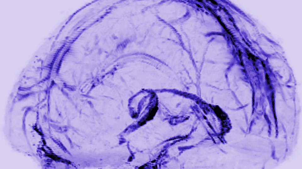
A transparent model of the brain with a network of vessels filled in
Daniel Reich / National Institute of Neurological Disorders and Stroke

You are now among the first people to see the brain’s lymphatic system. The vessels in the photo above transport fluid that is likely crucial to metabolic and inflammatory processes. Until now, no one knew for sure that they existed.

Doctors practicing today have been taught that there are no lymphatic vessels inside the skull. Those deep-purple vessels were seen for the first time in images published this week by researchers at the U.S. National Institute of Neurological Disorders and Stroke.

In the rest of the body, the lymphatic system collects and drains the fluid that bathes our cells, in the process exporting their waste. It also serves as a conduit for immune cells, which go out into the body looking for adversaries and learning how to distinguish self from other, and then travel back to lymph nodes and organs through lymphatic vessels.

So how was it even conceivable that this process wasn’t happening in our brains?

Reich (Daniel Reich, senior investigator) started his search in 2015, after a major study in Nature reported a similar conduit for lymph in mice. The University of Virginia team wrote at the time, “The discovery of the central-nervous-system lymphatic system may call for a reassessment of basic assumptions in neuroimmunology.” The study was regarded as a potential breakthrough in understanding how neurodegenerative disease is associated with the immune system.

Around the same time, researchers discovered fluid in the brains of mice and humans that would become known as the “glymphatic system.” [emphasis mine] It was described by a team at the University of Rochester in 2015 as not just the brain’s “waste-clearance system,” but as potentially helping fuel the brain by transporting glucose, lipids, amino acids, and neurotransmitters. Although since “the central nervous system completely lacks conventional lymphatic vessels,” the researchers wrote at the time, it remained unclear how this fluid communicated with the rest of the body.

There are occasional references to the idea of a lymphatic system in the brain in historic literature. Two centuries ago, the anatomist Paolo Mascagni made full-body models of the lymphatic system that included the brain, though this was dismissed as an error. [emphases mine]  A historical account in The Lancet in 2003 read: “Mascagni was probably so impressed with the lymphatic system that he saw lymph vessels even where they did not exist—in the brain.”

I couldn’t resist the reference to someone whose work had been dismissed summarily being proved right, eventually, and with the help of mouse brains. Do read Hamblin’s article in its entirety if you have time as these excerpts don’t do it justice.

Getting back to Princeton’s research, here’s their research paper,

Digital museum of retinal ganglion cells with dense anatomy and physiology,” by Alexander Bae, Shang Mu, Jinseop Kim, Nicholas Turner, Ignacio Tartavull, Nico Kemnitz, Chris Jordan, Alex Norton, William Silversmith, Rachel Prentki, Marissa Sorek, Celia David, Devon Jones, Doug Bland, Amy Sterling, Jungman Park, Kevin Briggman, Sebastian Seung and the Eyewirers, was published May 17 in the journal Cell with DOI 10.1016/j.cell.2018.04.040.

The research was supported by the Gatsby Charitable Foundation, National Institute of Health-National Institute of Neurological Disorders and Stroke (U01NS090562 and 5R01NS076467), Defense Advanced Research Projects Agency (HR0011-14-2- 0004), Army Research Office (W911NF-12-1-0594), Intelligence Advanced Research Projects Activity (D16PC00005), KT Corporation, Amazon Web Services Research Grants, Korea Brain Research Institute (2231-415) and Korea National Research Foundation Brain Research Program (2017M3C7A1048086).

This paper is behind a paywall. For the players amongst us, here’s the Eyewire website. Go forth,  play, and, maybe, discover new neurons!

Yes! Art, genetic modifications, gene editing, and xenotransplantation at the Vancouver Biennale (Canada)

Patricia Piccinini’s Curious Imaginings Courtesy: Vancouver Biennale [downloaded from http://dailyhive.com/vancouver/vancouver-biennale-unsual-public-art-2018/]

Up to this point, I’ve been a little jealous of the Art/Sci Salon’s (Toronto, Canada) January 2018 workshops for artists and discussions about CRISPR ((clustered regularly interspaced short palindromic repeats))/Cas9 and its social implications. (See my January 10, 2018 posting for more about the events.) Now, it seems Vancouver may be in line for its ‘own’ discussion about CRISPR and the implications of gene editing. The image you saw (above) represents one of the installations being hosted by the 2018 – 2020 edition of the Vancouver Biennale.

While this posting is mostly about the Biennale and Piccinini’s work, there is a ‘science’ subsection featuring the science of CRISPR and xenotransplantation. Getting back to the Biennale and Piccinini: A major public art event since 1988, the Vancouver Biennale has hosted over 91 outdoor sculptures and new media works by more than 78 participating artists from over 25 countries and from 4 continents.

Quickie description of the 2018 – 2020 Vancouver Biennale

The latest edition of the Vancouver Biennale was featured in a June 6, 2018 news item on the Daily Hive (Vancouver),

The Vancouver Biennale will be bringing new —and unusual— works of public art to the city beginning this June.

The theme for this season’s Vancouver Biennale exhibition is “re-IMAGE-n” and it kicks off on June 20 [2018] in Vanier Park with Saudi artist Ajlan Gharem’s Paradise Has Many Gates.

Gharem’s architectural chain-link sculpture resembles a traditional mosque, the piece is meant to challenge the notions of religious orthodoxy and encourages individuals to image a space free of Islamophobia.

Melbourne artist Patricia Piccinini’s Curious Imaginings is expected to be one of the most talked about installations of the exhibit. Her style of “oddly captivating, somewhat grotesque, human-animal hybrid creature” is meant to be shocking and thought-provoking.

Piccinini’s interactive [emphasis mine] experience will “challenge us to explore the social impacts of emerging biotechnology and our ethical limits in an age where genetic engineering and digital technologies are already pushing the boundaries of humanity.”

Piccinini’s work will be displayed in the 105-year-old Patricia Hotel in Vancouver’s Strathcona neighbourhood. The 90-day ticketed exhibition [emphasis mine] is scheduled to open this September [2018].

Given that this blog is focused on nanotechnology and other emerging technologies such as CRISPR, I’m focusing on Piccinini’s work and its art/science or sci-art status. This image from the GOMA Gallery where Piccinini’s ‘Curious Affection‘ installation is being shown from March 24 – Aug. 5, 2018 in Brisbane, Queensland, Australia may give you some sense of what one of her installations is like,

Courtesy: Queensland Art Gallery | Gallery of Modern Art (QAGOMA)

I spoke with Serena at the Vancouver Biennale office and asked about the ‘interactive’ aspect of Piccinini’s installation. She suggested the term ‘immersive’ as an alternative. In other words, you won’t be playing with the sculptures or pressing buttons and interacting with computer screens or robots. She also noted that the ticket prices have not been set yet and they are currently developing events focused on the issues raised by the installation. She knew that 2018 is the 200th anniversary of the publication of Mary Shelley’s Frankenstein but I’m not sure how the Biennale folks plan (or don’t plan)  to integrate any recognition of the novle’s impact on the discussions about ‘new’ technologies .They expect Piccinini will visit Vancouver. (Note 1: Piccinini’s work can  also be seen in a group exhibition titled: Frankenstein’s Birthday Party at the Hosfselt Gallery in San Francisco (California, US) from June 23 – August 11, 2018.  Note 2: I featured a number of international events commemorating the 200th anniversary of the publication of Mary Shelley’s novel, Frankenstein, in my Feb. 26, 2018 posting. Note 3: The term ‘Frankenfoods’ helped to shape the discussion of genetically modified organisms and food supply on this planet. It was a wildly successful campaign for activists affecting legislation in some areas of research. Scientists have not been as enthusiastic about the effects. My January 15, 2009 posting briefly traces a history of the term.)

The 2018 – 2020 Vancouver Biennale and science

A June 7, 2018 Vancouver Biennale news release provides more detail about the current series of exhibitions,

The Biennale is also committed to presenting artwork at the cutting edge of discussion and in keeping with the STEAM (science, technology, engineering, arts, math[ematics]) approach to integrating the arts and sciences. In August [2018], Colombian/American visual artist Jessica Angel will present her monumental installation Dogethereum Bridge at Hinge Park in Olympic Village. Inspired by blockchain technology, the artwork’s design was created through the integration of scientific algorithms, new developments in technology, and the arts. This installation, which will serve as an immersive space and collaborative hub for artists and technologists, will host a series of activations with blockchain as the inspirational jumping-off point.

In what is expected to become one of North America’s most talked-about exhibitions of the year, Melbourne artist Patricia Piccinini’s Curious Imaginings will see the intersection of art, science, and ethics. For the first time in the Biennale’s fifteen years of creating transformative experiences, and in keeping with the 2018-2020 theme of “re-IMAGE-n,” the Biennale will explore art in unexpected places by exhibiting in unconventional interior spaces.  The hyperrealist “world of oddly captivating, somewhat grotesque, human-animal hybrid creatures” will be the artist’s first exhibit in a non-museum setting, transforming a wing of the 105-year-old Patricia Hotel. Situated in Vancouver’s oldest neighbourbood of Strathcona, Piccinini’s interactive experience will “challenge us to explore the social impacts of emerging bio-technology and our ethical limits in an age where genetic engineering and digital technologies are already pushing the boundaries of humanity.” In this intimate hotel setting located in a neighborhood continually undergoing its own change, Curious Imaginings will empower visitors to personally consider questions posed by the exhibition, including the promises and consequences of genetic research and human interference. …

There are other pieces being presented at the Biennale but my special interest is in the art/sci pieces and, at this point, CRISPR.

Piccinini in more depth

You can find out more about Patricia Piccinini in her biography on the Vancouver Biennale website but I found this Char Larsson April 7, 2018 article for the Independent (UK) more informative (Note: A link has been removed),

Patricia Piccinini’s sculptures are deeply disquieting. Walking through Curious Affection, her new solo exhibition at Brisbane’s Gallery of Modern Art, is akin to entering a science laboratory full of DNA experiments. Made from silicone, fibreglass and even human hair, her sculptures are breathtakingly lifelike, however, we can’t be sure what life they are like. The artist creates an exuberant parallel universe where transgenic experiments flourish and human evolution has given way to genetic engineering and DNA splicing.

Curious Affection is a timely and welcome recognition of Piccinini’s enormous contribution to reaching back to the mid-1990s. Working across a variety of mediums including photography, video and drawing, she is perhaps best known for her hyperreal creations.

As a genre, hyperrealism depends on the skill of the artist to create the illusion of reality. To be truly successful, it must convince the spectator of its realness. Piccinini acknowledges this demand, but with a delightful twist. The excruciating attention to detail deliberately solicits our desire to look, only to generate unease, as her sculptures are imbued with a fascinating otherness. Part human, part animal, the works are uncannily familiar, but also alarmingly “other”.

Inspired by advances in genetically modified pigs to generate replacement organs for humans [also known as xenotransplantation], we are reminded that Piccinini has always been at the forefront of debates concerning the possibilities of science, technology and DNA cloning. She does so, however, with a warm affection and sense of humour, eschewing the hysterical anxiety frequently accompanying these scientific developments.

Beyond the astonishing level of detail achieved by working with silicon and fibreglass, there is an ethics at work here. Piccinini is asking us not to avert our gaze from the other, and in doing so, to develop empathy and understanding through the encounter.

I encourage anyone who’s interested to read Larsson’s entire piece (April 7, 2018 article).

According to her Wikipedia entry, Piccinini works in a variety of media including video, sound, sculpture, and more. She also has her own website.

Gene editing and xenotransplantation

Sarah Zhang’s June 8, 2018 article for The Atlantic provides a peek at the extraordinary degree of interest and competition in the field of gene editing and CRISPR ((clustered regularly interspaced short palindromic repeats))/Cas9 research (Note: A link has been removed),

China Is Genetically Engineering Monkeys With Brain Disorders

Guoping Feng applied to college the first year that Chinese universities reopened after the Cultural Revolution. It was 1977, and more than a decade’s worth of students—5.7 million—sat for the entrance exams. Feng was the only one in his high school to get in. He was assigned—by chance, essentially—to medical school. Like most of his contemporaries with scientific ambitions, he soon set his sights on graduate studies in the United States. “China was really like 30 to 50 years behind,” he says. “There was no way to do cutting-edge research.” So in 1989, he left for Buffalo, New York, where for the first time he saw snow piled several feet high. He completed his Ph.D. in genetics at the State University of New York at Buffalo.

Feng is short and slim, with a monk-like placidity and a quick smile, and he now holds an endowed chair in neuroscience at MIT, where he focuses on the genetics of brain disorders. His 45-person lab is part of the McGovern Institute for Brain Research, which was established in 2000 with the promise of a $350 million donation, the largest ever received by the university. In short, his lab does not lack for much.

Yet Feng now travels to China several times a year, because there, he can pursue research he has not yet been able to carry out in the United States. [emphasis mine] …

Feng had organized a symposium at SIAT [Shenzhen Institutes of Advanced Technology], and he was not the only scientist who traveled all the way from the United States to attend: He invited several colleagues as symposium speakers, including a fellow MIT neuroscientist interested in tree shrews, a tiny mammal related to primates and native to southern China, and Chinese-born neuroscientists who study addiction at the University of Pittsburgh and SUNY Upstate Medical University. Like Feng, they had left China in the ’80s and ’90s, part of a wave of young scientists in search of better opportunities abroad. Also like Feng, they were back in China to pursue a type of cutting-edge research too expensive and too impractical—and maybe too ethically sensitive—in the United States.

Here’s what precipitated Feng’s work in China, (from Zhang’s article; Note: Links have been removed)

At MIT, Feng’s lab worked on genetically engineering a monkey species called marmosets, which are very small and genuinely bizarre-looking. They are cheaper to keep due to their size, but they are a relatively new lab animal, and they can be difficult to train on lab tasks. For this reason, Feng also wanted to study Shank3 on macaques in China. Scientists have been cataloging the social behavior of macaques for decades, making it an obvious model for studies of disorders like autism that have a strong social component. Macaques are also more closely related to humans than marmosets, making their brains a better stand-in for those of humans.

The process of genetically engineering a macaque is not trivial, even with the advanced tools of CRISPR. Researchers begin by dosing female monkeys with the same hormones used in human in vitro fertilization. They then collect and fertilize the eggs, and inject the resulting embryos with CRISPR proteins using a long, thin glass needle. Monkey embryos are far more sensitive than mice embryos, and can be affected by small changes in the pH of the injection or the concentration of CRISPR proteins. Only some of the embryos will have the desired mutation, and only some will survive once implanted in surrogate mothers. It takes dozens of eggs to get to just one live monkey, so making even a few knockout monkeys required the support of a large breeding colony.

The first Shank3 macaque was born in 2015. Four more soon followed, bringing the total to five.

To visit his research animals, Feng now has to fly 8,000 miles across 12 time zones. It would be a lot more convenient to carry out his macaque research in the United States, of course, but so far, he has not been able to.

He originally inquired about making Shank3 macaques at the New England Primate Research Center, one of eight national primate research centers then funded by the National Institutes of Health in partnership with a local institution (Harvard Medical School, in this case). The center was conveniently located in Southborough, Massachusetts, just 20 miles west of the MIT campus. But in 2013, Harvard decided to shutter the center.

The decision came as a shock to the research community, and it was widely interpreted as a sign of waning interest in primate research in the United States. While the national primate centers have been important hubs of research on HIV, Zika, Ebola, and other diseases, they have also come under intense public scrutiny. Animal-rights groups like the Humane Society of the United States have sent investigators to work undercover in the labs, and the media has reported on monkey deaths in grisly detail. Harvard officially made its decision to close for “financial” reasons. But the announcement also came after the high-profile deaths of four monkeys from improper handling between 2010 and 2012. The deaths sparked a backlash; demonstrators showed up at the gates. The university gave itself two years to wind down their primate work, officially closing the center in 2015.

“They screwed themselves,” Michael Halassa, the MIT neuroscientist who spoke at Feng’s symposium, told me in Shenzhen. Wei-Dong Yao, another one of the speakers, chimed in, noting that just two years later CRISPR has created a new wave of interest in primate research. Yao was one of the researchers at Harvard’s primate center before it closed; he now runs a lab at SUNY Upstate Medical University that uses genetically engineered mouse and human stem cells, and he had come to Shenzhen to talk about restarting his addiction research on primates.

Here’s comes the competition (from Zhang’s article; Note: Links have been removed),

While the U.S. government’s biomedical research budget has been largely flat, both national and local governments in China are eager to raise their international scientific profiles, and they are shoveling money into research. A long-rumored, government-sponsored China Brain Project is supposed to give neuroscience research, and primate models in particular, a big funding boost. Chinese scientists may command larger salaries, too: Thanks to funding from the Shenzhen local government, a new principal investigator returning from overseas can get 3 million yuan—almost half a million U.S. dollars—over his or her first five years. China is even finding success in attracting foreign researchers from top U.S. institutions like Yale.

In the past few years, China has seen a miniature explosion of genetic engineering in monkeys. In Kunming, Shanghai, and Guangzhou, scientists have created monkeys engineered to show signs of Parkinson’s, Duchenne muscular dystrophy, autism, and more. And Feng’s group is not even the only one in China to have created Shank3 monkeys. Another group—a collaboration primarily between researchers at Emory University and scientists in China—has done the same.

Chinese scientists’ enthusiasm for CRISPR also extends to studies of humans, which are moving much more quickly, and in some cases under less oversight, than in the West. The first studies to edit human embryos and first clinical trials for cancer therapies using CRISPR have all happened in China. [emphases mine]

Some ethical issues are also covered (from Zhang’s article),

Parents with severely epileptic children had asked him if it would be possible to study the condition in a monkey. Feng told them what he thought would be technically possible. “But I also said, ‘I’m not sure I want to generate a model like this,’” he recalled. Maybe if there were a drug to control the monkeys’ seizures, he said: “I cannot see them seizure all the time.”

But is it ethical, he continued, to let these babies die without doing anything? Is it ethical to generate thousands or millions of mutant mice for studies of brain disorders, even when you know they will not elucidate much about human conditions?

Primates should only be used if other models do not work, says Feng, and only if a clear path forward is identified. The first step in his work, he says, is to use the Shank3 monkeys to identify the changes the mutations cause in the brain. Then, researchers might use that information to find targets for drugs, which could be tested in the same monkeys. He’s talking with the Oregon National Primate Research Center about carrying out similar work in the United States. ….[Note: I have a three-part series about CRISPR and germline editing* in the US, precipitated by research coming out of Oregon, Part 1, which links to the other parts, is here.]

Zhang’s June 8, 2018 article is excellent and I highly recommend reading it.

I touched on the topic of xenotransplanttaion in a commentary on a book about the science  of the television series, Orphan Black in a January 31,2018 posting (Note: A chimera is what you use to incubate a ‘human’ organ for transplantation or, more accurately, xenotransplantation),

On the subject of chimeras, the Canadian Broadcasting Corporation (CBC) featured a January 26, 2017 article about the pig-human chimeras on its website along with a video,

The end

I am very excited to see Piccinini’s work come to Vancouver. There have been a number of wonderful art and art/science installations and discussions here but this is the first one (I believe) to tackle the emerging gene editing technologies and the issues they raise. (It also fits in rather nicely with the 200th anniversary of the publication of Mary Shelley’s Frankenstein which continues to raise issues and stimulate discussion.)

In addition to the ethical issues raised in Zhang’s article, there are some other philosophical questions:

  • what does it mean to be human
  • if we are going to edit genes to create hybrid human/animals, what are they and how do they fit into our current animal/human schema
  • are you still human if you’ve had an organ transplant where the organ was incubated in a pig

There are also going to be legal issues. In addition to any questions about legal status, there are also fights about intellectual property such as the one involving Harvard & MIT’s [Massachusetts Institute of Technology] Broad Institute vs the University of California at Berkeley (March 15, 2017 posting)..

While I’m thrilled about the Piccinini installation, it should be noted the issues raised by other artworks hosted in this version of the Biennale are important. Happily, they have been broached here in Vancouver before and I suspect this will result in more nuanced  ‘conversations’ than are possible when a ‘new’ issue is introduced.

Bravo 2018 – 2020 Vancouver Biennale!

* Germline editing is when your gene editing will affect subsequent generations as opposed to editing out a mutated gene for the lifetime of a single individual.

Art/sci and CRISPR links

This art/science posting may prove of some interest:

The connectedness of living things: an art/sci project in Saskatchewan: evolutionary biology (February 16, 2018)

A selection of my CRISPR posts:

CRISPR and editing the germline in the US (part 1 of 3): In the beginning (August 15, 2017)

NOTE: An introductory CRISPR video describing how CRISPR/Cas9 works was embedded in part1.

Why don’t you CRISPR yourself? (January 25, 2018)

Editing the genome with CRISPR ((clustered regularly interspaced short palindromic repeats)-carrying nanoparticles (January 26, 2018)

Immune to CRISPR? (April 10, 2018)

Are plants and brains alike?

The answer to the question about whether brains and plants are alike is the standard ‘yes and no’. That said, there are some startling similarities from a statistical perspective (from a July 6, 2017 Salk Institute news release (also received via email; Note: Links have been removed),

Plants and brains are more alike than you might think: Salk scientists discovered that the mathematical rules governing how plants grow are similar to how brain cells sprout connections. The new work, published in Current Biology on July 6, 2017, and based on data from 3D laser scanning of plants, suggests there may be universal rules of logic governing branching growth across many biological systems.

“Our project was motivated by the question of whether, despite all the diversity we see in plant forms, there is some form or structure they all share,” says Saket Navlakha, assistant professor in Salk’s Center for Integrative Biology and senior author of the paper. “We discovered that there is—and, surprisingly, the variation in how branches are distributed in space can be described mathematically by something called a Gaussian function, which is also known as a bell curve.”

Being immobile, plants have to find creative strategies for adjusting their architecture to address environmental challenges, like being shaded by a neighbor. The diversity in plant forms, from towering redwoods to creeping thyme, is a visible sign of these strategies, but Navlakha wondered if there was some unseen organizing principle at work. To find out, his team used high-precision 3D scanning technology to measure the architecture of young plants over time and quantify their growth in ways that could be analyzed mathematically.

“This collaboration arose from a conversation that Saket and I had shortly after his arrival at Salk,” says Professor and Director of the Plant Molecular and Cellular Biology Laboratory Joanne Chory, who, along with being the Howard H. and Maryam R. Newman Chair in Plant Biology, is also a Howard Hughes Medical Investigator and one of the paper’s coauthors. “We were able to fund our studies thanks to Salk’s innovation grant program and the Howard Hughes Medical Institute.”

The team began with three agriculturally valuable crops: sorghum, tomato and tobacco. The researchers grew the plants from seeds under conditions the plants might experience naturally (shade, ambient light, high light, high heat and drought). Every few days for a month, first author Adam Conn scanned each plant to digitally capture its growth. In all, Conn scanned almost 600 plants.

“We basically scanned the plants like you would scan a piece of paper,” says Conn, a Salk research assistant. “But in this case the technology is 3D and allows us to capture a complete form—the full architecture of how the plant grows and distributes branches in space.”

From left: Adam Conn and Saket Navlakha
From left: Adam Conn and Saket Navlakha Credit: Salk Institute

Each plant’s digital representation is called a point cloud, a set of 3D coordinates in space that can be analyzed computationally. With the new data, the team built a statistical description of theoretically possible plant shapes by studying the plant’s branch density function. The branch density function depicts the likelihood of finding a branch at any point in the space surrounding a plant.

This model revealed three properties of growth: separability, self-similarity and a Gaussian branch density function. Separability means that growth in one spatial direction is independent of growth in other directions. According to Navlakha, this property means that growth is very simple and modular, which may let plants be more resilient to changes in their environment. Self-similarity means that all the plants have the same underlying shape, even though some plants may be stretched a little more in one direction, or squeezed in another direction. In other words, plants don’t use different statistical rules to grow in shade than they do to grow in bright light. Lastly, the team found that, regardless of plant species or growth conditions, branch density data followed a Gaussian distribution that is truncated at the boundary of the plant. Basically, this says that branch growth is densest near the plant’s center and gets less dense farther out following a bell curve.

The high level of evolutionary efficiency suggested by these properties is surprising. Even though it would be inefficient for plants to evolve different growth rules for every type of environmental condition, the researchers did not expect to find that plants would be so efficient as to develop only a single functional form. The properties they identified in this work may help researchers evaluate new strategies for genetically engineering crops.

Previous work by one of the paper’s authors, Charles Stevens, a professor in Salk’s Molecular Neurobiology Laboratory, found the same three mathematical properties at work in brain neurons. “The similarity between neuronal arbors and plant shoots is quite striking, and it seems like there must be an underlying reason,” says Stevens. “Probably, they both need to cover a territory as completely as possible but in a very sparse way so they don’t interfere with each other.”

The next challenge for the team is to identify what might be some of the mechanisms at the molecular level driving these changes. Navlakha adds, “We could see whether these principles deviate in other agricultural species and maybe use that knowledge in selecting plants to improve crop yields.”

Should you not be able to access the news release, you can find the information in a July 6, 2017 news item on ScienceDaily.

For the paper, here’s a link and a citation,

A Statistical Description of Plant Shoot Architecture by Adam Conn, Ullas V. Pedmale4, Joanne Chory, Charles F. Stevens, Saket Navlakha. Current Biology DOI: http://dx.doi.org/10.1016/j.cub.2017.06.009 Publication stage: In Press Corrected Proof July 2017

This paper is behind a paywall.

Here’s an image that illustrates the principles the researchers are attempting to establish,

This illustration represents how plants use the same rules to grow under widely different conditions (for example, cloudy versus sunny), and that the density of branches in space follows a Gaussian (“bell curve”) distribution, which is also true of neuronal branches in the brain. Credit: Salk Institute

Carbon nanotubes to repair nerve fibres (cyborg brains?)

Can cyborg brains be far behind now that researchers are looking at ways to repair nerve fibers with carbon nanotubes (CNTs)? A June 26, 2017 news item on ScienceDaily describes the scheme using carbon nanotubes as a material for repairing nerve fibers,

Carbon nanotubes exhibit interesting characteristics rendering them particularly suited to the construction of special hybrid devices — consisting of biological issue and synthetic material — planned to re-establish connections between nerve cells, for instance at spinal level, lost on account of lesions or trauma. This is the result of a piece of research published on the scientific journal Nanomedicine: Nanotechnology, Biology, and Medicine conducted by a multi-disciplinary team comprising SISSA (International School for Advanced Studies), the University of Trieste, ELETTRA Sincrotrone and two Spanish institutions, Basque Foundation for Science and CIC BiomaGUNE. More specifically, researchers have investigated the possible effects on neurons of the interaction with carbon nanotubes. Scientists have proven that these nanomaterials may regulate the formation of synapses, specialized structures through which the nerve cells communicate, and modulate biological mechanisms, such as the growth of neurons, as part of a self-regulating process. This result, which shows the extent to which the integration between nerve cells and these synthetic structures is stable and efficient, highlights the great potentialities of carbon nanotubes as innovative materials capable of facilitating neuronal regeneration or in order to create a kind of artificial bridge between groups of neurons whose connection has been interrupted. In vivo testing has actually already begun.

The researchers have included a gorgeous image to illustrate their work,

Caption: Scientists have proven that these nanomaterials may regulate the formation of synapses, specialized structures through which the nerve cells communicate, and modulate biological mechanisms, such as the growth of neurons, as part of a self-regulating process. Credit: Pixabay

A June 26, 2017 SISSA press release (also on EurekAlert), which originated the news item, describes the work in more detail while explaining future research needs,

“Interface systems, or, more in general, neuronal prostheses, that enable an effective re-establishment of these connections are under active investigation” explain Laura Ballerini (SISSA) and Maurizio Prato (UniTS-CIC BiomaGUNE), coordinating the research project. “The perfect material to build these neural interfaces does not exist, yet the carbon nanotubes we are working on have already proved to have great potentialities. After all, nanomaterials currently represent our best hope for developing innovative strategies in the treatment of spinal cord injuries”. These nanomaterials are used both as scaffolds, a supportive framework for nerve cells, and as means of interfaces releasing those signals that empower nerve cells to communicate with each other.

Many aspects, however, still need to be addressed. Among them, the impact on neuronal physiology of the integration of these nanometric structures with the cell membrane. “Studying the interaction between these two elements is crucial, as it might also lead to some undesired effects, which we ought to exclude”. Laura Ballerini explains: “If, for example, the mere contact provoked a vertiginous rise in the number of synapses, these materials would be essentially unusable”. “This”, Maurizio Prato adds, “is precisely what we have investigated in this study where we used pure carbon nanotubes”.

The results of the research are extremely encouraging: “First of all we have proved that nanotubes do not interfere with the composition of lipids, of cholesterol in particular, which make up the cellular membrane in neurons. Membrane lipids play a very important role in the transmission of signals through the synapses. Nanotubes do not seem to influence this process, which is very important”.

There is more, however. The research has also highlighted the fact that the nerve cells growing on the substratum of nanotubes, thanks to this interaction, develop and reach maturity very quickly, eventually reaching a condition of biological homeostasis. “Nanotubes facilitate the full growth of neurons and the formation of new synapses. This growth, however, is not indiscriminate and unlimited since, as we proved, after a few weeks a physiological balance is attained. Having established the fact that this interaction is stable and efficient is an aspect of fundamental importance”. Maurizio Prato and Laura Ballerini conclude as follows: “We are proving that carbon nanotubes perform excellently in terms of duration, adaptability and mechanical compatibility with the tissue. Now we know that their interaction with the biological material, too, is efficient. Based on this evidence, we are already studying the in vivo application, and preliminary results appear to be quite promising also in terms of recovery of the lost neurological functions”.

Here’s a link to and a citation for the paper,

Sculpting neurotransmission during synaptic development by 2D nanostructured interfaces by Niccolò Paolo Pampaloni, Denis Scaini, Fabio Perissinotto, Susanna Bosi, Maurizio Prato, Laura Ballerini. Nanomedicine: Nanotechnology, Biology and Medicine, DOI: http://dx.doi.org/10.1016/j.nano.2017.01.020 Published online: May 25, 2017

This paper is open access.

Nuclear magnetic resonance microscope breaks records

Dutch researchers have found a way to apply the principles underlying magnetic resonance imaging (MRI) to a microscope designed *for* examining matter and life at the nanoscale. From a July 15, 2016 news item on phys.org,

A new nuclear magnetic resonance (NMR) microscope gives researchers an improved instrument to study fundamental physical processes. It also offers new possibilities for medical science—for example, to better study proteins in Alzheimer’s patients’ brains. …

A Leiden Institute of Physics press release, which originated the news item, expands on the theme,

If you get a knee injury, physicians use an MRI machine to look right through the skin and see what exactly is the problem. For this trick, doctors make use of the fact that our body’s atomic nuclei are electrically charged and spin around their axis. Just like small electromagnets they induce their own magnetic field. By placing the knee in a uniform magnetic field, the nuclei line up with their axis pointing in the same direction. The MRI machine then sends a specific type of radio waves through the knee, causing some axes to flip. After turning off this signal, those nuclei flip back after some time, under excitation of a small radio wave. Those waves give away the atoms’ location, and provide physicians with an accurate image of the knee.

NMR

MRI is the medical application of Nuclear Magnetic Resonance (NMR), which is based on the same principle and was invented by physicists to conduct fundamental research on materials. One of the things they study with NMR is the so-called relaxation time. This is the time scale at which the nuclei flip back and it gives a lot of information about a material’s properties.

Microscope

To study materials on the smallest of scales as well, physicists go one step further and develop NMR microscopes, with which they study the mechanics behind physical processes at the level of a group of atoms. Now Leiden PhD students Jelmer Wagenaar and Arthur de Haan have built an NMR microscope, together with principal investigator Tjerk Oosterkamp, that operates at a record temperature of 42 milliKelvin—close to absolute zero. In their article in Physical Review Applied they prove it works by measuring the relaxation time of copper. They achieved a thousand times higher sensitivity than existing NMR microscopes—also a world record.

Alzheimer

With their microscope, they give physicists an instrument to conduct fundamental research on many physical phenomena, like systems displaying strange behavior in extreme cold. And like NMR eventually led to MRI machines in hospitals, NMR microscopes have great potential too. Wagenaar: ‘One example is that you might be able to use our technique to study Alzheimer patients’ brains at the molecular level, in order to find out how iron is locked up in proteins.’

Here’s a link to and a citation for the paper,

Probing the Nuclear Spin-Lattice Relaxation Time at the Nanoscale by J. J. T. Wagenaar, A. M. J. den Haan, J. M. de Voogd, L. Bossoni, T. A. de Jong, M. de Wit, K. M. Bastiaans, D. J. Thoen, A. Endo, T. M. Klapwijk, J. Zaanen, and T. H. Oosterkamp. Phys. Rev. Applied 6, 014007 DOI:http://dx.doi.org/10.1103/PhysRevApplied.6.014007 Published 15 July 2016

This paper is open access.

*’fro’ changed to ‘for’ on Aug. 3, 2016.