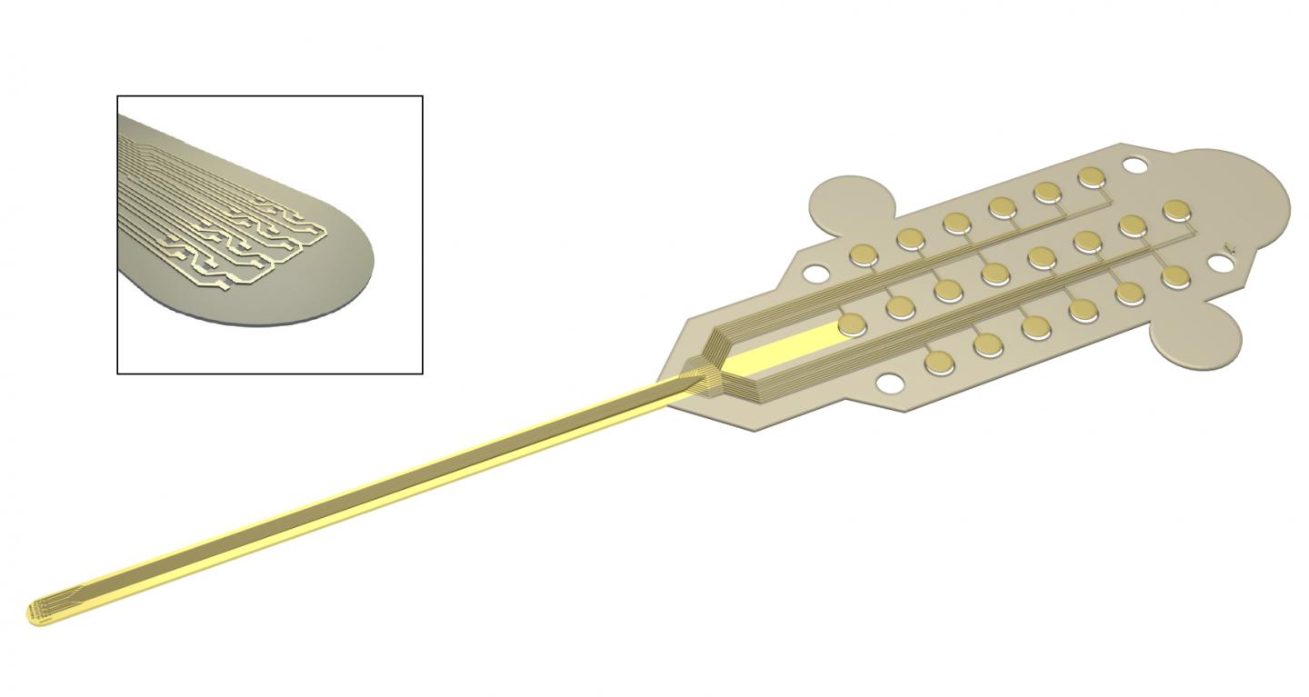
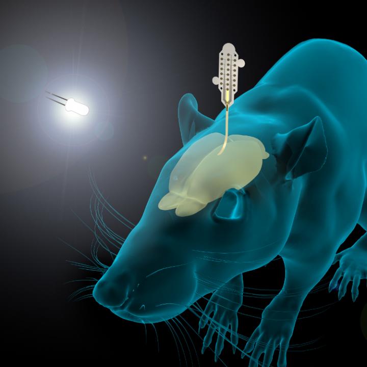
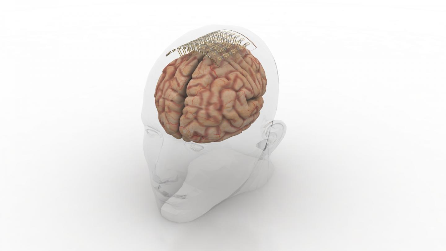
Tag Archives: epilepsy

Compact and affordable brain-computer interface (BCI)

This device could help people with disabilities to regain control of their limbs or provide advance warnings of seizures to people with epilepsy and it’s all based on technology that is a century old.

A January 19, 2022 Skolkovo Institute of Science and Technology (Skoltech) press release (also on EurekAlert) provides details about the device (Note: A link has been removed),

Scientists from Skoltech, South Ural State University, and elsewhere have developed a device for recording brain activity that is more compact and affordable than the solutions currently on the market. With its high signal quality and customizable configuration, the device could help people with restricted mobility regain control of their limbs or provide advance warnings of an impending seizure to patients with epilepsy. The article presenting the device and testing results came out in Experimental Brain Research.

Researchers and medics, as well as engineers working on futuristic gadgets, need tools that measure brain activity. Among their scientific applications are research on sleep, decision-making, memory, and attention. In a clinical setting, these tools allow doctors to assess the extent of damage to an injured brain and monitor coma patients. Further down cyberpunk lane, brain signals can be translated into commands and sent to an external or implanted device, either to make up for lost functions in the body or for plain fun. The commands could range from moving the arm of an exoskeleton worn by a paralyzed person to turning on the TV.

Invented about a century ago, electroencephalographers are devices that read the electrical activity of the brain via small electrodes placed on the scalp. The recorded signals are then used for research, diagnostics, or gadgetry. The problem with the existing systems used in labs and hospitals is they are bulky and/or expensive. And even then, the number of electrodes is limited, resulting in moderate signal quality. Amateur devices tend to be more affordable, but with even poorer sensitivity.

To fill that gap, researchers from South Ural State University, North Carolina State University, and Brainflow — led by electronic research engineer Ildar Rakhmatulin and Skoltech neuroscientist Professor Mikhail Lebedev — created a device you can build for just $350, compared with the $1,000 or more you would need for currently available analogs. Besides being less expensive, the new electroencephalographer has as many as 24 electrodes or more. Importantly, it also provides research-grade signal quality. At half a centimeter in diameter (about 1/5 inches), the processing unit is compact enough to be worn throughout the day or during the night. The entire device weighs about 150 grams (about 5 ounces).

The researchers have made the instructions for building the device and the accompanying documentation and software openly available on GitHub. The team hopes this will attract more enthusiasts involved in brain-computer interface development, giving an impetus to support and rehabilitation system development, cognitive research, and pushing the geek community to come up with new futuristic gizmos.

“The more convenient and affordable such devices become, the more chances there are this would drive the home lab movement, with some of the research on brain-computer interfaces migrating from large science centers to small-scale amateur projects,” Lebedev said.

“Or we could see people with limited mobility using do-it-yourself interfaces to train, say, a smartphone-based system that would electrically stimulate a biceps to flex the arm at the elbow,” the researcher went on. “That works on someone who has lost control over their arm due to spinal cord trauma or a stroke, where the commands are still generated in the brain — they just don’t reach the limb, and that’s where our little brain-computer interfacing comes in.”

According to the team, such interfaces could also help patients with epilepsy by detecting tell-tale brain activity patterns that indicate when a seizure is imminent, so they can prepare by lying down comfortably in a safe space or attempting to suppress the seizure via electrical stimulation.

Here’s a link to and a citation for the paper,

Low-cost brain computer interface for everyday use by Ildar Rakhmatulin, Andrey Parfenov, Zachary Traylor, Chang S. Nam & Mikhail Lebedev. Experimental Brain Research volume 239,Issue Date: December 2021, pages 3573–3583 (2021) DOI: https://doi.org/10.1007/s00221-021-06231-4 Published online: 29 September 2021

This paper is behind a paywall.

You can find Brainflow here and this description on its homepage: “BrainFlow is a library intended to obtain, parse and analyze EEG, EMG, ECG and other kinds of data from biosensors.”

Brainy and brainy: a novel synaptic architecture and a neuromorphic computing platform called SpiNNaker

I have two items about brainlike computing. The first item hearkens back to memristors, a topic I have been following since 2008. (If you’re curious about the various twists and turns just enter  the term ‘memristor’ in this blog’s search engine.) The latest on memristors is from a team than includes IBM (US), École Politechnique Fédérale de Lausanne (EPFL; Swizterland), and the New Jersey Institute of Technology (NJIT; US). The second bit comes from a Jülich Research Centre team in Germany and concerns an approach to brain-like computing that does not include memristors.

Multi-memristive synapses

In the inexorable march to make computers function more like human brains (neuromorphic engineering/computing), an international team has announced its latest results in a July 10, 2018 news item on Nanowerk,

Two New Jersey Institute of Technology (NJIT) researchers, working with collaborators from the IBM Research Zurich Laboratory and the École Polytechnique Fédérale de Lausanne, have demonstrated a novel synaptic architecture that could lead to a new class of information processing systems inspired by the brain.

The findings are an important step toward building more energy-efficient computing systems that also are capable of learning and adaptation in the real world. …

A July 10, 2018 NJIT news release (also on EurekAlert) by Tracey Regan, which originated by the news item, adds more details,

The researchers, Bipin Rajendran, an associate professor of electrical and computer engineering, and S. R. Nandakumar, a graduate student in electrical engineering, have been developing brain-inspired computing systems that could be used for a wide range of big data applications.

Over the past few years, deep learning algorithms have proven to be highly successful in solving complex cognitive tasks such as controlling self-driving cars and language understanding. At the heart of these algorithms are artificial neural networks – mathematical models of the neurons and synapses of the brain – that are fed huge amounts of data so that the synaptic strengths are autonomously adjusted to learn the intrinsic features and hidden correlations in these data streams.

However, the implementation of these brain-inspired algorithms on conventional computers is highly inefficient, consuming huge amounts of power and time. This has prompted engineers to search for new materials and devices to build special-purpose computers that can incorporate the algorithms. Nanoscale memristive devices, electrical components whose conductivity depends approximately on prior signaling activity, can be used to represent the synaptic strength between the neurons in artificial neural networks.

While memristive devices could potentially lead to faster and more power-efficient computing systems, they are also plagued by several reliability issues that are common to nanoscale devices. Their efficiency stems from their ability to be programmed in an analog manner to store multiple bits of information; however, their electrical conductivities vary in a non-deterministic and non-linear fashion.

In the experiment, the team showed how multiple nanoscale memristive devices exhibiting these characteristics could nonetheless be configured to efficiently implement artificial intelligence algorithms such as deep learning. Prototype chips from IBM containing more than one million nanoscale phase-change memristive devices were used to implement a neural network for the detection of hidden patterns and correlations in time-varying signals.

“In this work, we proposed and experimentally demonstrated a scheme to obtain high learning efficiencies with nanoscale memristive devices for implementing learning algorithms,” Nandakumar says. “The central idea in our demonstration was to use several memristive devices in parallel to represent the strength of a synapse of a neural network, but only chose one of them to be updated at each step based on the neuronal activity.”

Here’s a link to and a citation for the paper,

Neuromorphic computing with multi-memristive synapses by Irem Boybat, Manuel Le Gallo, S. R. Nandakumar, Timoleon Moraitis, Thomas Parnell, Tomas Tuma, Bipin Rajendran, Yusuf Leblebici, Abu Sebastian, & Evangelos Eleftheriou. Nature Communications volume 9, Article number: 2514 (2018) DOI: https://doi.org/10.1038/s41467-018-04933-y Published 28 June 2018

This is an open access paper.

Also they’ve got a couple of very nice introductory paragraphs which I’m including here, (from the June 28, 2018 paper in Nature Communications; Note: Links have been removed),

The human brain with less than 20 W of power consumption offers a processing capability that exceeds the petaflops mark, and thus outperforms state-of-the-art supercomputers by several orders of magnitude in terms of energy efficiency and volume. Building ultra-low-power cognitive computing systems inspired by the operating principles of the brain is a promising avenue towards achieving such efficiency. Recently, deep learning has revolutionized the field of machine learning by providing human-like performance in areas, such as computer vision, speech recognition, and complex strategic games1. However, current hardware implementations of deep neural networks are still far from competing with biological neural systems in terms of real-time information-processing capabilities with comparable energy consumption.

One of the reasons for this inefficiency is that most neural networks are implemented on computing systems based on the conventional von Neumann architecture with separate memory and processing units. There are a few attempts to build custom neuromorphic hardware that is optimized to implement neural algorithms2,3,4,5. However, as these custom systems are typically based on conventional silicon complementary metal oxide semiconductor (CMOS) circuitry, the area efficiency of such hardware implementations will remain relatively low, especially if in situ learning and non-volatile synaptic behavior have to be incorporated. Recently, a new class of nanoscale devices has shown promise for realizing the synaptic dynamics in a compact and power-efficient manner. These memristive devices store information in their resistance/conductance states and exhibit conductivity modulation based on the programming history6,7,8,9. The central idea in building cognitive hardware based on memristive devices is to store the synaptic weights as their conductance states and to perform the associated computational tasks in place.

The two essential synaptic attributes that need to be emulated by memristive devices are the synaptic efficacy and plasticity. …

It gets more complicated from there.

Now onto the next bit.

SpiNNaker

At a guess, those capitalized N’s are meant to indicate ‘neural networks’. As best I can determine, SpiNNaker is not based on the memristor. Moving on, a July 11, 2018 news item on phys.org announces work from a team examining how neuromorphic hardware and neuromorphic software work together,

A computer built to mimic the brain’s neural networks produces similar results to that of the best brain-simulation supercomputer software currently used for neural-signaling research, finds a new study published in the open-access journal Frontiers in Neuroscience. Tested for accuracy, speed and energy efficiency, this custom-built computer named SpiNNaker, has the potential to overcome the speed and power consumption problems of conventional supercomputers. The aim is to advance our knowledge of neural processing in the brain, to include learning and disorders such as epilepsy and Alzheimer’s disease.

A July 11, 2018 Frontiers Publishing news release on EurekAlert, which originated the news item, expands on the latest work,

“SpiNNaker can support detailed biological models of the cortex–the outer layer of the brain that receives and processes information from the senses–delivering results very similar to those from an equivalent supercomputer software simulation,” says Dr. Sacha van Albada, lead author of this study and leader of the Theoretical Neuroanatomy group at the Jülich Research Centre, Germany. “The ability to run large-scale detailed neural networks quickly and at low power consumption will advance robotics research and facilitate studies on learning and brain disorders.”

The human brain is extremely complex, comprising 100 billion interconnected brain cells. We understand how individual neurons and their components behave and communicate with each other and on the larger scale, which areas of the brain are used for sensory perception, action and cognition. However, we know less about the translation of neural activity into behavior, such as turning thought into muscle movement.

Supercomputer software has helped by simulating the exchange of signals between neurons, but even the best software run on the fastest supercomputers to date can only simulate 1% of the human brain.

“It is presently unclear which computer architecture is best suited to study whole-brain networks efficiently. The European Human Brain Project and Jülich Research Centre have performed extensive research to identify the best strategy for this highly complex problem. Today’s supercomputers require several minutes to simulate one second of real time, so studies on processes like learning, which take hours and days in real time are currently out of reach.” explains Professor Markus Diesmann, co-author, head of the Computational and Systems Neuroscience department at the Jülich Research Centre.

He continues, “There is a huge gap between the energy consumption of the brain and today’s supercomputers. Neuromorphic (brain-inspired) computing allows us to investigate how close we can get to the energy efficiency of the brain using electronics.”

Developed over the past 15 years and based on the structure and function of the human brain, SpiNNaker — part of the Neuromorphic Computing Platform of the Human Brain Project — is a custom-built computer composed of half a million of simple computing elements controlled by its own software. The researchers compared the accuracy, speed and energy efficiency of SpiNNaker with that of NEST–a specialist supercomputer software currently in use for brain neuron-signaling research.

“The simulations run on NEST and SpiNNaker showed very similar results,” reports Steve Furber, co-author and Professor of Computer Engineering at the University of Manchester, UK. “This is the first time such a detailed simulation of the cortex has been run on SpiNNaker, or on any neuromorphic platform. SpiNNaker comprises 600 circuit boards incorporating over 500,000 small processors in total. The simulation described in this study used just six boards–1% of the total capability of the machine. The findings from our research will improve the software to reduce this to a single board.”

Van Albada shares her future aspirations for SpiNNaker, “We hope for increasingly large real-time simulations with these neuromorphic computing systems. In the Human Brain Project, we already work with neuroroboticists who hope to use them for robotic control.”

Before getting to the link and citation for the paper, here’s a description of SpiNNaker’s hardware from the ‘Spiking neural netowrk’ Wikipedia entry, Note: Links have been removed,

Neurogrid, built at Stanford University, is a board that can simulate spiking neural networks directly in hardware. SpiNNaker (Spiking Neural Network Architecture) [emphasis mine], designed at the University of Manchester, uses ARM processors as the building blocks of a massively parallel computing platform based on a six-layer thalamocortical model.[5]

Now for the link and citation,

Performance Comparison of the Digital Neuromorphic Hardware SpiNNaker and the Neural Network Simulation Software NEST for a Full-Scale Cortical Microcircuit Model by
Sacha J. van Albada, Andrew G. Rowley, Johanna Senk, Michael Hopkins, Maximilian Schmidt, Alan B. Stokes, David R. Lester, Markus Diesmann, and Steve B. Furber. Neurosci. 12:291. doi: 10.3389/fnins.2018.00291 Published: 23 May 2018

As noted earlier, this is an open access paper.

Soft things for your brain

A March 5, 2018 news item on Nanowerk describes the latest stretchable electrode (Note: A link has been removed),

Klas Tybrandt, principal investigator at the Laboratory of Organic Electronics at Linköping University [Sweden], has developed new technology for long-term stable neural recording. It is based on a novel elastic material composite, which is biocompatible and retains high electrical conductivity even when stretched to double its original length.

The result has been achieved in collaboration with colleagues in Zürich and New York. The breakthrough, which is crucial for many applications in biomedical engineering, is described in an article published in the prestigious scientific journal Advanced Materials (“High-Density Stretchable Electrode Grids for Chronic Neural Recording”).

A March 5, 2018 Linköping University press release, which originated the news item, gives more detail but does not mention that the nanowires are composed of titanium dioxide (you can find additional details in the abstract for the paper; link and citation will be provided later in this posting)),

The coupling between electronic components and nerve cells is crucial not only to collect information about cell signalling, but also to diagnose and treat neurological disorders and diseases, such as epilepsy.

It is very challenging to achieve long-term stable connections that do not damage neurons or tissue, since the two systems, the soft and elastic tissue of the body and the hard and rigid electronic components, have completely different mechanical properties.

Stretchable soft electrodeThe soft electrode stretched to twice its length Photo credit: Thor Balkhed

“As human tissue is elastic and mobile, damage and inflammation arise at the interface with rigid electronic components. It not only causes damage to tissue; it also attenuates neural signals,” says Klas Tybrandt, leader of the Soft Electronics group at the Laboratory of Organic Electronics, Linköping University, Campus Norrköping.

New conductive material

Klas Tybrandt has developed a new conductive material that is as soft as human tissue and can be stretched to twice its length. The material consists of gold coated titanium dioxide nanowires, embedded into silicone rubber. The material is biocompatible – which means it can be in contact with the body without adverse effects – and its conductivity remains stable over time.

“The microfabrication of soft electrically conductive composites involves several challenges. We have developed a process to manufacture small electrodes that also preserves the biocompatibility of the materials. The process uses very little material, and this means that we can work with a relatively expensive material such as gold, without the cost becoming prohibitive,” says Klas Tybrandt.

The electrodes are 50 µm [microns or micrometres] in size and are located at a distance of 200 µm from each other. The fabrication procedure allows 32 electrodes to be placed onto a very small surface. The final probe, shown in the photograph, has a width of 3.2 mm and a thickness of 80 µm.

The soft microelectrodes have been developed at Linköping University and ETH Zürich, and researchers at New York University and Columbia University have subsequently implanted them in the brain of rats. The researchers were able to collect high-quality neural signals from the freely moving rats for 3 months. The experiments have been subject to ethical review, and have followed the strict regulations that govern animal experiments.

Important future applications

Klas Tybrandt, researcher at Laboratory for Organic ElectronicsKlas Tybrandt, researcher at Laboratory for Organic Electronics Photo credit: Thor Balkhed

“When the neurons in the brain transmit signals, a voltage is formed that the electrodes detect and transmit onwards through a tiny amplifier. We can also see which electrodes the signals came from, which means that we can estimate the location in the brain where the signals originated. This type of spatiotemporal information is important for future applications. We hope to be able to see, for example, where the signal that causes an epileptic seizure starts, a prerequisite for treating it. Another area of application is brain-machine interfaces, by which future technology and prostheses can be controlled with the aid of neural signals. There are also many interesting applications involving the peripheral nervous system in the body and the way it regulates various organs,” says Klas Tybrandt.

The breakthrough is the foundation of the research area Soft Electronics, currently being established at Linköping University, with Klas Tybrandt as principal investigator.
liu.se/soft-electronics

A video has been made available (Note: For those who find any notion of animal testing disturbing; don’t watch the video even though it is an animation and does not feature live animals),

Here’s a link to and a citation for the paper,

High-Density Stretchable Electrode Grids for Chronic Neural Recording by Klas Tybrandt, Dion Khodagholy, Bernd Dielacher, Flurin Stauffer, Aline F. Renz, György Buzsáki, and János Vörös. Advanced Materials 2018. DOI: 10.1002/adma.201706520
 First published 28 February 2018

This paper is open access.

Yes! Art, genetic modifications, gene editing, and xenotransplantation at the Vancouver Biennale (Canada)

Patricia Piccinini’s Curious Imaginings Courtesy: Vancouver Biennale [downloaded from http://dailyhive.com/vancouver/vancouver-biennale-unsual-public-art-2018/]

Up to this point, I’ve been a little jealous of the Art/Sci Salon’s (Toronto, Canada) January 2018 workshops for artists and discussions about CRISPR ((clustered regularly interspaced short palindromic repeats))/Cas9 and its social implications. (See my January 10, 2018 posting for more about the events.) Now, it seems Vancouver may be in line for its ‘own’ discussion about CRISPR and the implications of gene editing. The image you saw (above) represents one of the installations being hosted by the 2018 – 2020 edition of the Vancouver Biennale.

While this posting is mostly about the Biennale and Piccinini’s work, there is a ‘science’ subsection featuring the science of CRISPR and xenotransplantation. Getting back to the Biennale and Piccinini: A major public art event since 1988, the Vancouver Biennale has hosted over 91 outdoor sculptures and new media works by more than 78 participating artists from over 25 countries and from 4 continents.

Quickie description of the 2018 – 2020 Vancouver Biennale

The latest edition of the Vancouver Biennale was featured in a June 6, 2018 news item on the Daily Hive (Vancouver),

The Vancouver Biennale will be bringing new —and unusual— works of public art to the city beginning this June.

The theme for this season’s Vancouver Biennale exhibition is “re-IMAGE-n” and it kicks off on June 20 [2018] in Vanier Park with Saudi artist Ajlan Gharem’s Paradise Has Many Gates.

Gharem’s architectural chain-link sculpture resembles a traditional mosque, the piece is meant to challenge the notions of religious orthodoxy and encourages individuals to image a space free of Islamophobia.

Melbourne artist Patricia Piccinini’s Curious Imaginings is expected to be one of the most talked about installations of the exhibit. Her style of “oddly captivating, somewhat grotesque, human-animal hybrid creature” is meant to be shocking and thought-provoking.

Piccinini’s interactive [emphasis mine] experience will “challenge us to explore the social impacts of emerging biotechnology and our ethical limits in an age where genetic engineering and digital technologies are already pushing the boundaries of humanity.”

Piccinini’s work will be displayed in the 105-year-old Patricia Hotel in Vancouver’s Strathcona neighbourhood. The 90-day ticketed exhibition [emphasis mine] is scheduled to open this September [2018].

Given that this blog is focused on nanotechnology and other emerging technologies such as CRISPR, I’m focusing on Piccinini’s work and its art/science or sci-art status. This image from the GOMA Gallery where Piccinini’s ‘Curious Affection‘ installation is being shown from March 24 – Aug. 5, 2018 in Brisbane, Queensland, Australia may give you some sense of what one of her installations is like,

Courtesy: Queensland Art Gallery | Gallery of Modern Art (QAGOMA)

I spoke with Serena at the Vancouver Biennale office and asked about the ‘interactive’ aspect of Piccinini’s installation. She suggested the term ‘immersive’ as an alternative. In other words, you won’t be playing with the sculptures or pressing buttons and interacting with computer screens or robots. She also noted that the ticket prices have not been set yet and they are currently developing events focused on the issues raised by the installation. She knew that 2018 is the 200th anniversary of the publication of Mary Shelley’s Frankenstein but I’m not sure how the Biennale folks plan (or don’t plan)  to integrate any recognition of the novle’s impact on the discussions about ‘new’ technologies .They expect Piccinini will visit Vancouver. (Note 1: Piccinini’s work can  also be seen in a group exhibition titled: Frankenstein’s Birthday Party at the Hosfselt Gallery in San Francisco (California, US) from June 23 – August 11, 2018.  Note 2: I featured a number of international events commemorating the 200th anniversary of the publication of Mary Shelley’s novel, Frankenstein, in my Feb. 26, 2018 posting. Note 3: The term ‘Frankenfoods’ helped to shape the discussion of genetically modified organisms and food supply on this planet. It was a wildly successful campaign for activists affecting legislation in some areas of research. Scientists have not been as enthusiastic about the effects. My January 15, 2009 posting briefly traces a history of the term.)

The 2018 – 2020 Vancouver Biennale and science

A June 7, 2018 Vancouver Biennale news release provides more detail about the current series of exhibitions,

The Biennale is also committed to presenting artwork at the cutting edge of discussion and in keeping with the STEAM (science, technology, engineering, arts, math[ematics]) approach to integrating the arts and sciences. In August [2018], Colombian/American visual artist Jessica Angel will present her monumental installation Dogethereum Bridge at Hinge Park in Olympic Village. Inspired by blockchain technology, the artwork’s design was created through the integration of scientific algorithms, new developments in technology, and the arts. This installation, which will serve as an immersive space and collaborative hub for artists and technologists, will host a series of activations with blockchain as the inspirational jumping-off point.

In what is expected to become one of North America’s most talked-about exhibitions of the year, Melbourne artist Patricia Piccinini’s Curious Imaginings will see the intersection of art, science, and ethics. For the first time in the Biennale’s fifteen years of creating transformative experiences, and in keeping with the 2018-2020 theme of “re-IMAGE-n,” the Biennale will explore art in unexpected places by exhibiting in unconventional interior spaces.  The hyperrealist “world of oddly captivating, somewhat grotesque, human-animal hybrid creatures” will be the artist’s first exhibit in a non-museum setting, transforming a wing of the 105-year-old Patricia Hotel. Situated in Vancouver’s oldest neighbourbood of Strathcona, Piccinini’s interactive experience will “challenge us to explore the social impacts of emerging bio-technology and our ethical limits in an age where genetic engineering and digital technologies are already pushing the boundaries of humanity.” In this intimate hotel setting located in a neighborhood continually undergoing its own change, Curious Imaginings will empower visitors to personally consider questions posed by the exhibition, including the promises and consequences of genetic research and human interference. …

There are other pieces being presented at the Biennale but my special interest is in the art/sci pieces and, at this point, CRISPR.

Piccinini in more depth

You can find out more about Patricia Piccinini in her biography on the Vancouver Biennale website but I found this Char Larsson April 7, 2018 article for the Independent (UK) more informative (Note: A link has been removed),

Patricia Piccinini’s sculptures are deeply disquieting. Walking through Curious Affection, her new solo exhibition at Brisbane’s Gallery of Modern Art, is akin to entering a science laboratory full of DNA experiments. Made from silicone, fibreglass and even human hair, her sculptures are breathtakingly lifelike, however, we can’t be sure what life they are like. The artist creates an exuberant parallel universe where transgenic experiments flourish and human evolution has given way to genetic engineering and DNA splicing.

Curious Affection is a timely and welcome recognition of Piccinini’s enormous contribution to reaching back to the mid-1990s. Working across a variety of mediums including photography, video and drawing, she is perhaps best known for her hyperreal creations.

As a genre, hyperrealism depends on the skill of the artist to create the illusion of reality. To be truly successful, it must convince the spectator of its realness. Piccinini acknowledges this demand, but with a delightful twist. The excruciating attention to detail deliberately solicits our desire to look, only to generate unease, as her sculptures are imbued with a fascinating otherness. Part human, part animal, the works are uncannily familiar, but also alarmingly “other”.

Inspired by advances in genetically modified pigs to generate replacement organs for humans [also known as xenotransplantation], we are reminded that Piccinini has always been at the forefront of debates concerning the possibilities of science, technology and DNA cloning. She does so, however, with a warm affection and sense of humour, eschewing the hysterical anxiety frequently accompanying these scientific developments.

Beyond the astonishing level of detail achieved by working with silicon and fibreglass, there is an ethics at work here. Piccinini is asking us not to avert our gaze from the other, and in doing so, to develop empathy and understanding through the encounter.

I encourage anyone who’s interested to read Larsson’s entire piece (April 7, 2018 article).

According to her Wikipedia entry, Piccinini works in a variety of media including video, sound, sculpture, and more. She also has her own website.

Gene editing and xenotransplantation

Sarah Zhang’s June 8, 2018 article for The Atlantic provides a peek at the extraordinary degree of interest and competition in the field of gene editing and CRISPR ((clustered regularly interspaced short palindromic repeats))/Cas9 research (Note: A link has been removed),

China Is Genetically Engineering Monkeys With Brain Disorders

Guoping Feng applied to college the first year that Chinese universities reopened after the Cultural Revolution. It was 1977, and more than a decade’s worth of students—5.7 million—sat for the entrance exams. Feng was the only one in his high school to get in. He was assigned—by chance, essentially—to medical school. Like most of his contemporaries with scientific ambitions, he soon set his sights on graduate studies in the United States. “China was really like 30 to 50 years behind,” he says. “There was no way to do cutting-edge research.” So in 1989, he left for Buffalo, New York, where for the first time he saw snow piled several feet high. He completed his Ph.D. in genetics at the State University of New York at Buffalo.

Feng is short and slim, with a monk-like placidity and a quick smile, and he now holds an endowed chair in neuroscience at MIT, where he focuses on the genetics of brain disorders. His 45-person lab is part of the McGovern Institute for Brain Research, which was established in 2000 with the promise of a $350 million donation, the largest ever received by the university. In short, his lab does not lack for much.

Yet Feng now travels to China several times a year, because there, he can pursue research he has not yet been able to carry out in the United States. [emphasis mine] …

Feng had organized a symposium at SIAT [Shenzhen Institutes of Advanced Technology], and he was not the only scientist who traveled all the way from the United States to attend: He invited several colleagues as symposium speakers, including a fellow MIT neuroscientist interested in tree shrews, a tiny mammal related to primates and native to southern China, and Chinese-born neuroscientists who study addiction at the University of Pittsburgh and SUNY Upstate Medical University. Like Feng, they had left China in the ’80s and ’90s, part of a wave of young scientists in search of better opportunities abroad. Also like Feng, they were back in China to pursue a type of cutting-edge research too expensive and too impractical—and maybe too ethically sensitive—in the United States.

Here’s what precipitated Feng’s work in China, (from Zhang’s article; Note: Links have been removed)

At MIT, Feng’s lab worked on genetically engineering a monkey species called marmosets, which are very small and genuinely bizarre-looking. They are cheaper to keep due to their size, but they are a relatively new lab animal, and they can be difficult to train on lab tasks. For this reason, Feng also wanted to study Shank3 on macaques in China. Scientists have been cataloging the social behavior of macaques for decades, making it an obvious model for studies of disorders like autism that have a strong social component. Macaques are also more closely related to humans than marmosets, making their brains a better stand-in for those of humans.

The process of genetically engineering a macaque is not trivial, even with the advanced tools of CRISPR. Researchers begin by dosing female monkeys with the same hormones used in human in vitro fertilization. They then collect and fertilize the eggs, and inject the resulting embryos with CRISPR proteins using a long, thin glass needle. Monkey embryos are far more sensitive than mice embryos, and can be affected by small changes in the pH of the injection or the concentration of CRISPR proteins. Only some of the embryos will have the desired mutation, and only some will survive once implanted in surrogate mothers. It takes dozens of eggs to get to just one live monkey, so making even a few knockout monkeys required the support of a large breeding colony.

The first Shank3 macaque was born in 2015. Four more soon followed, bringing the total to five.

To visit his research animals, Feng now has to fly 8,000 miles across 12 time zones. It would be a lot more convenient to carry out his macaque research in the United States, of course, but so far, he has not been able to.

He originally inquired about making Shank3 macaques at the New England Primate Research Center, one of eight national primate research centers then funded by the National Institutes of Health in partnership with a local institution (Harvard Medical School, in this case). The center was conveniently located in Southborough, Massachusetts, just 20 miles west of the MIT campus. But in 2013, Harvard decided to shutter the center.

The decision came as a shock to the research community, and it was widely interpreted as a sign of waning interest in primate research in the United States. While the national primate centers have been important hubs of research on HIV, Zika, Ebola, and other diseases, they have also come under intense public scrutiny. Animal-rights groups like the Humane Society of the United States have sent investigators to work undercover in the labs, and the media has reported on monkey deaths in grisly detail. Harvard officially made its decision to close for “financial” reasons. But the announcement also came after the high-profile deaths of four monkeys from improper handling between 2010 and 2012. The deaths sparked a backlash; demonstrators showed up at the gates. The university gave itself two years to wind down their primate work, officially closing the center in 2015.

“They screwed themselves,” Michael Halassa, the MIT neuroscientist who spoke at Feng’s symposium, told me in Shenzhen. Wei-Dong Yao, another one of the speakers, chimed in, noting that just two years later CRISPR has created a new wave of interest in primate research. Yao was one of the researchers at Harvard’s primate center before it closed; he now runs a lab at SUNY Upstate Medical University that uses genetically engineered mouse and human stem cells, and he had come to Shenzhen to talk about restarting his addiction research on primates.

Here’s comes the competition (from Zhang’s article; Note: Links have been removed),

While the U.S. government’s biomedical research budget has been largely flat, both national and local governments in China are eager to raise their international scientific profiles, and they are shoveling money into research. A long-rumored, government-sponsored China Brain Project is supposed to give neuroscience research, and primate models in particular, a big funding boost. Chinese scientists may command larger salaries, too: Thanks to funding from the Shenzhen local government, a new principal investigator returning from overseas can get 3 million yuan—almost half a million U.S. dollars—over his or her first five years. China is even finding success in attracting foreign researchers from top U.S. institutions like Yale.

In the past few years, China has seen a miniature explosion of genetic engineering in monkeys. In Kunming, Shanghai, and Guangzhou, scientists have created monkeys engineered to show signs of Parkinson’s, Duchenne muscular dystrophy, autism, and more. And Feng’s group is not even the only one in China to have created Shank3 monkeys. Another group—a collaboration primarily between researchers at Emory University and scientists in China—has done the same.

Chinese scientists’ enthusiasm for CRISPR also extends to studies of humans, which are moving much more quickly, and in some cases under less oversight, than in the West. The first studies to edit human embryos and first clinical trials for cancer therapies using CRISPR have all happened in China. [emphases mine]

Some ethical issues are also covered (from Zhang’s article),

Parents with severely epileptic children had asked him if it would be possible to study the condition in a monkey. Feng told them what he thought would be technically possible. “But I also said, ‘I’m not sure I want to generate a model like this,’” he recalled. Maybe if there were a drug to control the monkeys’ seizures, he said: “I cannot see them seizure all the time.”

But is it ethical, he continued, to let these babies die without doing anything? Is it ethical to generate thousands or millions of mutant mice for studies of brain disorders, even when you know they will not elucidate much about human conditions?

Primates should only be used if other models do not work, says Feng, and only if a clear path forward is identified. The first step in his work, he says, is to use the Shank3 monkeys to identify the changes the mutations cause in the brain. Then, researchers might use that information to find targets for drugs, which could be tested in the same monkeys. He’s talking with the Oregon National Primate Research Center about carrying out similar work in the United States. ….[Note: I have a three-part series about CRISPR and germline editing* in the US, precipitated by research coming out of Oregon, Part 1, which links to the other parts, is here.]

Zhang’s June 8, 2018 article is excellent and I highly recommend reading it.

I touched on the topic of xenotransplanttaion in a commentary on a book about the science  of the television series, Orphan Black in a January 31,2018 posting (Note: A chimera is what you use to incubate a ‘human’ organ for transplantation or, more accurately, xenotransplantation),

On the subject of chimeras, the Canadian Broadcasting Corporation (CBC) featured a January 26, 2017 article about the pig-human chimeras on its website along with a video,

The end

I am very excited to see Piccinini’s work come to Vancouver. There have been a number of wonderful art and art/science installations and discussions here but this is the first one (I believe) to tackle the emerging gene editing technologies and the issues they raise. (It also fits in rather nicely with the 200th anniversary of the publication of Mary Shelley’s Frankenstein which continues to raise issues and stimulate discussion.)

In addition to the ethical issues raised in Zhang’s article, there are some other philosophical questions:

  • what does it mean to be human
  • if we are going to edit genes to create hybrid human/animals, what are they and how do they fit into our current animal/human schema
  • are you still human if you’ve had an organ transplant where the organ was incubated in a pig

There are also going to be legal issues. In addition to any questions about legal status, there are also fights about intellectual property such as the one involving Harvard & MIT’s [Massachusetts Institute of Technology] Broad Institute vs the University of California at Berkeley (March 15, 2017 posting)..

While I’m thrilled about the Piccinini installation, it should be noted the issues raised by other artworks hosted in this version of the Biennale are important. Happily, they have been broached here in Vancouver before and I suspect this will result in more nuanced  ‘conversations’ than are possible when a ‘new’ issue is introduced.

Bravo 2018 – 2020 Vancouver Biennale!

* Germline editing is when your gene editing will affect subsequent generations as opposed to editing out a mutated gene for the lifetime of a single individual.

Art/sci and CRISPR links

This art/science posting may prove of some interest:

The connectedness of living things: an art/sci project in Saskatchewan: evolutionary biology (February 16, 2018)

A selection of my CRISPR posts:

CRISPR and editing the germline in the US (part 1 of 3): In the beginning (August 15, 2017)

NOTE: An introductory CRISPR video describing how CRISPR/Cas9 works was embedded in part1.

Why don’t you CRISPR yourself? (January 25, 2018)

Editing the genome with CRISPR ((clustered regularly interspaced short palindromic repeats)-carrying nanoparticles (January 26, 2018)

Immune to CRISPR? (April 10, 2018)
Graphene-based neural probes

I have two news bits (dated almost one month apart) about the use of graphene in neural probes, one from the European Union and the other from Korea.

European Union (EU)

This work is being announced by the European Commission’s (a subset of the EU) Graphene Flagship (one of two mega-funding projects announced in 2013; 1B Euros each over ten years for the Graphene Flagship and the Human Brain Project).

According to a March 27, 2017 news item on ScienceDaily, researchers have developed a graphene-based neural probe that has been tested on rats,

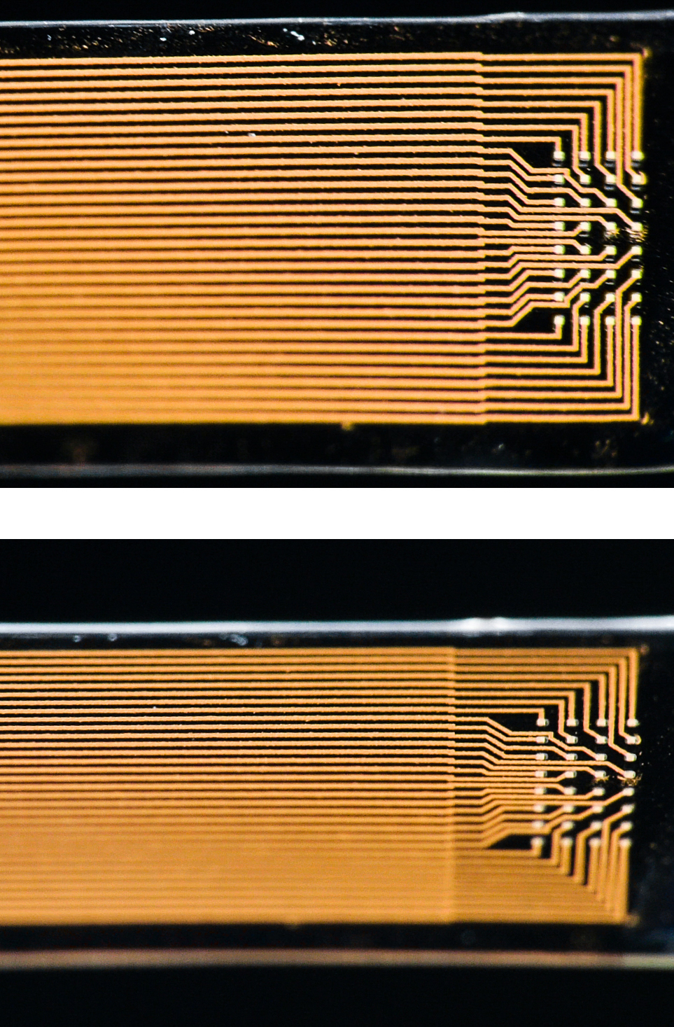
Measuring brain activity with precision is essential to developing further understanding of diseases such as epilepsy and disorders that affect brain function and motor control. Neural probes with high spatial resolution are needed for both recording and stimulating specific functional areas of the brain. Now, researchers from the Graphene Flagship have developed a new device for recording brain activity in high resolution while maintaining excellent signal to noise ratio (SNR). Based on graphene field-effect transistors, the flexible devices open up new possibilities for the development of functional implants and interfaces.

The research, published in 2D Materials, was a collaborative effort involving Flagship partners Technical University of Munich (TU Munich; Germany), Institut d’Investigacions Biomèdiques August Pi i Sunyer (IDIBAPS; Spain), Spanish National Research Council (CSIC; Spain), The Biomedical Research Networking Center in Bioengineering, Biomaterials and Nanomedicine (CIBER-BBN; Spain) and the Catalan Institute of Nanoscience and Nanotechnology (ICN2; Spain).

Caption: Graphene transistors integrated in a flexible neural probe enables electrical signals from neurons to be measured with high accuracy and density. Inset: The tip of the probe contains 16 flexible graphene transistors. Credit: ICN2

A March 27, 2017 Graphene Flagship press release on EurekAlert, which originated the news item, describes the work,  in more detail,

The devices were used to record the large signals generated by pre-epileptic activity in rats, as well as the smaller levels of brain activity during sleep and in response to visual light stimulation. These types of activities lead to much smaller electrical signals, and are at the level of typical brain activity. Neural activity is detected through the highly localised electric fields generated when neurons fire, so densely packed, ultra-small measuring devices is important for accurate brain readings.

The neural probes are placed directly on the surface of the brain, so safety is of paramount importance for the development of graphene-based neural implant devices. Importantly, the researchers determined that the graphene-based probes are non-toxic, and did not induce any significant inflammation.

Devices implanted in the brain as neural prosthesis for therapeutic brain stimulation technologies and interfaces for sensory and motor devices, such as artificial limbs, are an important goal for improving quality of life for patients. This work represents a first step towards the use of graphene in research as well as clinical neural devices, showing that graphene-based technologies can deliver the high resolution and high SNR needed for these applications.

First author Benno Blaschke (TU Munich) said “Graphene is one of the few materials that allows recording in a transistor configuration and simultaneously complies with all other requirements for neural probes such as flexibility, biocompability and chemical stability. Although graphene is ideally suited for flexible electronics, it was a great challenge to transfer our fabrication process from rigid substrates to flexible ones. The next step is to optimize the wafer-scale fabrication process and improve device flexibility and stability.”

Jose Antonio Garrido (ICN2), led the research. He said “Mechanical compliance is an important requirement for safe neural probes and interfaces. Currently, the focus is on ultra-soft materials that can adapt conformally to the brain surface. Graphene neural interfaces have shown already great potential, but we have to improve on the yield and homogeneity of the device production in order to advance towards a real technology. Once we have demonstrated the proof of concept in animal studies, the next goal will be to work towards the first human clinical trial with graphene devices during intraoperative mapping of the brain. This means addressing all regulatory issues associated to medical devices such as safety, biocompatibility, etc.”

Caption: The graphene-based neural probes were used to detect rats’ responses to visual stimulation, as well as neural signals during sleep. Both types of signals are small, and typically difficult to measure. Credit: ICN2

Here’s a link to and a citation for the paper,

Mapping brain activity with flexible graphene micro-transistors by Benno M Blaschke, Núria Tort-Colet, Anton Guimerà-Brunet, Julia Weinert, Lionel Rousseau, Axel Heimann, Simon Drieschner, Oliver Kempski, Rosa Villa, Maria V Sanchez-Vives. 2D Materials, Volume 4, Number 2 DOI https://doi.org/10.1088/2053-1583/aa5eff Published 24 February 2017

© 2017 IOP Publishing Ltd

This paper is behind a paywall.

Korea

While this research from Korea was published more recently, the probe itself has not been subjected to in vivo (animal testing). From an April 19, 2017 news item on ScienceDaily,

Electrodes placed in the brain record neural activity, and can help treat neural diseases like Parkinson’s and epilepsy. Interest is also growing in developing better brain-machine interfaces, in which electrodes can help control prosthetic limbs. Progress in these fields is hindered by limitations in electrodes, which are relatively stiff and can damage soft brain tissue.

Designing smaller, gentler electrodes that still pick up brain signals is a challenge because brain signals are so weak. Typically, the smaller the electrode, the harder it is to detect a signal. However, a team from the Daegu Gyeongbuk Institute of Science & Technology [DGIST} in Korea developed new probes that are small, flexible and read brain signals clearly.

This is a pretty interesting way to illustrate the research,

Caption: Graphene and gold make a better brain probe. Credit: DGIST

An April 19, 2017 DGIST press release (also on EurekAlert), which originated the news item, expands on the theme (Note: A link has been removed),

The probe consists of an electrode, which records the brain signal. The signal travels down an interconnection line to a connector, which transfers the signal to machines measuring and analysing the signals.

The electrode starts with a thin gold base. Attached to the base are tiny zinc oxide nanowires, which are coated in a thin layer of gold, and then a layer of conducting polymer called PEDOT. These combined materials increase the probe’s effective surface area, conducting properties, and strength of the electrode, while still maintaining flexibility and compatibility with soft tissue.

Packing several long, thin nanowires together onto one probe enables the scientists to make a smaller electrode that retains the same effective surface area of a larger, flat electrode. This means the electrode can shrink, but not reduce signal detection. The interconnection line is made of a mix of graphene and gold. Graphene is flexible and gold is an excellent conductor. The researchers tested the probe and found it read rat brain signals very clearly, much better than a standard flat, gold electrode.

“Our graphene and nanowires-based flexible electrode array can be useful for monitoring and recording the functions of the nervous system, or to deliver electrical signals to the brain,” the researchers conclude in their paper recently published in the journal ACS Applied Materials and Interfaces.

The probe requires further clinical tests before widespread commercialization. The researchers are also interested in developing a wireless version to make it more convenient for a variety of applications.

Here’s a link to and a citation for the paper,

Enhancement of Interface Characteristics of Neural Probe Based on Graphene, ZnO Nanowires, and Conducting Polymer PEDOT by Mingyu Ryu, Jae Hoon Yang, Yumi Ahn, Minkyung Sim, Kyung Hwa Lee, Kyungsoo Kim, Taeju Lee, Seung-Jun Yoo, So Yeun Kim, Cheil Moon, Minkyu Je, Ji-Woong Choi, Youngu Lee, and Jae Eun Jang. ACS Appl. Mater. Interfaces, 2017, 9 (12), pp 10577–10586 DOI: 10.1021/acsami.7b02975 Publication Date (Web): March 7, 2017

Copyright © 2017 American Chemical Society

This paper is behind a paywall.

A bionic hybrid neurochip from the University of Calgary (Canada)

The University of Calgary is publishing some very exciting work these days as can be seen in my Sept. 21, 2016 posting about quantum teleportation. Today, the university announced this via an Oct. 26, 2016 news item on Nanowerk (Note: A link has been removed),

Brain functions are controlled by millions of brain cells. However, in order to understand how the brain controls functions, such as simple reflexes or learning and memory, we must be able to record the activity of large networks and groups of neurons. Conventional methods have allowed scientists to record the activity of neurons for minutes, but a new technology, developed by University of Calgary researchers, known as a bionic hybrid neuro chip, is able to record activity in animal brain cells for weeks at a much higher resolution. The technological advancement was published in the journal Scientific Reports(“A novel bio-mimicking, planar nano-edge microelectrode enables enhanced long-term neural recording”).

There’s more from an Oct. 26, 2016 University of Calgary news release on EurekAlert, which originated the news item,

“These chips are 15 times more sensitive than conventional neuro chips,” says Naweed Syed, PhD, scientific director of the University of Calgary, Cumming School of Medicine’s Alberta Children’s Hospital Research Institute, member of the Hotchkiss Brain Institute and senior author on the study. “This allows brain cell signals to be amplified more easily and to see real time recordings of brain cell activity at a resolution that has never been achieved before.”

The development of this technology will allow researchers to investigate and understand in greater depth, in animal models, the origins of neurological diseases and conditions such as epilepsy, as well as other cognitive functions such as learning and memory.

“Recording this activity over a long period of time allows you to see changes that occur over time, in the activity itself,” says Pierre Wijdenes, a PhD student in the Biomedical Engineering Graduate Program and the study’s first author. “This helps to understand why certain neurons form connections with each other and why others won’t.”

The cross-faculty team created the chip to mimic the natural biological contact between brain cells, essentially tricking the brain cells into believing that they are connecting with other brain cells. As a result, the cells immediately connect with the chip, thereby allowing researchers to view and record the two-way communication that would go on between two normal functioning brain cells.

“We simulated what mother-nature does in nature and provided brain cells with an environment where they feel as if they are at home,” says Syed. “This has allowed us to increase the sensitivity of our readings and help neurons build a long-term relationship with our electronic chip.”

While the chip is currently used to analyze animal brain cells, this increased resolution and the ability to make long-term recordings is bringing the technology one step closer to being effective in the recording of human brain cell activity.

“Human brain cell signals are smaller and therefore require more sensitive electronic tools to be designed to pick up the signals,” says Colin Dalton, Adjunct Professor in the Department of Electrical and Computer Engineering at the Schulich School of Engineering and a co-author on this study. Dalton is also the Facility Manager of the University of Calgary’s Advanced Micro/nanosystems Integration Facility (AMIF), where the chips were designed and fabricated.

Researchers hope the technology will one day be used as a tool to bring personalized therapeutic options to patients facing neurological disease.

Here’s a link to and a citation for the paper,

A novel bio-mimicking, planar nano-edge microelectrode enables enhanced long-term neural recording by Pierre Wijdenes, Hasan Ali, Ryden Armstrong, Wali Zaidi, Colin Dalton & Naweed I. Syed. Scientific Reports 6, Article number: 34553 (2016) doi:10.1038/srep34553
Published online: 12 October 2016

This paper is  open access.

Calming a synapse (part of a neuron) with graphene flakes

As we continue to colonize our own brains, there’s more news of graphene and neurons (see my Feb. 1, 2016 post featuring research from the same team in Italy featured in this post). A May 10, 2016 news item on ScienceDaily highlights work that could be used for epilepsy,

Innovative graphene technology to buffer the activity of synapses– this is the idea behind a recently-published study in the journal ACS Nano coordinated by the International School for Advanced Studies in Trieste (SISSA) and the University of Trieste. In particular, the study showed how effective graphene oxide flakes are at interfering with excitatory synapses, an effect that could prove useful in new treatments for diseases like epilepsy.

I guess the press release took a while to make its way through translation, here’s more from the April 10, 2016 SISSA (International School for Advanced Studies) press release (also on EurekAlert),

The laboratory of SISSA’s Laura Ballerini in collaboration with the University of Trieste, the University of Manchester and the University of Castilla -la Mancha, has discovered a new approach to modulating synapses. This methodology could be useful for treating diseases in which electrical nerve activity is altered. Ballerini and Maurizio Prato (University of Trieste) are the principal investigators of the project within the European flagship on graphene, a far-reaching 10-year international collaboration (one billion euros in funding) that studies innovative uses of the material.

Traditional treatments for neurological diseases generally include drugs that act on the brain or neurosurgery. Today however, graphene technology is showing promise for these types of applications, and is receiving increased attention from the scientific community. The method studied by Ballerini and colleagues uses “graphene nano-ribbons” (flakes) which buffer activity of synapses simply by being present.

“We administered aqueous solutions of graphene flakes to cultured neurons in ‘chronic’ exposure conditions, repeating the operation every day for a week. Analyzing functional neuronal electrical activity, we then traced the effect on synapses” says Rossana Rauti, SISSA researcher and first author of the study.

In the experiments, size of the flakes varied (10 microns or 80 nanometers) as well as the type of graphene: in one condition graphene was used, in another, graphene oxide. “The ‘buffering’ effect on synaptic activity happens only with smaller flakes of graphene oxide and not in other conditions,” says Ballerini. “The effect, in the system we tested, is selective for the excitatory synapses, while it is absent in inhibitory ones”

A Matter of Size

What is the origin of this selectivity? “We know that in principle graphene does not interact chemically with synapses in a significant way- its effect is likely due to the mere presence of synapses,” explains SISSA researcher and one of the study’s authors, Denis Scaini. “We do not yet have direct evidence, but our hypothesis is that there is a link with the sub-cellular organization of the synaptic space.”

A synapse is a contact point between one neuron and another where the nervous electrical signal “jumps” between a pre and post-synaptic unit. [emphasis mine] There is a small gap or discontinuity where the electrical signal is “translated” by a neurotransmitter and released by pre-synaptic termination into the extracellular space and reabsorbed by the postsynaptic space, to be translated again into an electrical signal. The access to this space varies depending on the type of synapses: “For the excitatory synapses, the structure’s organization allows higher exposure for the graphene flakes interaction, unlike inhibitory synapses, which are less physically accessible in this experimental model,” says Scaini.

Another clue that distance and size could be crucial in the process is found in the observation that graphene performs its function only in the oxidized form. “Normal graphene looks like a stretched and stiff sheet while graphene oxide appears crumpled, and thus possibly favoring interface with the synaptic space, ” adds Rauti.

Administering graphene flake solutions leaves the neurons alive and intact. For this reason the team thinks they could be used in biomedical applications for treating certain diseases. “We may imagine to target a drug by exploiting the apparent flakes’ selectivity for synapses, thus targeting directly the basic functional unit of neurons”concludes Ballerini.

That’s a nice description of neurons, synapses, and neurotransmitters.

Here’s a link to and a citation for the paper,

Graphene Oxide Nanosheets Reshape Synaptic Function in Cultured Brain Networks by Rossana Rauti, Neus Lozano, Veronica León, Denis Scaini†, Mattia Musto, Ilaria Rago, Francesco P. Ulloa Severino, Alessandra Fabbro, Loredana Casalis, Ester Vázquez, Kostas Kostarelos, Maurizio Prato, and Laura Ballerini. ACS Nano, 2016, 10 (4), pp 4459–4471
DOI: 10.1021/acsnano.6b00130 Publication Date (Web): March 31, 2016

Copyright © 2016 American Chemical Society

This paper is behind a paywall.

With over 150 partners from over 20 countries, the European Union’s Graphene Flagship research initiative unveils its work package devoted to biomedical technologies

An April 11, 2016 news item on Nanowerk announces the Graphene Flagship’s latest work package,

With a budget of €1 billion, the Graphene Flagship represents a new form of joint, coordinated research on an unprecedented scale, forming Europe’s biggest ever research initiative. It was launched in 2013 to bring together academic and industrial researchers to take graphene from the realm of academic laboratories into European society in the timeframe of 10 years. The initiative currently involves over 150 partners from more than 20 European countries. The Graphene Flagship, coordinated by Chalmers University of Technology (Sweden), is implemented around 15 scientific Work Packages on specific science and technology topics, such as fundamental science, materials, health and environment, energy, sensors, flexible electronics and spintronics.

Today [April 11, 2016], the Graphene Flagship announced in Barcelona the creation of a new Work Package devoted to Biomedical Technologies, one emerging application area for graphene and other 2D materials. This initiative is led by Professor Kostas Kostarelos, from the University of Manchester (United Kingdom), and ICREA Professor Jose Antonio Garrido, from the Catalan Institute of Nanoscience and Nanotechnology (ICN2, Spain). The Kick-off event, held in the Casa Convalescència of the Universitat Autònoma de Barcelona (UAB), is co-organised by ICN2 (ICREA Prof Jose Antonio Garrido), Centro Nacional de Microelectrónica (CNM-IMB-CSIC, CIBER-BBN; CSIC Tenured Scientist Dr Rosa Villa), and Institut d’Investigacions Biomèdiques August Pi i Sunyer (IDIBAPS; ICREA Prof Mavi Sánchez-Vives).

An April 11, 2016 ICN2 press release, which originated the news item, provides more detail about the Biomedical Technologies work package and other work packages,

The new Work Package will focus on the development of implants based on graphene and 2D-materials that have therapeutic functionalities for specific clinical outcomes, in disciplines such as neurology, ophthalmology and surgery. It will include research in three main areas: Materials Engineering; Implant Technology & Engineering; and Functionality and Therapeutic Efficacy. The objective is to explore novel implants with therapeutic capacity that will be further developed in the next phases of the Graphene Flagship.

The Materials Engineering area will be devoted to the production, characterisation, chemical modification and optimisation of graphene materials that will be adopted for the design of implants and therapeutic element technologies. Its results will be applied by the Implant Technology and Engineering area on the design of implant technologies. Several teams will work in parallel on retinal, cortical, and deep brain implants, as well as devices to be applied in the periphery nerve system. Finally, The Functionality and Therapeutic Efficacy area activities will centre on development of devices that, in addition to interfacing the nerve system for recording and stimulation of electrical activity, also have therapeutic functionality.

Stimulation therapies will focus on the adoption of graphene materials in implants with stimulation capabilities in Parkinson’s, blindness and epilepsy disease models. On the other hand, biological therapies will focus on the development of graphene materials as transport devices of biological molecules (nucleic acids, protein fragments, peptides) for modulation of neurophysiological processes. Both approaches involve a transversal innovation environment that brings together the efforts of different Work Packages within the Graphene Flagship.

A leading role for Barcelona in Graphene and 2D-Materials

The kick-off meeting of the new Graphene Flagship Work Package takes place in Barcelona because of the strong involvement of local institutions and the high international profile of Catalonia in 2D-materials and biomedical research. Institutions such as the Catalan Institute of Nanoscience and Nanotechnology (ICN2) develop frontier research in a supportive environment which attracts talented researchers from abroad, such as ICREA Research Prof Jose Antonio Garrido, Group Leader of the ICN2 Advanced Electronic Materials and Devices Group and now also Deputy Leader of the Biomedical Technologies Work Package. Until summer 2015 he was leading a research group at the Technische Universität München (Germany).

Further Graphene Flagship events in Barcelona are planned; in May 2016 ICN2 will also host a meeting of the Spintronics Work Package. ICREA Prof Stephan Roche, Group Leader of the ICN2 Theoretical and Computational Nanoscience Group, is the deputy leader of this Work Package led by Prof Bart van Wees, from the University of Groningen (The Netherlands). Another Work Package, on optoelectronics, is led by Prof Frank Koppens from the Institute of Photonic Sciences (ICFO, Spain), with Prof Andrea Ferrari from the University of Cambridge (United Kingdom) as deputy. Thus a number of prominent research institutes in Barcelona are deeply involved in the coordination of this European research initiative.

Kostas Kostarelos, the leader of the Biomedical Technologies Graphene Flagship work package, has been mentioned here before in the context of his blog posts for The Guardian science blog network (see my Aug. 7, 2014 post for a link to his post on metaphors used in medicine).

Electronic organic micropump for direct drug delivery to the brain

I can understand the appeal but have some questions about this micropump in the brain concept. First, here’s more about the research from an April 16, 2015 news item on Nanowerk,

Many potentially efficient drugs have been created to treat neurological disorders, but they cannot be used in practice. Typically, for a condition such as epilepsy, it is essential to act at exactly the right time and place in the brain. For this reason, the team of researchers led by Christophe Bernard at Inserm Unit 1106, “Institute of Systems Neuroscience” (INS), with the help of scientists at the École des Mines de Saint-Étienne and Linköping University (Sweden) have developed an organic electronic micropump which, when combined with an anticonvulsant drug, enables localised inhibition of epileptic seizure in brain tissue in vitro.

An April 16, 2015 INSERM (Institut national de la santé et de la recherche médicale) press release on EurekAlert, which originated the news item, goes on to describe the problem the researchers are attempting to solve and their solution to it,

Drugs constitute the most widely used approach for treating brain disorders. However, many promising drugs failed during clinical testing for several reasons:

  • they are diluted in potentially toxic solutions,
  • they may themselves be toxic when they reach organs to which they were not initially directed,
  • the blood-brain barrier, which separates the brain from the blood circulation, prevents most drugs from reaching their targets in the brain,
  • drugs that succeed in penetrating the brain will act in a non-specific manner, i.e. on healthy regions of the brain, altering their functions.

Epilepsy is a typical example of a condition for which many drugs could not be commercialised because of their harmful effects, when they might have been effective for treating patients resistant to conventional treatments [1].

During an epileptic seizure, the nerve cells in a specific area of the brain are suddenly activated in an excessive manner. How can this phenomenon be controlled without affecting healthy brain regions? To answer this question, Christophe Bernard’s team, in collaboration with a team led by George Malliaras at the Georges Charpak-Provence Campus of the École des Mines of Saint-Étienne and Swedish scientists led by Magnus Berggren from Linköping University, have developed a biocompatible micropump that makes it possible to deliver therapeutic substances directly to the relevant areas of the brain.

The micropump (20 times thinner than a hair) is composed of a membrane known as “cation exchange,” i.e., it has negative ions attached to its surface. It thus attracts small positively charged molecules, whether these are ions or drugs. When an electrical current is applied to it, the flow of electrons generated projects the molecules of interest toward the target area.

To enable validation of this new technique, the researchers reproduced the hyperexcitability of epileptic neurons in mouse brains in vitro. They then injected GABA, a compound naturally produced in the brain and that inhibits neurons, into this hyperactive region using the micropump. The scientists then observed that the compound not only stopped this abnormal activity in the target region, but, most importantly, did not interfere with the functioning of the neighbouring regions.

This technology may thus resolve all the above-mentioned problems, by allowing very localised action, directly in the brain and without peripheral toxicity.

“By combining electrodes, such as those used to treat Parkinson’s disease, with this micropump, it may be possible to use this technology to treat patients with epilepsy who are resistant to conventional treatments, and those for whom the side-effects are too great,” explains Christophe Bernard, Inserm Research Director.

Based on these initial results, the researchers are now working to move on to an in vivo animal model and the possibility of combining this high-technology system with the microchip they previously developed in 2013. The device could be embedded and autonomous. The chip would be used to detect the imminent occurrence of a seizure, in order to activate the pump to inject the drug at just the right moment. It may therefore be possible to control brain activity where and when it is needed.

In addition to epilepsy, this state-of-the-art technology, combined with existing drugs, offers new opportunities for many brain diseases that remain difficult to treat at this time.

###

[1] Epilepsy in brief

This disease, which affects nearly 50 million people in the world, is the most common neurological disorder after migraine.

The neuronal dysfunctions associated with epilepsy lead to attacks with variable symptoms, from loss of consciousness to disorders of movement, sensation or mood.

Despite advances in medicine, 30% of those affected are resistant to all treatments.

Here’s a link to and a citation for the paper,

Controlling Epileptiform Activity with Organic Electronic Ion Pumps by Adam Williamson, Jonathan Rivnay, Loïg Kergoat, Amanda Jonsson, Sahika Inal, Ilke Uguz, Marc Ferro, Anton Ivanov, Theresia Arbring-Sjöström, Daniel T. Simon, Magnus Berggren, George G. Malliaras, and Christophe Bernardi. Advanced Materials First published: 11 April 2015Full publication history DOI: 10.1002/adma.201500482

This paper is behind a paywall.

Finally, my questions. How does the pump get refilled once the drugs are used up? Do you get a warning when the drug supply is almost nil? How does that warning work? Does implanting the pump require brain surgery or is there a less intrusive fashion of placing this pump exactly where you want it to be? Once it’s been implanted, how do you find a pump  20 times thinner than a human hair?

For some reason this micropump brought back memories of working in high tech environments where developers would come up with all kinds of nifty ideas but put absolutely no thought into how these ideas might actually work once human human beings got their hands on the product. In any event, the micropump seems exciting and I hope researchers work out the kinks, implementationwise, before they’re implanted.