Tag Archives: neurological disorders

Brain surgery with no scalpel or incisions

A December 3, 2021 news item on ScienceDaily announces some very exciting work from the University of Virginia UVA) and Stanford University,

University of Virginia School of Medicine researchers have developed a noninvasive way to remove faulty brain circuits that could allow doctors to treat debilitating neurological diseases without the need for conventional brain surgery.

The UVA team, together with colleagues at Stanford University, indicate that the approach, if successfully translated to the operating room, could revolutionize the treatment of some of the most challenging and complex neurological diseases, including epilepsy, movement disorders and more. The approach uses low-intensity focused ultrasound waves combined with microbubbles to briefly penetrate the brain’s natural defenses and allow the targeted delivery of a neurotoxin. This neurotoxin kills the culprit brain cells while sparing other healthy cells and preserving the surrounding brain architecture.

A November 22, 2021 University of Virginia news release (also on EurekAlert but published on December 3, 2021), which originated the news item, offers technical details (Note: Links have been removed),

“This novel surgical strategy has the potential to supplant existing neurosurgical procedures used for the treatment of neurological disorders that don’t respond to medication,” said researcher Kevin S. Lee of UVA’s Departments of Neuroscience and Neurosurgery and the Center for Brain Immunology and Glia, or BIG. “This unique approach eliminates the diseased brain cells, spares adjacent healthy cells and achieves these outcomes without even having to cut into the scalp.”

The Power of PING

The new approach, called “PING,” has already demonstrated exciting potential in laboratory studies. For instance, one of the promising applications for PING could be for the surgical treatment of epilepsies that do not respond to medication. Approximately a third of patients with epilepsy do not respond to anti-seizure drugs, and surgery can reduce or eliminate seizures for some of them. Lee and his team, along with their collaborators at Stanford, have shown that PING can reduce or eliminate seizures in two research models of epilepsy. The findings raise the possibility of treating epilepsy in a carefully targeted and noninvasive manner without the need for traditional brain surgery. 

Another important potential advantage of PING is that it could encourage the surgical treatment of appropriate patients with epilepsy who are reluctant to undergo conventional invasive or ablative surgery.

In a scientific paper newly published in the Journal of Neurosurgery, Lee and his collaborators detail the ability of PING to focally eliminate neurons in a brain region, while sparing non-target cells in the same area. In contrast, currently available surgical approaches damage all cells in a treated brain region. 

A key advantage of the approach is its incredible precision. PING harnesses the power of magnetic-resonance imaging to let scientists peer inside the skull so that they can precisely guide sound waves to open the body’s natural blood-brain barrier exactly where needed. This barrier is designed to keep harmful cells and molecules out of the brain, but it also prevents the delivery of potentially beneficial treatments.

The UVA group’s new paper concludes that PING allows the delivery of a highly targeted neurotoxin, cleanly wiping out problematic neurons, a type of brain cell, without causing collateral damage. 

Another key advantage of the precision of this approach is that it can be used on irregularly shaped targets in areas that would be extremely difficult or impossible to reach through regular brain surgery. “If this strategy translates to the clinic,” the researchers write in their new paper, “the noninvasive nature and specificity of the procedure could positively influence both physician referrals for, and patient confidence in, surgery for medically intractable neurological disorders.”

“Our hope is that the PING strategy will become a key element in the next generation of very precise, noninvasive, neurosurgical approaches to treat major neurological disorders,” said Lee, who is part of the UVA Brain Institute.

About the Research

Lee’s groundbreaking research has been supported by the National Institutes of Health, the Chester Fund and the Charlottesville-based Focused Ultrasound Foundation. The work is part of an expansive effort at UVA Health to explore the potential of scalpel-free focused ultrasound to treat complex diseases throughout the body.

UVA’s pioneering research has already paved the way for the federal Food and Drug Administration to approve focused ultrasound for the treatment of essential tremor, a common movement disorder, and Parkinson’s disease symptoms. Research is underway on its potential applications for many more conditions, including breast cancer and glioblastoma, a deadly form of brain tumor. Learn more about UVA’s focused ultrasound research.

The research team included Yi Wang, Matthew J. Anzivino, Yanrong Zhang, Edward H. Bertram, James Woznak, Alexander L. Klibanov, Erik Dumont and Max Wintermark. 

An application to patent the PING procedure has been submitted by members of the research group. 

The research was funded by the National Institutes of Health, grants R01 NS102194 and R01 CA217953-01; the Chester Fund; and the Focused Ultrasound Foundation.

To keep up with the latest medical research news from UVA, subscribe to the Making of Medicine blog at http://makingofmedicine.virginia.edu.

Here’s a link to and a citation for the paper,

Noninvasive disconnection of targeted neuronal circuitry sparing axons of passage and nonneuronal cells by Yi Wang, Matthew J. Anzivino, Yanrong Zhang, Edward H. Bertram, James Woznak, Alexander L. Klibanov, Erik Dumont, Max Wintermark, and Kevin S. Lee. Journal of Neurosurgery DOI: https://doi.org/10.3171/2021.7.JNS21123 Online Publication Date: 19 Nov 2021

This paper is behind a paywall.

Neurons and graphene carpets

I don’t entirely grasp the carpet analogy. Actually, I have no why they used a carpet analogy but here’s the June 12, 2018 ScienceDaily news item about the research,

A work led by SISSA [Scuola Internazionale Superiore di Studi Avanzati] and published on Nature Nanotechnology reports for the first time experimentally the phenomenon of ion ‘trapping’ by graphene carpets and its effect on the communication between neurons. The researchers have observed an increase in the activity of nerve cells grown on a single layer of graphene. Combining theoretical and experimental approaches they have shown that the phenomenon is due to the ability of the material to ‘trap’ several ions present in the surrounding environment on its surface, modulating its composition. Graphene is the thinnest bi-dimensional material available today, characterised by incredible properties of conductivity, flexibility and transparency. Although there are great expectations for its applications in the biomedical field, only very few works have analysed its interactions with neuronal tissue.

A June 12, 2018 SISSA press release (also on EurekAlert), which originated the news item, provides more detail,

A study conducted by SISSA – Scuola Internazionale Superiore di Studi Avanzati, in association with the University of Antwerp (Belgium), the University of Trieste and the Institute of Science and Technology of Barcelona (Spain), has analysed the behaviour of neurons grown on a single layer of graphene, observing a strengthening in their activity. Through theoretical and experimental approaches the researchers have shown that such behaviour is due to reduced ion mobility, in particular of potassium, to the neuron-graphene interface. This phenomenon is commonly called ‘ion trapping’, already known at theoretical level, but observed experimentally for the first time only now. “It is as if graphene behaves as an ultra-thin magnet on whose surface some of the potassium ions present in the extra cellular solution between the cells and the graphene remain trapped. It is this small variation that determines the increase in neuronal excitability” comments Denis Scaini, researcher at SISSA who has led the research alongside Laura Ballerini.

The study has also shown that this strengthening occurs when the graphene itself is supported by an insulator, like glass, or suspended in solution, while it disappears when lying on a conductor. “Graphene is a highly conductive material which could potentially be used to coat any surface. Understanding how its behaviour varies according to the substratum on which it is laid is essential for its future applications, above all in the neurological field” continues Scaini, “considering the unique properties of graphene it is natural to think for example about the development of innovative electrodes of cerebral stimulation or visual devices”.

It is a study with a double outcome. Laura Ballerini comments as follows: “This ‘ion trap’ effect was described only in theory. Studying the impact of the ‘technology of materials’ on biological systems, we have documented a mechanism to regulate membrane excitability, but at the same time we have also experimentally described a property of the material through the biology of neurons.”

Dexter Johnson in a June 13, 2018 posting, on his Nanoclast blog (on the IEEE [Institute of Electrical and Electronics Engineers] website), provides more context for the work (Note: Links have been removed),

While graphene has been tapped to deliver on everything from electronics to optoelectronics, it’s a bit harder to picture how it may offer a key tool for addressing neurological damage and disorders. But that’s exactly what researchers have been looking at lately because of the wonder material’s conductivity and transparency.

In the most recent development, a team from Europe has offered a deeper understanding of how graphene can be combined with neurological tissue and, in so doing, may have not only given us an additional tool for neurological medicine, but also provided a tool for gaining insights into other biological processes.

“The results demonstrate that, depending on how the interface with [single-layer graphene] is engineered, the material may tune neuronal activities by altering the ion mobility, in particular potassium, at the cell/substrate interface,” said Laura Ballerini, a researcher in neurons and nanomaterials at SISSA.

Ballerini provided some context for this most recent development by explaining that graphene-based nanomaterials have come to represent potential tools in neurology and neurosurgery.

“These materials are increasingly engineered as components of a variety of applications such as biosensors, interfaces, or drug-delivery platforms,” said Ballerini. “In particular, in neural electrode or interfaces, a precise requirement is the stable device/neuronal electrical coupling, which requires governing the interactions between the electrode surface and the cell membrane.”

This neuro-electrode hybrid is at the core of numerous studies, she explained, and graphene, thanks to its electrical properties, transparency, and flexibility represents an ideal material candidate.

In all of this work, the real challenge has been to investigate the ability of a single atomic layer to tune neuronal excitability and to demonstrate unequivocally that graphene selectively modifies membrane-associated neuronal functions.

I encourage you to read Dexter’s posting as it clarifies the work described in the SISSA press release for those of us (me) who may fail to grasp the implications.

Here’s a link to and a citation for the paper,

Single-layer graphene modulates neuronal communication and augments membrane ion currents by Niccolò Paolo Pampaloni, Martin Lottner, Michele Giugliano, Alessia Matruglio, Francesco D’Amico, Maurizio Prato, Josè Antonio Garrido, Laura Ballerini, & Denis Scaini. Nature Nanotechnology (2018) DOI: https://doi.org/10.1038/s41565-018-0163-6 Published online June 13, 2018

This paper is behind a paywall.

All this brings to mind a prediction made about the Graphene Flagship and the Human Brain Project shortly after the European Commission announced in January 2013 that each project had won funding of 1B Euros to be paid out over a period of 10 years. The prediction was that scientists would work on graphene/human brain research.

Soft things for your brain

A March 5, 2018 news item on Nanowerk describes the latest stretchable electrode (Note: A link has been removed),

Klas Tybrandt, principal investigator at the Laboratory of Organic Electronics at Linköping University [Sweden], has developed new technology for long-term stable neural recording. It is based on a novel elastic material composite, which is biocompatible and retains high electrical conductivity even when stretched to double its original length.

The result has been achieved in collaboration with colleagues in Zürich and New York. The breakthrough, which is crucial for many applications in biomedical engineering, is described in an article published in the prestigious scientific journal Advanced Materials (“High-Density Stretchable Electrode Grids for Chronic Neural Recording”).

A March 5, 2018 Linköping University press release, which originated the news item, gives more detail but does not mention that the nanowires are composed of titanium dioxide (you can find additional details in the abstract for the paper; link and citation will be provided later in this posting)),

The coupling between electronic components and nerve cells is crucial not only to collect information about cell signalling, but also to diagnose and treat neurological disorders and diseases, such as epilepsy.

It is very challenging to achieve long-term stable connections that do not damage neurons or tissue, since the two systems, the soft and elastic tissue of the body and the hard and rigid electronic components, have completely different mechanical properties.

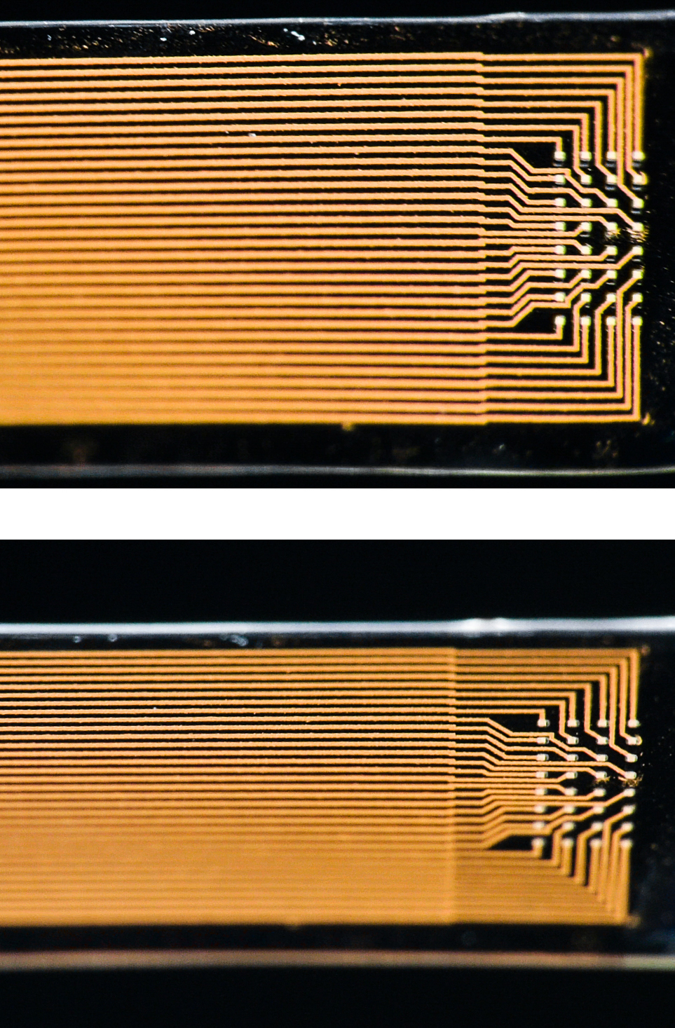
Stretchable soft electrodeThe soft electrode stretched to twice its length Photo credit: Thor Balkhed

“As human tissue is elastic and mobile, damage and inflammation arise at the interface with rigid electronic components. It not only causes damage to tissue; it also attenuates neural signals,” says Klas Tybrandt, leader of the Soft Electronics group at the Laboratory of Organic Electronics, Linköping University, Campus Norrköping.

New conductive material

Klas Tybrandt has developed a new conductive material that is as soft as human tissue and can be stretched to twice its length. The material consists of gold coated titanium dioxide nanowires, embedded into silicone rubber. The material is biocompatible – which means it can be in contact with the body without adverse effects – and its conductivity remains stable over time.

“The microfabrication of soft electrically conductive composites involves several challenges. We have developed a process to manufacture small electrodes that also preserves the biocompatibility of the materials. The process uses very little material, and this means that we can work with a relatively expensive material such as gold, without the cost becoming prohibitive,” says Klas Tybrandt.

The electrodes are 50 µm [microns or micrometres] in size and are located at a distance of 200 µm from each other. The fabrication procedure allows 32 electrodes to be placed onto a very small surface. The final probe, shown in the photograph, has a width of 3.2 mm and a thickness of 80 µm.

The soft microelectrodes have been developed at Linköping University and ETH Zürich, and researchers at New York University and Columbia University have subsequently implanted them in the brain of rats. The researchers were able to collect high-quality neural signals from the freely moving rats for 3 months. The experiments have been subject to ethical review, and have followed the strict regulations that govern animal experiments.

Important future applications

Klas Tybrandt, researcher at Laboratory for Organic ElectronicsKlas Tybrandt, researcher at Laboratory for Organic Electronics Photo credit: Thor Balkhed

“When the neurons in the brain transmit signals, a voltage is formed that the electrodes detect and transmit onwards through a tiny amplifier. We can also see which electrodes the signals came from, which means that we can estimate the location in the brain where the signals originated. This type of spatiotemporal information is important for future applications. We hope to be able to see, for example, where the signal that causes an epileptic seizure starts, a prerequisite for treating it. Another area of application is brain-machine interfaces, by which future technology and prostheses can be controlled with the aid of neural signals. There are also many interesting applications involving the peripheral nervous system in the body and the way it regulates various organs,” says Klas Tybrandt.

The breakthrough is the foundation of the research area Soft Electronics, currently being established at Linköping University, with Klas Tybrandt as principal investigator.
liu.se/soft-electronics

A video has been made available (Note: For those who find any notion of animal testing disturbing; don’t watch the video even though it is an animation and does not feature live animals),

Here’s a link to and a citation for the paper,

High-Density Stretchable Electrode Grids for Chronic Neural Recording by Klas Tybrandt, Dion Khodagholy, Bernd Dielacher, Flurin Stauffer, Aline F. Renz, György Buzsáki, and János Vörös. Advanced Materials 2018. DOI: 10.1002/adma.201706520
 First published 28 February 2018

This paper is open access.

Emergence in Toronto and Ottawa and brains in Vancouver (Canada): three April 2018 events

April 2018 is shaping up to be quite the month where art/sci events are concerned. I just published a March 27, 2018 posting titled ‘Curiosity collides with the quantum and with the Science Writers and Communicators of Canada in Vancouver (Canada)‘ and I’ve now received news about more happenings in Toronto and Ottawa.  Plus, there’s a science-themed meeting organized by ARPICO (Society of Italian Researchers &; Professionals in Western Canada) featuring brains and brain imaging in Vancouver.

Toronto’s and Ottawa’s Emergence

There’s an art/sci exhibit opening, from a March 27, 2018 Art/Sci Salon announcement (received via email),
You are invited!

FaceBook event:

The Oakwood Village Library and Arts Centre event:

341 Oakwood Avenue, Toronto, ON  M6E 2W1

I check the library webpage listed in the above and found this artist’s statement,

Artist / Scientist Statement [Stephen Morris]

I am interested in self-organized, emergent patterns and textures. I make images of patterns both from the natural world and of experiments in my laboratory in the Department of Physics at the University of Toronto. Patterns naturally attract casual attention but are also the subject of serious scientific research. Some things just evolve all by themselves into strikingly regular shapes and textures. Why? These shapes emerge spontaneously from a dynamic process of growing, folding, cracking, wrinkling, branching, flowing and other kinds of morphological development. My photos are informed by the scientific aesthetic of nonlinear physics, and celebrate the subtle interplay of order and complexity in emergent patterns. They are a kind of “Scientific Folk Art” of the science of Emergence.

While the official opening is April 5, 2018, the event itself runs from April 1 – 30, 2018.

Next, there’s another March 27, 2018 announcement (received via email) from the Art/Sci Salon but this one concerns a series of talks about ’emergence’, Note: Some of the event information was a little difficult to decipher so I’ve added a note to the relevant section).

What is Emergent Form?

Nature teems with self-organized forms that seem to spring spontaneously from the smooth background of things, by mechanisms that are not always apparent. Think of rippled sand on a beach or regular stripes in the clouds.  Plants, insects and animals exhibit spirals and spots and stripes in an exuberant riot of colours.  Fluid flows in amazingly regular swirls and eddies.  The emergence of form is ubiquitous, and presents a challenge and an inspiration to both artists and scientists. In mathematics, patterns appear as solutions of the nonlinear partial differential equations in the continuum limit of classical physics, chemistry and biology. In the arts and humanities, “emergent form” addresses the entangled ways in which humans, plants animals, microorganisms inevitably co-exist in the universe; the way that human intervention and natural transformation can generate new landscapes and new forms of life.

With Emergent Form, we want to question the idea of a fixed world.

For us, Emergent Form is not just a series of natural and human phenomena too complicated to understand, measure or predict, but also a concept to help us identify ways in which we can come to term with, and embrace their complexity as a source of inspiration.

Join us in Toronto and Ottawa for a series of interdisciplinary discussions, performances and exhibitions on Emergent Form on Apr 10, 11, 12 (Toronto) and Apr. 14 [2018] (Ottawa).

This series is the result of a collaboration among several parties. Each event of the series is different and has its dedicated RSVP 

Tue. Apr 10 The Fields Institute, 222 College Street

Emergent form: an interdisciplinary concept 6:00-8:00 pm Pier Luigi Capucci, Accademia di Belle Arti Urbino. Founder and director, Noemalab*, Charles Sowers, Independent artist and exhibit designer, the Exploratorium, Stephen Morris, Professor of of Physics University of Toronto, Ron Wild, smART Maps

CLICK HERE FOR MORE AND TO RSVP

Wed. Apr 11 The Fields Institute6:00-8:00 pm

Anatomy of an Interconnected SystemA Performative Lecture with Margherita Pevere, Aalto University, Helsinki

CLICK HERE FOR MORE AND TO RSVP

Thu. Apr 12 (Note: I believe that from 5 – 6 pm, you’re invited to see Pevere’s exhibit and then proceed to Luella Massey Studio Theatre for performances)

5:00 pm  Cabinets in the Koffler Student Centre [I believe this is at the University of Toronto] Anatomy of an Interconnected System An exhibition by Margherita Pevere

6:00 pm Luella Massey Studio Theatre, 4 Glen Morris Ave., Toronto biopoetriX – conFiGURing AI

6:00-8:00 pm Performance: 

6:00pm Performance “Corpus Nil. A Ritual of Birth for a Modified Body” conceived and performed by Marco Donnarumma

6.30pm LAB dance: Blitz media posters on labs in the arts, sciences and engineering

7.10pm Panel: Performing AI, hybrid media and humans in/as technologyMarco Donnarumma, Doug van Nort (Dispersion Lab, York U.), Jane Tingley (Stratford User Research & Gameful Experiences Lab –SURGE-, U of Waterloo), Angela Schoellig (Dynamic Systems Lab, U of T)

Panel animators: Antje Budde (Digital Dramaturgy Lab) and Roberta Buiani (ArtSci Salon)

8.15pm Reception at the Italian Cultural Institute, 496 Huron St, Toronto

CLICK HERE FOR MORE AND TO RSVP

Ottawa. Sat. Apr. 14 National Arts Centre, 1 Elgin Street11:00 am-1:00 pm

Emergent Form and complex phenomenaA creative panel discussion and surprise demonstrationsWith Pier Luigi Capucci, Margherita Pevere, Marco Donnarumma, Stephen Morris

CLICK HERE FOR MORE AND TO RSVP

This event would not be possible without the support of The Fields Institute for Research in Mathematical Science, The Italian Embassy, the Centre for Drama, Theatre and Performance Studies at the University of Toronto, the Digital Dramaturgy Lab, and the Istituto Italiano di Cultura. Many thanks to our community partner BYOR (Bring your own Robot)

I wonder if some of the funding from Italy is in support of Italian Research in World Day. This is the inaugural year for the event, which will be held annually on April 15.

Vancouver’s brains

The Society of Italian Researchers and Professionals in Western Canada (ARPICO) is hosting an event in Vancouver (from a March 22, 2018 ARICO announcement received via email),

Our second speaking event of the year, in collaboration with the Consulate General of Italy in Vancouver, has been scheduled for Wednesday, April 11th, 2018 at the Roundhouse Community Centre. Professor Vesna Sossi’s talk will be examining how positron emission tomography (PET) imaging has contributed to better understanding of the brain function and disease with particular focus on Parkinson’s disease. You can read a summary of Prof. Sossi’s lecture as well as her short professional biography at the bottom of this message.

This event is organized in collaboration with the Consulate General of Italy in Vancouver to celebrate the newly instituted Italian Research in the World Day, as part of the Piano Straordinario “Vivere all’Italiana” – Giornata della ricerca Italiana nel mondo. You can read more on our website event page.

We look forward to seeing everyone there.

Please register for the event by visiting the EventBrite link or RSVPing to info@arpico.ca.

The evening agenda is as follows:

  • 6:45 pm – Doors Open
  • 7:00 pm – Lecture by Prof. Vesna Sossi
  • ~8:00 pm – Q & A Period
  • Mingling & Refreshments until about 9:30 pm

If you have not yet RSVP’d, please do so on our EventBrite page.

Further details are also available at arpico.ca, our facebook page, and Eventbrite.


Imaging: A Window into the Brain

Brain illness, comprising neurological disorders, mental illness and addiction, is considered the major health challenge in the 21st century with a socio-economic cost greater than cancer and cardiovascular disease combined. There are at least three unique challenges hampering brain disease management: relative inaccessibility, disease onset often preceding the onset of clinical symptoms by many years and overlap between clinical and pathological symptoms that makes accurate disease identification often difficult. This talk will give examples of how positron emission tomography (PET) imaging has contributed to better understanding of the brain function and disease with particular focus on Parkinson’s disease. Emphasis will be placed on the interplay between scientific discoveries and instrumentation and data analysis development as exemplified by the current understanding of the brain function as comprised by interactions between connectivity networks and neurochemistry and advancement in multi-modal imaging such as simultaneous PET and magnetic resonance imaging (MRI).

Vesna Sossi is a Professor in the University of British Columbia (UBC) Physics and Astronomy Department and at the UBC Djavad Mowafaghian Center for Brain Health. She directs the UBC Positron Emission Tomography (PET) imaging centre, which is known for its use of imaging as applied to neurodegeneration with emphasis on Parkinson’s disease. Her main areas of interest comprise development of imaging methods to enhance the investigation of neurochemical mechanisms that lead to an increased risk of Parkinson’s disease (PD) and mechanisms that contribute to treatment-related complications. She uses PET imaging to explore how alterations of the different neurotransmitter systems contribute to different trajectories of disease progression. Her other areas of interest are PET image analysis, instrumentation and multi-modal, multi-parameter data analysis. She published more than 180 peer review papers, is funded by several granting agencies, including the Michael J Fox Foundation, and sits on several national and international review panels.


WHEN: Wednesday, April 11th, 2018 at 7:00pm (doors open at 6:45pm)
WHERE: Roundhouse Community Centre, Room B – 181 Roundhouse Mews, Vancouver, BC, V6Z 2W3
RSVP: Please RSVP at EventBrite (https://imaging-a-window-into-the-brain.eventbrite.ca) or email info@arpico.ca


Tickets are Needed

  • Tickets are FREE, but all individuals are requested to obtain “free-admission” tickets on EventBrite site due to limited seating at the venue. Organizers need accurate registration numbers to manage wait lists and prepare name tags.
  • All ARPICO events are 100% staffed by volunteer organizers and helpers, however, room rental, stationery, and guest refreshments are costs incurred and underwritten by members of ARPICO. Therefore to be fair, all audience participants are asked to donate to the best of their ability at the door or via EventBrite to “help” defray costs of the event.

You can find directions for the Roundhouse Community Centre here

I have one idle question. What’s going to happen these groups if Canadians change their use of  Facebook or abandon the platform as they are threatening to do in the face of Cambridge Analytica’s use of their data? A March 25, 2018 article on huffingtonpost.ca outlines the latest about Canadians’ reaction to the Cambridge Analytical news according to an Angus Reid poll,

A survey by Angus Reid Institute suggests 73 per cent of Canadian Facebook users say they will make changes, while 27 per cent say it will be “business as usual.”

Nearly a quarter (23 per cent) said they would use Facebook less in the future, and 41 per cent of users said they would check and/or change their privacy settings.

The survey also found that one in 10 say they plan to abandon the platform, at least temporarily.

Facebook has been under fire for its ability to protect user privacy after Cambridge Analytica was accused of lifting the Facebook profiles of more than 50 million users without their permission.

There you have it.

*Well, a bit more information about one of the “Emergent’ speakers was received in an April 4, 2018 ArtSci Salon email announcement,

Do make sure to check out Pier Luigi Capucci’s EU-based (but with international breadth) Noemalab platform. https://noemalab.eu/ since the mid-nineties, this platform has been an important node of information for New Media Art and the relation between the arts and science.

noemalab’s blog regularly hosts reviews of events and conferences occurring around the world, including  the Subtle Technologies Festival between 2007 and 2014. you can search its archives here http://blogs.noemalab.eu/

Capucci has been writing several reflections on emergent forms of Life and theorized what he called the “third life”. See a recent essay https://noemalab.eu/memo/events/evolutionary-creativity-the-inner-life-and-meaning-of-art/ here is a picture which I would love him to explain during Emergent Form. Intrigued? come listen to him!
What is a multiregional brain-on-a-chip?

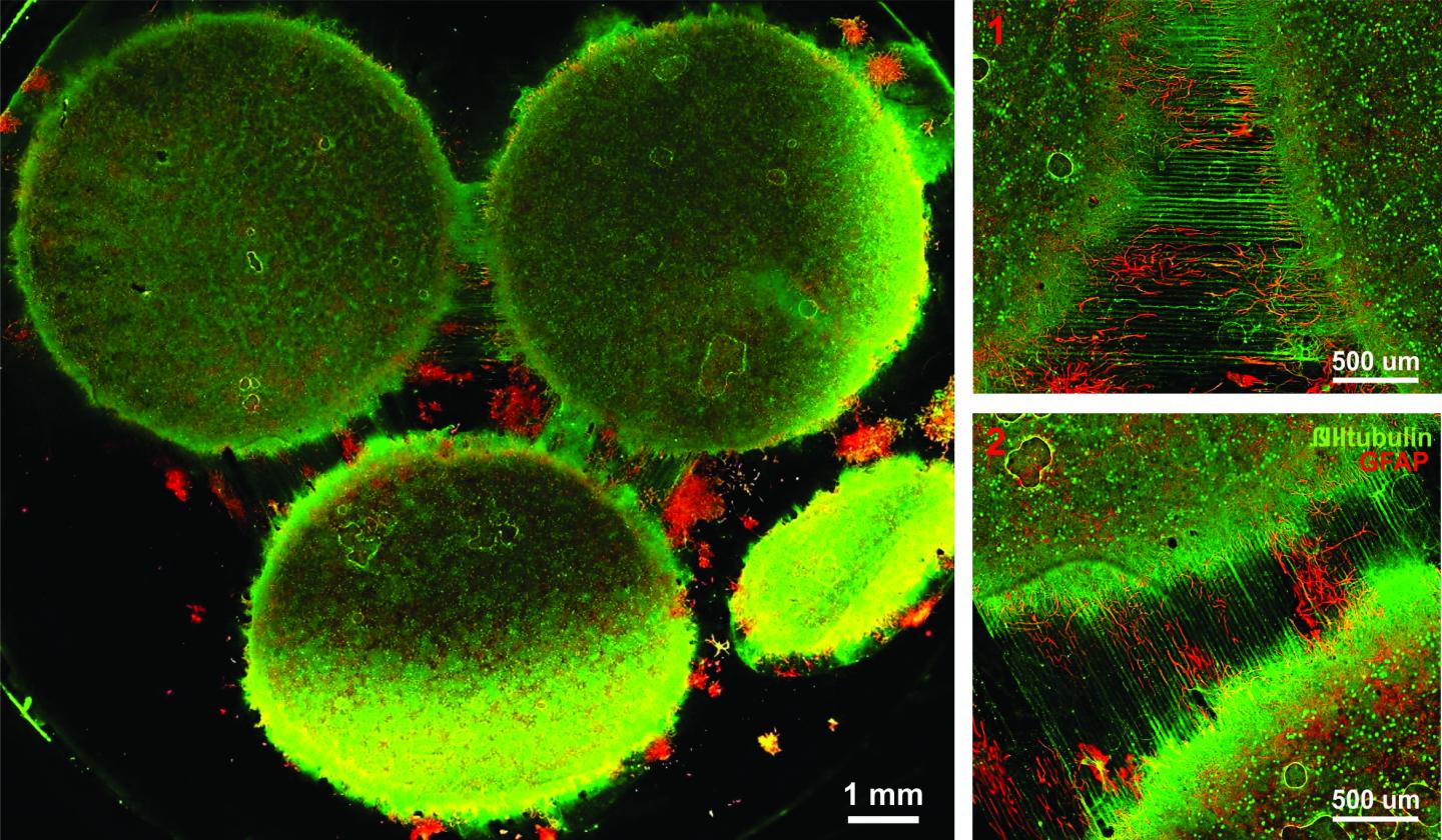
In response to having created a multiregional brain-on-a-chip, there’s an explanation from the team at Harvard University (which answers my question) in a Jan. 13, 2017 Harvard John A. Paulson School of Engineering and Applied Sciences news release (also on EurekAlert) by Leah Burrows,

Harvard University researchers have developed a multiregional brain-on-a-chip that models the connectivity between three distinct regions of the brain. The in vitro model was used to extensively characterize the differences between neurons from different regions of the brain and to mimic the system’s connectivity.

“The brain is so much more than individual neurons,” said Ben Maoz, co-first author of the paper and postdoctoral fellow in the Disease Biophysics Group in the Harvard John A. Paulson School of Engineering and Applied Sciences (SEAS). “It’s about the different types of cells and the connectivity between different regions of the brain. When modeling the brain, you need to be able to recapitulate that connectivity because there are many different diseases that attack those connections.”

“Roughly twenty-six percent of the US healthcare budget is spent on neurological and psychiatric disorders,” said Kit Parker, the Tarr Family Professor of Bioengineering and Applied Physics Building at SEAS and Core Faculty Member of the Wyss Institute for Biologically Inspired Engineering at Harvard University. “Tools to support the development of therapeutics to alleviate the suffering of these patients is not only the human thing to do, it is the best means of reducing this cost.”

Researchers from the Disease Biophysics Group at SEAS and the Wyss Institute modeled three regions of the brain most affected by schizophrenia — the amygdala, hippocampus and prefrontal cortex.

They began by characterizing the cell composition, protein expression, metabolism, and electrical activity of neurons from each region in vitro.

“It’s no surprise that neurons in distinct regions of the brain are different but it is surprising just how different they are,” said Stephanie Dauth, co-first author of the paper and former postdoctoral fellow in the Disease Biophysics Group. “We found that the cell-type ratio, the metabolism, the protein expression and the electrical activity all differ between regions in vitro. This shows that it does make a difference which brain region’s neurons you’re working with.”

Next, the team looked at how these neurons change when they’re communicating with one another. To do that, they cultured cells from each region independently and then let the cells establish connections via guided pathways embedded in the chip.

The researchers then measured cell composition and electrical activity again and found that the cells dramatically changed when they were in contact with neurons from different regions.

“When the cells are communicating with other regions, the cellular composition of the culture changes, the electrophysiology changes, all these inherent properties of the neurons change,” said Maoz. “This shows how important it is to implement different brain regions into in vitro models, especially when studying how neurological diseases impact connected regions of the brain.”

To demonstrate the chip’s efficacy in modeling disease, the team doped different regions of the brain with the drug Phencyclidine hydrochloride — commonly known as PCP — which simulates schizophrenia. The brain-on-a-chip allowed the researchers for the first time to look at both the drug’s impact on the individual regions as well as its downstream effect on the interconnected regions in vitro.

The brain-on-a-chip could be useful for studying any number of neurological and psychiatric diseases, including drug addiction, post traumatic stress disorder, and traumatic brain injury.

“To date, the Connectome project has not recognized all of the networks in the brain,” said Parker. “In our studies, we are showing that the extracellular matrix network is an important part of distinguishing different brain regions and that, subsequently, physiological and pathophysiological processes in these brain regions are unique. This advance will not only enable the development of therapeutics, but fundamental insights as to how we think, feel, and survive.”

Here’s an image from the researchers,

Caption: Image of the in vitro model showing three distinct regions of the brain connected by axons. Credit: Disease Biophysics Group/Harvard University

Here’s a link to and a citation for the paper,

Neurons derived from different brain regions are inherently different in vitro: A novel multiregional brain-on-a-chip by Stephanie Dauth, Ben M Maoz, Sean P Sheehy, Matthew A Hemphill, Tara Murty, Mary Kate Macedonia, Angie M Greer, Bogdan Budnik, Kevin Kit Parker. Journal of Neurophysiology Published 28 December 2016 Vol. no. [?] , DOI: 10.1152/jn.00575.2016

This paper is behind a paywall and they haven’t included the vol. no. in the citation I’ve found.