Tag Archives: Sean P Sheehy

New wound dressings with nanofibres for tissue regeneration

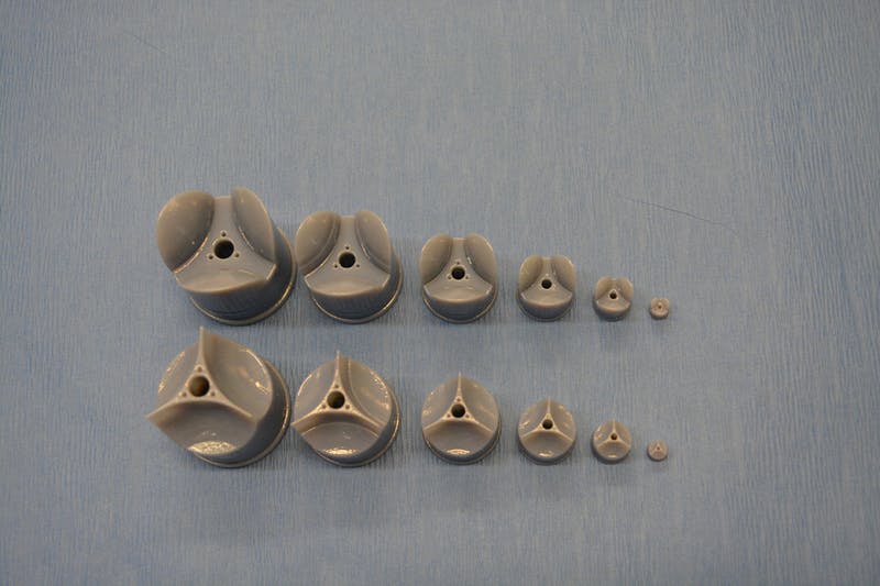
The Rotary Jet-Spinning manufacturing system was developed specifically as a therapeutic for the wounds of war. The dressings could be a good option for large wounds, such as burns, as well as smaller wounds on the face and hands, where preventing scarring is important. Illustration courtesy of Michael Rosnach/Harvard University

This image really gets the idea of regeneration across to the viewer while also informing you that this is medicine that comes from the military. A March 19,2018 news item on phys.org announces the work,

Researchers from the Harvard John A. Paulson School of Engineering and Applied Sciences (SEAS) and the Wyss Institute for Biologically Inspired Engineering have developed new wound dressings that dramatically accelerate healing and improve tissue regeneration. The two different types of nanofiber dressings, described in separate papers, use naturally-occurring proteins in plants and animals to promote healing and regrow tissue.

Our fiber manufacturing system was developed specifically for the purpose of developing therapeutics for the wounds of war,” said Kit Parker, the Tarr Family Professor of Bioengineering and Applied Physics at SEAS and senior author of the research. “As a soldier in Afghanistan, I witnessed horrible wounds and, at times, the healing process for those wounds was a horror unto itself. This research is a years-long effort by many people on my team to help with these problems.”

Parker is also a Core Faculty Member of the Wyss Institute.

The most recent paper, published in Biomaterials, describes a wound dressing inspired by fetal tissue.

A March 19, 2018 Harvard University John A. Paulson School of Engineering and Applied Science news release by Leah Burrows (also on EurekAlert), which originated the news item, provides some background information before launching into more detail about this latest work,

In the late 1970s, when scientists first started studying the wound-healing process early in development, they discovered something unexpected: Wounds incurred before the third trimester left no scars. This opened a range of possibilities for regenerative medicine. But for decades, researchers have struggled to replicate those unique properties of fetal skin.

Unlike adult skin, fetal skin has high levels of a protein called fibronectin, which assembles into the extracellular matrix and promotes cell binding and adhesion. Fibronectin has two structures: globular, which is found in blood, and fibrous, which is found in tissue. Even though fibrous fibronectin holds the most promise for wound healing, previous research focused on the globular structure, in part because manufacturing fibrous fibronectin was a major engineering challenge.

But Parker and his team are pioneers in the field of nanofiber engineering.

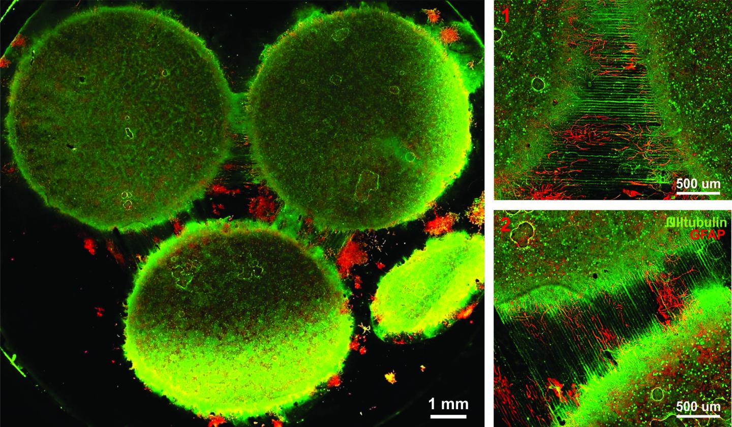
The researchers made fibrous fibronectin using a fiber-manufacturing platform called Rotary Jet-Spinning (RJS), developed by Parker’s Disease Biophysics Group. RJS works likes a cotton-candy machine — a liquid polymer solution, in this case globular fibronectin dissolved in a solvent, is loaded into a reservoir and pushed out through a tiny opening by centrifugal force as the device spins. As the solution leaves the reservoir, the solvent evaporates and the polymers solidify. The centrifugal force unfolds the globular protein into small, thin fibers. These fibers — less than one micrometer in diameter — can be collected to form a large-scale wound dressing or bandage.

“The dressing integrates into the wound and acts like an instructive scaffold, recruiting different stem cells that are relevant for regeneration and assisting in the healing process before being absorbed into the body,” said Christophe Chantre, a graduate student in the Disease Biophysics Group and first author of the paper.

In in vivo testing, the researchers found that wounds treated with the fibronectin dressing showed 84 percent tissue restoration within 20 days, compared with 55.6 percent restoration in wounds treated with a standard dressing.

The researchers also demonstrated that wounds treated with the fibronectin dressing had almost normal epidermal thickness and dermal architecture, and even regrew hair follicles — often considered one of the biggest challenges in the field of wound healing.

“This is an important step forward,” said Chantre. “Most work done on skin regeneration to date involves complex treatments combining scaffolds, cells, and even growth factors. Here we were able to demonstrate tissue repair and hair follicle regeneration using an entirely material approach. This has clear advantages for clinical translation.”

In another paper published in Advanced Healthcare Materials, the Disease Biophysics Group demonstrated a soy-based nanofiber that also enhances and promotes wound healing.

Soy protein contains both estrogen-like molecules — which have been shown to accelerate wound healing — and bioactive molecules similar to those that build and support human cells.

“Both the soy- and fibronectin-fiber technologies owe their success to keen observations in reproductive medicine,” said Parker. “During a woman’s cycle, when her estrogen levels go high, a cut will heal faster. If you do a surgery on a baby still in the womb, they have scar-less wound healing. Both of these new technologies are rooted in the most fascinating of all the topics in human biology — how we reproduce.”

In a similar way to fibronectin fibers, the research team used RJS to spin ultrathin soy fibers into wound dressings. In experiments, the soy- and cellulose-based dressing demonstrated a 72 percent increase in healing over wounds with no dressing and a 21 percent increase in healing over wounds dressed without soy protein.

“These findings show the great promise of soy-based nanofibers for wound healing,” said Seungkuk Ahn, a graduate student in the Disease Biophysics Group and first author of the paper. “These one-step, cost-effective scaffolds could be the next generation of regenerative dressings and push the envelope of nanofiber technology and the wound-care market.”

Both kinds of dressing, according to researchers, have advantages in the wound-healing space. The soy-based nanofibers — consisting of cellulose acetate and soy protein hydrolysate — are inexpensive, making them a good option for large-scale use, such as on burns. The fibronectin dressings, on the other hand, could be used for smaller wounds on the face and hands, where preventing scarring is important.

Here’s are links and citations for both papers mentioned in the news release,

Soy Protein/Cellulose Nanofiber Scaffolds Mimicking Skin Extracellular Matrix for Enhanced Wound Healing by Seungkuk Ahn, Christophe O. Chantre, Alanna R. Gannon, Johan U. Lind, Patrick H. Campbell, Thomas Grevesse, Blakely B. O’Connor, Kevin Kit Parker. Advanced Healthcare Materials https://doi.org/10.1002/adhm.201701175 First published: 23 January 2018

Production-scale fibronectin nanofibers promote wound closure and tissue repair in a dermal mouse model by Christophe O. Chantre, Patrick H. Campbell, Holly M. Golecki, Adrian T. Buganza, Andrew K. Capulli, Leila F. Deravi, Stephanie Dauth, Sean P. Sheehy, Jeffrey A.Paten. KarlGledhill, Yanne S. Doucet, Hasan E.Abaci, Seungkuk Ahn, Benjamin D.Pope, Jeffrey W.Ruberti, Simon P.Hoerstrup, Angela M.Christiano, Kevin Kit Parker. Biomaterials Volume 166, June 2018, Pages 96-108 https://doi.org/10.1016/j.biomaterials.2018.03.006 Available online 5 March 2018

Both papers are behind paywalls although you may want to check with ResearchGate where many researchers make their papers available for free.

One last comment, I noticed this at the end of Burrows’ news release,

The Harvard Office of Technology Development has protected the intellectual property relating to these projects and is exploring commercialization opportunities.

It reminded me of the patent battle between the Broad Institute (a Harvard University and Massachusetts Institute of Technology joint venture) and the University of California at Berkeley over CRISPR (clustered regularly interspaced short palindromic repeats) technology. (My March 15, 2017 posting describes the battle’s outcome.)

Lest we forget, there could be major financial rewards from this work.

A nano fabrication technique used to create next generation heart valve

I am going to have take the researchers’ word that these somehow lead to healthy heart valve tissue,

In rotary jet spinning technology, a rotating nozzle extrudes a solution of extracellular matrix (ECM) into nanofibers that wrap themselves around heart valve-shaped mandrels. By using a series of mandrels with different sizes, the manufacturing process becomes fully scalable and is able to provide JetValves for all age groups and heart sizes. Credit: Wyss Institute at Harvard University

From a May 18, 2017 news item on ScienceDaily,

The human heart beats approximately 35 million times every year, effectively pumping blood into the circulation via four different heart valves. Unfortunately, in over four million people each year, these delicate tissues malfunction due to birth defects, age-related deteriorations, and infections, causing cardiac valve disease.

Today, clinicians use either artificial prostheses or fixed animal and cadaver-sourced tissues to replace defective valves. While these prostheses can restore the function of the heart for a while, they are associated with adverse comorbidity and wear down and need to be replaced during invasive and expensive surgeries. Moreover, in children, implanted heart valve prostheses need to be replaced even more often as they cannot grow with the child.

A team lead by Kevin Kit Parker, Ph.D. at Harvard University’s Wyss Institute for Biologically Inspired Engineering recently developed a nanofiber fabrication technique to rapidly manufacture heart valves with regenerative and growth potential. In a paper published in Biomaterials, Andrew Capulli, Ph.D. and colleagues fabricated a valve-shaped nanofiber network that mimics the mechanical and chemical properties of the native valve extracellular matrix (ECM). To achieve this, the team used the Parker lab’s proprietary rotary jet spinning technology — in which a rotating nozzle extrudes an ECM solution into nanofibers that wrap themselves around heart valve-shaped mandrels. “Our setup is like a very fast cotton candy machine that can spin a range of synthetic and natural occurring materials. In this study, we used a combination of synthetic polymers and ECM proteins to fabricate biocompatible JetValves that are hemodynamically competent upon implantation and support cell migration and re-population in vitro. Importantly, we can make human-sized JetValves in minutes — much faster than possible for other regenerative prostheses,” said Parker.

A May 18,2017 Wyss Institute for Biologically Inspired Engineering news release (also on EurekAlert), which originated the news item, expands on the theme of Jetvalves,

To further develop and test the clinical potential of JetValves, Parker’s team collaborated with the translational team of Simon P. Hoerstrup, M.D., Ph.D., at the University of Zurich in Switzerland, which is a partner institution with the Wyss Institute. As a leader in regenerative heart prostheses, Hoerstrup and his team in Zurich have previously developed regenerative, tissue-engineered heart valves to replace mechanical and fixed-tissue heart valves. In Hoerstrup’s approach, human cells directly deposit a regenerative layer of complex ECM on biodegradable scaffolds shaped as heart valves and vessels. The living cells are then eliminated from the scaffolds resulting in an “off-the-shelf” human matrix-based prostheses ready for implantation.

In the paper, the cross-disciplinary team successfully implanted JetValves in sheep using a minimally invasive technique and demonstrated that the valves functioned properly in the circulation and regenerated new tissue. “In our previous studies, the cell-derived ECM-coated scaffolds could recruit cells from the receiving animal’s heart and support cell proliferation, matrix remodeling, tissue regeneration, and even animal growth. While these valves are safe and effective, their manufacturing remains complex and expensive as human cells must be cultured for a long time under heavily regulated conditions. The JetValve’s much faster manufacturing process can be a game-changer in this respect. If we can replicate these results in humans, this technology could have invaluable benefits in minimizing the number of pediatric re-operations,” said Hoerstrup.

In support of these translational efforts, the Wyss Institute for Biologically Inspired Engineering and the University of Zurich announced today a cross-institutional team effort to generate a functional heart valve replacement with the capacity for repair, regeneration, and growth. The team is also working towards a GMP-grade version of their customizable, scalable, and cost-effective manufacturing process that would enable deployment to a large patient population. In addition, the new heart valve would be compatible with minimally invasive procedures to serve both pediatric and adult patients.

The project will be led jointly by Parker and Hoerstrup. Parker is a Core Faculty member of the Wyss Institute and the Tarr Family Professor of Bioengineering and Applied Physics at the Harvard John A. Paulson School of Engineering and Applied Sciences (SEAS). Hoerstrup is Chair and Director of the University of Zurich’s Institute for Regenerative Medicine (IREM), Co-Director of the recently founded Wyss Translational Center Zurich and a Wyss Institute Associate Faculty member.

Since JetValves can be manufactured in all desired shapes and sizes, and take seconds to minutes to produce, the team’s goal is to provide customized, ready-to-use, regenerative heart valves much faster and at much lower cost than currently possible.

“Achieving the goal of minimally invasive, low-cost regenerating heart valves could have tremendous impact on patients’ lives across age-, social- and geographical boundaries. Once again, our collaborative team structure that combines unique and leading expertise in bioengineering, regenerative medicine, surgical innovation and business development across the Wyss Institute and our partner institutions, makes it possible for us to advance technology development in ways not possible in a conventional academic laboratory,” said Wyss Institute Founding Director Donald Ingber, M.D., Ph.D., who is also the Judah Folkman Professor of Vascular Biology at HMS and the Vascular Biology Program at Boston Children’s Hospital, as well as Professor of Bioengineering at SEAS.

This scanning electron microscopy image shows how extracellular matrix (ECM) nanofibers generated with JetValve technology are arranged in parallel networks with physical properties comparable to those found in native heart tissue. Credit: Wyss Institute at Harvard University

Here’s a link to and a citation for the paper,

JetValve: Rapid manufacturing of biohybrid scaffolds for biomimetic heart valve replacement by Andrew K. Capulli, Maximillian Y. Emmert, Francesco S. Pasqualini, b, Debora Kehl, Etem Caliskan, Johan U. Lind, Sean P. Sheehy, Sung Jin Park, Seungkuk Ahn, Benedikt Webe, Josue A. Goss. Biomaterials Volume 133, July 2017, Pages 229–241  https://doi.org/10.1016/j.biomaterials.2017.04.033

This paper is behind a paywall.

What is a multiregional brain-on-a-chip?

In response to having created a multiregional brain-on-a-chip, there’s an explanation from the team at Harvard University (which answers my question) in a Jan. 13, 2017 Harvard John A. Paulson School of Engineering and Applied Sciences news release (also on EurekAlert) by Leah Burrows,

Harvard University researchers have developed a multiregional brain-on-a-chip that models the connectivity between three distinct regions of the brain. The in vitro model was used to extensively characterize the differences between neurons from different regions of the brain and to mimic the system’s connectivity.

“The brain is so much more than individual neurons,” said Ben Maoz, co-first author of the paper and postdoctoral fellow in the Disease Biophysics Group in the Harvard John A. Paulson School of Engineering and Applied Sciences (SEAS). “It’s about the different types of cells and the connectivity between different regions of the brain. When modeling the brain, you need to be able to recapitulate that connectivity because there are many different diseases that attack those connections.”

“Roughly twenty-six percent of the US healthcare budget is spent on neurological and psychiatric disorders,” said Kit Parker, the Tarr Family Professor of Bioengineering and Applied Physics Building at SEAS and Core Faculty Member of the Wyss Institute for Biologically Inspired Engineering at Harvard University. “Tools to support the development of therapeutics to alleviate the suffering of these patients is not only the human thing to do, it is the best means of reducing this cost.”

Researchers from the Disease Biophysics Group at SEAS and the Wyss Institute modeled three regions of the brain most affected by schizophrenia — the amygdala, hippocampus and prefrontal cortex.

They began by characterizing the cell composition, protein expression, metabolism, and electrical activity of neurons from each region in vitro.

“It’s no surprise that neurons in distinct regions of the brain are different but it is surprising just how different they are,” said Stephanie Dauth, co-first author of the paper and former postdoctoral fellow in the Disease Biophysics Group. “We found that the cell-type ratio, the metabolism, the protein expression and the electrical activity all differ between regions in vitro. This shows that it does make a difference which brain region’s neurons you’re working with.”

Next, the team looked at how these neurons change when they’re communicating with one another. To do that, they cultured cells from each region independently and then let the cells establish connections via guided pathways embedded in the chip.

The researchers then measured cell composition and electrical activity again and found that the cells dramatically changed when they were in contact with neurons from different regions.

“When the cells are communicating with other regions, the cellular composition of the culture changes, the electrophysiology changes, all these inherent properties of the neurons change,” said Maoz. “This shows how important it is to implement different brain regions into in vitro models, especially when studying how neurological diseases impact connected regions of the brain.”

To demonstrate the chip’s efficacy in modeling disease, the team doped different regions of the brain with the drug Phencyclidine hydrochloride — commonly known as PCP — which simulates schizophrenia. The brain-on-a-chip allowed the researchers for the first time to look at both the drug’s impact on the individual regions as well as its downstream effect on the interconnected regions in vitro.

The brain-on-a-chip could be useful for studying any number of neurological and psychiatric diseases, including drug addiction, post traumatic stress disorder, and traumatic brain injury.

“To date, the Connectome project has not recognized all of the networks in the brain,” said Parker. “In our studies, we are showing that the extracellular matrix network is an important part of distinguishing different brain regions and that, subsequently, physiological and pathophysiological processes in these brain regions are unique. This advance will not only enable the development of therapeutics, but fundamental insights as to how we think, feel, and survive.”

Here’s an image from the researchers,

Caption: Image of the in vitro model showing three distinct regions of the brain connected by axons. Credit: Disease Biophysics Group/Harvard University

Here’s a link to and a citation for the paper,

Neurons derived from different brain regions are inherently different in vitro: A novel multiregional brain-on-a-chip by Stephanie Dauth, Ben M Maoz, Sean P Sheehy, Matthew A Hemphill, Tara Murty, Mary Kate Macedonia, Angie M Greer, Bogdan Budnik, Kevin Kit Parker. Journal of Neurophysiology Published 28 December 2016 Vol. no. [?] , DOI: 10.1152/jn.00575.2016

This paper is behind a paywall and they haven’t included the vol. no. in the citation I’ve found.