Tag Archives: Kevin Kit Parker

Biohybrid fish made from human cardiac cells could lead to artificial hearts

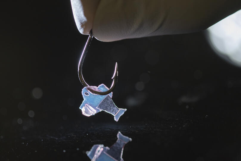
Biohybrid fish on a hook (Photo credit to Michael Rosnach, Keel Yong Lee, Sung-Jin Park, Kevin Kit Parker)

A February 10, 2022 news item on ScienceDaily announces research on a biohybrid fish,

Harvard University researchers, in collaboration with colleagues from Emory University, have developed the first fully autonomous biohybrid fish from human stem-cell derived cardiac muscle cells. The artificial fish swims by recreating the muscle contractions of a pumping heart, bringing researchers one step closer to developing a more complex artificial muscular pump and providing a platform to study heart disease like arrhythmia.

A February 10, 2022 Harvard University John A. Paulson School of Engineering and Applied Sciences news release (also on EurekAlert) by Leah Burrows explains how this research could lead to an artificial heart (Note: Links have been removed),

“Our ultimate goal is to build an artificial heart to replace a malformed heart in a child,” said Kit Parker, the Tarr Family Professor of Bioengineering and Applied Physics at the Harvard John A. Paulson School of Engineering and Applied Sciences (SEAS) and senior author of the paper.  “Most of the work in building heart tissue or hearts, including some work we have done, is focused on replicating the anatomical features or replicating the simple beating of the heart in the engineered tissues. But here, we are drawing design inspiration from the biophysics of the heart, which is harder to do. Now, rather than using heart imaging as a blueprint, we are identifying the key biophysical principles that make the heart work, using them as design criteria, and replicating them in a system, a living, swimming fish, where it is much easier to see if we are successful.”

The research is published in Science

The biohybrid fish developed by the team builds off previous research from Parker’s Disease Biophysics Group. In 2012, the lab used cardiac muscle cells from rats to build a jellyfish-like biohybrid pump and in 2016 the researchers developed a swimming, artificial stingray also from rat heart muscle cells.

In this research, the team built the first autonomous biohybrid device made from human stem-cell derived cardiomyocytes. This device was inspired by the shape and swimming motion of a zebrafish. Unlike previous devices, the biohybrid zebrafish has two layers of muscle cells, one on each side of the tail fin. When one side contracts, the other stretches. That stretch triggers the opening of a mechanosensitive protein channel, which causes a contraction, which triggers a stretch and so on and so forth, leading to a closed loop system that can propel the fish for more than 100 days. 

“By leveraging cardiac mechano-electrical signaling between two layers of muscle, we recreated the cycle where each contraction results automatically as a response to the stretching on the opposite side,” said Keel Yong Lee, a postdoctoral fellow at SEAS and co-first author of the study. “The results highlight the role of feedback mechanisms in muscular pumps such as the heart.”

The researchers also engineered an autonomous pacing node, like a pacemaker, which controls the frequency and rhythm of these spontaneous contractions. Together, the two layers of muscle and the autonomous pacing node enabled the generation of continuous, spontaneous, and coordinated, back-and-forth fin movements.

“Because of the two internal pacing mechanisms, our fish can live longer, move faster and swim more efficiently than previous work,” said Sung-Jin Park, a former postdoctoral fellow in the Disease Biophysics Group at SEAS and co-first author of the study. “This new research provides a model to investigate mechano-electrical signaling as a therapeutic target of heart rhythm management and for understanding pathophysiology in sinoatrial node dysfunctions and cardiac arrhythmia.”

Park is currently an Assistant Professor at the Coulter Department of Biomedical Engineering at Georgia Institute of Technology and Emory University School of Medicine.

Unlike a fish in your refrigerator, this biohybrid fish improves with age. Its muscle contraction amplitude, maximum swimming speed, and muscle coordination all increased for the first month as the cardiomyocyte cells matured.  Eventually, the biohybrid fish reached speeds and swimming efficacy similar to zebrafish in the wild. 

Next, the team aims to build even more complex biohybrid devices from human heart cells. 

“I could build a model heart out of Play-Doh, it doesn’t mean I can build a heart,” said Parker. “You can grow some random tumor cells in a dish until they curdle into a throbbing lump and call it a cardiac organoid. Neither of those efforts is going to, by design, recapitulate the physics of a system that beats over a billion times during your lifetime while simultaneously rebuilding its cells on the fly. That is the challenge. That is where we go to work.”

The research was co-authored by David G. Matthews, Sean L. Kim, Carlos Antonio Marquez, John F. Zimmerman, Herdeline Ann M. Ardona, Andre G. Kleber and George V. Lauder. 

It was supported in part by National Institutes of Health National Center for Advancing Translational Sciences grant UH3TR000522, and National Science Foundation Materials Research Science and Engineering Center grant DMR-142057.

Before giving you a link and a citation for the paper, here’s a little more information about the work from a February 10, 2022 American Association for the Advancement of Science (AAAS) news release on EurekAlert announcing publication of the paper in their journal Science, Note: A link has been removed,

An autonomously swimming biohybrid fish, designed with a focus on two key regulatory features of the human heart, has revealed the importance of feedback mechanisms in muscular pumps (such as the heart). The findings could one day help inform the development of an artificial heart made from living muscle cells. Biohybrid systems – devices containing both biological and artificial components – are an effective way to investigate the physiological control mechanisms in biological organisms and to discover bio-inspired robotic solutions to a host of pressing concerns, including those related to human health. When it comes to natural fluid transport pumps, like those that circulate blood, the performance of biohybrid systems has been lacking, however.  Here, researchers considered whether two functional regulatory features of the heart — mechanoelectrical signaling and automaticity — could be transferred to a synthetic analog of another fluid transport system: a swimming fish. Lee et al. developed an autonomously swimming fish constructed from a bilayer of human cardiac cells; the muscular bilayer was integrated using tissue engineering techniques. Lee and team were able to control muscle contractions in the biohybrid fish using external optogenetic stimulation, allowing the fish analog to swim. In tests, the biohybrid fish outperformed the locomotory speed of previous biohybrid muscular systems, the authors say. It maintained spontaneous activity for 108 days. By contrast, say the authors, biohybrid fish equipped with single-layered muscle showed deteriorating activity within the first month. The data in this study demonstrate the potential of muscular bilayer systems and mechanoelectrical signaling as a means to promote maturation of in vitro muscle tissues, write Lee and colleagues. “Taken together,” the authors conclude, “the technology described here may represent foundational work toward the goal of creating autonomous systems capable of homeostatic regulation and adaptive behavioral control.”

For reporters interested in trends, this work builds upon previous work published in a July 2016 study in Science, in which Sung-jin Park et al. used cardiac cells from rats to develop a self-propelling ray fish analog.

Here’s a link to and a citation for the paper,

An autonomously swimming biohybrid fish designed with human cardiac biophysics by Keel Yong Lee, Sung-Jin Park, David G. Matthews. Sean L. Kim, Carlos Antonio Marquez, John F. Zimmerman, Herdeline Ann M. Ardoña, Andre G. Kleber, George V. Lauder and Kevin Kit Parker. Science • 10 Feb 2022 • Vol 375, Issue 6581 • pp. 639-647 • DOI: 10.1126/science.abh0474

This paper is behind a paywall.

Suit up with nanofiber for protection against explosions and high temperatures

Where explosions are concerned you might expect to see some army research and you would be right. A June 29, 2020 news item on ScienceDaily breaks the news,

Since World War I, the vast majority of American combat casualties has come not from gunshot wounds but from explosions. Today, most soldiers wear a heavy, bullet-proof vest to protect their torso but much of their body remains exposed to the indiscriminate aim of explosive fragments and shrapnel.

Designing equipment to protect extremities against the extreme temperatures and deadly projectiles that accompany an explosion has been difficult because of a fundamental property of materials. Materials that are strong enough to protect against ballistic threats can’t protect against extreme temperatures and vice versa. As a result, much of today’s protective equipment is composed of multiple layers of different materials, leading to bulky, heavy gear that, if worn on the arms and legs, would severely limit a soldier’s mobility.

Now, Harvard University researchers, in collaboration with the U.S. Army Combat Capabilities Development Command Soldier Center (CCDC SC) and West Point, have developed a lightweight, multifunctional nanofiber material that can protect wearers from both extreme temperatures and ballistic threats.

A June 29, 2020 Harvard University news release (also on EurekAlert) by Leah Burrows, which originated the news item, expands on the theme,

“When I was in combat in Afghanistan, I saw firsthand how body armor could save lives,” said senior author Kit Parker, the Tarr Family Professor of Bioengineering and Applied Physics at the Harvard John A. Paulson School of Engineering and Applied Sciences (SEAS) and a lieutenant colonel in the United States Army Reserve. “I also saw how heavy body armor could limit mobility. As soldiers on the battlefield, the three primary tasks are to move, shoot, and communicate. If you limit one of those, you decrease survivability and you endanger mission success.”

“Our goal was to design a multifunctional material that could protect someone working in an extreme environment, such as an astronaut, firefighter or soldier, from the many different threats they face,” said Grant M. Gonzalez, a postdoctoral fellow at SEAS and first author of the paper.

In order to achieve this practical goal, the researchers needed to explore the tradeoff between mechanical protection and thermal insulation, properties rooted in a material’s molecular structure and orientation.

Materials with strong mechanical protection, such as metals and ceramics, have a highly ordered and aligned molecular structure. This structure allows them to withstand and distribute the energy of a direct blow. Insulating materials, on the other hand, have a much less ordered structure, which prevents the transmission of heat through the material.

Kevlar and Twaron are commercial products used extensively in protective equipment and can provide either ballistic or thermal protection, depending on how they are manufactured. Woven Kevlar, for example, has a highly aligned crystalline structure and is used in protective bulletproof vests. Porous Kevlar aerogels, on the other hand, have been shown to have high thermal insulation.

“Our idea was to use this Kevlar polymer to combine the woven, ordered structure of fibers with the porosity of aerogels to make long, continuous fibers with porous spacing in between,” said Gonzalez. “In this system, the long fibers could resist a mechanical impact while the pores would limit heat diffusion.”

The research team used immersion Rotary Jet-Spinning (iRJS), a technique developed by Parker’s Disease Biophysics Group, to manufacture the fibers. In this technique, a liquid polymer solution is loaded into a reservoir and pushed out through a tiny opening by centrifugal force as the device spins. When the polymer solution shoots out of the reservoir, it first passes through an area of open air, where the polymers elongate and the chains align. Then the solution hits a liquid bath that removes the solvent and precipitates the polymers to form solid fibers. Since the bath is also spinning — like water in a salad spinner — the nanofibers follow the stream of the vortex and wrap around a rotating collector at the base of the device.

By tuning the viscosity of the liquid polymer solution, the researchers were able to spin long, aligned nanofibers into porous sheets — providing enough order to protect against projectiles but enough disorder to protect against heat. In about 10 minutes, the team could spin sheets about 10 by 30 centimeters in size.

To test the sheets, the Harvard team turned to their collaborators to perform ballistic tests. Researchers at CCDC SC in Natick, Massachusetts simulated shrapnel impact by shooting large, BB-like projectiles at the sample. The team performed tests by sandwiching the nanofiber sheets between sheets of woven Twaron. They observed little difference in protection between a stack of all woven Twaron sheets and a combined stack of woven Twaron and spun nanofibers.

“The capabilities of the CCDC SC allow us to quantify the successes of our fibers from the perspective of protective equipment for warfighters, specifically,” said Gonzalez.

“Academic collaborations, especially those with distinguished local universities such as Harvard, provide CCDC SC the opportunity to leverage cutting-edge expertise and facilities to augment our own R&D capabilities,” said Kathleen Swana, a researcher at CCDC SC and one of the paper’s authors. “CCDC SC, in return, provides valuable scientific and soldier-centric expertise and testing capabilities to help drive the research forward.”

In testing for thermal protection, the researchers found that the nanofibers provided 20 times the heat insulation capability of commercial Twaron and Kevlar.

“While there are improvements that could be made, we have pushed the boundaries of what’s possible and started moving the field towards this kind of multifunctional material,” said Gonzalez.

“We’ve shown that you can develop highly protective textiles for people that work in harm’s way,” said Parker. “Our challenge now is to evolve the scientific advances to innovative products for my brothers and sisters in arms.”

Harvard’s Office of Technology Development has filed a patent application for the technology and is actively seeking commercialization opportunities.

Here’s a link to and a citation for the paper,

para-Aramid Fiber Sheets for Simultaneous Mechanical and Thermal Protection in Extreme Environments by Grant M. Gonzalez, Janet Ward, John Song, Kathleen Swana, Stephen A. Fossey, Jesse L. Palmer, Felita W. Zhang, Veronica M. Lucian, Luca Cera, John F. Zimmerman, F. John Burpo, Kevin Kit Parker. Matter DOI: https://doi.org/10.1016/j.matt.2020.06.001 Published:June 29, 2020

This paper is behind a paywall.

While this is the first time I’ve featured clothing/armour that’s protective against explosions I have on at least two occasions featured bulletproof clothing in a Canadian context. A November 4, 2013 posting had a story about a Toronto-based tailoring establishment, Garrison Bespoke, that was going to publicly test a bulletproof business suit. Should you be interested, it is possible to order the suit here. There’s also a February 11, 2020 posting announcing research into “Comfortable, bulletproof clothing for Canada’s Department of National Defence.”

New wound dressings with nanofibres for tissue regeneration

The Rotary Jet-Spinning manufacturing system was developed specifically as a therapeutic for the wounds of war. The dressings could be a good option for large wounds, such as burns, as well as smaller wounds on the face and hands, where preventing scarring is important. Illustration courtesy of Michael Rosnach/Harvard University

This image really gets the idea of regeneration across to the viewer while also informing you that this is medicine that comes from the military. A March 19,2018 news item on phys.org announces the work,

Researchers from the Harvard John A. Paulson School of Engineering and Applied Sciences (SEAS) and the Wyss Institute for Biologically Inspired Engineering have developed new wound dressings that dramatically accelerate healing and improve tissue regeneration. The two different types of nanofiber dressings, described in separate papers, use naturally-occurring proteins in plants and animals to promote healing and regrow tissue.

Our fiber manufacturing system was developed specifically for the purpose of developing therapeutics for the wounds of war,” said Kit Parker, the Tarr Family Professor of Bioengineering and Applied Physics at SEAS and senior author of the research. “As a soldier in Afghanistan, I witnessed horrible wounds and, at times, the healing process for those wounds was a horror unto itself. This research is a years-long effort by many people on my team to help with these problems.”

Parker is also a Core Faculty Member of the Wyss Institute.

The most recent paper, published in Biomaterials, describes a wound dressing inspired by fetal tissue.

A March 19, 2018 Harvard University John A. Paulson School of Engineering and Applied Science news release by Leah Burrows (also on EurekAlert), which originated the news item, provides some background information before launching into more detail about this latest work,

In the late 1970s, when scientists first started studying the wound-healing process early in development, they discovered something unexpected: Wounds incurred before the third trimester left no scars. This opened a range of possibilities for regenerative medicine. But for decades, researchers have struggled to replicate those unique properties of fetal skin.

Unlike adult skin, fetal skin has high levels of a protein called fibronectin, which assembles into the extracellular matrix and promotes cell binding and adhesion. Fibronectin has two structures: globular, which is found in blood, and fibrous, which is found in tissue. Even though fibrous fibronectin holds the most promise for wound healing, previous research focused on the globular structure, in part because manufacturing fibrous fibronectin was a major engineering challenge.

But Parker and his team are pioneers in the field of nanofiber engineering.

The researchers made fibrous fibronectin using a fiber-manufacturing platform called Rotary Jet-Spinning (RJS), developed by Parker’s Disease Biophysics Group. RJS works likes a cotton-candy machine — a liquid polymer solution, in this case globular fibronectin dissolved in a solvent, is loaded into a reservoir and pushed out through a tiny opening by centrifugal force as the device spins. As the solution leaves the reservoir, the solvent evaporates and the polymers solidify. The centrifugal force unfolds the globular protein into small, thin fibers. These fibers — less than one micrometer in diameter — can be collected to form a large-scale wound dressing or bandage.

“The dressing integrates into the wound and acts like an instructive scaffold, recruiting different stem cells that are relevant for regeneration and assisting in the healing process before being absorbed into the body,” said Christophe Chantre, a graduate student in the Disease Biophysics Group and first author of the paper.

In in vivo testing, the researchers found that wounds treated with the fibronectin dressing showed 84 percent tissue restoration within 20 days, compared with 55.6 percent restoration in wounds treated with a standard dressing.

The researchers also demonstrated that wounds treated with the fibronectin dressing had almost normal epidermal thickness and dermal architecture, and even regrew hair follicles — often considered one of the biggest challenges in the field of wound healing.

“This is an important step forward,” said Chantre. “Most work done on skin regeneration to date involves complex treatments combining scaffolds, cells, and even growth factors. Here we were able to demonstrate tissue repair and hair follicle regeneration using an entirely material approach. This has clear advantages for clinical translation.”

In another paper published in Advanced Healthcare Materials, the Disease Biophysics Group demonstrated a soy-based nanofiber that also enhances and promotes wound healing.

Soy protein contains both estrogen-like molecules — which have been shown to accelerate wound healing — and bioactive molecules similar to those that build and support human cells.

“Both the soy- and fibronectin-fiber technologies owe their success to keen observations in reproductive medicine,” said Parker. “During a woman’s cycle, when her estrogen levels go high, a cut will heal faster. If you do a surgery on a baby still in the womb, they have scar-less wound healing. Both of these new technologies are rooted in the most fascinating of all the topics in human biology — how we reproduce.”

In a similar way to fibronectin fibers, the research team used RJS to spin ultrathin soy fibers into wound dressings. In experiments, the soy- and cellulose-based dressing demonstrated a 72 percent increase in healing over wounds with no dressing and a 21 percent increase in healing over wounds dressed without soy protein.

“These findings show the great promise of soy-based nanofibers for wound healing,” said Seungkuk Ahn, a graduate student in the Disease Biophysics Group and first author of the paper. “These one-step, cost-effective scaffolds could be the next generation of regenerative dressings and push the envelope of nanofiber technology and the wound-care market.”

Both kinds of dressing, according to researchers, have advantages in the wound-healing space. The soy-based nanofibers — consisting of cellulose acetate and soy protein hydrolysate — are inexpensive, making them a good option for large-scale use, such as on burns. The fibronectin dressings, on the other hand, could be used for smaller wounds on the face and hands, where preventing scarring is important.

Here’s are links and citations for both papers mentioned in the news release,

Soy Protein/Cellulose Nanofiber Scaffolds Mimicking Skin Extracellular Matrix for Enhanced Wound Healing by Seungkuk Ahn, Christophe O. Chantre, Alanna R. Gannon, Johan U. Lind, Patrick H. Campbell, Thomas Grevesse, Blakely B. O’Connor, Kevin Kit Parker. Advanced Healthcare Materials https://doi.org/10.1002/adhm.201701175 First published: 23 January 2018

Production-scale fibronectin nanofibers promote wound closure and tissue repair in a dermal mouse model by Christophe O. Chantre, Patrick H. Campbell, Holly M. Golecki, Adrian T. Buganza, Andrew K. Capulli, Leila F. Deravi, Stephanie Dauth, Sean P. Sheehy, Jeffrey A.Paten. KarlGledhill, Yanne S. Doucet, Hasan E.Abaci, Seungkuk Ahn, Benjamin D.Pope, Jeffrey W.Ruberti, Simon P.Hoerstrup, Angela M.Christiano, Kevin Kit Parker. Biomaterials Volume 166, June 2018, Pages 96-108 https://doi.org/10.1016/j.biomaterials.2018.03.006 Available online 5 March 2018

Both papers are behind paywalls although you may want to check with ResearchGate where many researchers make their papers available for free.

One last comment, I noticed this at the end of Burrows’ news release,

The Harvard Office of Technology Development has protected the intellectual property relating to these projects and is exploring commercialization opportunities.

It reminded me of the patent battle between the Broad Institute (a Harvard University and Massachusetts Institute of Technology joint venture) and the University of California at Berkeley over CRISPR (clustered regularly interspaced short palindromic repeats) technology. (My March 15, 2017 posting describes the battle’s outcome.)

Lest we forget, there could be major financial rewards from this work.

A nano fabrication technique used to create next generation heart valve

I am going to have take the researchers’ word that these somehow lead to healthy heart valve tissue,

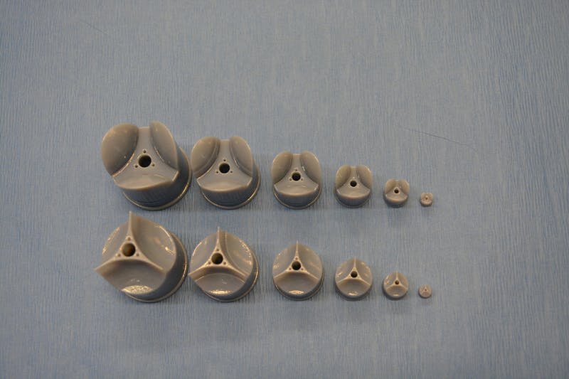
In rotary jet spinning technology, a rotating nozzle extrudes a solution of extracellular matrix (ECM) into nanofibers that wrap themselves around heart valve-shaped mandrels. By using a series of mandrels with different sizes, the manufacturing process becomes fully scalable and is able to provide JetValves for all age groups and heart sizes. Credit: Wyss Institute at Harvard University

From a May 18, 2017 news item on ScienceDaily,

The human heart beats approximately 35 million times every year, effectively pumping blood into the circulation via four different heart valves. Unfortunately, in over four million people each year, these delicate tissues malfunction due to birth defects, age-related deteriorations, and infections, causing cardiac valve disease.

Today, clinicians use either artificial prostheses or fixed animal and cadaver-sourced tissues to replace defective valves. While these prostheses can restore the function of the heart for a while, they are associated with adverse comorbidity and wear down and need to be replaced during invasive and expensive surgeries. Moreover, in children, implanted heart valve prostheses need to be replaced even more often as they cannot grow with the child.

A team lead by Kevin Kit Parker, Ph.D. at Harvard University’s Wyss Institute for Biologically Inspired Engineering recently developed a nanofiber fabrication technique to rapidly manufacture heart valves with regenerative and growth potential. In a paper published in Biomaterials, Andrew Capulli, Ph.D. and colleagues fabricated a valve-shaped nanofiber network that mimics the mechanical and chemical properties of the native valve extracellular matrix (ECM). To achieve this, the team used the Parker lab’s proprietary rotary jet spinning technology — in which a rotating nozzle extrudes an ECM solution into nanofibers that wrap themselves around heart valve-shaped mandrels. “Our setup is like a very fast cotton candy machine that can spin a range of synthetic and natural occurring materials. In this study, we used a combination of synthetic polymers and ECM proteins to fabricate biocompatible JetValves that are hemodynamically competent upon implantation and support cell migration and re-population in vitro. Importantly, we can make human-sized JetValves in minutes — much faster than possible for other regenerative prostheses,” said Parker.

A May 18,2017 Wyss Institute for Biologically Inspired Engineering news release (also on EurekAlert), which originated the news item, expands on the theme of Jetvalves,

To further develop and test the clinical potential of JetValves, Parker’s team collaborated with the translational team of Simon P. Hoerstrup, M.D., Ph.D., at the University of Zurich in Switzerland, which is a partner institution with the Wyss Institute. As a leader in regenerative heart prostheses, Hoerstrup and his team in Zurich have previously developed regenerative, tissue-engineered heart valves to replace mechanical and fixed-tissue heart valves. In Hoerstrup’s approach, human cells directly deposit a regenerative layer of complex ECM on biodegradable scaffolds shaped as heart valves and vessels. The living cells are then eliminated from the scaffolds resulting in an “off-the-shelf” human matrix-based prostheses ready for implantation.

In the paper, the cross-disciplinary team successfully implanted JetValves in sheep using a minimally invasive technique and demonstrated that the valves functioned properly in the circulation and regenerated new tissue. “In our previous studies, the cell-derived ECM-coated scaffolds could recruit cells from the receiving animal’s heart and support cell proliferation, matrix remodeling, tissue regeneration, and even animal growth. While these valves are safe and effective, their manufacturing remains complex and expensive as human cells must be cultured for a long time under heavily regulated conditions. The JetValve’s much faster manufacturing process can be a game-changer in this respect. If we can replicate these results in humans, this technology could have invaluable benefits in minimizing the number of pediatric re-operations,” said Hoerstrup.

In support of these translational efforts, the Wyss Institute for Biologically Inspired Engineering and the University of Zurich announced today a cross-institutional team effort to generate a functional heart valve replacement with the capacity for repair, regeneration, and growth. The team is also working towards a GMP-grade version of their customizable, scalable, and cost-effective manufacturing process that would enable deployment to a large patient population. In addition, the new heart valve would be compatible with minimally invasive procedures to serve both pediatric and adult patients.

The project will be led jointly by Parker and Hoerstrup. Parker is a Core Faculty member of the Wyss Institute and the Tarr Family Professor of Bioengineering and Applied Physics at the Harvard John A. Paulson School of Engineering and Applied Sciences (SEAS). Hoerstrup is Chair and Director of the University of Zurich’s Institute for Regenerative Medicine (IREM), Co-Director of the recently founded Wyss Translational Center Zurich and a Wyss Institute Associate Faculty member.

Since JetValves can be manufactured in all desired shapes and sizes, and take seconds to minutes to produce, the team’s goal is to provide customized, ready-to-use, regenerative heart valves much faster and at much lower cost than currently possible.

“Achieving the goal of minimally invasive, low-cost regenerating heart valves could have tremendous impact on patients’ lives across age-, social- and geographical boundaries. Once again, our collaborative team structure that combines unique and leading expertise in bioengineering, regenerative medicine, surgical innovation and business development across the Wyss Institute and our partner institutions, makes it possible for us to advance technology development in ways not possible in a conventional academic laboratory,” said Wyss Institute Founding Director Donald Ingber, M.D., Ph.D., who is also the Judah Folkman Professor of Vascular Biology at HMS and the Vascular Biology Program at Boston Children’s Hospital, as well as Professor of Bioengineering at SEAS.

This scanning electron microscopy image shows how extracellular matrix (ECM) nanofibers generated with JetValve technology are arranged in parallel networks with physical properties comparable to those found in native heart tissue. Credit: Wyss Institute at Harvard University

Here’s a link to and a citation for the paper,

JetValve: Rapid manufacturing of biohybrid scaffolds for biomimetic heart valve replacement by Andrew K. Capulli, Maximillian Y. Emmert, Francesco S. Pasqualini, b, Debora Kehl, Etem Caliskan, Johan U. Lind, Sean P. Sheehy, Sung Jin Park, Seungkuk Ahn, Benedikt Webe, Josue A. Goss. Biomaterials Volume 133, July 2017, Pages 229–241  https://doi.org/10.1016/j.biomaterials.2017.04.033

This paper is behind a paywall.

Portable nanofibre fabrication device (point-of-use manufacturing)

A portable nanofiber fabrication device is quite an achievement although it seems it’s not quite ready for prime time yet. From a March 1, 2017 news item on Nanowerk (Note: A link has been removed),

Harvard researchers have developed a lightweight, portable nanofiber fabrication device that could one day be used to dress wounds on a battlefield or dress shoppers in customizable fabrics. The research was published recently in Macromolecular Materials and Engineering (“Design and Fabrication of Fibrous Nanomaterials Using Pull Spinning”)

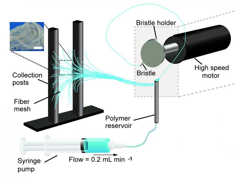
A schematic of the pull spinning apparatus with a side view illustration of a fiber being pulled from the polymer reservoir. The pull spinning system consists of a rotating bristle that dips and pulls a polymer jet in a spiral trajectory (Leila Deravi/Harvard University)

A March 1, 2017 Harvard University news release (also on EurekAlert) by Leah Burrow,, which originated the news item, describes the current process for nanofiber fabrication and explains how this technique is an improvement,

There are many ways to make nanofibers. These versatile materials — whose target applications include everything from tissue engineering to bullet proof vests — have been made using centrifugal force, capillary force, electric field, stretching, blowing, melting, and evaporation.

Each of these fabrication methods has pros and cons. For example, Rotary Jet-Spinning (RJS) and Immersion Rotary Jet-Spinning (iRJS) are novel manufacturing techniques developed in the Disease Biophysics Group at the Harvard John A. Paulson School of Engineering and Applied Sciences (SEAS) and the Wyss Institute for Biologically Inspired Engineering. Both RJS and iRJS dissolve polymers and proteins in a liquid solution and use centrifugal force or precipitation to elongate and solidify polymer jets into nanoscale fibers. These methods are great for producing large amounts of a range of materials – including DNA, nylon, and even Kevlar – but until now they haven’t been particularly portable.

The Disease Biophysics Group recently announced the development of a hand-held device that can quickly produce nanofibers with precise control over fiber orientation. Regulating fiber alignment and deposition is crucial when building nanofiber scaffolds that mimic highly aligned tissue in the body or designing point-of-use garments that fit a specific shape.

“Our main goal for this research was to make a portable machine that you could use to achieve controllable deposition of nanofibers,” said Nina Sinatra, a graduate student in the Disease Biophysics Group and co-first author of the paper. “In order to develop this kind of point-and-shoot device, we needed a technique that could produce highly aligned fibers with a reasonably high throughput.”

The new fabrication method, called pull spinning, uses a high-speed rotating bristle that dips into a polymer or protein reservoir and pulls a droplet from solution into a jet. The fiber travels in a spiral trajectory and solidifies before detaching from the bristle and moving toward a collector. Unlike other processes, which involve multiple manufacturing variables, pull spinning requires only one processing parameter — solution viscosity — to regulate nanofiber diameter. Minimal process parameters translate to ease of use and flexibility at the bench and, one day, in the field.

Pull spinning works with a range of different polymers and proteins. The researchers demonstrated proof-of-concept applications using polycaprolactone and gelatin fibers to direct muscle tissue growth and function on bioscaffolds, and nylon and polyurethane fibers for point-of-wear apparel.

“This simple, proof-of-concept study demonstrates the utility of this system for point-of-use manufacturing,” said Kit Parker, the Tarr Family Professor of Bioengineering and Applied Physics and director of the Disease Biophysics Group. “Future applications for directed production of customizable nanotextiles could extend to spray-on sportswear that gradually heats or cools an athlete’s body, sterile bandages deposited directly onto a wound, and fabrics with locally varying mechanical properties.”

Here’s a link to and a citation for the paper,

Design and Fabrication of Fibrous Nanomaterials Using Pull Spinning by Leila F. Deravi, Nina R. Sinatra, Christophe O. Chantre, Alexander P. Nesmith, Hongyan Yuan, Sahm K. Deravi, Josue A. Goss, Luke A. MacQueen, Mohammad R. Badrossamy, Grant M. Gonzalez, Michael D. Phillips, and Kevin Kit Parker. Macromolecular Materials and Engineering DOI: 10.1002/mame.201600404 Version of Record online: 17 JAN 2017

© 2017 WILEY-VCH Verlag GmbH & Co. KGaA, Weinheim

This paper is behind a paywall.

What is a multiregional brain-on-a-chip?

In response to having created a multiregional brain-on-a-chip, there’s an explanation from the team at Harvard University (which answers my question) in a Jan. 13, 2017 Harvard John A. Paulson School of Engineering and Applied Sciences news release (also on EurekAlert) by Leah Burrows,

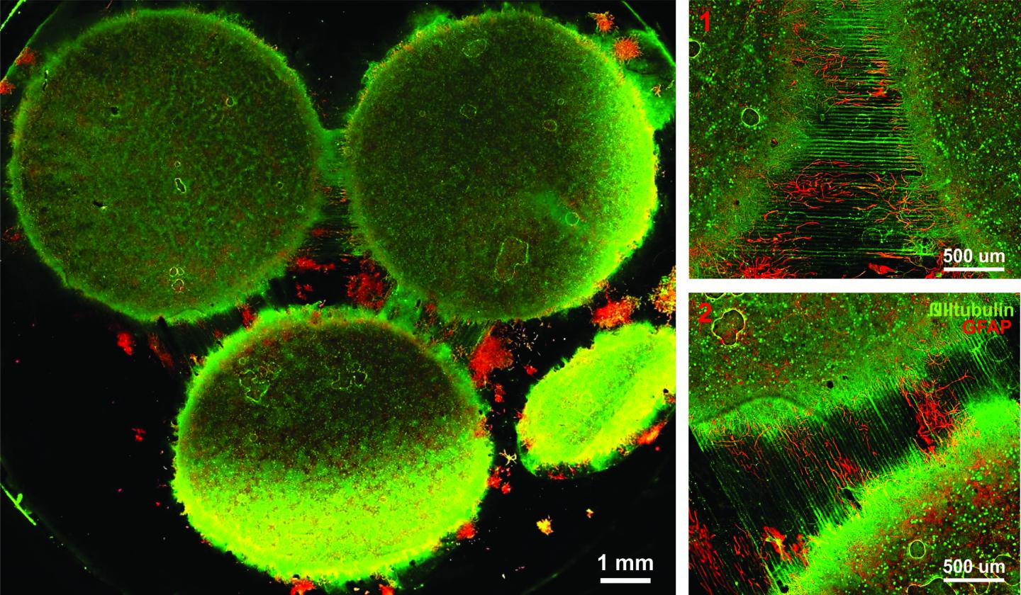
Harvard University researchers have developed a multiregional brain-on-a-chip that models the connectivity between three distinct regions of the brain. The in vitro model was used to extensively characterize the differences between neurons from different regions of the brain and to mimic the system’s connectivity.

“The brain is so much more than individual neurons,” said Ben Maoz, co-first author of the paper and postdoctoral fellow in the Disease Biophysics Group in the Harvard John A. Paulson School of Engineering and Applied Sciences (SEAS). “It’s about the different types of cells and the connectivity between different regions of the brain. When modeling the brain, you need to be able to recapitulate that connectivity because there are many different diseases that attack those connections.”

“Roughly twenty-six percent of the US healthcare budget is spent on neurological and psychiatric disorders,” said Kit Parker, the Tarr Family Professor of Bioengineering and Applied Physics Building at SEAS and Core Faculty Member of the Wyss Institute for Biologically Inspired Engineering at Harvard University. “Tools to support the development of therapeutics to alleviate the suffering of these patients is not only the human thing to do, it is the best means of reducing this cost.”

Researchers from the Disease Biophysics Group at SEAS and the Wyss Institute modeled three regions of the brain most affected by schizophrenia — the amygdala, hippocampus and prefrontal cortex.

They began by characterizing the cell composition, protein expression, metabolism, and electrical activity of neurons from each region in vitro.

“It’s no surprise that neurons in distinct regions of the brain are different but it is surprising just how different they are,” said Stephanie Dauth, co-first author of the paper and former postdoctoral fellow in the Disease Biophysics Group. “We found that the cell-type ratio, the metabolism, the protein expression and the electrical activity all differ between regions in vitro. This shows that it does make a difference which brain region’s neurons you’re working with.”

Next, the team looked at how these neurons change when they’re communicating with one another. To do that, they cultured cells from each region independently and then let the cells establish connections via guided pathways embedded in the chip.

The researchers then measured cell composition and electrical activity again and found that the cells dramatically changed when they were in contact with neurons from different regions.

“When the cells are communicating with other regions, the cellular composition of the culture changes, the electrophysiology changes, all these inherent properties of the neurons change,” said Maoz. “This shows how important it is to implement different brain regions into in vitro models, especially when studying how neurological diseases impact connected regions of the brain.”

To demonstrate the chip’s efficacy in modeling disease, the team doped different regions of the brain with the drug Phencyclidine hydrochloride — commonly known as PCP — which simulates schizophrenia. The brain-on-a-chip allowed the researchers for the first time to look at both the drug’s impact on the individual regions as well as its downstream effect on the interconnected regions in vitro.

The brain-on-a-chip could be useful for studying any number of neurological and psychiatric diseases, including drug addiction, post traumatic stress disorder, and traumatic brain injury.

“To date, the Connectome project has not recognized all of the networks in the brain,” said Parker. “In our studies, we are showing that the extracellular matrix network is an important part of distinguishing different brain regions and that, subsequently, physiological and pathophysiological processes in these brain regions are unique. This advance will not only enable the development of therapeutics, but fundamental insights as to how we think, feel, and survive.”

Here’s an image from the researchers,

Caption: Image of the in vitro model showing three distinct regions of the brain connected by axons. Credit: Disease Biophysics Group/Harvard University

Here’s a link to and a citation for the paper,

Neurons derived from different brain regions are inherently different in vitro: A novel multiregional brain-on-a-chip by Stephanie Dauth, Ben M Maoz, Sean P Sheehy, Matthew A Hemphill, Tara Murty, Mary Kate Macedonia, Angie M Greer, Bogdan Budnik, Kevin Kit Parker. Journal of Neurophysiology Published 28 December 2016 Vol. no. [?] , DOI: 10.1152/jn.00575.2016

This paper is behind a paywall and they haven’t included the vol. no. in the citation I’ve found.

Stronger more robust nanofibers for everything from bulletproof vests to cellular scaffolds (tissue engineering)

This work on a new technique for producing nanofibers comes from Harvard University’s School of Engineering and Applied Sciences and the Wyss Institute for Biologically Inspired Engineering (also at Harvard University). From an Oct. 10, 2016 news item on phys.org,

Fibrous materials—known for their toughness, durability and pliability—are used in everything from bulletproof vests to tires, filtration systems and cellular scaffolds for tissue engineering and regenerative medicine.

The properties of these materials are such that the smaller the fibers are, the stronger and tougher they become. But making certain fibers very small has been an engineering challenge.

Now, researchers from the Harvard John A. Paulson School of Engineering and Applied Sciences (SEAS) and the Wyss Institute for Biologically Inspired Engineering at Harvard have developed a new method to make and collect nanofibers and control their size and morphology. This could lead to stronger, more durable bulletproof vests and armor and more robust cellular scaffolding for tissue repair.

An Oct. 7, 2016 Harvard University press release by Leah Burrows, which originated the news item, describes the research in more detail (Note: A link has been removed),

Nanofibers are smaller than one micrometer in diameter.  Most nanofiber production platforms rely on dissolving polymers in a solution, which then evaporates as the fiber forms.

Rotary Jet-Spinning (RJS), the technique developed by Kit Parker’s Disease Biophysics Group, works likes a cotton candy machine. Parker is Tarr Family Professor of Bioengineering and Applied Physics at SEAS and a Core Member of the Wyss Institute. A liquid polymer solution is loaded into a reservoir and pushed out through a tiny opening by centrifugal force as the device spins. As the solution leaves the reservoir, the solvent evaporates and the polymers solidify and elongate into small, thin fibers.

“This advance is important because it allows us to manufacture ballistic protection that is much lighter, more flexible and more functional than what is available today,” said Parker, who in addition to his Harvard role is a lieutenant colonel in the United States Army Reserve and was motivated by his own combat experiences in Afghanistan. “Not only could it save lives but for the warfighter, it also could help reduce the repetitive injury motions that soldiers, sailors, marines and airmen have suffered over the last 15 years of the war on terror.”

“Rotary Jet-Spinning is great for most polymer fibers you want to make,” said Grant Gonzalez, a graduate student at SEAS and first author of the paper.  “However, some fibers require a solvent that doesn’t evaporate easily. Para-aramid, the polymer used in Kevlar® for example, is dissolved in sulfuric acid, which doesn’t evaporate off. The solution just splashes against the walls of the device without forming fibers.”

Nanofibers are smaller than one micrometer in diameter.  Most nanofiber production platforms rely on dissolving polymers in a solution, which then evaporates as the fiber forms.

Rotary Jet-Spinning (RJS), the technique developed by Kit Parker’s Disease Biophysics Group, works likes a cotton candy machine. Parker is Tarr Family Professor of Bioengineering and Applied Physics at SEAS and a Core Member of the Wyss Institute. A liquid polymer solution is loaded into a reservoir and pushed out through a tiny opening by centrifugal force as the device spins. As the solution leaves the reservoir, the solvent evaporates and the polymers solidify and elongate into small, thin fibers.

“This advance is important because it allows us to manufacture ballistic protection that is much lighter, more flexible and more functional than what is available today,” said Parker, who in addition to his Harvard role is a lieutenant colonel in the United States Army Reserve and was motivated by his own combat experiences in Afghanistan. “Not only could it save lives but for the warfighter, it also could help reduce the repetitive injury motions that soldiers, sailors, marines and airmen have suffered over the last 15 years of the war on terror.”

“Rotary Jet-Spinning is great for most polymer fibers you want to make,” said Grant Gonzalez, a graduate student at SEAS and first author of the paper.  “However, some fibers require a solvent that doesn’t evaporate easily. Para-aramid, the polymer used in Kevlar® for example, is dissolved in sulfuric acid, which doesn’t evaporate off. The solution just splashes against the walls of the device without forming fibers.”

Other methods, such as electrospinning, which uses an electric field to pull the polymer into a thin fiber, also have poor results with Kevlar and other polymers such as alginate used for tissue scaffolding and DNA.

The Harvard team overcame these challenges by developing a wet-spinning platform, which uses the same principles as the RJS system but relies on precipitation rather than evaporation to separate the solvent from the polymer.

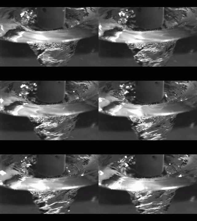
In this system, called immersion Rotary Jet-Spinning (iRJS), when the polymer solution shoots out of the reservoir, it first passes through an area of open air, where the polymers elongate and the chains align. Then the solution hits a liquid bath that removes the solvent and precipitates the polymers to form solid fibers. Since the bath is also spinning — like water in a salad spinner — the nanofibers follow the stream of the vortex and wrap around a rotating collector at the base of the device.

Using this system, the team produced Nylon, DNA, alginate and ballistic resistant para-aramid nanofibers. The team could tune the fiber’s diameter by changing the solution concentration, the rotational speed and the distance the polymer traveled from the reservoir to the bath.

“By being able to modulate fiber strength, we can create a cellular scaffold that can mimic skeleton muscle and native tissues,” said Gonzalez.  “This platform could enable us to create a wound dressing out of alginate material or seed and mature cells on scaffolding for tissue engineering.”

Because the fibers were collected by a spinning vortex, the system also produced well-aligned sheets of nanofibers, which is important for scaffolding and ballistic resistant materials.

This is the ‘candy floss’ technique at work,

Rotary Jet-Spinning (RJS) works likes a cotton candy machine. A liquid polymer solution is loaded into a reservoir and pushed out through a tiny opening by centrifugal force as the device spins. As the solution leaves the reservoir, the solvent evaporates and the polymers solidify and elongate into small, thin fibers. Courtesy: Harvard University

Rotary Jet-Spinning (RJS) works likes a cotton candy machine. A liquid polymer solution is loaded into a reservoir and pushed out through a tiny opening by centrifugal force as the device spins. As the solution leaves the reservoir, the solvent evaporates and the polymers solidify and elongate into small, thin fibers. Courtesy: Harvard University

Here’s a link to and a citation for the paper,

Production of Synthetic, Para-Aramid and Biopolymer Nanofibers by Immersion Rotary Jet-Spinning by Grant M. Gonzalez, Luke A. MacQueen, Johan U. Lind, Stacey A. Fitzgibbons, Christophe O. Chantre, Isabelle Huggler, Holly M. Golecki, Josue A. Goss, Kevin Kit Parker. Macromolecular Materials and Engineering DOI: 10.1002/mame.201600365 Version of Record online: 7 OCT 2016

© 2016 WILEY-VCH Verlag GmbH & Co. KGaA, Weinheim

This paper is behind a paywall.

Asthma on a chip

Harvard University’s Wyss Institute for Biologically Inspired Engineering has found a way to mimic the lung’s muscle action when an asthma attack is being experienced according to a Sept. 23, 2014 news item on Nanowerk,

The majority of drugs used to treat asthma today are the same ones that were used 50 years ago. New drugs are urgently needed to treat this chronic respiratory disease, which causes nearly 25 million people in the United States alone to wheeze, cough, and find it difficult at best to take a deep breath.

But finding new treatments is tough: asthma is a patient-specific disease, so what works for one person doesn’t necessarily work for another, and the animal models traditionally used to test new drug candidates often fail to mimic human responses–costing tremendous money and time.

Hope for healthier airways may be on the horizon thanks to a Harvard University team that has developed a human airway muscle-on-a-chip that could be used to test new drugs because it accurately mimics the way smooth muscle contracts in the human airway, under normal circumstances and when exposed to asthma triggers. [emphasis mine]

A Sept. 23, 2014 Wyss Institute news release (also on EurekAlert*), which originated the news item, provides more details about the technology and its advantages,

The chip, a soft polymer well that is mounted on a glass substrate, contains a planar array of microscale, engineered human airway muscles, designed to mimic the laminar structure of the muscular layers of the human airway.

To mimic a typical allergic asthma response, the team first introduced interleukin-13 (IL-13) to the chip. IL-13 is a natural protein often found in the airway of asthmatic patients that mediates the response of smooth muscle to an allergen.

Then they introduced acetylcholine, a neurotransmitter that causes smooth muscle to contract. Sure enough, the airway muscle on the chip hypercontracted – and the soft chip curled up – in response to higher doses of the neurotransmitter.

They achieved the reverse effect as well and triggered the muscle to relax using drugs called β-agonists, which are used in inhalers.

Significantly, they were able to measure the contractile stress of the muscle tissue as it responded to varying doses of the drugs, said lead author Alexander Peyton Nesmith, a Ph.D./M.D. student at Harvard SEAS and the University of Alabama at Birmingham. “Our chip offers a simple, reliable and direct way to measure human responses to an asthma trigger,” he said.

The team then investigated what happened on a cellular level in response to the IL-13 and confirmed, for example, that the smooth muscle cells grew larger in the presence of IL-13 over time – a structural hallmark of the airways in asthma patients as well. They also documented an increased alignment of actin fibers within smooth muscle cells, which is consistent with the muscle in the airway of asthma patients. Actin fibers are super-thin cellular components involved in muscle contraction.

Next they observed how IL-13 changes the expression of contractile proteins called RhoA proteins, which have been implicated in the asthmatic response, although the details of their activation and signaling have remained elusive. To do this they introduced a drug called HA1077, which is not currently used to treat asthmatic patients – but targets the RhoA pathway. It turns out that the drug made the asthmatic tissue on the chip less sensitive to the asthma trigger – and preliminary tests indicated that using a combined therapy of HA1077 plus a currently approved asthma drug worked better than the single drug alone.

“Asthma is one of the top reasons for trips to the emergency room – particularly for children, and a large segment of the asthmatic population doesn’t respond to currently available treatments,” said Wyss Institute Founding Director Don Ingber, M.D., Ph.D. “The airway muscle-on-a-chip provides an important and exciting new tool for discovering new therapeutic agents.”

The scientists have provided an illustration of healthy and asthmatic airways,

Schematic comparing a healthy airway (few immune cells, normal airway diameter) to an asthmatic airway (many immune cells, constricted airway). Credit: Harvard's Wyss Institute and Harvard SEAS [School of Engineering and Applied Sciences]

Schematic comparing a healthy airway (few immune cells, normal airway diameter) to an asthmatic airway (many immune cells, constricted airway). Credit: Harvard’s Wyss Institute and Harvard SEAS [School of Engineering and Applied Sciences]

Here’s link to and a citation for the paper,

Human airway musculature on a chip: an in vitro model of allergic asthmatic bronchoconstriction and bronchodilation by Alexander Peyton Nesmith, Ashutosh Agarwal, Megan Laura McCain and Kevin Kit Parker.Lab Chip, 2014,14, 3925-3936 DOI: 10.1039/C4LC00688G First published online 05 Aug 2014

This paper is open access provided you have registered yourself for free at the site.

* EurekAlert link added Sept. 24, 2014.

Organ chips for DARPA (Defense Advanced Research Projects Agency)

The Wyss Institute will receive up to  $37M US for a project that integrates ten different organ-on-a-chip projects into one system. From the July 24, 2012 news release on EurekAlert,

With this new DARPA funding, Institute researchers and a multidisciplinary team of collaborators seek to build 10 different human organs-on-chips, to link them together to more closely mimic whole body physiology, and to engineer an automated instrument that will control fluid flow and cell viability while permitting real-time analysis of complex biochemical functions. As an accurate alternative to traditional animal testing models that often fail to predict human responses, this instrumented “human-on-a-chip” will be used to rapidly assess responses to new drug candidates, providing critical information on their safety and efficacy.

This unique platform could help ensure that safe and effective therapeutics are identified sooner, and ineffective or toxic ones are rejected early in the development process. As a result, the quality and quantity of new drugs moving successfully through the pipeline and into the clinic may be increased, regulatory decision-making could be better informed, and patient outcomes could be improved.

Jesse Goodman, FDA Chief Scientist and Deputy Commissioner for Science and Public Health, commented that the automated human-on-chip instrument being developed “has the potential to be a better model for determining human adverse responses. FDA looks forward to working with the Wyss Institute in its development of this model that may ultimately be used in therapeutic development.”

Wyss Founding Director, Donald Ingber, M.D., Ph.D., and Wyss Core Faculty member, Kevin Kit Parker, Ph.D., will co-lead this five-year project.

I note that Kevin Kit Parker was mentioned in an earlier posting today (July 26, 2012) titled, Medusa, jellyfish, and tissue engineering, and Donald Ingber in my Dec.1e, 2011 posting about Shrilk and insect skeletons.

As for the Wyss Institute, here’s a description from the news release,

The Wyss Institute for Biologically Inspired Engineering at Harvard University (http://wyss.harvard.edu) uses Nature’s design principles to develop bioinspired materials and devices that will transform medicine and create a more sustainable world. Working as an alliance among Harvard’s Schools of Medicine, Engineering, and Arts & Sciences, and in partnership with Beth Israel Deaconess Medical Center, Boston Children’s Hospital, Brigham and Women’s Hospital, , Dana Farber Cancer Institute, Massachusetts General Hospital, the University of Massachusetts Medical School, Spaulding Rehabilitation Hospital, Tufts University, and Boston University, the Institute crosses disciplinary and institutional barriers to engage in high-risk research that leads to transformative technological breakthroughs. By emulating Nature’s principles for self-organizing and self-regulating, Wyss researchers are developing innovative new engineering solutions for healthcare, energy, architecture, robotics, and manufacturing. These technologies are translated into commercial products and therapies through collaborations with clinical investigators, corporate alliances, and new start-ups.

I hadn’t thought of an organ-on-a-chip as particularly bioinspired so I’ll have to think about that one for a while.

Medusa, jellyfish, and tissue engineering

The ‘Medusoid’ is a reverse- tissue-engineered jellyfish designed by a collaborative team of researchers based, respectively, at the California Institute of Technology (Caltech) and Harvard University. From the July 22, 2012 news item on ScienceDaily,

When one observes a colorful jellyfish pulsating through the ocean, Greek mythology probably doesn’t immediately come to mind. But the animal once was known as the medusa, after the snake-haired mythological creature its tentacles resemble. The mythological Medusa’s gaze turned people into stone, and now, thanks to recent advances in bio-inspired engineering, a team led by researchers at the California Institute of Technology (Caltech) and Harvard University have flipped that fable on its head: turning a solid element—silicon—and muscle cells into a freely swimming “jellyfish.”

“A big goal of our study was to advance tissue engineering,” says Janna Nawroth, a doctoral student in biology at Caltech and lead author of the study. “In many ways, it is still a very qualitative art [emphasis mine], with people trying to copy a tissue or organ just based on what they think is important or what they see as the major components—without necessarily understanding if those components are relevant to the desired function or without analyzing first how different materials could be used.” Because a particular function—swimming, say—doesn’t necessarily emerge just from copying every single element of a swimming organism into a design, “our idea,” she says, “was that we would make jellyfish functions—swimming and creating feeding currents—as our target and then build a structure based on that information.”

Oops! I’m not sure why Nawroth uses the word ‘qualitative’ here. It’s certainly inappropriate given my understanding of the word. Here’s my rough definition, if anyone has anything better or can explain why Nawroth used ‘qualitative’  in that context, please do comment. I’m going to start by contrasting qualitative with quantitative, both of which I’m going to hugely oversimplify. Quantitative data offers numbers, e.g. 50,000 people committed suicide last year. Qualitative data helps offer insight into why. Researchers can obtain the quantitative data from police records, vital statistics, surveys, etc. where qualitative data is gathered from ‘story-oriented’ or highly detailed personal interviews. ( I would have used ‘hit or miss,’ ‘guesswork,’ or simply used the word art without qualifying it  in this context.)

The originating July 22, 2012 news release from Caltech goes on to describe why jellyfish were selected and how the collaboration between Harvard and Caltech came about,

Jellyfish are believed to be the oldest multi-organ animals in the world, possibly existing on Earth for the past 500 million years. Because they use a muscle to pump their way through the water, their function—on a very basic level—is similar to that of a human heart, which makes the animal a good biological system to analyze for use in tissue engineering.

“It occurred to me in 2007 that we might have failed to understand the fundamental laws of muscular pumps,” says Kevin Kit Parker, Tarr Family Professor of Bioengineering and Applied Physics at Harvard and a coauthor of the study. “I started looking at marine organisms that pump to survive. Then I saw a jellyfish at the New England Aquarium, and I immediately noted both similarities and differences between how the jellyfish pumps and the human heart. The similarities help reveal what you need to do to design a bio-inspired pump.”

Parker contacted John Dabiri, professor of aeronautics and bioengineering at Caltech—and Nawroth’s advisor—and a partnership was born. Together, the two groups worked for years to understand the key factors that contribute to jellyfish propulsion, including the arrangement of their muscles, how their bodies contract and recoil, and how fluid-dynamic effects help or hinder their movements. Once these functions were well understood, the researchers began to design the artificial jellyfish.

Here’s how they created the ‘Medusoid’ (artificial jellyfish, from the July 22, 2012 Harvard University news release on EurekAlert,

To reverse engineer a medusa jellyfish, the investigators used analysis tools borrowed from the fields of law enforcement biometrics and crystallography to make maps of the alignment of subcellular protein networks within all of the muscle cells within the animal. They then conducted studies to understand the electrophysiological triggering of jellyfish propulsion and the biomechanics of the propulsive stroke itself.

Based on such understanding, it turned out that a sheet of cultured rat heart muscle tissue that would contract when electrically stimulated in a liquid environment was the perfect raw material to create an ersatz jellyfish. The team then incorporated a silicone polymer that fashions the body of the artificial creature into a thin membrane that resembles a small jellyfish, with eight arm-like appendages.

Using the same analysis tools, the investigators were able to quantitatively match the subcellular, cellular, and supracellular architecture of the jellyfish musculature with the rat heart muscle cells.

The artificial construct was placed in container of ocean-like salt water and shocked into swimming with synchronized muscle contractions that mimic those of real jellyfish. (In fact, the muscle cells started to contract a bit on their own even before the electrical current was applied.)

“I was surprised that with relatively few components—a silicone base and cells that we arranged—we were able to reproduce some pretty complex swimming and feeding behaviors that you see in biological jellyfish,” says Dabiri.

Their design strategy, they say, will be broadly applicable to the reverse engineering of muscular organs in humans.

For future research direction I’ve excerpted this from the Caltech news release,

The team’s next goal is to design a completely self-contained system that is able to sense and actuate on its own using internal signals, as human hearts do. Nawroth and Dabiri would also like for the Medusoid to be able to go out and gather food on its own. Then, researchers could think about systems that could live in the human body for years at a time without having to worry about batteries because the system would be able to fend for itself. For example, these systems could be the basis for a pacemaker made with biological elements.

“We’re reimagining how much we can do in terms of synthetic biology,” says Dabiri. “A lot of work these days is done to engineer molecules, but there is much less effort to engineer organisms. I think this is a good glimpse into the future of re-engineering entire organisms for the purposes of advancing biomedical technology. We may also be able to engineer applications where these biological systems give us the opportunity to do things more efficiently, with less energy usage.”

I think this excerpt from the Harvard news release provides some insight into at least some of the motivations behind this work,

In addition to advancing the field of tissue engineering, Parker adds that he took on the challenge of building a creature to challenge the traditional view of synthetic biology which is “focused on genetic manipulations of cells.” Instead of building just a cell, he sought to “build a beast.”

A little competitive, eh?

For anyone who’s interested in reading the research (which is behind a paywall), from the ScienceDaily news item,

Janna C Nawroth, Hyungsuk Lee, Adam W Feinberg, Crystal M Ripplinger, Megan L McCain, Anna Grosberg, John O Dabiri & Kevin Kit Parker. A tissue-engineered jellyfish with biomimetic propulsion. Nature Biotechnology, 22 July 2012 DOI: 10.1038/nbt.2269

Andrew Maynard weighs in on the matter with his July 22, 2012 posting titled, We took a rat apart and rebuilt it as a jellyfish, on the 2020Science blog (Note: I have removed links),

 Sometimes you read a science article and it sends a tingle down your spine. That was my reaction this afternoon reading Ed Yong’s piece on a paper just published in Nature Biotechnology by Janna Nawroth, Kevin Kit Parker and colleagues.

The gist of the work is that Parker’s team have created a hybrid biological machine that “swims” like a jellyfish by growing rat heart muscle cells on a patterned sheet of polydimethylsiloxane.  The researchers are using the technique to explore muscular pumps, but the result opens the door to new technologies built around biological-non biological hybrids.

Ed Yong’s July 22, 2012 article for Nature (as mentioned by Andrew) offers a wider perspective on the work than is immediately evident in either of the news releases (Note: I have removed a footnote),

Bioengineers have made an artificial jellyfish using silicone and muscle cells from a rat’s heart. The synthetic creature, dubbed a medusoid, looks like a flower with eight petals. When placed in an electric field, it pulses and swims exactly like its living counterpart.

“Morphologically, we’ve built a jellyfish. Functionally, we’ve built a jellyfish. Genetically, this thing is a rat,” says Kit Parker, a biophysicist at Harvard University in Cambridge, Massachusetts, who led the work. The project is described today in Nature Biotechnology.

….

“I think that this is terrific,” says Joseph Vacanti, a tissue engineer at Massachusetts General Hospital in Boston. “It is a powerful demonstration of engineering chimaeric systems of living and non-living components.”

Here’s a video from the researchers demonstrating the artificial jellyfish in action,

There’s a lot of material for contemplation but what I’m going to note here is the difference in the messaging. The news releases from the ‘universities’ are very focused on the medical application where the discussion in the science community revolves primarily around the synthetic biology/bioengineering elements. It seems to me that this strategy can lead to future problems with a population that is largely unprepared to deal with the notion of mixing and recombining  genetic material or demonstrations of “of engineering chimaeric systems of living and non-living components.”