Tag Archives: Boston Children’s Hospital

Israeli startup (Nanomedic) and a ‘ray’ gun that shoots wound-healing skin

[downloaded from https://uploads.neatorama.com/images/posts/967/107/107967/Spray-on-Nanofiber-Skin-May-Improve-Burn-and-Wound-Care_0-x.jpg?v=10727]

Where I see a ‘ray’ gun, Rina Raphael, author of a July 6, 2019 article for Fast tCompany, sees a water pistol (Note: Links have been removed),

Imagine if bandaging looked a little more like, well, a water gun?

Israeli startup Nanomedic Technologies Ltd., a subsidiary of medical device company Nicast, has invented a new mechanical contraption to treat burns, wounds, and surgical injuries by mimicking human tissue. Shaped like a children’s toy, the lightweight SpinCare emits a proprietary nanofiber “second skin” that completely covers the area that needs to heal.

All one needs to do is aim, squeeze the two triggers, and fire off an electrospun polymer material that attaches to the skin.

The Nanomedic spray method avoids any need to come into direct contact with the wound. In that sense, it completely sidesteps painful routine bandage dressings. The transient skin then fully develops into a secure physical barrier with tough adherence. Once new skin is regenerated, usually between two to three weeks (depending on the individual’s heal time), the layer naturally peels off.

“You don’t replace it,” explains Nanomedic CEO Dr. Chen Barak. “You put it only once—on the day of application—and it remains there until it feels the new layer of skin healed.”

“It’s the same model as an espresso machine,” says Barak.

The SpinCare holds single-use ampoules containing Nanomedic’s polymer formulation. Once the capsule is firmly in place, one activates the device roughly eight inches towards the wound. Pressing the trigger activates the electron-spinning process, which sprays a web-like a layer of nano fibers directly on the wound.

The solution adjusts to the morphology of the wound, thereby creating a transient skin layer that imitates the skin structure’s human tissue. It’s a transparent, protective film that then allows the patient and doctor to monitor progress. Once the wound has healed and developed a new layer of skin, the SpinCare “bandage” falls off on its own.

The product is already being tested in hospitals. In the coming year, following FDA clearance, Nanomedic plans to expand to emergency rooms, ambulances, military use, and disaster relief response like fire truck companies. The global wound healing market is expected to hit $35 billion by 2025, according to a report by Transparency Market Research.

Nanomedic joins other researchers attempting to reimagine the wound healing process. Engineers at the University of Wisconsin-Madison, for example, created a new kind of protective bandage that sends a mild electrical stimulation, thereby “dramatically” reducing the time deep surgical wounds take to heal.

As for the the playful (yet functional) design, it resembles other medical tools utilizing the point-and-shoot feature. Researchers at the Technion-Israel Institute of Technology and Boston Children’s Hospital recently revealed a “hot-glue gun” that melds torn human tissues together. The medical glue is meant to replace painful and often scarring stitches and staples.

Down the line, Nanomedic plans to enter the in-home care market, where it believes it can better assist caretakers for treatment of chronic wounds, such as pressure ulcers. The chronic wounds segment is projected to hold the dominant share in the wound healing market due to aging populations.

But a bigger opportunity lies in the multiple uses the SpinCare can ultimately provide. It is, in essence, a platform technology that could benefit multiple categories, not just medical wound care. Currently, the SpinCare’s capsules do not contain any active ingredients.

Nanomedic is already researching how to add different additives, such as antibacterial compliments, collagen, silicone, cannabinoids—and, eventually, stem cells and cellular treatments.

Such advancements would propel the device to new markets, like plastic surgery, aesthetics, and dermatology. The latter, for example, spans “burns” caused by deep, cosmetic laser peels.

“Because it is a solution, we can combine additives inside,” explains Katz. “By that, we are transforming the transient skin into a drug delivery system and slow release system.”

Nanomedic is still at the premarket phase, [emphasis mine] having concluded one clinical trial related to the treatment of split graft donor site wounds and currently engaged in two ongoing burn studies. Barak anticipates FDA approval will take between nine to 12 months, during which the company will focus on building manufacturing lines and preparing for a European launch in early 2020.

According to the startup’s estimates, the product’s final price (not yet determined) will be far more affordable than traditional dressings. Nanomedic has raised $7 million in funding to date, including a grant by the EU’s Horizon 2020 SME Instrument program.

Barak believes Nanocare [sic] brings a highly cost-effective alternative to the healthcare system, but more than anything, she’s proud that SpinCare, above all else, mitigates patient pain and hassle. Some users, the company reports, are able to return to work and physical activity right away.

The Nanomedic website can be found here. The company has also produced a video featuring SpinCare,

There’s a bit more about the technology (I’m especially interested in the electrospinning) on Nanomedic’s Technology webpage,

Electrospinning technology allows the development of a wide range of products and devices, with tailored composition, geometry and morphology.

Almost any natural or synthetic polymer can be electrospun to create a nanofibrous mat. The intrinsic structure of the electrospun products, which mimics the natural extra cellular matrix (ECM), encourages quick and efficient tissue integration and minimizes medical complications.

Raphael’s article and the Nanomedic website offer more detail to what you can see in the excerpts provided here. If you have the time, I recommend checking out both.

Breakthrough with Alpaca nanobodies

Caption: Bryson and Sanchez, two alpacas who produce unusually small antibodies. These ‘nanobodies’ could help highly promising CAR T-cell therapies kill solid tumors, where right now they work only in blood cancers. Credit: Courtesy of Boston Children’s Hospital

Bryson and Sanchez are not the first camelids to grace this blog. ‘Llam’ me lend you some antibodies—antibody particles extracted from camels and llamas, a June 12, 2014 posting, and Llama-derived nanobodies are good for solving crystal structure, a December 14, 2017 posting, both feature news about medical breakthroughs with regard to the antibodies found in Llamas, camels, and other camelids (including alpacas) could enable.

The latest camelid-oriented medical research story is in an April 11, 2019 news item on phys.org (Note: A link has been removed),

In 1989, two undergraduate students at the Free University of Brussels were asked to test frozen blood serum from camels, and stumbled on a previously unknown kind of antibody. It was a miniaturized version of a human antibody, made up only of two heavy protein chains, rather than two light and two heavy chains. As they eventually reported, the antibodies’ presence was confirmed not only in camels, but also in llamas and alpacas.

Fast forward 30 years. In the journal PNAS [Proceedings of the National Academy of Science] this week [April 8 – 12, 2019], researchers at Boston Children’s Hospital and MIT [Massachusetts Institute of Technology] show that these mini-antibodies, shrunk further to create so-called nanobodies, may help solve a problem in the cancer field: making CAR T-cell therapies work in solid tumors.

An April 11, 2019 Boston Children’s Hospital news release on EurekAlert, which originated the news item, explores the technology,

Highly promising for blood cancers, chimeric antigen receptor (CAR) T-cell therapy genetically engineers a patient’s own T cells to make them better at attacking cancer cells. The Dana-Farber/Boston Children’s Cancer and Blood Disorders Center is currently using CAR T-cell therapy for relapsed acute lymphocytic leukemia (ALL), for example.

But CAR T cells haven’t been good at eliminating solid tumors. It’s been hard to find cancer-specific proteins on solid tumors that could serve as safe targets. Solid tumors are also protected by an extracellular matrix, a supportive web of proteins that acts as a barrier, as well as immunosuppressive molecules that weaken the T-cell attack.

Rethinking CAR T cells

That’s where nanobodies come in. For two decades, they largely remained in the hands of the Belgian team. But that changed after the patent expired in 2013. [emphases mine]

“A lot of people got into the game and began to appreciate nanobodies’ unique properties,” says Hidde Ploegh, PhD, an immunologist in the Program in Cellular and Molecular Medicine at Boston Children’s and senior investigator on the PNAS study.

One useful attribute is their enhanced targeting abilities. Ploegh and his team at Boston Children’s, in collaboration with Noo Jalikhani, PhD, and Richard Hynes, PhD at MIT’s Koch Institute for Integrative Cancer Research, have harnessed nanobodies to carry imaging agents, allowing precise visualization of metastatic cancers.

The Hynes team targeted the nanobodies to the tumors’ extracellular matrix, or ECM — aiming imaging agents not at the cancer cells themselves, but at the environment that surrounds them. Such markers are common to many tumors, but don’t typically appear on normal cells.

“Our lab and the Hynes lab are among the few actively pursuing this approach of targeting the tumor micro-environment,” says Ploegh. “Most labs are looking for tumor-specific antigens.”

Targeting tumor protectors

Ploegh’s lab took this idea to CAR T-cell therapy. His team, including members of the Hynes lab, took aim at the very factors that make solid tumors difficult to treat.

The CAR T cells they created were studded with nanobodies that recognize specific proteins in the tumor environment, bearing signals directing them to kill any cell they bound to. One protein, EIIIB, a variant of fibronectin, is found only on newly formed blood vessels that supply tumors with nutrients. Another, PD-L1, is an immunosuppressive protein that most cancers use to silence approaching T cells.

Biochemist Jessica Ingram, PhD of the Dana-Farber Cancer Institute, Ploegh’s partner and a coauthor on the paper, led the manufacturing pipeline. She would drive to Amherst, Mass., to gather T cells from two alpacas, Bryson and Sanchez, inject them with the antigen of interest and harvest their blood for further processing back in Boston to generate mini-antibodies.

Taking down melanoma and colon cancer

Tested in two separate melanoma mouse models, as well as a colon adenocarcinoma model in mice, the nanobody-based CAR T cells killed tumor cells, significantly slowed tumor growth and improved the animals’ survival, with no readily apparent side effects.

Ploegh thinks that the engineered T cells work through a combination of factors. They caused damage to tumor tissue, which tends to stimulate inflammatory immune responses. Targeting EIIIB may damage blood vessels in a way that decreases blood supply to tumors, while making them more permeable to cancer drugs.

“If you destroy the local blood supply and cause vascular leakage, you could perhaps improve the delivery of other things that might have a harder time getting in,” says Ploegh. “I think we should look at this as part of a combination therapy.”

Future directions

Ploegh thinks his team’s approach could be useful in many solid tumors. He’s particularly interested in testing nanobody-based CAR T cells in models of pancreatic cancer and cholangiocarcinoma, a bile duct cancer from which Ingram passed away in 2018.

The technology itself can be pushed even further, says Ploegh.

“Nanobodies could potentially carry a cytokine to boost the immune response to the tumor, toxic molecules that kill tumor and radioisotopes to irradiate the tumor at close range,” he says. “CAR T cells are the battering ram that would come in to open the door; the other elements would finish the job. In theory, you could equip a single T cell with multiple chimeric antigen receptors and achieve even more precision. That’s something we would like to pursue.”

So, the Belgian researchers have a patent for two decades and, after it expires, more researchers could help to take the work further. Hmm …

Moving on, here’s a link to and a citation for the paper,

Nanobody-based CAR T cells that target the tumor microenvironment inhibit the growth of solid tumors in immunocompetent mice by Yushu Joy Xie, Michael Dougan, Noor Jailkhani, Jessica Ingram, Tao Fang, Laura Kummer, Noor Momin, Novalia Pishesha, Steffen Rickelt, Richard O. Hynes, and Hidde Ploegh. PNAS DOI: https://doi.org/10.1073/pnas.1817147116
First published April 1, 2019

This paper is behind a paywall

The Hedy Lamarr of international research: Canada’s Third assessment of The State of Science and Technology and Industrial Research and Development in Canada (1 of 2)

Before launching into the assessment, a brief explanation of my theme: Hedy Lamarr was considered to be one of the great beauties of her day,

“Ziegfeld Girl” Hedy Lamarr 1941 MGM *M.V.
Titles: Ziegfeld Girl
People: Hedy Lamarr
Image courtesy mptvimages.com [downloaded from https://www.imdb.com/title/tt0034415/mediaviewer/rm1566611456]

Aside from starring in Hollywood movies and, before that, movies in Europe, she was also an inventor and not just any inventor (from a Dec. 4, 2017 article by Laura Barnett for The Guardian), Note: Links have been removed,

Let’s take a moment to reflect on the mercurial brilliance of Hedy Lamarr. Not only did the Vienna-born actor flee a loveless marriage to a Nazi arms dealer to secure a seven-year, $3,000-a-week contract with MGM, and become (probably) the first Hollywood star to simulate a female orgasm on screen – she also took time out to invent a device that would eventually revolutionise mobile communications.

As described in unprecedented detail by the American journalist and historian Richard Rhodes in his new book, Hedy’s Folly, Lamarr and her business partner, the composer George Antheil, were awarded a patent in 1942 for a “secret communication system”. It was meant for radio-guided torpedoes, and the pair gave to the US Navy. It languished in their files for decades before eventually becoming a constituent part of GPS, Wi-Fi and Bluetooth technology.

(The article goes on to mention other celebrities [Marlon Brando, Barbara Cartland, Mark Twain, etc] and their inventions.)

Lamarr’s work as an inventor was largely overlooked until the 1990’s when the technology community turned her into a ‘cultish’ favourite and from there her reputation grew and acknowledgement increased culminating in Rhodes’ book and the documentary by Alexandra Dean, ‘Bombshell: The Hedy Lamarr Story (to be broadcast as part of PBS’s American Masters series on May 18, 2018).

Canada as Hedy Lamarr

There are some parallels to be drawn between Canada’s S&T and R&D (science and technology; research and development) and Ms. Lamarr. Chief amongst them, we’re not always appreciated for our brains. Not even by people who are supposed to know better such as the experts on the panel for the ‘Third assessment of The State of Science and Technology and Industrial Research and Development in Canada’ (proper title: Competing in a Global Innovation Economy: The Current State of R&D in Canada) from the Expert Panel on the State of Science and Technology and Industrial Research and Development in Canada.

A little history

Before exploring the comparison to Hedy Lamarr further, here’s a bit more about the history of this latest assessment from the Council of Canadian Academies (CCA), from the report released April 10, 2018,

This assessment of Canada’s performance indicators in science, technology, research, and innovation comes at an opportune time. The Government of Canada has expressed a renewed commitment in several tangible ways to this broad domain of activity including its Innovation and Skills Plan, the announcement of five superclusters, its appointment of a new Chief Science Advisor, and its request for the Fundamental Science Review. More specifically, the 2018 Federal Budget demonstrated the government’s strong commitment to research and innovation with historic investments in science.

The CCA has a decade-long history of conducting evidence-based assessments about Canada’s research and development activities, producing seven assessments of relevance:

The State of Science and Technology in Canada (2006) [emphasis mine]
•Innovation and Business Strategy: Why Canada Falls Short (2009)
•Catalyzing Canada’s Digital Economy (2010)
•Informing Research Choices: Indicators and Judgment (2012)
The State of Science and Technology in Canada (2012) [emphasis mine]
The State of Industrial R&D in Canada (2013) [emphasis mine]
•Paradox Lost: Explaining Canada’s Research Strength and Innovation Weakness (2013)

Using similar methods and metrics to those in The State of Science and Technology in Canada (2012) and The State of Industrial R&D in Canada (2013), this assessment tells a similar and familiar story: Canada has much to be proud of, with world-class researchers in many domains of knowledge, but the rest of the world is not standing still. Our peers are also producing high quality results, and many countries are making significant commitments to supporting research and development that will position them to better leverage their strengths to compete globally. Canada will need to take notice as it determines how best to take action. This assessment provides valuable material for that conversation to occur, whether it takes place in the lab or the legislature, the bench or the boardroom. We also hope it will be used to inform public discussion. [p. ix Print, p. 11 PDF]

This latest assessment succeeds the general 2006 and 2012 reports, which were mostly focused on academic research, and combines it with an assessment of industrial research, which was previously separate. Also, this third assessment’s title (Competing in a Global Innovation Economy: The Current State of R&D in Canada) makes what was previously quietly declared in the text, explicit from the cover onwards. It’s all about competition, despite noises such as the 2017 Naylor report (Review of fundamental research) about the importance of fundamental research.

One other quick comment, I did wonder in my July 1, 2016 posting (featuring the announcement of the third assessment) how combining two assessments would impact the size of the expert panel and the size of the final report,

Given the size of the 2012 assessment of science and technology at 232 pp. (PDF) and the 2013 assessment of industrial research and development at 220 pp. (PDF) with two expert panels, the imagination boggles at the potential size of the 2016 expert panel and of the 2016 assessment combining the two areas.

I got my answer with regard to the panel as noted in my Oct. 20, 2016 update (which featured a list of the members),

A few observations, given the size of the task, this panel is lean. As well, there are three women in a group of 13 (less than 25% representation) in 2016? It’s Ontario and Québec-dominant; only BC and Alberta rate a representative on the panel. I hope they will find ways to better balance this panel and communicate that ‘balanced story’ to the rest of us. On the plus side, the panel has representatives from the humanities, arts, and industry in addition to the expected representatives from the sciences.

The imbalance I noted then was addressed, somewhat, with the selection of the reviewers (from the report released April 10, 2018),

The CCA wishes to thank the following individuals for their review of this report:

Ronald Burnett, C.M., O.B.C., RCA, Chevalier de l’ordre des arts et des
lettres, President and Vice-Chancellor, Emily Carr University of Art and Design
(Vancouver, BC)

Michelle N. Chretien, Director, Centre for Advanced Manufacturing and Design
Technologies, Sheridan College; Former Program and Business Development
Manager, Electronic Materials, Xerox Research Centre of Canada (Brampton,
ON)

Lisa Crossley, CEO, Reliq Health Technologies, Inc. (Ancaster, ON)
Natalie Dakers, Founding President and CEO, Accel-Rx Health Sciences
Accelerator (Vancouver, BC)

Fred Gault, Professorial Fellow, United Nations University-MERIT (Maastricht,
Netherlands)

Patrick D. Germain, Principal Engineering Specialist, Advanced Aerodynamics,
Bombardier Aerospace (Montréal, QC)

Robert Brian Haynes, O.C., FRSC, FCAHS, Professor Emeritus, DeGroote
School of Medicine, McMaster University (Hamilton, ON)

Susan Holt, Chief, Innovation and Business Relationships, Government of
New Brunswick (Fredericton, NB)

Pierre A. Mohnen, Professor, United Nations University-MERIT and Maastricht
University (Maastricht, Netherlands)

Peter J. M. Nicholson, C.M., Retired; Former and Founding President and
CEO, Council of Canadian Academies (Annapolis Royal, NS)

Raymond G. Siemens, Distinguished Professor, English and Computer Science
and Former Canada Research Chair in Humanities Computing, University of
Victoria (Victoria, BC) [pp. xii- xiv Print; pp. 15-16 PDF]

The proportion of women to men as reviewers jumped up to about 36% (4 of 11 reviewers) and there are two reviewers from the Maritime provinces. As usual, reviewers external to Canada were from Europe. Although this time, they came from Dutch institutions rather than UK or German institutions. Interestingly and unusually, there was no one from a US institution. When will they start using reviewers from other parts of the world?

As for the report itself, it is 244 pp. (PDF). (For the really curious, I have a  December 15, 2016 post featuring my comments on the preliminary data for the third assessment.)

To sum up, they had a lean expert panel tasked with bringing together two inquiries and two reports. I imagine that was daunting. Good on them for finding a way to make it manageable.

Bibliometrics, patents, and a survey

I wish more attention had been paid to some of the issues around open science, open access, and open data, which are changing how science is being conducted. (I have more about this from an April 5, 2018 article by James Somers for The Atlantic but more about that later.) If I understand rightly, they may not have been possible due to the nature of the questions posed by the government when requested the assessment.

As was done for the second assessment, there is an acknowledgement that the standard measures/metrics (bibliometrics [no. of papers published, which journals published them; number of times papers were cited] and technometrics [no. of patent applications, etc.] of scientific accomplishment and progress are not the best and new approaches need to be developed and adopted (from the report released April 10, 2018),

It is also worth noting that the Panel itself recognized the limits that come from using traditional historic metrics. Additional approaches will be needed the next time this assessment is done. [p. ix Print; p. 11 PDF]

For the second assessment and as a means of addressing some of the problems with metrics, the panel decided to take a survey which the panel for the third assessment has also done (from the report released April 10, 2018),

The Panel relied on evidence from multiple sources to address its charge, including a literature review and data extracted from statistical agencies and organizations such as Statistics Canada and the OECD. For international comparisons, the Panel focused on OECD countries along with developing countries that are among the top 20 producers of peer-reviewed research publications (e.g., China, India, Brazil, Iran, Turkey). In addition to the literature review, two primary research approaches informed the Panel’s assessment:
•a comprehensive bibliometric and technometric analysis of Canadian research publications and patents; and,
•a survey of top-cited researchers around the world.

Despite best efforts to collect and analyze up-to-date information, one of the Panel’s findings is that data limitations continue to constrain the assessment of R&D activity and excellence in Canada. This is particularly the case with industrial R&D and in the social sciences, arts, and humanities. Data on industrial R&D activity continue to suffer from time lags for some measures, such as internationally comparable data on R&D intensity by sector and industry. These data also rely on industrial categories (i.e., NAICS and ISIC codes) that can obscure important trends, particularly in the services sector, though Statistics Canada’s recent revisions to how this data is reported have improved this situation. There is also a lack of internationally comparable metrics relating to R&D outcomes and impacts, aside from those based on patents.

For the social sciences, arts, and humanities, metrics based on journal articles and other indexed publications provide an incomplete and uneven picture of research contributions. The expansion of bibliometric databases and methodological improvements such as greater use of web-based metrics, including paper views/downloads and social media references, will support ongoing, incremental improvements in the availability and accuracy of data. However, future assessments of R&D in Canada may benefit from more substantive integration of expert review, capable of factoring in different types of research outputs (e.g., non-indexed books) and impacts (e.g., contributions to communities or impacts on public policy). The Panel has no doubt that contributions from the humanities, arts, and social sciences are of equal importance to national prosperity. It is vital that such contributions are better measured and assessed. [p. xvii Print; p. 19 PDF]

My reading: there’s a problem and we’re not going to try and fix it this time. Good luck to those who come after us. As for this line: “The Panel has no doubt that contributions from the humanities, arts, and social sciences are of equal importance to national prosperity.” Did no one explain that when you use ‘no doubt’, you are introducing doubt? It’s a cousin to ‘don’t take this the wrong way’ and ‘I don’t mean to be rude but …’ .

Good news

This is somewhat encouraging (from the report released April 10, 2018),

Canada’s international reputation for its capacity to participate in cutting-edge R&D is strong, with 60% of top-cited researchers surveyed internationally indicating that Canada hosts world-leading infrastructure or programs in their fields. This share increased by four percentage points between 2012 and 2017. Canada continues to benefit from a highly educated population and deep pools of research skills and talent. Its population has the highest level of educational attainment in the OECD in the proportion of the population with
a post-secondary education. However, among younger cohorts (aged 25 to 34), Canada has fallen behind Japan and South Korea. The number of researchers per capita in Canada is on a par with that of other developed countries, andincreased modestly between 2004 and 2012. Canada’s output of PhD graduates has also grown in recent years, though it remains low in per capita terms relative to many OECD countries. [pp. xvii-xviii; pp. 19-20]

Don’t let your head get too big

Most of the report observes that our international standing is slipping in various ways such as this (from the report released April 10, 2018),

In contrast, the number of R&D personnel employed in Canadian businesses
dropped by 20% between 2008 and 2013. This is likely related to sustained and
ongoing decline in business R&D investment across the country. R&D as a share
of gross domestic product (GDP) has steadily declined in Canada since 2001,
and now stands well below the OECD average (Figure 1). As one of few OECD
countries with virtually no growth in total national R&D expenditures between
2006 and 2015, Canada would now need to more than double expenditures to
achieve an R&D intensity comparable to that of leading countries.

Low and declining business R&D expenditures are the dominant driver of this
trend; however, R&D spending in all sectors is implicated. Government R&D
expenditures declined, in real terms, over the same period. Expenditures in the
higher education sector (an indicator on which Canada has traditionally ranked
highly) are also increasing more slowly than the OECD average. Significant
erosion of Canada’s international competitiveness and capacity to participate
in R&D and innovation is likely to occur if this decline and underinvestment
continue.

Between 2009 and 2014, Canada produced 3.8% of the world’s research
publications, ranking ninth in the world. This is down from seventh place for
the 2003–2008 period. India and Italy have overtaken Canada although the
difference between Italy and Canada is small. Publication output in Canada grew
by 26% between 2003 and 2014, a growth rate greater than many developed
countries (including United States, France, Germany, United Kingdom, and
Japan), but below the world average, which reflects the rapid growth in China
and other emerging economies. Research output from the federal government,
particularly the National Research Council Canada, dropped significantly
between 2009 and 2014.(emphasis mine)  [p. xviii Print; p. 20 PDF]

For anyone unfamiliar with Canadian politics,  2009 – 2014 were years during which Stephen Harper’s Conservatives formed the government. Justin Trudeau’s Liberals were elected to form the government in late 2015.

During Harper’s years in government, the Conservatives were very interested in changing how the National Research Council of Canada operated and, if memory serves, the focus was on innovation over research. Consequently, the drop in their research output is predictable.

Given my interest in nanotechnology and other emerging technologies, this popped out (from the report released April 10, 2018),

When it comes to research on most enabling and strategic technologies, however, Canada lags other countries. Bibliometric evidence suggests that, with the exception of selected subfields in Information and Communication Technologies (ICT) such as Medical Informatics and Personalized Medicine, Canada accounts for a relatively small share of the world’s research output for promising areas of technology development. This is particularly true for Biotechnology, Nanotechnology, and Materials science [emphasis mine]. Canada’s research impact, as reflected by citations, is also modest in these areas. Aside from Biotechnology, none of the other subfields in Enabling and Strategic Technologies has an ARC rank among the top five countries. Optoelectronics and photonics is the next highest ranked at 7th place, followed by Materials, and Nanoscience and Nanotechnology, both of which have a rank of 9th. Even in areas where Canadian researchers and institutions played a seminal role in early research (and retain a substantial research capacity), such as Artificial Intelligence and Regenerative Medicine, Canada has lost ground to other countries.

Arguably, our early efforts in artificial intelligence wouldn’t have garnered us much in the way of ranking and yet we managed some cutting edge work such as machine learning. I’m not suggesting the expert panel should have or could have found some way to measure these kinds of efforts but I’m wondering if there could have been some acknowledgement in the text of the report. I’m thinking a couple of sentences in a paragraph about the confounding nature of scientific research where areas that are ignored for years and even decades then become important (e.g., machine learning) but are not measured as part of scientific progress until after they are universally recognized.

Still, point taken about our diminishing returns in ’emerging’ technologies and sciences (from the report released April 10, 2018),

The impression that emerges from these data is sobering. With the exception of selected ICT subfields, such as Medical Informatics, bibliometric evidence does not suggest that Canada excels internationally in most of these research areas. In areas such as Nanotechnology and Materials science, Canada lags behind other countries in levels of research output and impact, and other countries are outpacing Canada’s publication growth in these areas — leading to declining shares of world publications. Even in research areas such as AI, where Canadian researchers and institutions played a foundational role, Canadian R&D activity is not keeping pace with that of other countries and some researchers trained in Canada have relocated to other countries (Section 4.4.1). There are isolated exceptions to these trends, but the aggregate data reviewed by this Panel suggest that Canada is not currently a world leader in research on most emerging technologies.

The Hedy Lamarr treatment

We have ‘good looks’ (arts and humanities) but not the kind of brains (physical sciences and engineering) that people admire (from the report released April 10, 2018),

Canada, relative to the world, specializes in subjects generally referred to as the
humanities and social sciences (plus health and the environment), and does
not specialize as much as others in areas traditionally referred to as the physical
sciences and engineering. Specifically, Canada has comparatively high levels
of research output in Psychology and Cognitive Sciences, Public Health and
Health Services, Philosophy and Theology, Earth and Environmental Sciences,
and Visual and Performing Arts. [emphases mine] It accounts for more than 5% of world researchin these fields. Conversely, Canada has lower research output than expected
in Chemistry, Physics and Astronomy, Enabling and Strategic Technologies,
Engineering, and Mathematics and Statistics. The comparatively low research
output in core areas of the natural sciences and engineering is concerning,
and could impair the flexibility of Canada’s research base, preventing research
institutions and researchers from being able to pivot to tomorrow’s emerging
research areas. [p. xix Print; p. 21 PDF]

Couldn’t they have used a more buoyant tone? After all, science was known as ‘natural philosophy’ up until the 19th century. As for visual and performing arts, let’s include poetry as a performing and literary art (both have been the case historically and cross-culturally) and let’s also note that one of the great physics texts, (De rerum natura by Lucretius) was a multi-volume poem (from Lucretius’ Wikipedia entry; Note: Links have been removed).

His poem De rerum natura (usually translated as “On the Nature of Things” or “On the Nature of the Universe”) transmits the ideas of Epicureanism, which includes Atomism [the concept of atoms forming materials] and psychology. Lucretius was the first writer to introduce Roman readers to Epicurean philosophy.[15] The poem, written in some 7,400 dactylic hexameters, is divided into six untitled books, and explores Epicurean physics through richly poetic language and metaphors. Lucretius presents the principles of atomism; the nature of the mind and soul; explanations of sensation and thought; the development of the world and its phenomena; and explains a variety of celestial and terrestrial phenomena. The universe described in the poem operates according to these physical principles, guided by fortuna, “chance”, and not the divine intervention of the traditional Roman deities.[16]

Should you need more proof that the arts might have something to contribute to physical sciences, there’s this in my March 7, 2018 posting,

It’s not often you see research that combines biologically inspired engineering and a molecular biophysicist with a professional animator who worked at Peter Jackson’s (Lord of the Rings film trilogy, etc.) Park Road Post film studio. An Oct. 18, 2017 news item on ScienceDaily describes the project,

Like many other scientists, Don Ingber, M.D., Ph.D., the Founding Director of the Wyss Institute, [emphasis mine] is concerned that non-scientists have become skeptical and even fearful of his field at a time when technology can offer solutions to many of the world’s greatest problems. “I feel that there’s a huge disconnect between science and the public because it’s depicted as rote memorization in schools, when by definition, if you can memorize it, it’s not science,” says Ingber, who is also the Judah Folkman Professor of Vascular Biology at Harvard Medical School and the Vascular Biology Program at Boston Children’s Hospital, and Professor of Bioengineering at the Harvard Paulson School of Engineering and Applied Sciences (SEAS). [emphasis mine] “Science is the pursuit of the unknown. We have a responsibility to reach out to the public and convey that excitement of exploration and discovery, and fortunately, the film industry is already great at doing that.”

“Not only is our physics-based simulation and animation system as good as other data-based modeling systems, it led to the new scientific insight [emphasis mine] that the limited motion of the dynein hinge focuses the energy released by ATP hydrolysis, which causes dynein’s shape change and drives microtubule sliding and axoneme motion,” says Ingber. “Additionally, while previous studies of dynein have revealed the molecule’s two different static conformations, our animation visually depicts one plausible way that the protein can transition between those shapes at atomic resolution, which is something that other simulations can’t do. The animation approach also allows us to visualize how rows of dyneins work in unison, like rowers pulling together in a boat, which is difficult using conventional scientific simulation approaches.”

It comes down to how we look at things. Yes, physical sciences and engineering are very important. If the report is to be believed we have a very highly educated population and according to PISA scores our students rank highly in mathematics, science, and reading skills. (For more information on Canada’s latest PISA scores from 2015 see this OECD page. As for PISA itself, it’s an OECD [Organization for Economic Cooperation and Development] programme where 15-year-old students from around the world are tested on their reading, mathematics, and science skills, you can get some information from my Oct. 9, 2013 posting.)

Is it really so bad that we choose to apply those skills in fields other than the physical sciences and engineering? It’s a little bit like Hedy Lamarr’s problem except instead of being judged for our looks and having our inventions dismissed, we’re being judged for not applying ourselves to physical sciences and engineering and having our work in other closely aligned fields dismissed as less important.

Canada’s Industrial R&D: an oft-told, very sad story

Bemoaning the state of Canada’s industrial research and development efforts has been a national pastime as long as I can remember. Here’s this from the report released April 10, 2018,

There has been a sustained erosion in Canada’s industrial R&D capacity and competitiveness. Canada ranks 33rd among leading countries on an index assessing the magnitude, intensity, and growth of industrial R&D expenditures. Although Canada is the 11th largest spender, its industrial R&D intensity (0.9%) is only half the OECD average and total spending is declining (−0.7%). Compared with G7 countries, the Canadian portfolio of R&D investment is more concentrated in industries that are intrinsically not as R&D intensive. Canada invests more heavily than the G7 average in oil and gas, forestry, machinery and equipment, and finance where R&D has been less central to business strategy than in many other industries. …  About 50% of Canada’s industrial R&D spending is in high-tech sectors (including industries such as ICT, aerospace, pharmaceuticals, and automotive) compared with the G7 average of 80%. Canadian Business Enterprise Expenditures on R&D (BERD) intensity is also below the OECD average in these sectors. In contrast, Canadian investment in low and medium-low tech sectors is substantially higher than the G7 average. Canada’s spending reflects both its long-standing industrial structure and patterns of economic activity.

R&D investment patterns in Canada appear to be evolving in response to global and domestic shifts. While small and medium-sized enterprises continue to perform a greater share of industrial R&D in Canada than in the United States, between 2009 and 2013, there was a shift in R&D from smaller to larger firms. Canada is an increasingly attractive place to conduct R&D. Investment by foreign-controlled firms in Canada has increased to more than 35% of total R&D investment, with the United States accounting for more than half of that. [emphasis mine]  Multinational enterprises seem to be increasingly locating some of their R&D operations outside their country of ownership, possibly to gain proximity to superior talent. Increasing foreign-controlled R&D, however, also could signal a long-term strategic loss of control over intellectual property (IP) developed in this country, ultimately undermining the government’s efforts to support high-growth firms as they scale up. [pp. xxii-xxiii Print; pp. 24-25 PDF]

Canada has been known as a ‘branch plant’ economy for decades. For anyone unfamiliar with the term, it means that companies from other countries come here, open up a branch and that’s how we get our jobs as we don’t have all that many large companies here. Increasingly, multinationals are locating R&D shops here.

While our small to medium size companies fund industrial R&D, it’s large companies (multinationals) which can afford long-term and serious investment in R&D. Luckily for companies from other countries, we have a well-educated population of people looking for jobs.

In 2017, we opened the door more widely so we can scoop up talented researchers and scientists from other countries, from a June 14, 2017 article by Beckie Smith for The PIE News,

Universities have welcomed the inclusion of the work permit exemption for academic stays of up to 120 days in the strategy, which also introduces expedited visa processing for some highly skilled professions.

Foreign researchers working on projects at a publicly funded degree-granting institution or affiliated research institution will be eligible for one 120-day stay in Canada every 12 months.

And universities will also be able to access a dedicated service channel that will support employers and provide guidance on visa applications for foreign talent.

The Global Skills Strategy, which came into force on June 12 [2017], aims to boost the Canadian economy by filling skills gaps with international talent.

As well as the short term work permit exemption, the Global Skills Strategy aims to make it easier for employers to recruit highly skilled workers in certain fields such as computer engineering.

“Employers that are making plans for job-creating investments in Canada will often need an experienced leader, dynamic researcher or an innovator with unique skills not readily available in Canada to make that investment happen,” said Ahmed Hussen, Minister of Immigration, Refugees and Citizenship.

“The Global Skills Strategy aims to give those employers confidence that when they need to hire from abroad, they’ll have faster, more reliable access to top talent.”

Coincidentally, Microsoft, Facebook, Google, etc. have announced, in 2017, new jobs and new offices in Canadian cities. There’s a also Chinese multinational telecom company Huawei Canada which has enjoyed success in Canada and continues to invest here (from a Jan. 19, 2018 article about security concerns by Matthew Braga for the Canadian Broadcasting Corporation (CBC) online news,

For the past decade, Chinese tech company Huawei has found no shortage of success in Canada. Its equipment is used in telecommunications infrastructure run by the country’s major carriers, and some have sold Huawei’s phones.

The company has struck up partnerships with Canadian universities, and say it is investing more than half a billion dollars in researching next generation cellular networks here. [emphasis mine]

While I’m not thrilled about using patents as an indicator of progress, this is interesting to note (from the report released April 10, 2018),

Canada produces about 1% of global patents, ranking 18th in the world. It lags further behind in trademark (34th) and design applications (34th). Despite relatively weak performance overall in patents, Canada excels in some technical fields such as Civil Engineering, Digital Communication, Other Special Machines, Computer Technology, and Telecommunications. [emphases mine] Canada is a net exporter of patents, which signals the R&D strength of some technology industries. It may also reflect increasing R&D investment by foreign-controlled firms. [emphasis mine] [p. xxiii Print; p. 25 PDF]

Getting back to my point, we don’t have large companies here. In fact, the dream for most of our high tech startups is to build up the company so it’s attractive to buyers, sell, and retire (hopefully before the age of 40). Strangely, the expert panel doesn’t seem to share my insight into this matter,

Canada’s combination of high performance in measures of research output and impact, and low performance on measures of industrial R&D investment and innovation (e.g., subpar productivity growth), continue to be viewed as a paradox, leading to the hypothesis that barriers are impeding the flow of Canada’s research achievements into commercial applications. The Panel’s analysis suggests the need for a more nuanced view. The process of transforming research into innovation and wealth creation is a complex multifaceted process, making it difficult to point to any definitive cause of Canada’s deficit in R&D investment and productivity growth. Based on the Panel’s interpretation of the evidence, Canada is a highly innovative nation, but significant barriers prevent the translation of innovation into wealth creation. The available evidence does point to a number of important contributing factors that are analyzed in this report. Figure 5 represents the relationships between R&D, innovation, and wealth creation.

The Panel concluded that many factors commonly identified as points of concern do not adequately explain the overall weakness in Canada’s innovation performance compared with other countries. [emphasis mine] Academia-business linkages appear relatively robust in quantitative terms given the extent of cross-sectoral R&D funding and increasing academia-industry partnerships, though the volume of academia-industry interactions does not indicate the nature or the quality of that interaction, nor the extent to which firms are capitalizing on the research conducted and the resulting IP. The educational system is high performing by international standards and there does not appear to be a widespread lack of researchers or STEM (science, technology, engineering, and mathematics) skills. IP policies differ across universities and are unlikely to explain a divergence in research commercialization activity between Canadian and U.S. institutions, though Canadian universities and governments could do more to help Canadian firms access university IP and compete in IP management and strategy. Venture capital availability in Canada has improved dramatically in recent years and is now competitive internationally, though still overshadowed by Silicon Valley. Technology start-ups and start-up ecosystems are also flourishing in many sectors and regions, demonstrating their ability to build on research advances to develop and deliver innovative products and services.

You’ll note there’s no mention of a cultural issue where start-ups are designed for sale as soon as possible and this isn’t new. Years ago, there was an accounting firm that published a series of historical maps (the last one I saw was in 2005) of technology companies in the Vancouver region. Technology companies were being developed and sold to large foreign companies from the 19th century to present day.

Part 2

Robotics where and how you don’t expect them: a wearable robot and a robot implant for regeneration

Generally I  expect robots to be machines that are external to my body but recently there were two news bits about some different approaches. First, the wearable robot.

A robot that supports your hip

A January 10, 2018 news item on ScienceDaily describes research into muscles that can be worn,

Scientists are one step closer to artificial muscles. Orthotics have come a long way since their initial wood and strap designs, yet innovation lapsed when it came to compensating for muscle power — until now.

A collaborative research team has designed a wearable robot to support a person’s hip joint while walking. The team, led by Minoru Hashimoto, a professor of textile science and technology at Shinshu University in Japan, published the details of their prototype in Smart Materials and Structures, a journal published by the Institute of Physics.

A January 9, 2018 Shinshu University press release on EurekAlert, which originated the news item, provides more detail,

“With a rapidly aging society, an increasing number of elderly people require care after suffering from stroke, and other-age related disabilities. Various technologies, devices, and robots are emerging to aid caretakers,” wrote Hashimoto, noting that several technologies meant to assist a person with walking are often cumbersome to the user. “[In our] current study, [we] sought to develop a lightweight, soft, wearable assist wear for supporting activities of daily life for older people with weakened muscles and those with mobility issues.”

The wearable system consists of plasticized polyvinyl chloride (PVC) gel, mesh electrodes, and applied voltage. The mesh electrodes sandwich the gel, and when voltage is applied, the gel flexes and contracts, like a muscle. It’s a wearable actuator, the mechanism that causes movement.

“We thought that the electrical mechanical properties of the PVC gel could be used for robotic artificial muscles, so we started researching the PVC gel,” said Hashimoto. “The ability to add voltage to PVC gel is especially attractive for high speed movement, and the gel moves with high speed with just a few hundred volts.”

In a preliminary evaluation, a stroke patient with some paralysis on one side of his body walked with and without the wearable system.

“We found that the assist wear enabled natural movement, increasing step length and decreasing muscular activity during straight line walking,” wrote Hashimoto. The researchers also found that adjusting the charge could change the level of assistance the actuator provides.

The robotic system earned first place in demonstrations with their multilayer PVC gel artificial muscle at the, “24th International Symposium on Smart Structures and Materials & Nondestructive Evaluation and Health Monitoring” for SPIE the international society for optics and photonics.

Next, the researchers plan to create a string actuator using the PVC gel, which could potentially lead to the development of fabric capable of providing more manageable external muscular support with ease.

Here’s a link to and a citation for the paper,

PVC gel soft actuator-based wearable assist wear for hip joint support during walking by Yi Li and Minoru Hashimoto. Smart Materials and Structures, Volume 26, Number 12 DOI: 10.1088/1361-665X/aa9315 Published 30 October 2017

© 2017 IOP Publishing Ltd

This paper is behind a paywall and I see it was published in the Fall of 2017. Either they postponed the publicity or this is the second wave. In any event, it was timely as it allowed me to post this along with the robotic research on regeneration.

Robotic implants and tissue regeneration

Boston Children’s Hospital in a January 10, 2018 news release on EurekAlert describes a new (to me) method for tissue regeneration,

An implanted, programmable medical robot can gradually lengthen tubular organs by applying traction forces — stimulating tissue growth in stunted organs without interfering with organ function or causing apparent discomfort, report researchers at Boston Children’s Hospital.

The robotic system, described today in Science Robotics, induced cell proliferation and lengthened part of the esophagus in a large animal by about 75 percent, while the animal remained awake and mobile. The researchers say the system could treat long-gap esophageal atresia, a rare birth defect in which part of the esophagus is missing, and could also be used to lengthen the small intestine in short bowel syndrome.

The most effective current operation for long-gap esophageal atresia, called the Foker process, uses sutures anchored on the patient’s back to gradually pull on the esophagus. To prevent the esophagus from tearing, patients must be paralyzed in a medically induced coma and placed on mechanical ventilation in the intensive care unit for one to four weeks. The long period of immobilization can also cause medical complications such as bone fractures and blood clots.

“This project demonstrates proof-of-concept that miniature robots can induce organ growth inside a living being for repair or replacement, while avoiding the sedation and paralysis currently required for the most difficult cases of esophageal atresia,” says Russell Jennings, MD, surgical director of the Esophageal and Airway Treatment Center at Boston Children’s Hospital, and a co-investigator on the study. “The potential uses of such robots are yet to be fully explored, but they will certainly be applied to many organs in the near future.”

The motorized robotic device is attached only to the esophagus, so would allow a patient to move freely. Covered by a smooth, biocompatible, waterproof “skin,” it includes two attachment rings, placed around the esophagus and sewn into place with sutures. A programmable control unit outside the body applies adjustable traction forces to the rings, slowly and steadily pulling the tissue in the desired direction.

The device was tested in the esophagi of pigs (five received the implant and three served as controls). The distance between the two rings (pulling the esophagus in opposite directions) was increased by small, 2.5-millimeter increments each day for 8 to 9 days. The animals were able to eat normally even with the device applying traction to its esophagus, and showed no sign of discomfort.

On day 10, the segment of esophagus had increased in length by 77 percent on average. Examination of the tissue showed a proliferation of the cells that make up the esophagus. The organ also maintained its normal diameter.

“This shows we didn’t simply stretch the esophagus — it lengthened through cell growth,” says Pierre Dupont, PhD, the study’s senior investigator and Chief of Pediatric Cardiac Bioengineering at Boston Children’s.

The research team is now starting to test the robotic system in a large animal model of short bowel syndrome. While long-gap esophageal atresia is quite rare, the prevalence of short bowel syndrome is much higher. Short bowel can be caused by necrotizing enterocolitis in the newborn, Crohn’s disease in adults, or a serious infection or cancer requiring a large segment of intestine to be removed.

“Short bowel syndrome is a devastating illness requiring patients to be fed intravenously,” says gastroenterologist Peter Ngo, MD, a coauthor on the study. “This, in turn, can lead to liver failure, sometimes requiring a liver or multivisceral (liver-intestine) transplant, outcomes that are both devastating and costly.”

The team hopes to get support to continue its tests of the device in large animal models, and eventually conduct clinical trials. They will also test other features.

“No one knows the best amount of force to apply to an organ to induce growth,” explains Dupont. “Today, in fact, we don’t even know what forces we are applying clinically. It’s all based on surgeon experience. A robotic device can figure out the best forces to apply and then apply those forces precisely.”

Here’s a link to and a citation for the paper,

In vivo tissue regeneration with robotic implants by Dana D. Damian, Karl Price, Slava Arabagi, Ignacio Berra, Zurab Machaidze, Sunil Manjila, Shogo Shimada, Assunta Fabozzo, Gustavo Arnal, David Van Story, Jeffrey D. Goldsmith, Agoston T. Agoston, Chunwoo Kim, Russell W. Jennings, Peter D. Ngo, Michael Manfredi, and Pierre E. Dupont. Science Robotics 10 Jan 2018: Vol. 3, Issue 14, eaaq0018 DOI: 10.1126/scirobotics.aaq0018

This paper is behind a paywall.

Hollywood and neurosurgery

Usually a story about Hollywood and science (in this case, neurosurgery) is focused on how scientifically accurate the portrayal is. This time the situation has been reversed and science has borrowed from Hollywood. From an April 25, 2017 Johns Hopkins University School of Medicine news release (also on EurekAlert), Note: A link has been removed,

A team of computer engineers and neurosurgeons, with an assist from Hollywood special effects experts, reports successful early tests of a novel, lifelike 3D simulator designed to teach surgeons to perform a delicate, minimally invasive brain operation.

A report on the simulator that guides trainees through an endoscopic third ventriculostomy (ETV) was published in the Journal of Neurosurgery: Pediatrics on April 25 [2017]. The procedure uses endoscopes, which are small, computer-guided tubes and instruments, to treat certain forms of hydrocephalus, a condition marked by an excessive accumulation of cerebrospinal fluid and pressure on the brain. ETV is a minimally invasive procedure that short-circuits the fluid back into normal channels in the brain, eliminating the need for implantation of a shunt, a lifelong device with the associated complications of a foreign body.

“For surgeons, the ability to practice a procedure is essential for accurate and safe performance of the procedure. Surgical simulation is akin to a golfer taking a practice swing,” says Alan R. Cohen, M.D., professor of neurosurgery at the Johns Hopkins University School of Medicine and a senior author of the report. “With surgical simulation, we can practice the operation before performing it live.”

While cadavers are the traditional choice for such surgical training, Cohen says they are scarce, expensive, nonreusable, and most importantly, unable to precisely simulate the experience of operating on the problem at hand, which Cohen says requires a special type of hand-eye coordination he dubs “Nintendo Neurosurgery.”

In an effort to create a more reliable, realistic and cost-effective way for surgeons to practice ETV, the research team worked with 3D printing and special effects professionals to create a lifelike, anatomically correct, full-size head and brain with the touch and feel of human skull and brain tissue.

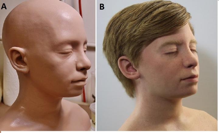
The fusion of 3D printing and special effects resulted in a full-scale reproduction of a 14-year-old child’s head, modeled after a real patient with hydrocephalus, one of the most common problems seen in the field of pediatric neurosurgery. Special features include an electronic pump to reproduce flowing cerebrospinal fluid and brain pulsations. One version of the simulator is so realistic that it has facial features, hair, eyelashes and eyebrows.

To test the model, Cohen and his team randomly paired four neurosurgery fellows and 13 medical residents to perform ETV on either the ultra-realistic simulator or a lower-resolution simulator, which had no hair, lashes or brows.

After completing the simulation, fellows and residents each rated the simulator using a five-point scale. On average, both the surgical fellows and the residents rated the simulator more highly (4.88 out of 5) on its effectiveness for ETV training than on its aesthetic features (4.69). The procedures performed by the trainees were also recorded and later watched and graded by two fully trained neurosurgeons in a way that they could not identify who the trainees were or at what stage they were in their training.

The neurosurgeons assessed the trainees’ performance using criteria such as “flow of operation,” “instrument handling” and “time and motion.”

Neurosurgeons consistently rated the fellows higher than residents on all criteria measured, which accurately reflected their advanced training and knowledge, and demonstrated the simulator’s ability to distinguish between novice and expert surgeons.

Cohen says that further tests are needed to determine whether the simulator will actually improve performance in the operating room. “With this unique assortment of investigators, we were able to develop a high-fidelity simulator for minimally invasive neurosurgery that is realistic, reliable, reusable and cost-effective. The models can be designed to be patient-specific, enabling the surgeon to practice the operation before going into the operating room,” says Cohen.

Other authors on this paper include Roberta Rehder from the Johns Hopkins School of Medicine, and Peter Weinstock, Sanjay P. Parbhu, Peter W. Forbes and Christopher Roussin from Boston Children’s Hospital.

Funding for the study was provided by a grant from the Boston Investment Conference. The research team acknowledges the contribution of FracturedFX, an Emmy Award-winning special effects group from Hollywood, California, in the development of the surgical models.

The investigators report no financial stake or interests in the success of the simulator.

Here’s what the model looks like,

Caption: A. Low-fidelity simulated surgical model for ETV. B. High-fidelity model with hair, eyelashes and eyebrows. Credit: Copyright AANS. Used with permission.

An April 25, 2017 Journal of Neurosurgery news release on EurekAlert details the refinements applied to this replica (Note: There is some repetitive material),

….

A neurosurgery residency training program generally lasts seven years–longer than any other medical specialty. Trainees log countless hours observing surgeries performed by experienced neurosurgeons and developing operative skills in practice labs before touching patients. It is challenging to create a realistic surgical experience outside an operating room. Cadaveric specimens and virtual reality programs have been used, but they are costly and do not provide as realistic an experience as desired.

The new training simulation model described in this paper is a full-scale reproduction of the head of an adolescent patient with hydrocephalus. The external appearance of the head is uncannily accurate, as is the internal neuroanatomy.

One failing of 3D models is the stiffness of most sculpting material. This problem was overcome by addition of special-effects materials that reproduce the textures of external skin and internal brain structures. In addition, the operative environment in this training model is amazingly alive, with pulsations of a simulated basilar artery and ventricles as well as movement of cerebrospinal fluid. These advances provide visual and tactile feedback to the trainee that closely resembles that of the surgical experience.

The procedure selected to test the new training model was endoscopic third ventriculostomy (ETV), a minimally invasive surgical procedure increasingly used to treat hydrocephalus. The goal of ETV is to create a hole in the floor of the third ventricle. This provides a new pathway by which excess cerebrospinal fluid can circulate.

During ETV, the surgeon drills a small hole in the skull of the patient and inserts an endoscope into the ventricular system. The endoscope accommodates a lighted miniature video camera to visualize the operative site and specialized surgical instruments suited to perform operative tasks through the endoscope. The video camera sends a direct feed to external monitors in the operating room so that surgeons can clearly see what they are doing.

To evaluate the usefulness of the training simulation model of ETV, the researchers solicited feedback from users (neurosurgical residents and fellows) and their teachers. Trainees were asked to respond to a 14-item questionnaire focused on the external and internal appearances of the model and its tactile feel during simulated surgery (face validity) as well as on how closely the simulated procedure reproduced an actual ETV (content validity). The usefulness of the model in assessing trainees’ performances was then evaluated by two attending neurosurgeons blinded to the identity and training status (post-graduate year of training) of the residents and fellows (construct validity).

The neurosurgical residents and fellows gave high scores to the training model for both face and content validity (mean scores of 4.69 and 4.88, respectively; 5 would be a perfect score). The performance scores given to individual trainees by the attending neurosurgeons clearly distinguished novice surgeons from more experienced surgeons, accurately reflecting the trainees’ post-graduate years of training.

The training model described in this paper is not limited to hydrocephalus or treatment with ETV. The simulated head accommodates replaceable plug-and-play components to provide a fresh operative field for each training session. A variety of diseased or injured brain scenarios could be tested using different plug-and-play components. In addition, the ability to pop in new components between practice sessions greatly reduces training costs compared to other models.

When asked about the paper, the senior author, Alan R. Cohen, MD, at Johns Hopkins Hospital, said, “This unique collaboration of interdisciplinary experts resulted in the creation of an ultra-realistic 3D surgical training model. Simulation has become increasingly important for training in minimally invasive neurosurgery. It also has the potential to revolutionize training for all surgical procedures.

Here’s a link to and a citation for the paper,

Creation of a novel simulator for minimally invasive neurosurgery: fusion of 3D printing and special effects by Peter Weinstock, Roberta Rehder, Sanjay P. Parbhu, Peter W. Forbes, Christopher J. Roussin, and Alan R. Cohen. Journal of Neurosurgery: Pediatrics, published online, ahead of print, April 25, 2017; DOI: 10.3171/2017.1.PEDS16568

This paper appears to be open access.

Mesenchymal condensation (a process embryos use to begin forming a variety of organs, including teeth, cartilage, bone, muscle, tendon, and kidney) for complex 3D tissue engineering

It seems that there are three strategies for creating complex 3D tissues and until now scientists have used only two of the three. From a March 5, 2014 news item on ScienceDaily,

A bit of pressure from a new shrinking, sponge-like gel is all it takes to turn transplanted unspecialized cells into cells that lay down minerals and begin to form teeth.

The bioinspired gel material could one day help repair or replace damaged organs, such as teeth and bone, and possibly other organs as well, scientists from the Wyss Institute for Biologically Inspired Engineering at Harvard University, Harvard School of Engineering and Applied Sciences (SEAS), and Boston Children’s Hospital report recently in Advanced Materials.

“Tissue engineers have long raised the idea of using synthetic materials to mimic the inductive power of the embryo,” said Don Ingber, M.D., Ph.D., Founding Director of the Wyss Institute, …, Professor of Bioengineering at SEAS, and senior author of the study. “We’re excited about this work because it shows that it really is possible.”

The March 5, 2014 Wyss Institute news release, which originated the news item, delves into the nature of the research,

Embryonic tissues have the power to drive cells and tissues to specialize and form organs. To do that, they employ biomolecules called growth factors to stimulate growth; gene-activating chemicals that cause the cells to specialize, and mechanical forces that modulate cell responses to these other factors.

But so far tissue engineers who want to build organs in the laboratory have employed only two of the three strategies — growth factors and gene-activating chemicals. Perhaps as a result, they have not yet succeeded in producing complex three-dimensional tissues.

A few years ago, Ingber and Tadanori Mammoto, M.D., Ph.D., Instructor in Surgery at Boston Children’s Hospital and Harvard Medical School, investigated a process called mesenchymal condensation that embryos use to begin forming a variety of organs, including teeth, cartilage, bone, muscle, tendon, and kidney.

In mesenchymal condensation, two adjacent tissue layers — loosely packed connective-tissue cells called mesenchyme and sheet-like tissue called an epithelium that covers it — exchange biochemical signals. This exchange causes the mesenchymal cells to squeeze themselves tightly into a small knot directly below where the new organ will form.

Here’s a video from the Wyss Institute illustrating the squeezing process,

When the temperature rises to just below body temperature, this biocompatible gel shrinks dramatically within minutes, bringing tooth-precursor cells (green) closer together. Credit: Basma Hashmi

Getting back to the research (from the news release),

By examining tissues isolated from the jaws of embryonic mice, Mammoto and Ingber showed that when the compressed mesenchymal cells turn on genes that stimulate them to generate whole teeth composed of mineralized tissues, including dentin and enamel.

Inspired by this embryonic induction mechanism, Ingber and Basma Hashmi, a Ph.D. candidate at SEAS who is the lead author of the current paper, set out to develop a way to engineer artificial teeth by creating a tissue-friendly material that accomplishes the same goal. Specifically, they wanted a porous sponge-like gel that could be impregnated with mesenchymal cells, then, when implanted into the body, induced to shrink in 3D to physically compact the cells inside it.

To develop such a material, Ingber and Hashmi teamed up with researchers led by Joanna Aizenberg, Ph.D., a Wyss Institute Core Faculty member who leads the Institute’s Adaptive Materials Technologies platform. Aizenberg is the Amy Smith Berylson Professor of Materials Science at SEAS and Professor of Chemistry and Chemical Biology at Harvard University.

They chemically modified a special gel-forming polymer called PNIPAAm that scientists have used to deliver drugs to the body’s tissues. PNIPAAm gels have an unusual property: they contract abruptly when they warm.

But they do this at a lukewarm temperature, whereas the researchers wanted them to shrink specifically at 37°C — body temperature — so that they’d squeeze their contents as soon as they were injected into the body. Hashmi worked with Lauren Zarzar, Ph.D., a former SEAS graduate student who’s now a postdoctoral associate at Massachusetts Institute of Technology, for more than a year, modifying PNIPAAm and testing the resulting materials. Ultimately, they developed a polymer that forms a tissue-friendly gel with two key properties: cells stick to it, and it compresses abruptly when warmed to body temperature.

As an initial test, Hashmi implanted mesenchymal cells in the gel and warmed it in the lab. Sure enough, when the temperature reached 37°C, the gel shrank within 15 minutes, causing the cells inside the gel to round up, shrink, and pack tightly together.

“The reason that’s cool is that the cells are alive,” Hashmi said. “Usually when this happens, cells are dead or dying.”

Not only were they alive — they activated three genes that drive tooth formation.

To see if the shrinking gel also worked its magic in the body, Hashmi worked with Mammoto to load mesenchymal cells into the gel, then implant the gel beneath the mouse kidney capsule — a tissue that is well supplied with blood and often used for transplantation experiments.

The implanted cells not only expressed tooth-development genes — they laid down calcium and minerals, just as mesenchymal cells do in the body as they begin to form teeth.

“They were in full-throttle tooth-development mode,” Hashmi said.

The researchers have future plans (from the news release),

In the embryo, mesenchymal cells can’t build teeth alone — they need to be combined with cells that form the epithelium. In the future, the scientists plan to test whether the shrinking gel can stimulate both tissues to generate an entire functional tooth.

Here’s a link to and a citation for the paper about the successful attempt to stimulate mesenchymal cells into the beginnings of tooth formation,

Developmentally-Inspired Shrink-Wrap Polymers for Mechanical Induction of Tissue Differentiation by Basma Hashmi, Lauren D. Zarzar, Tadanori Mammoto, Akiko Mammoto, Amanda Jiang, Joanna Aizenberg, and Donald E. Ingber. Advanced Materials Article first published online: 18 FEB 2014 DOI: 10.1002/adma.201304995

© 2014 WILEY-VCH Verlag GmbH & Co. KGaA, Weinheim

This paper is behind a paywall.

Gluing a broken heart back together

The Jan. 8, 2014 news item on ScienceDaily doesn’t identify which creature(s) may have inspired the heart glue developed by researchers from Brigham and Women’s Hospital (BWH), Boston Children’s Hospital, and Massachusetts Institute of Technology (MIT),

When a child is born with a heart defect such as a hole in the heart, the highly invasive therapies are challenging due to an inability to quickly and safely secure devices inside the heart. Sutures take too much time to stitch and can cause stress on fragile heart tissue, and currently available clinical adhesives are either too toxic or tend to lose their sticking power in the presence of blood or under dynamic conditions, such as in a beating heart.

“About 40,000 babies are born with congenital heart defects in the United States annually, and those that require treatment are plagued with multiple surgeries to deliver or replace non-degradable implants that do not grow with young patients,” says Jeffrey Karp, PhD, Division of Biomedical Engineering, BWH Department of Medicine, co-senior study author of a new study that may improve how surgeons treat congenital heart defects.

In the preclinical study, researchers from Boston Children’s Hospital, BWH and Massachusetts Institute of Technology (MIT) developed a bio-inspired adhesive that could rapidly attach biodegradable patches inside a beating heart — in the exact place where congenital holes in the heart occur, such as with ventricular heart defects.

The Jan. 8, 2014 BWH news release on EurekAlert, which originated the news item, discusses the use of adhesives for repair in the body and some of the specifics of this particular application,

Recognizing that many creatures in nature have secretions that are viscous and repel water, enabling them to attach under wet and dynamic conditions, the researchers developed a material with these properties that also is biodegradable, elastic and biocompatible. According to the study authors, the degradable patches secured with the glue remained attached even at increased heart rates and blood pressure.

“This adhesive platform addresses all of the drawbacks of previous systems in that it works in the presence of blood and moving structures,” says Pedro del Nido, MD, Chief of Cardiac Surgery, Boston Children’s Hospital, co-senior study author. “It should provide the physician with a completely new, much simpler technology and a new paradigm for tissue reconstruction to improve the quality of life of patients following surgical procedures.”

Unlike current surgical adhesives, this new adhesive maintains very strong sticking power when in the presence of blood, and even in active environments.

“This study demonstrated that the adhesive was strong enough to hold tissue and patches onto the heart equivalent to suturing,” says the study’s co-first author Nora Lang, MD, Department of Cardiac Surgery, Boston Children’s Hospital. “Also, the adhesive patch is biodegradable and biocompatible, so nothing foreign or toxic stays in the bodies of these patients.”

Importantly, its adhesive abilities are activated with ultraviolent (UV) light, providing an on-demand, anti-bleeding seal within five seconds of UV light application when applied to high-pressure large blood vessels and cardiac wall defects.

“When we attached patches coated with our adhesive to the walls of a beating heart, the patches remained despite the high pressures of blood flowing through the heart and blood vessels,” says Maria N. Pereira, PhD, Division of Biomedical Engineering, BWH Department of Medicine, co-first study author.

The researchers note that their waterproof, light-activated adhesive will be useful in reducing the invasiveness of surgical procedures, as well as operating times, in addition to improving heart surgery outcomes.

As to which creature(s) may have inspired the glue, perhaps this offers a hint,

The adhesive technology (and other related platforms) has been licensed to a start-up company, Gecko Biomedical, based in Paris. [emphasis mine] The company has raised 8 million Euros in their recently announced Series A financing round and expects to bring the adhesive to the market within two to three years.

The last time geckos and adhesives were mentioned here was in a Jan. 2, 2014 posting titled: Simon Fraser University’s (Canada) gecko-type robots and the European Space Agency.

Getting back to the heart glue, here’s an image illustrating the researchers’ work,

Caption: The waterproof, light-activated glue developed by researchers at Brigham and Women's Hospital, Boston Children's Hospital and Massachusetts Institute of Technology can successfully secure biodegradable patches to seal holes in a beating heart. Credit: Karp Laboratory

Caption: The waterproof, light-activated glue developed by researchers at Brigham and Women’s Hospital, Boston Children’s Hospital and Massachusetts Institute of Technology can successfully secure biodegradable patches to seal holes in a beating heart.
Credit: Karp Laboratory

For the interested, here’s a link to and a citation for the paper,

A Blood-Resistant Surgical Glue for Minimally Invasive Repair of Vessels and Heart Defects by Nora Lang, Maria J. Pereira, Yuhan Lee, Ingeborg Friehs, Nikolay V. Vasilyev, Eric N. Feins, Klemens Ablasser, Eoin D. O’Cearbhaill, Chenjie Xu, Assunta Fabozzo, Robert Padera, Steve Wasserman, Franz Freudenthal, Lino S. Ferreira, Robert Langer, Jeffrey M. Karp, and Pedro J. del Nido. Sci Transl Med 8 January 2014: Vol. 6, Issue 218, p. 218ra6 Sci. Transl. Med. DOI: 10.1126/scitranslmed.3006557

This paper is behind a paywall.

No need for eye drops when your contact lenses can dispense your eye medication

Anyone who has difficulty getting or allowing drops into their eyes (I once slid out of an ophthalmologist’s examination chair trying to avoid the eye drops he was administering at the end of my appointment) is going appreciate this Dec. 9, 2013 news item on Nanowerk,

For nearly half a century, contact lenses have been proposed as a means of ocular drug delivery that may someday replace eye drops, but achieving controlled drug release has been a significant challenge. Researchers at Massachusetts Eye and Ear/Harvard Medical School Department of Ophthalmology, Boston Children’s Hospital, and the Massachusetts Institute of Technology are one step closer to an eye drop-free reality with the development of a drug-eluting contact lens designed for prolonged delivery of latanoprost, a common drug used for the treatment of glaucoma, the leading cause of irreversible blindness worldwide.

The Dec. 9, 2013 Massachusetts Eye and Ear Infirmary press release (also on EurekAlert), which originated the news item, notes that a lot of people have problems with eye drops and gives a general description of the research,

“In general, eye drops are an inefficient method of drug delivery that has notoriously poor patient adherence. This contact lens design can potentially be used as a treatment for glaucoma and as a platform for other ocular drug delivery applications,” said Joseph Ciolino, M.D, Mass. Eye and Ear cornea specialist and lead author of the paper.

The contacts were designed with materials that are FDA-approved for use on the eye. The latanoprost-eluting contact lenses were created by encapsulating latanoprost-polymer films in commonly used contact lens hydrogel. Their findings are described online and will be in the January 2014 printed issue of Biomaterials.

“The lens we have developed is capable of delivering large amounts of drug at substantially constant rates over weeks to months,” said Professor Daniel Kohane, director of the Laboratory for Biomaterials and Drug Delivery at Boston Children’s Hospital.

In vivo, single contact lenses were able to achieve, for one month, latanoprost concentrations in the aqueous humor that were comparable to those achieved with daily topical latanoprost solution, the current first-line treatment for glaucoma.

The lenses appeared safe in cell culture and animal studies. This is the first contact lens that has been shown to release drugs for this long in animal models.

The newly designed contact lens has a clear central aperture and contains a drug-polymer film in the periphery, which helps to control drug release. The lenses can be made with no refractive power or with the ability to correct the refractive error in near sided or far sided eyes.

Here’s a link to and a citation for the researchers’ published paper,

In vivo performance of a drug-eluting contact lens to treat glaucoma for a month by Joseph B. Ciolino, Cristina F. Stefanescu, Amy E. Ross, Borja Salvador-Culla, Priscila Cortez, Eden M. Ford, Kate A. Wymbs, Sarah L. Sprague, Daniel R. Mascoop, Shireen S. Rudina, Sunia A. Trauger, Fabiano Cade, Daniel S. Kohane. Biomaterials Volume 35, Issue 1, January 2014, Pages 432–439 DOI: S0142961213011150

This article is behind a paywall.

The body as an electronic device—adding electronics to biological tissue

What makes this particular combination of electronic s  and living tissue special is t that it was achieved in 3-D rather than 2-D.  From the Boston Children’s Hospital Aug. 26, 2012 news release on EurekAlert,

A multi-institutional research team has developed a method for embedding networks of biocompatible nanoscale wires within engineered tissues. These networks—which mark the first time that electronics and tissue have been truly merged in 3D—allow direct tissue sensing and potentially stimulation, a potential boon for development of engineered tissues that incorporate capabilities for monitoring and stimulation, and of devices for screening new drugs.

The Aug. 27, 2012 news item on Nanowerk provides more detail about integration of the cells and electronics,

Until now, the only cellular platforms that incorporated electronic sensors consisted of flat layers of cells grown on planar metal electrodes or transistors. Those two-dimensional systems do not accurately replicate natural tissue, so the research team set out to design a 3-D scaffold that could monitor electrical activity, allowing them to see how cells inside the structure would respond to specific drugs.

The researchers built their new scaffold out of epoxy, a nontoxic material that can take on a porous, 3-D structure. Silicon nanowires embedded in the scaffold carry electrical signals to and from cells grown within the structure.

“The scaffold is not just a mechanical support for cells, it contains multiple sensors. We seed cells into the scaffold and eventually it becomes a 3-D engineered tissue,” Tian says [Bozhi Tian, a former postdoc at MIT {Massachusetts Institute of Technology} and Children’s Hospital and a lead author of the paper ].

The team chose silicon nanowires for electronic sensors because they are small, stable, can be safely implanted into living tissue and are more electrically sensitive than metal electrodes. The nanowires, which range in diameter from 30 to 80 nanometers (about 1,000 times smaller than a human hair), can detect voltages less than one-thousandth of a watt, which is the level of electricity that might be seen in a cell.

Here’s more about why the researchers want to integrate living tissue and electronics, from the Harvard University Aug. 26, 2012 news release on EurekAlert,

“The current methods we have for monitoring or interacting with living systems are limited,” said Lieber [Charles M. Lieber, the Mark Hyman, Jr. Professor of Chemistry at Harvard and one of the study’s team leaders]. “We can use electrodes to measure activity in cells or tissue, but that damages them. With this technology, for the first time, we can work at the same scale as the unit of biological system without interrupting it. Ultimately, this is about merging tissue with electronics in a way that it becomes difficult to determine where the tissue ends and the electronics begin.”

The research addresses a concern that has long been associated with work on bioengineered tissue – how to create systems capable of sensing chemical or electrical changes in the tissue after it has been grown and implanted. The system might also represent a solution to researchers’ struggles in developing methods to directly stimulate engineered tissues and measure cellular reactions.

“In the body, the autonomic nervous system keeps track of pH, chemistry, oxygen and other factors, and triggers responses as needed,” Kohane [Daniel Kohane, a Harvard Medical School professor in the Department of Anesthesia at Children’s Hospital Boston and a team leader] explained. “We need to be able to mimic the kind of intrinsic feedback loops the body has evolved in order to maintain fine control at the cellular and tissue level.”

Here’s a citation and a link to the paper (which is behind a paywall),

Macroporous nanowire nanoelectronic scaffolds for synthetic tissues by Bozhi Tian, Jia Lin, Tal Dvir, Lihua Jin, Jonathan H. Tsui, Quan  Qing, Zhigang Suo, Robert Langer, Daniel S. Kohane, and Charles M. Lieber in Nature Materials (2012) doi:10.1038/nmat3404 Published onlin26 August 2012.

This is the image MIT included with its Aug 27, 2012 news release (which originated the news item on Nanowerk),

A 3-D reconstructed confocal fluorescence micrograph of a tissue scaffold.
Image: Charles M. Lieber and Daniel S. Kohane.

At this point they’re discussing therapeutic possibilities but I expect that ‘enhancement’ is also being considered although not mentioned for public consumption.

Organ chips for DARPA (Defense Advanced Research Projects Agency)

The Wyss Institute will receive up to  $37M US for a project that integrates ten different organ-on-a-chip projects into one system. From the July 24, 2012 news release on EurekAlert,

With this new DARPA funding, Institute researchers and a multidisciplinary team of collaborators seek to build 10 different human organs-on-chips, to link them together to more closely mimic whole body physiology, and to engineer an automated instrument that will control fluid flow and cell viability while permitting real-time analysis of complex biochemical functions. As an accurate alternative to traditional animal testing models that often fail to predict human responses, this instrumented “human-on-a-chip” will be used to rapidly assess responses to new drug candidates, providing critical information on their safety and efficacy.

This unique platform could help ensure that safe and effective therapeutics are identified sooner, and ineffective or toxic ones are rejected early in the development process. As a result, the quality and quantity of new drugs moving successfully through the pipeline and into the clinic may be increased, regulatory decision-making could be better informed, and patient outcomes could be improved.

Jesse Goodman, FDA Chief Scientist and Deputy Commissioner for Science and Public Health, commented that the automated human-on-chip instrument being developed “has the potential to be a better model for determining human adverse responses. FDA looks forward to working with the Wyss Institute in its development of this model that may ultimately be used in therapeutic development.”

Wyss Founding Director, Donald Ingber, M.D., Ph.D., and Wyss Core Faculty member, Kevin Kit Parker, Ph.D., will co-lead this five-year project.

I note that Kevin Kit Parker was mentioned in an earlier posting today (July 26, 2012) titled, Medusa, jellyfish, and tissue engineering, and Donald Ingber in my Dec.1e, 2011 posting about Shrilk and insect skeletons.

As for the Wyss Institute, here’s a description from the news release,

The Wyss Institute for Biologically Inspired Engineering at Harvard University (http://wyss.harvard.edu) uses Nature’s design principles to develop bioinspired materials and devices that will transform medicine and create a more sustainable world. Working as an alliance among Harvard’s Schools of Medicine, Engineering, and Arts & Sciences, and in partnership with Beth Israel Deaconess Medical Center, Boston Children’s Hospital, Brigham and Women’s Hospital, , Dana Farber Cancer Institute, Massachusetts General Hospital, the University of Massachusetts Medical School, Spaulding Rehabilitation Hospital, Tufts University, and Boston University, the Institute crosses disciplinary and institutional barriers to engage in high-risk research that leads to transformative technological breakthroughs. By emulating Nature’s principles for self-organizing and self-regulating, Wyss researchers are developing innovative new engineering solutions for healthcare, energy, architecture, robotics, and manufacturing. These technologies are translated into commercial products and therapies through collaborations with clinical investigators, corporate alliances, and new start-ups.

I hadn’t thought of an organ-on-a-chip as particularly bioinspired so I’ll have to think about that one for a while.