Tag Archives: Massachusetts General Hospital

Quadriplegic man reanimates a limb with implanted brain-recording and muscle-stimulating systems

It took me a few minutes to figure out why this item about a quadriplegic (also known as, tetraplegic) man is news. After all, I have a May 17, 2012 posting which features a video and information about a quadri(tetra)plegic woman who was drinking her first cup of coffee, independently, in many years. The difference is that she was using an external robotic arm and this man is using *his own arm*,

This Case Western Reserve University (CRWU) video accompanies a March 28, 2017 CRWU news release, (h/t ScienceDaily March 28, 2017 news item)

Bill Kochevar grabbed a mug of water, drew it to his lips and drank through the straw.

His motions were slow and deliberate, but then Kochevar hadn’t moved his right arm or hand for eight years.

And it took some practice to reach and grasp just by thinking about it.

Kochevar, who was paralyzed below his shoulders in a bicycling accident, is believed to be the first person with quadriplegia in the world to have arm and hand movements restored with the help of two temporarily implanted technologies.

A brain-computer interface with recording electrodes under his skull, and a functional electrical stimulation (FES) system* activating his arm and hand, reconnect his brain to paralyzed muscles.

Holding a makeshift handle pierced through a dry sponge, Kochevar scratched the side of his nose with the sponge. He scooped forkfuls of mashed potatoes from a bowl—perhaps his top goal—and savored each mouthful.

“For somebody who’s been injured eight years and couldn’t move, being able to move just that little bit is awesome to me,” said Kochevar, 56, of Cleveland. “It’s better than I thought it would be.”

Kochevar is the focal point of research led by Case Western Reserve University, the Cleveland Functional Electrical Stimulation (FES) Center at the Louis Stokes Cleveland VA Medical Center and University Hospitals Cleveland Medical Center (UH). A study of the work was published in the The Lancet March 28 [2017] at 6:30 p.m. U.S. Eastern time.

“He’s really breaking ground for the spinal cord injury community,” said Bob Kirsch, chair of Case Western Reserve’s Department of Biomedical Engineering, executive director of the FES Center and principal investigator (PI) and senior author of the research. “This is a major step toward restoring some independence.”

When asked, people with quadriplegia say their first priority is to scratch an itch, feed themselves or perform other simple functions with their arm and hand, instead of relying on caregivers.

“By taking the brain signals generated when Bill attempts to move, and using them to control the stimulation of his arm and hand, he was able to perform personal functions that were important to him,” said Bolu Ajiboye, assistant professor of biomedical engineering and lead study author.

Technology and training

The research with Kochevar is part of the ongoing BrainGate2* pilot clinical trial being conducted by a consortium of academic and VA institutions assessing the safety and feasibility of the implanted brain-computer interface (BCI) system in people with paralysis. Other investigational BrainGate research has shown that people with paralysis can control a cursor on a computer screen or a robotic arm (braingate.org).

“Every day, most of us take for granted that when we will to move, we can move any part of our body with precision and control in multiple directions and those with traumatic spinal cord injury or any other form of paralysis cannot,” said Benjamin Walter, associate professor of neurology at Case Western Reserve School of Medicine, clinical PI of the Cleveland BrainGate2 trial and medical director of the Deep Brain Stimulation Program at UH Cleveland Medical Center.

“The ultimate hope of any of these individuals is to restore this function,” Walter said. “By restoring the communication of the will to move from the brain directly to the body this work will hopefully begin to restore the hope of millions of paralyzed individuals that someday they will be able to move freely again.”

Jonathan Miller, assistant professor of neurosurgery at Case Western Reserve School of Medicine and director of the Functional and Restorative Neurosurgery Center at UH, led a team of surgeons who implanted two 96-channel electrode arrays—each about the size of a baby aspirin—in Kochevar’s motor cortex, on the surface of the brain.

The arrays record brain signals created when Kochevar imagines movement of his own arm and hand. The brain-computer interface extracts information from the brain signals about what movements he intends to make, then passes the information to command the electrical stimulation system.

To prepare him to use his arm again, Kochevar first learned how to use his brain signals to move a virtual-reality arm on a computer screen.

“He was able to do it within a few minutes,” Kirsch said. “The code was still in his brain.”

As Kochevar’s ability to move the virtual arm improved through four months of training, the researchers believed he would be capable of controlling his own arm and hand.

Miller then led a team that implanted the FES systems’ 36 electrodes that animate muscles in the upper and lower arm.

The BCI decodes the recorded brain signals into the intended movement command, which is then converted by the FES system into patterns of electrical pulses.

The pulses sent through the FES electrodes trigger the muscles controlling Kochevar’s hand, wrist, arm, elbow and shoulder. To overcome gravity that would otherwise prevent him from raising his arm and reaching, Kochevar uses a mobile arm support, which is also under his brain’s control.

New Capabilities

Eight years of muscle atrophy required rehabilitation. The researchers exercised Kochevar’s arm and hand with cyclical electrical stimulation patterns. Over 45 weeks, his strength, range of motion and endurance improved. As he practiced movements, the researchers adjusted stimulation patterns to further his abilities.

Kochevar can make each joint in his right arm move individually. Or, just by thinking about a task such as feeding himself or getting a drink, the muscles are activated in a coordinated fashion.

When asked to describe how he commanded the arm movements, Kochevar told investigators, “I’m making it move without having to really concentrate hard at it…I just think ‘out’…and it goes.”

Kocehvar is fitted with temporarily implanted FES technology that has a track record of reliable use in people. The BCI and FES system together represent early feasibility that gives the research team insights into the potential future benefit of the combined system.

Advances needed to make the combined technology usable outside of a lab are not far from reality, the researchers say. Work is underway to make the brain implant wireless, and the investigators are improving decoding and stimulation patterns needed to make movements more precise. Fully implantable FES systems have already been developed and are also being tested in separate clinical research.

Kochevar welcomes new technology—even if it requires more surgery—that will enable him to move better. “This won’t replace caregivers,” he said. “But, in the long term, people will be able, in a limited way, to do more for themselves.”

There is more about the research in a March 29, 2017 article by Sarah Boseley for The Guardian,

Bill Kochevar, 53, has had electrical implants in the motor cortex of his brain and sensors inserted in his forearm, which allow the muscles of his arm and hand to be stimulated in response to signals from his brain, decoded by computer. After eight years, he is able to drink and feed himself without assistance.

“I think about what I want to do and the system does it for me,” Kochevar told the Guardian. “It’s not a lot of thinking about it. When I want to do something, my brain does what it does.”

The experimental technology, pioneered by the Case Western Reserve University in Cleveland, Ohio, is the first in the world to restore brain-controlled reaching and grasping in a person with complete paralysis.

For now, the process is relatively slow, but the scientists behind the breakthrough say this is proof of concept and that they hope to streamline the technology until it becomes a routine treatment for people with paralysis. In the future, they say, it will also be wireless and the electrical arrays and sensors will all be implanted under the skin and invisible.

A March 28, 2017 Lancet news release on EurekAlert provides a little more technical insight into the research and Kochevar’s efforts,

Although only tested with one participant, the study is a major advance and the first to restore brain-controlled reaching and grasping in a person with complete paralysis. The technology, which is only for experimental use in the USA, circumvents rather than repairs spinal injuries, meaning the participant relies on the device being implanted and switched on to move.

“Our research is at an early stage, but we believe that this neuro-prosthesis could offer individuals with paralysis the possibility of regaining arm and hand functions to perform day-to-day activities, offering them greater independence,” said lead author Dr Bolu Ajiboye, Case Western Reserve University, USA. “So far it has helped a man with tetraplegia to reach and grasp, meaning he could feed himself and drink. With further development, we believe the technology could give more accurate control, allowing a wider range of actions, which could begin to transform the lives of people living with paralysis.” [1]

Previous research has used similar elements of the neuro-prosthesis. For example, a brain-computer interface linked to electrodes on the skin has helped a person with less severe paralysis open and close his hand, while other studies have allowed participants to control a robotic arm using their brain signals. However, this is the first to restore reaching and grasping via the system in a person with a chronic spinal cord injury.

In this study, a 53 year-old man who had been paralysed below the shoulders for eight years underwent surgery to have the neuro-prosthesis fitted.

This involved brain surgery to place sensors in the motor cortex area of his brain responsible for hand movement – creating a brain-computer interface that learnt which movements his brain signals were instructing for. This initial stage took four months and included training using a virtual reality arm.

He then underwent another procedure placing 36 muscle stimulating electrodes into his upper and lower arm, including four that helped restore finger and thumb, wrist, elbow and shoulder movements. These were switched on 17 days after the procedure, and began stimulating the muscles for eight hours a week over 18 weeks to improve strength, movement and reduce muscle fatigue.

The researchers then wired the brain-computer interface to the electrical stimulators in his arm, using a decoder (mathematical algorithm) to translate his brain signals into commands for the electrodes in his arm. The electrodes stimulated the muscles to produce contractions, helping the participant intuitively complete the movements he was thinking of. The system also involved an arm support to stop gravity simply pulling his arm down.

During his training, the participant described how he controlled the neuro-prosthesis: “It’s probably a good thing that I’m making it move without having to really concentrate hard at it. I just think ‘out’ and it just goes.”

After 12 months of having the neuro-prosthesis fitted, the participant was asked to complete day-to-day tasks, including drinking a cup of coffee and feeding himself. First of all, he observed while his arm completed the action under computer control. During this, he thought about making the same movement so that the system could recognise the corresponding brain signals. The two systems were then linked and he was able to use it to drink a coffee and feed himself.

He successfully drank in 11 out of 12 attempts, and it took him roughly 20-40 seconds to complete the task. When feeding himself, he did so multiple times – scooping forkfuls of food and navigating his hand to his mouth to take several bites.

“Although similar systems have been used before, none of them have been as easy to adopt for day-to-day use and they have not been able to restore both reaching and grasping actions,” said Dr Ajiboye. “Our system builds on muscle stimulating electrode technology that is already available and will continue to improve with the development of new fully implanted and wireless brain-computer interface systems. This could lead to enhanced performance of the neuro-prosthesis with better speed, precision and control.” [1]

At the time of the study, the participant had had the neuro-prosthesis implanted for almost two years (717 days) and in this time experienced four minor, non-serious adverse events which were treated and resolved.

Despite its achievements, the neuro-prosthesis still had some limitations, including that movements made using it were slower and less accurate than those made using the virtual reality arm the participant used for training. When using the technology, the participant also needed to watch his arm as he lost his sense of proprioception – the ability to intuitively sense the position and movement of limbs – as a result of the paralysis.

Writing in a linked Comment, Dr Steve Perlmutter, University of Washington, USA, said: “The goal is futuristic: a paralysed individual thinks about moving her arm as if her brain and muscles were not disconnected, and implanted technology seamlessly executes the desired movement… This study is groundbreaking as the first report of a person executing functional, multi-joint movements of a paralysed limb with a motor neuro-prosthesis. However, this treatment is not nearly ready for use outside the lab. The movements were rough and slow and required continuous visual feedback, as is the case for most available brain-machine interfaces, and had restricted range due to the use of a motorised device to assist shoulder movements… Thus, the study is a proof-of-principle demonstration of what is possible, rather than a fundamental advance in neuro-prosthetic concepts or technology. But it is an exciting demonstration nonetheless, and the future of motor neuro-prosthetics to overcome paralysis is brighter.”

[1] Quote direct from author and cannot be found in the text of the Article.

Here’s a link to and a citation for the paper,

Restoration of reaching and grasping movements through brain-controlled muscle stimulation in a person with tetraplegia: a proof-of-concept demonstration by A Bolu Ajiboye, Francis R Willett, Daniel R Young, William D Memberg, Brian A Murphy, Jonathan P Miller, Benjamin L Walter, Jennifer A Sweet, Harry A Hoyen, Michael W Keith, Prof P Hunter Peckham, John D Simeral, Prof John P Donoghue, Prof Leigh R Hochberg, Prof Robert F Kirsch. The Lancet DOI: http://dx.doi.org/10.1016/S0140-6736(17)30601-3 Published: 28 March 2017 [online?]

This paper is behind a paywall.

For anyone  who’s interested, you can find the BrainGate website here.

*I initially misidentified the nature of the achievement and stated that Kochevar used a “robotic arm, which is attached to his body” when it was his own reanimated arm. Corrected on April 25, 2017.
New iron oxide nanoparticle as an MRI (magnetic resonance imaging) contrast agent

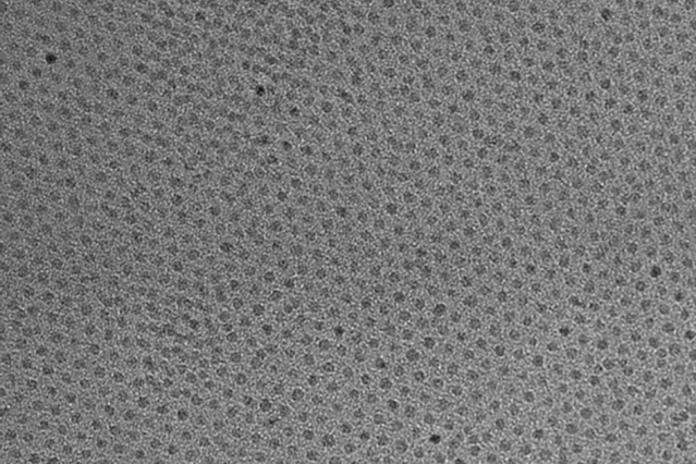
This high-resolution transmission electron micrograph of particles made by the research team shows the particles’ highly uniform size and shape. These are iron oxide particles just 3 nanometers across, coated with a zwitterion layer. Their small size means they can easily be cleared through the kidneys after injection. Courtesy of the researchers

A Feb. 14, 2017 news item on ScienceDaily announces a new MRI (magnetic resonance imaging) contrast agent,

A new, specially coated iron oxide nanoparticle developed by a team at MIT [Massachusetts Institute of Technology] and elsewhere could provide an alternative to conventional gadolinium-based contrast agents used for magnetic resonance imaging (MRI) procedures. In rare cases, the currently used gadolinium agents have been found to produce adverse effects in patients with impaired kidney function.

A Feb. 14, 2017 MIT news release (also on EurekAlert), which originated the news item, provides more technical detail,


The advent of MRI technology, which is used to observe details of specific organs or blood vessels, has been an enormous boon to medical diagnostics over the last few decades. About a third of the 60 million MRI procedures done annually worldwide use contrast-enhancing agents, mostly containing the element gadolinium. While these contrast agents have mostly proven safe over many years of use, some rare but significant side effects have shown up in a very small subset of patients. There may soon be a safer substitute thanks to this new research.

In place of gadolinium-based contrast agents, the researchers have found that they can produce similar MRI contrast with tiny nanoparticles of iron oxide that have been treated with a zwitterion coating. (Zwitterions are molecules that have areas of both positive and negative electrical charges, which cancel out to make them neutral overall.) The findings are being published this week in the Proceedings of the National Academy of Sciences, in a paper by Moungi Bawendi, the Lester Wolfe Professor of Chemistry at MIT; He Wei, an MIT postdoc; Oliver Bruns, an MIT research scientist; Michael Kaul at the University Medical Center Hamburg-Eppendorf in Germany; and 15 others.

Contrast agents, injected into the patient during an MRI procedure and designed to be quickly cleared from the body by the kidneys afterwards, are needed to make fine details of organ structures, blood vessels, and other specific tissues clearly visible in the images. Some agents produce dark areas in the resulting image, while others produce light areas. The primary agents for producing light areas contain gadolinium.

Iron oxide particles have been largely used as negative (dark) contrast agents, but radiologists vastly prefer positive (light) contrast agents such as gadolinium-based agents, as negative contrast can sometimes be difficult to distinguish from certain imaging artifacts and internal bleeding. But while the gadolinium-based agents have become the standard, evidence shows that in some very rare cases they can lead to an untreatable condition called nephrogenic systemic fibrosis, which can be fatal. In addition, evidence now shows that the gadolinium can build up in the brain, and although no effects of this buildup have yet been demonstrated, the FDA is investigating it for potential harm.

“Over the last decade, more and more side effects have come to light” from the gadolinium agents, Bruns says, so that led the research team to search for alternatives. “None of these issues exist for iron oxide,” at least none that have yet been detected, he says.

The key new finding by this team was to combine two existing techniques: making very tiny particles of iron oxide, and attaching certain molecules (called surface ligands) to the outsides of these particles to optimize their characteristics. The iron oxide inorganic core is small enough to produce a pronounced positive contrast in MRI, and the zwitterionic surface ligand, which was recently developed by Wei and coworkers in the Bawendi research group, makes the iron oxide particles water-soluble, compact, and biocompatible.

The combination of a very tiny iron oxide core and an ultrathin ligand shell leads to a total hydrodynamic diameter of 4.7 nanometers, below the 5.5-nanometer renal clearance threshold. This means that the coated iron oxide should quickly clear through the kidneys and not accumulate. This renal clearance property is an important feature where the particles perform comparably to gadolinium-based contrast agents.

Now that initial tests have demonstrated the particles’ effectiveness as contrast agents, Wei and Bruns say the next step will be to do further toxicology testing to show the particles’ safety, and to continue to improve the characteristics of the material. “It’s not perfect. We have more work to do,” Bruns says. But because iron oxide has been used for so long and in so many ways, even as an iron supplement, any negative effects could likely be treated by well-established protocols, the researchers say. If all goes well, the team is considering setting up a startup company to bring the material to production.

For some patients who are currently excluded from getting MRIs because of potential side effects of gadolinium, the new agents “could allow those patients to be eligible again” for the procedure, Bruns says. And, if it does turn out that the accumulation of gadolinium in the brain has negative effects, an overall phase-out of gadolinium for such uses could be needed. “If that turned out to be the case, this could potentially be a complete replacement,” he says.

Ralph Weissleder, a physician at Massachusetts General Hospital who was not involved in this work, says, “The work is of high interest, given the limitations of gadolinium-based contrast agents, which typically have short vascular half-lives and may be contraindicated in renally compromised patients.”

The research team included researchers in MIT’s chemistry, biological engineering, nuclear science and engineering, brain and cognitive sciences, and materials science and engineering departments and its program in Health Sciences and Technology; and at the University Medical Center Hamburg-Eppendorf; Brown University; and the Massachusetts General Hospital. It was supported by the MIT-Harvard NIH Center for Cancer Nanotechnology, the Army Research Office through MIT’s Institute for Soldier Nanotechnologies, the NIH-funded Laser Biomedical Research Center, the MIT Deshpande Center, and the European Union Seventh Framework Program.

Here’s a link to and a citation for the paper,

Exceedingly small iron oxide nanoparticles as positive MRI contrast agents by He Wei, Oliver T. Bruns, Michael G. Kaul, Eric C. Hansen, Mariya Barch, Agata Wiśniowsk, Ou Chen, Yue Chen, Nan Li, Satoshi Okada, Jose M. Cordero, Markus Heine, Christian T. Farrar, Daniel M. Montana, Gerhard Adam, Harald Ittrich, Alan Jasanoff, Peter Nielsen, and Moungi G. Bawendi. PNAS February 13, 2017 doi: 10.1073/pnas.1620145114 Published online before print February 13, 2017

This paper is behind a paywall.

Bionic pancreas tested at home

This news about a bionic pancreas must be exciting for diabetics as it would eliminate the need for constant blood sugar testing throughout the day. From a Dec. 19, 2016 Massachusetts General Hospital news release (also on EurekAlert), Note: Links have been removed,

The bionic pancreas system developed by Boston University (BU) investigators proved better than either conventional or sensor-augmented insulin pump therapy at managing blood sugar levels in patients with type 1 diabetes living at home, with no restrictions, over 11 days. The report of a clinical trial led by a Massachusetts General Hospital (MGH) physician is receiving advance online publication in The Lancet.

“For study participants living at home without limitations on their activity and diet, the bionic pancreas successfully reduced average blood glucose, while at the same time decreasing the risk of hypoglycemia,” says Steven Russell, MD, PhD, of the MGH Diabetes Unit. “This system requires no information other than the patient’s body weight to start, so it will require much less time and effort by health care providers to initiate treatment. And since no carbohydrate counting is required, it significantly reduces the burden on patients associated with diabetes management.”

Developed by Edward Damiano, PhD, and Firas El-Khatib, PhD, of the BU Department of Biomedical Engineering, the bionic pancreas controls patients’ blood sugar with both insulin and glucagon, a hormone that increases glucose levels. After a 2010 clinical trial confirmed that the original version of the device could maintain near-normal blood sugar levels for more than 24 hours in adult patients, two follow-up trials – reported in a 2014 New England Journal of Medicine paper – showed that an updated version of the system successfully controlled blood sugar levels in adults and adolescents for five days.  Another follow-up trial published in The Lancet Diabetes and Endocrinology in 2016  showed it could do the same for children as young as 6 years of age.

While minimal restrictions were placed on participants in the 2014 trials, participants in both spent nights in controlled settings and were accompanied at all times by either a nurse for the adult trial or remained in a diabetes camp for the adolescent and pre-adolescent trials. Participants in the current trial had no such restrictions placed upon them, as they were able to pursue normal activities at home or at work with no imposed limitations on diet or exercise. Patients needed to live within a 30-minute drive of one of the trial sites – MGH, the University of Massachusetts Medical School, Stanford University, and the University of North Carolina at Chapel Hill – and needed to designate a contact person who lived with them and could be contacted by study staff, if necessary.

The bionic pancreas system – the same as that used in the 2014 studies – consisted of a smartphone (iPhone 4S) that could wirelessly communicate with two pumps delivering either insulin or glucagon. Every five minutes the smartphone received a reading from an attached continuous glucose monitor, which was used to calculate and administer a dose of either insulin or glucagon. The algorighms controlling the system were updated for the current trial to better respond to blood sugar variations.

While the device allows participants to enter information about each upcoming meal into a smartphone app, allowing the system to deliver an anticipatory insulin dose, such entries were optional in the current trial. If participants’ blood sugar dropped to dangerous levels or if the monitor or one of the pumps was disconnected for more than 15 minutes, the system would alerted study staff, allowing them to check with the participants or their contact persons.

Study participants were adults who had been diagnosed with type 1 diabetes for a year or more and had used an insulin pump to manage their care for at least six months. Each of 39 participants that finished the study completed two 11-day study periods, one using the bionic pancreas and one using their usual insulin pump and any continous glucose monitor they had been using. In addition to the automated monitoring of glucose levels and administered doses of insulin or glucagon, participants completed daily surveys regarding any episodes of symptomatic hypoglycemia, carbohydrates consumed to treat those episodes, and any episodes of nausea.

On days when participants were on the bionic pancreas, their average blood glucose levels were significantly lower – 141 mg/dl versus 162 mg/dl – than when on their standard treatment. Blood sugar levels were at levels indicating hypoglycemia (less than 60 mg/dl) for 0.6 percent of the time when participants were on the bionic pancreas, versus 1.9 percent of the time on standard treatment. Participants reported fewer episodes of symptomatic hypoglycemia while on the bionic pancreas, and no episodes of severe hypoglycemia were associated with the system.

The system performed even better during the overnight period, when the risk of hypoglycemia is particularly concerning. “Patients with type 1 diabetes worry about developing hypoglycemia when they are sleeping and tend to let their blood sugar run high at night to reduce that risk,” explains Russell, an assistant professor of Medicine at Harvard Medical School. “Our study showed that the bionic pancreas reduced the risk of overnight hypoglycemia to almost nothing without raising the average glucose level. In fact the improvement in average overnight glucose was greater than the improvement in average glucose over the full 24-hour period.”

Damiano, whose work on this project is inspired by his own 17-year-old son’s type 1 diabetes, adds, “The availability of the bionic pancreas would dramatically change the life of people with diabetes by reducing average glucose levels – thereby reducing the risk of diabetes complications – reducing the risk of hypoglycemia, which is a constant fear of patients and their families, and reducing the emotional burden of managing type 1 diabetes.” A co-author of the Lancet report, Damiano is a professor of Biomedical Engineering at Boston University.

The BU patents covering the bionic pancreas have been licensed to Beta Bionics, a startup company co-founded by Damiano and El-Khatib. The company’s latest version of the bionic pancreas, called the iLet, integrates all components into a single unit, which will be tested in future clinical trials. People interested in participating in upcoming trials may contact Russell’s team at the MGH Diabetes Research Center in care of Llazar Cuko (LCUKO@mgh.harvard.edu ).

Here`s a link to and a citation for the paper,

Home use of a bihormonal bionic pancreas versus insulin pump therapy in adults with type 1 diabetes: a multicentre randomised crossover trial by Firas H El-Khatib, Courtney Balliro, Mallory A Hillard, Kendra L Magyar, Laya Ekhlaspour, Manasi Sinha, Debbie Mondesir, Aryan Esmaeili, Celia Hartigan, Michael J Thompson, Samir Malkani, J Paul Lock, David M Harlan, Paula Clinton, Eliana Frank, Darrell M Wilson, Daniel DeSalvo, Lisa Norlander, Trang Ly, Bruce A Buckingham, Jamie Diner, Milana Dezube, Laura A Young, April Goley, M Sue Kirkman, John B Buse, Hui Zheng, Rajendranath R Selagamsetty, Edward R Damiano, Steven J Russell. Lancet DOI: http://dx.doi.org/10.1016/S0140-6736(16)32567-3  Published: 19 December 2016

This paper is behind a paywall.

You can find out more about Beta Bionics and iLet here.

Tightening the skin (and protecting it and removing wrinkles, temporarily)

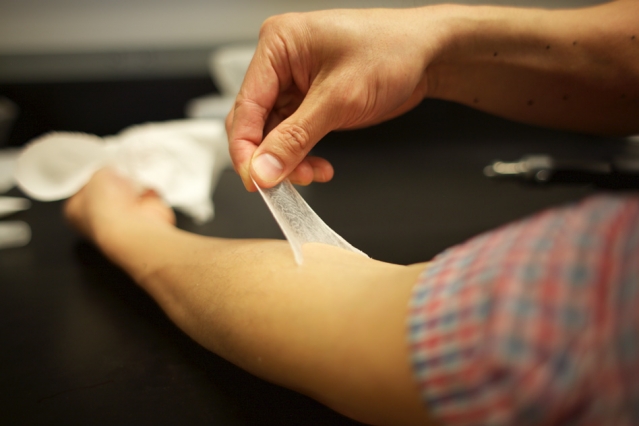
“It’s an invisible layer that can provide a barrier, provide cosmetic improvement, and potentially deliver a drug locally to the area that’s being treated. Those three things together could really make it ideal for use in humans,” Daniel Anderson says. Photo: Melanie Gonick/MIT

“It’s an invisible layer that can provide a barrier, provide cosmetic improvement, and potentially deliver a drug locally to the area that’s being treated. Those three things together could really make it ideal for use in humans,” Daniel Anderson says. Photo: Melanie Gonick/MIT

It almost looks like he’s peeling off his own skin and I imagine that’s the secret to this polymer’s success. A May 9, 2016 news item on phys.org describes the work being done at the Massachusetts Institute of Technology (MIT) and elsewhere with collaborators,

Scientists at MIT, Massachusetts General Hospital, Living Proof, and Olivo Labs have developed a new material that can temporarily protect and tighten skin, and smooth wrinkles. With further development, it could also be used to deliver drugs to help treat skin conditions such as eczema and other types of dermatitis.

A May 9, 2016 MIT news release (also on EurekAlert), which originated the news item, provides more detail,

The material, a silicone-based polymer that could be applied on the skin as a thin, imperceptible coating, mimics the mechanical and elastic properties of healthy, youthful skin. In tests with human subjects, the researchers found that the material was able to reshape “eye bags” under the lower eyelids and also enhance skin hydration. This type of “second skin” could also be adapted to provide long-lasting ultraviolet protection, the researchers say.

“It’s an invisible layer that can provide a barrier, provide cosmetic improvement, and potentially deliver a drug locally to the area that’s being treated. Those three things together could really make it ideal for use in humans,” says Daniel Anderson, an associate professor in MIT’s Department of Chemical Engineering and a member of MIT’s Koch Institute for Integrative Cancer Research and Institute for Medical Engineering and Science (IMES).

Anderson is one of the authors of a paper describing the polymer in the May 9 online issue of Nature Materials. Robert Langer, the David H. Koch Institute Professor at MIT and a member of the Koch Institute, is the paper’s senior author, and the paper’s lead author is Betty Yu SM ’98, ScD ’02, former vice president at Living Proof. Langer and Anderson are co-founders of Living Proof and Olivo Labs, and Yu earned her master’s and doctorate at MIT.

Mimicking skin

As skin ages, it becomes less firm and less elastic — problems that can be exacerbated by sun exposure. This impairs skin’s ability to protect against extreme temperatures, toxins, microorganisms, radiation, and injury. About 10 years ago, the research team set out to develop a protective coating that could restore the properties of healthy skin, for both medical and cosmetic applications.

“We started thinking about how we might be able to control the properties of skin by coating it with polymers that would impart beneficial effects,” Anderson says. “We also wanted it to be invisible and comfortable.”

The researchers created a library of more than 100 possible polymers, all of which contained a chemical structure known as siloxane — a chain of alternating atoms of silicon and oxygen. These polymers can be assembled into a network arrangement known as a cross-linked polymer layer (XPL). The researchers then tested the materials in search of one that would best mimic the appearance, strength, and elasticity of healthy skin.

“It has to have the right optical properties, otherwise it won’t look good, and it has to have the right mechanical properties, otherwise it won’t have the right strength and it won’t perform correctly,” Langer says.

The best-performing material has elastic properties very similar to those of skin. In laboratory tests, it easily returned to its original state after being stretched more than 250 percent (natural skin can be elongated about 180 percent). In laboratory tests, the novel XPL’s elasticity was much better than that of two other types of wound dressings now used on skin — silicone gel sheets and polyurethane films.

“Creating a material that behaves like skin is very difficult,” says Barbara Gilchrest, a dermatologist at MGH and an author of the paper. “Many people have tried to do this, and the materials that have been available up until this have not had the properties of being flexible, comfortable, nonirritating, and able to conform to the movement of the skin and return to its original shape.”

The XPL is currently delivered in a two-step process. First, polysiloxane components are applied to the skin, followed by a platinum catalyst that induces the polymer to form a strong cross-linked film that remains on the skin for up to 24 hours. This catalyst has to be added after the polymer is applied because after this step the material becomes too stiff to spread. Both layers are applied as creams or ointments, and once spread onto the skin, XPL becomes essentially invisible.

High performance

The researchers performed several studies in humans to test the material’s safety and effectiveness. In one study, the XPL was applied to the under-eye area where “eye bags” often form as skin ages. These eye bags are caused by protrusion of the fat pad underlying the skin of the lower lid. When the material was applied, it applied a steady compressive force that tightened the skin, an effect that lasted for about 24 hours.

In another study, the XPL was applied to forearm skin to test its elasticity. When the XPL-treated skin was distended with a suction cup, it returned to its original position faster than untreated skin.

The researchers also tested the material’s ability to prevent water loss from dry skin. Two hours after application, skin treated with the novel XPL suffered much less water loss than skin treated with a high-end commercial moisturizer. Skin coated with petrolatum was as effective as XPL in tests done two hours after treatment, but after 24 hours, skin treated with XPL had retained much more water. None of the study participants reported any irritation from wearing XPL.

“I think it has great potential for both cosmetic and noncosmetic applications, especially if you could incorporate antimicrobial agents or medications,” says Thahn Nga Tran, a dermatologist and instructor at Harvard Medical School, who was not involved in the research.

Living Proof has spun out the XPL technology to Olivo Laboratories, LLC, a new startup formed to focus on the further development of the XPL technology. Initially, Olivo’s team will focus on medical applications of the technology for treating skin conditions such as dermatitis.


This video supplied by MIT shows how to apply the polymer and offers a description and demonstration of its properties once applied,

Here’s a link to and a citation for the paper,

An elastic second skin by Betty Yu, Soo-Young Kang, Ariya Akthakul, Nithin Ramadurai, Morgan Pilkenton, Alpesh Patel, Amir Nashat, Daniel G. Anderson, Fernanda H. Sakamoto, Barbara A. Gilchrest, R. Rox Anderson & Robert Langer. Nature Materials (2016) doi:10.1038/nmat4635 Published online 09 May 2016

This paper is behind a paywall.

One final comment, I wonder who’s lining up to invest in this product.

Using scientific methods and technology to explore living systems as artistic subjects: bioart

There is a fascinating set of stories about bioart designed to whet your appetite for more (*) in a Nov. 23, 2015 Cell Press news release on EurekAlert (Note: A link has been removed),

Joe Davis is an artist who works not only with paints or pastels, but also with genes and bacteria. In 1986, he collaborated with geneticist Dan Boyd to encode a symbol for life and femininity into an E. coli bacterium. The piece, called Microvenus, was the first artwork to use the tools and techniques of molecular biology. Since then, bioart has become one of several contemporary art forms (including reclamation art and nanoart) that apply scientific methods and technology to explore living systems as artistic subjects. A review of the field, published November 23, can be found in Trends in Biotechnology.

Bioart ranges from bacterial manipulation to glowing rabbits, cellular sculptures, and–in the case of Australian-British artist Nina Sellars–documentation of an ear prosthetic that was implanted onto fellow artist Stelarc’s arm. In the pursuit of creating art, practitioners have generated tools and techniques that have aided researchers, while sometimes crossing into controversy, such as by releasing invasive species into the environment, blurring the lines between art and modern biology, raising philosophical, societal, and environmental issues that challenge scientific thinking.

“Most people don’t know that bioart exists, but it can enable scientists to produce new ideas and give us opportunities to look differently at problems,” says author Ali K. Yetisen, who works at Harvard Medical School and the Wellman Center for Photomedicine, Massachusetts General Hospital. “At the same time there’s been a lot of ethical and safety concerns happening around bioart and artists who wanted to get involved in the past have made mistakes.”

Here’s a sample of Joe Davis’s work,

 Caption This photograph shows polyptich paintings by Joe Davis of his 28-mer Microvenus DNA molecule (2006 Exhibition in Greece at Athens School of Fine Arts). Credit: Courtesy of Joe Davis

This photograph shows polyptich paintings by Joe Davis of his 28-mer Microvenus DNA molecule (2006 Exhibition in Greece at Athens School of Fine Arts). Credit: Courtesy of Joe Davis

The news release goes on to recount a brief history of bioart, which stretches back to 1928 and then further back into the 19th and 18th centuries,

In between experiments, Alexander Fleming would paint stick figures and landscapes on paper and in Petri dishes using bacteria. In 1928, after taking a brief hiatus from the lab, he noticed that portions of his “germ paintings,” had been killed. The culprit was a fungus, penicillin–a discovery that would revolutionize medicine for decades to come.

In 1938, photographer Edward Steichen used a chemical to genetically alter and produce interesting variations in flowering delphiniums. This chemical, colchicine, would later be used by horticulturalists to produce desirable mutations in crops and ornamental plants.

In the late 18th and early 19th centuries, the arts and sciences moved away from traditionally shared interests and formed secular divisions that persisted well into the 20th century. “Appearance of environmental art in the 1970s brought about renewed awareness of special relationships between art and the natural world,” Yetisen says.

To demonstrate how we change landscapes, American sculptor Robert Smithsonian paved a hillside with asphalt, while Bulgarian artist Christo Javacheffa (of Christo and Jeanne-Claude) surrounded resurfaced barrier islands with bright pink plastic.

These pieces could sometimes be destructive, however, such as in Ten Turtles Set Free by German-born Hans Haacke. To draw attention to the excesses of the pet trade, he released what he thought were endangered tortoises back to their natural habitat in France, but he inadvertently released the wrong subspecies, thus compromising the genetic lineages of the endangered tortoises as the two varieties began to mate.

By the late 1900s, technological advances began to draw artists’ attention to biology, and by the 2000s, it began to take shape as an artistic identity. Following Joe Davis’ transgenic Microvenus came a miniaturized leather jacket made of skin cells, part of the Tissue Culture & Art Project (initiated in 1996) by duo Oran Catts and Ionat Zurr. Other examples of bioart include: the use of mutant cacti to simulate appearance of human hair in the place of cactus spines by Laura Cinti of University College London’s C-Lab; modification of butterfly wings for artistic purposes by Marta de Menezes of Portugal; and photographs of amphibian deformation by American Brandon Ballengée.

“Bioart encourages discussions about societal, philosophical, and environmental issues and can help enhance public understanding of advances in biotechnology and genetic engineering,” says co-author Ahmet F. Coskun, who works in the Division of Chemistry and Chemical Engineering at California Institute of Technology.

Life as a Bioartist

Today, Joe Davis is a research affiliate at MIT Biology and “Artist-Scientist” at the George Church Laboratory at Harvard–a place that fosters creativity and technological development around genetic engineering and synthetic biology. “It’s Oz, pure and simple,” Davis says. “The total amount of resources in this environment and the minds that are accessible, it’s like I come to the city of Oz every day.”

But it’s not a one-way street. “My particular lab depends on thinking outside the box and not dismissing things because they sound like science fiction,” says [George M.] Church, who is also part of the Wyss Institute for Biologically Inspired Engineering. “Joe is terrific at keeping us flexible and nimble in that regard.”

For example, Davis is working with several members of the Church lab to perform metagenomics analyses of the dust that accumulates at the bottom of money-counting machines. Another project involves genetically engineering silk worms to spin metallic gold–an homage to the fairy tale of Rumpelstiltskin.

“I collaborate with many colleagues on projects that don’t necessarily have direct scientific results, but they’re excited to pursue these avenues of inquiry that they might not or would not look into ordinarily–they might try to hide it, but a lot of scientists have poetic souls,” Davis says. “Art, like science, has to describe the whole word and you can’t describe something you’re basically clueless about. The most exciting part of these activities is satiating overwhelming curiosity about everything around you.”

The number of bioartists is still small, Davis says, partly because of a lack of federal funding of the arts in general. Accessibility to the types of equipment bioartists want to experiment with can also be an issue. While Davis has partnered with labs over the past few decades, other artists affiliate themselves with community access laboratories that are run by do-it-yourself biologists. One way that universities can help is to create departmental-wide positions for bioartists to collaborate with scientists.

“In the past, there have been artists affiliated with departments in a very utilitarian way to produce figures or illustrations,” Church says. “Having someone like Joe stimulates our lab to come together in new ways and if we had more bioartists, I think thinking out of the box would be a more common thing.”

“In the era of genetic engineering, bioart will gain new meanings and annotations in social and scientific contexts,” says Yetisen. “Bioartists will surely take up new roles in science laboratories, but this will be subject to ethical criticism and controversy as a matter of course.”

Here’s a link to and a citation for the paper,

Bioart by Ali K. Yetisen, Joe Davis, Ahmet F. Coskun, George M. Church, Seok Hyun. Trends in Biotechnology,  DOI: http://dx.doi.org/10.1016/j.tibtech.2015.09.011 Published Online: November 23, 2015

This paper appears to be open access.

*Removed the word ‘featured’ on Dec. 1, 2015 at 1030 hours PDT.

Saving lives at birth 2013: Round 3 award nominees and their technologies

As I have noted before (most recently in a Feb. 13, 2013 posting) there are at least two Grand Challenges, one is a Bill & Melinda Gates Foundation program and the other, Grand Challenges Canada, is funded by the Canadian government. Both organizations along with the U.S. Agency for International Development (USAID), the Government of Norway, and the U.K’s Department for International Development (DFID) have combined their efforts on maternal health in a partnership, Saving Lives at Birth: A Grand Challenge for Development. 2013 is the third year for this competitive funding program, which attracts entries from around the world.

The organization’s July 31, 2013 news release announces the latest funding nominees,

The Saving Lives at Birth: A Grand Challenge for Development today announced 22 Round 3 award nominees from a pool of 53 finalists – innovators who descended on Washington for three days (DevelopmentXChange) to showcase bold, new ideas to save the lives of mothers and newborns in developing countries with aspirations of international funding to realize their vision.

The award nominees cut across maternal and neonatal health, family planning, nutrition and HIV and they present not only cutting-edge technologies that can be used in resource-poor settings, but innovative approaches to delivering services and the adoption of healthy behaviors. The announcement was made at the closing forum of the DevelopmentXChange by the Saving Lives at Birth partners. The nominees will now enter into final negotiations before awards are issued. [emphasis mine]

If I read this rightly, the nominees do not receive a set amount but negotiate for the money they need to implement and/or develop their ‘solution’. The news release provides more details about the process that applicants undertake when they reach the finalist stage,

The Saving Lives at Birth DevelopmentXChange provided a platform for top global innovators to present their ideas in an open, dynamic marketplace and exchange ideas with development experts and potential funders to help meet the immense challenge of protecting mothers and newborns in the poorest places on earth, during their most vulnerable hours. Other promising ideas will be considered for “incubator awards” to assist innovators in further developing their ideas through dialogue and mentorship.

….

The Saving Lives at Birth DevelopmentXChange featured discussions focused on meeting the needs and realities of women and children in low-resource settings as well as workshops that explored business planning, market research, impact investing, and strategies for scaling their innovations.  The three-day event concluded with a forum featuring Ambassador Susan E. Rice, National Security Advisor; Dr. Rajiv Shah, Administrator, USAID; HRH Princess Sarah Zeid of Jordan; New York Times best-selling author Dan Heath and NASA astronaut Col. Ron Garan (ret.).

Leading into the DevelopmentXChange, existing Saving Lives at Birth grantees participated in a three-day, customized training program – a focal point of the global health Xcelerator.  This eight-month program, offered through a partnership between National Collegiate Inventors and Innovators Alliance (NCIIA), the Lemelson Foundation and USAID, provides grantees the tools and knowledge to scale their ideas and maximize the impact of their innovations.

Here’s the list of nominees who emerged from the process (there is one overtly nanotechnology project listed and I suspect others are also enabled by nanotechnology),

Award nominees of Saving Lives at Birth Round 3 include 4 transition-to-scale grant nominees:

· Africare – Dakar, Senegal: A collaborative community-based technology that integrates community support services with mobile and telemedicine platforms to increase demand for, and access to, quality prenatal care services in Senegal.  More: http://savinglivesatbirth.net/summaries/232

· Epidemiological Research Center in Sexual and Reproductive Health – Guatemala City, Guatemala: An integrated approach to reduce maternal and perinatal mortality in Northern Guatemala through simulation-based training, social marketing campaigns and formal health care system engagement.  More: http://savinglivesatbirth.net/summaries/246

· Massachusetts General Hospital – Boston, MA, USA: A next-generation uterine balloon tamponade (UBT) device to treat postpartum hemorrhage (PPH) in Kenya and South Sudan.  More: http://savinglivesatbirth.net/summaries/255

· The Research Institute at Nationwide Children’s Hospital – Columbus, OH, USA: A low-cost paper-based urine test for early diagnosis of pre-eclampsia to reduce pre-eclampsia morbidity and mortality in resource-limited areas.  http://savinglivesatbirth.net/summaries/275

And 18 seed grant nominees:

· BILIMETRIX SRL – Trieste, Italy: An inexpensive system to rapidly test for markers of hyperbilirubinemia (kernicterus)-an often fatal form of brain damage caused by excessive jaundice- in low resource settings in Nigeria, Egypt, and Indonesia.  More: http://savinglivesatbirth.net/summaries/235

· JustMilk – Dept. of Chemical Engineering, University of Cambridge – Cambridge, UK: A low-cost system that aids the administration of drugs and nutrients to breastfeeding infants via easily disintegrating tablets housed within a modified Nipple Shield Delivery System (NSDS).  http://savinglivesatbirth.net/summaries/241

· The University of Melbourne – Melbourne, Australia: A low-cost, electricity-free oxygen concentrator suitable for providing provisional oxygen for neonates in low-resource settings.  http://savinglivesatbirth.net/summaries/277

· University of Toronto – Toronto, Canada: A spray-encapsulated iron premix that will be attached to tea leaves to reduce rates of iron deficiency of pregnant women in South Asia.  http://savinglivesatbirth.net/summaries/279

· University of Valencia – Valencia, Spain: A rapid point-of-care test strips for early diagnosis of sepsis in pregnancy and childbirth. More: http://savinglivesatbirth.net/summaries/281

· Mbarara University of Science and Technology – Mbarara, Uganda: The Augmented Infant Resuscitator (AIR) which gives instant feedback to healthcare professionals performing newborn resuscitation to reduce neonatal deaths from intrapartum birth asphyxia or prematurity.  http://savinglivesatbirth.net/summaries/256

· Bioceptive, Inc. – New Orleans, LA, USA: A low-cost, reusable, and intuitive intrauterine device (IUD) inserter to make the IUD insertion procedure easier and safer in low-resource settings. http://savinglivesatbirth.net/summaries/236

· Convergent Engineering Inc. – Newberry, FL, USA: An inexpensive, easy-to-use, handheld early-warning system that detects pre-eclampsia 10-12 weeks before the onset symptoms. The system pairs a wrist strap embedded with inexpensive ECG and photoplethysmography sensors with a smart phone for processing, data aggregation, and communication.  http://savinglivesatbirth.net/summaries/239

· Dimagi, Inc. (CommTrack) – Cambridge, MA, USA: An open-source distribution management system integrating mobile and GPS technology to improve transparency, supply chain functioning, communication, and the timely delivery of medicine to hard to reach, low-income areas in Africa.  http://savinglivesatbirth.net/summaries/243

· Duke University– Durham, NC, USA:  Healthcare system integration of the “Pratt Pouch”-a tiny ketchup-like packet that stores antiretroviral AIDS medication for a year-to enable the pouch to be used in home-birth settings to prevent transmission of HIV from mother to child. Testing taking place in Zambia.  http://savinglivesatbirth.net/summaries/244

· Emory University – Atlanta, GA, USA: A micro-needle patch that co-administers the influenza and tetanus toxoid vaccines to pregnant mothers and children in developing countries.  http://savinglivesatbirth.net/summaries/245

· Nanobiosym, Inc – Cambridge, MA, USA: A nanotech platform which enables rapid, accurate and mobile HIV diagnosis at point-of-care, allowing for timely treatment with antiretroviral therapy to reduce HIV-related mortality in infants in Rwanda.  http://savinglivesatbirth.net/summaries/259

· Oregon Health and Science University – Portland, OR, USA: The Xstat mini-sponge applicator for the treatment of postpartum hemorrhage (PPH).  http://savinglivesatbirth.net/summaries/260

· Population Services International – Washington DC, USA: A new inserter for immediate postpartum intrauterine device (PPIUD) insertions to increase contraceptive uptake in developing countries.  http://savinglivesatbirth.net/summaries/263

· President and Fellows of Harvard College – Boston, MA, USA: A handheld vital sign monitor for the rapid diagnosis of frail and sick newborns.  http://savinglivesatbirth.net/summaries/264

· Program for Appropriate Technology in Health (PATH) – Seattle, WA, USA: A heat-stable oxytocin in a fast-dissolving oral tablet to treat postpartum hemorrhage (PPH).  http://savinglivesatbirth.net/summaries/268

· Program for Appropriate Technology in Health (PATH) – Seattle, WA, USA: A magnesium sulfate (MgSO4) gel that simplifies treatment of pre-eclampsia and eclampsia.  http://savinglivesatbirth.net/summaries/267

· The Board of Regents of the University of Wisconsin System – Madison, WI, USA: A Lactobacillus casei strain that enables the sustainable home production of beta-Carotene enriched dairy products for at-risk mothers and families in Southern Asia.  http://savinglivesatbirth.net/summaries/272

While it’s not stated explicitly, the main focus for Saving Lives at Birth appears to be the continent of Africa as per this video animation which represents the organization’s goals and focus,

Organ chips for DARPA (Defense Advanced Research Projects Agency)

The Wyss Institute will receive up to  $37M US for a project that integrates ten different organ-on-a-chip projects into one system. From the July 24, 2012 news release on EurekAlert,

With this new DARPA funding, Institute researchers and a multidisciplinary team of collaborators seek to build 10 different human organs-on-chips, to link them together to more closely mimic whole body physiology, and to engineer an automated instrument that will control fluid flow and cell viability while permitting real-time analysis of complex biochemical functions. As an accurate alternative to traditional animal testing models that often fail to predict human responses, this instrumented “human-on-a-chip” will be used to rapidly assess responses to new drug candidates, providing critical information on their safety and efficacy.

This unique platform could help ensure that safe and effective therapeutics are identified sooner, and ineffective or toxic ones are rejected early in the development process. As a result, the quality and quantity of new drugs moving successfully through the pipeline and into the clinic may be increased, regulatory decision-making could be better informed, and patient outcomes could be improved.

Jesse Goodman, FDA Chief Scientist and Deputy Commissioner for Science and Public Health, commented that the automated human-on-chip instrument being developed “has the potential to be a better model for determining human adverse responses. FDA looks forward to working with the Wyss Institute in its development of this model that may ultimately be used in therapeutic development.”

Wyss Founding Director, Donald Ingber, M.D., Ph.D., and Wyss Core Faculty member, Kevin Kit Parker, Ph.D., will co-lead this five-year project.

I note that Kevin Kit Parker was mentioned in an earlier posting today (July 26, 2012) titled, Medusa, jellyfish, and tissue engineering, and Donald Ingber in my Dec.1e, 2011 posting about Shrilk and insect skeletons.

As for the Wyss Institute, here’s a description from the news release,

The Wyss Institute for Biologically Inspired Engineering at Harvard University (http://wyss.harvard.edu) uses Nature’s design principles to develop bioinspired materials and devices that will transform medicine and create a more sustainable world. Working as an alliance among Harvard’s Schools of Medicine, Engineering, and Arts & Sciences, and in partnership with Beth Israel Deaconess Medical Center, Boston Children’s Hospital, Brigham and Women’s Hospital, , Dana Farber Cancer Institute, Massachusetts General Hospital, the University of Massachusetts Medical School, Spaulding Rehabilitation Hospital, Tufts University, and Boston University, the Institute crosses disciplinary and institutional barriers to engage in high-risk research that leads to transformative technological breakthroughs. By emulating Nature’s principles for self-organizing and self-regulating, Wyss researchers are developing innovative new engineering solutions for healthcare, energy, architecture, robotics, and manufacturing. These technologies are translated into commercial products and therapies through collaborations with clinical investigators, corporate alliances, and new start-ups.

I hadn’t thought of an organ-on-a-chip as particularly bioinspired so I’ll have to think about that one for a while.

Brain, brains, brains: a roundup

I’ve decided to do a roundup of the various brain-related projects I’ve been coming across in the last several months. I was inspired by this article (Real-life Jedi: Pushing the limits of mind control) by Katia Moskvitch,

You don’t have to be a Jedi to make things move with your mind.

Granted, we may not be able to lift a spaceship out of a swamp like Yoda does in The Empire Strikes Back, but it is possible to steer a model car, drive a wheelchair and control a robotic exoskeleton with just your thoughts.

We are standing in a testing room at IBM’s Emerging Technologies lab in Winchester, England.

On my head is a strange headset that looks like a black plastic squid. Its 14 tendrils, each capped with a moistened electrode, are supposed to detect specific brain signals.

In front of us is a computer screen, displaying an image of a floating cube.

As I think about pushing it, the cube responds by drifting into the distance.

Moskvitch goes on to discuss a number of projects that translate thought into movement via various pieces of equipment before she mentions a project at Brown University (US) where researchers are implanting computer chips into brains,

Headsets and helmets offer cheap, easy-to-use ways of tapping into the mind. But there are other,

Imagine some kind of a wireless computer device in your head that you’ll use for mind control – what if people hacked into that”

At Brown Institute for Brain Science in the US, scientists are busy inserting chips right into the human brain.

The technology, dubbed BrainGate, sends mental commands directly to a PC.

Subjects still have to be physically “plugged” into a computer via cables coming out of their heads, in a setup reminiscent of the film The Matrix. However, the team is now working on miniaturising the chips and making them wireless.

The researchers are recruiting for human clinical trials, from the BrainGate Clinical Trials webpage,

Clinical Trials – Now Recruiting

The purpose of the first phase of the pilot clinical study of the BrainGate2 Neural Interface System is to obtain preliminary device safety information and to demonstrate the feasibility of people with tetraplegia using the System to control a computer cursor and other assistive devices with their thoughts. Another goal of the study is to determine the participants’ ability to operate communication software, such as e-mail, simply by imagining the movement of their own hand. The study is invasive and requires surgery.

Individuals with limited or no ability to use both hands due to cervical spinal cord injury, brainstem stroke, muscular dystrophy, or amyotrophic lateral sclerosis (ALS) or other motor neuron diseases are being recruited into a clinical study at Massachusetts General Hospital (MGH) and Stanford University Medical Center. Clinical trial participants must live within a three-hour drive of Boston, MA or Palo Alto, CA. Clinical trial sites at other locations may be opened in the future. The study requires a commitment of 13 months.

They have been recruiting since at least November 2011, from the Nov. 14, 2011 news item by Tanya Lewis on MedicalXpress,

Stanford University researchers are enrolling participants in a pioneering study investigating the feasibility of people with paralysis using a technology that interfaces directly with the brain to control computer cursors, robotic arms and other assistive devices.

The pilot clinical trial, known as BrainGate2, is based on technology developed at Brown University and is led by researchers at Massachusetts General Hospital, Brown and the Providence Veterans Affairs Medical Center. The researchers have now invited the Stanford team to establish the only trial site outside of New England.

Under development since 2002, BrainGate is a combination of hardware and software that directly senses electrical signals in the brain that control movement. The device — a baby-aspirin-sized array of electrodes — is implanted in the cerebral cortex (the outer layer of the brain) and records its signals; computer algorithms then translate the signals into digital instructions that may allow people with paralysis to control external devices.

Confusingly, there seemto be two BrainGate organizations. One appears to be a research entity where a number of institutions collaborate and the other is some sort of jointly held company. From the About Us webpage of the BrainGate research entity,

In the late 1990s, the initial translation of fundamental neuroengineering research from “bench to bedside” – that is, to pilot clinical testing – would require a level of financial commitment ($10s of millions) available only from private sources. In 2002, a Brown University spin-off/startup medical device company, Cyberkinetics, Inc. (later, Cyberkinetics Neurotechnology Systems, Inc.) was formed to collect the regulatory permissions and financial resources required to launch pilot clinical trials of a first-generation neural interface system. The company’s efforts and substantial initial capital investment led to the translation of the preclinical research at Brown University to an initial human device, the BrainGate Neural Interface System [Caution: Investigational Device. Limited by Federal Law to Investigational Use]. The BrainGate system uses a brain-implantable sensor to detect neural signals that are then decoded to provide control signals for assistive technologies. In 2004, Cyberkinetics received from the U.S. Food and Drug Administration (FDA) the first of two Investigational Device Exemptions (IDEs) to perform this research. Hospitals in Rhode Island, Massachusetts, and Illinois were established as clinical sites for the pilot clinical trial run by Cyberkinetics. Four trial participants with tetraplegia (decreased ability to use the arms and legs) were enrolled in the study and further helped to develop the BrainGate device. Initial results from these trials have been published or presented, with additional publications in preparation.

While scientific progress towards the creation of this promising technology has been steady and encouraging, Cyberkinetics’ financial sponsorship of the BrainGate research – without which the research could not have been started – began to wane. In 2007, in response to business pressures and changes in the capital markets, Cyberkinetics turned its focus to other medical devices. Although Cyberkinetics’ own funds became unavailable for BrainGate research, the research continued through grants and subcontracts from federal sources. By early 2008 it became clear that Cyberkinetics would eventually need to withdraw completely from directing the pilot clinical trials of the BrainGate device. Also in 2008, Cyberkinetics spun off its device manufacturing to new ownership, BlackRock Microsystems, Inc., which now produces and is further developing research products as well as clinically-validated (510(k)-cleared) implantable neural recording devices.

Beginning in mid 2008, with the agreement of Cyberkinetics, a new, fully academically-based IDE application (for the “BrainGate2 Neural Interface System”) was developed to continue this important research. In May 2009, the FDA provided a new IDE for the BrainGate2 pilot clinical trial. [Caution: Investigational Device. Limited by Federal Law to Investigational Use.] The BrainGate2 pilot clinical trial is directed by faculty in the Department of Neurology at Massachusetts General Hospital, a teaching affiliate of Harvard Medical School; the research is performed in close scientific collaboration with Brown University’s Department of Neuroscience, School of Engineering, and Brown Institute for Brain Sciences, and the Rehabilitation Research and Development Service of the U.S. Department of Veteran’s Affairs at the Providence VA Medical Center. Additionally, in late 2011, Stanford University joined the BrainGate Research Team as a clinical site and is currently enrolling participants in the clinical trial. This interdisciplinary research team includes scientific partners from the Functional Electrical Stimulation Center at Case Western Reserve University and the Cleveland VA Medical Center. As was true of the decades of fundamental, preclinical research that provided the basis for the recent clinical studies, funding for BrainGate research is now entirely from federal and philanthropic sources.

The BrainGate Research Team at Brown University, Massachusetts General Hospital, Stanford University, and Providence VA Medical Center comprises physicians, scientists, and engineers working together to advance understanding of human brain function and to develop neurotechnologies for people with neurologic disease, injury, or limb loss.

I think they’re saying there was a reverse takeover of Cyberkinetics, from the BrainGate company About webpage,

The BrainGate™ Co. is a privately-held firm focused on the advancement of the BrainGate™ Neural Interface System.  The Company owns the Intellectual property of the BrainGate™ system as well as new technology being developed by the BrainGate company.  In addition, the Company also owns  the intellectual property of Cyberkinetics which it purchased in April 2009.

Meanwhile, in Europe there are two projects BrainAble and the Human Brain Project. The BrainAble project is similar to BrainGate in that it is intended for people with injuries but they seem to be concentrating on a helmet or cap for thought transmission (as per Moskovitch’s experience at the beginning of this posting). From the Feb. 28, 2012 news item on Science Daily,

In the 2009 film Surrogates, humans live vicariously through robots while safely remaining in their own homes. That sci-fi future is still a long way off, but recent advances in technology, supported by EU funding, are bringing this technology a step closer to reality in order to give disabled people more autonomy and independence than ever before.

“Our aim is to give people with motor disabilities as much autonomy as technology currently allows and in turn greatly improve their quality of life,” says Felip Miralles at Barcelona Digital Technology Centre, a Spanish ICT research centre.

Mr. Miralles is coordinating the BrainAble* project (http://www.brainable.org/), a three-year initiative supported by EUR 2.3 million in funding from the European Commission to develop and integrate a range of different technologies, services and applications into a commercial system for people with motor disabilities.

Here’s more from the BrainAble home page,

In terms of HCI [human-computer interface], BrainAble improves both direct and indirect interaction between the user and his smart home. Direct control is upgraded by creating tools that allow controlling inner and outer environments using a “hybrid” Brain Computer Interface (BNCI) system able to take into account other sources of information such as measures of boredom, confusion, frustration by means of the so-called physiological and affective sensors.

Furthermore, interaction is enhanced by means of Ambient Intelligence (AmI) focused on creating a proactive and context-aware environments by adding intelligence to the user’s surroundings. AmI’s main purpose is to aid and facilitate the user’s living conditions by creating proactive environments to provide assistance.

Human-Computer Interfaces are complemented by an intelligent Virtual Reality-based user interface with avatars and scenarios that will help the disabled move around freely, and interact with any sort of devices. Even more the VR will provide self-expression assets using music, pictures and text, communicate online and offline with other people, play games to counteract cognitive decline, and get trained in new functionalities and tasks.

Perhaps this video helps,

Another European project, NeuroCare, which I discussed in my March 5, 2012 posting, is focused on creating neural implants to replace damaged and/or destroyed sensory cells in the eye or the ear.

The Human Brain Project is, despite its title, a neuromorphic engineering project (although the researchers do mention some medical applications on the project’s home page)  in common with the work being done at the University of Michigan/HRL Labs mentioned in my April 19, 2012 posting (A step closer to artificial synapses courtesy of memritors) about that project. From the April 11, 2012 news item about the Human Brain Project on Science Daily,

Researchers at the EPFL [Ecole Polytechnique Fédérale de Lausanne] have discovered rules that relate the genes that a neuron switches on and off, to the shape of that neuron, its electrical properties and its location in the brain.

The discovery, using state-of-the-art informatics tools, increases the likelihood that it will be possible to predict much of the fundamental structure and function of the brain without having to measure every aspect of it. That in turn makes the Holy Grail of modelling the brain in silico — the goal of the proposed Human Brain Project — a more realistic, less Herculean, prospect. “It is the door that opens to a world of predictive biology,” says Henry Markram, the senior author on the study, which is published this week in PLoS ONE.

Here’s a bit more about the Human Brain Project (from the home page),

Today, simulating a single neuron requires the full power of a laptop computer. But the brain has billions of neurons and simulating all them simultaneously is a huge challenge. To get round this problem, the project will develop novel techniques of multi-level simulation in which only groups of neurons that are highly active are simulated in detail. But even in this way, simulating the complete human brain will require a computer a thousand times more powerful than the most powerful machine available today. This means that some of the key players in the Human Brain Project will be specialists in supercomputing. Their task: to work with industry to provide the project with the computing power it will need at each stage of its work.

The Human Brain Project will impact many different areas of society. Brain simulation will provide new insights into the basic causes of neurological diseases such as autism, depression, Parkinson’s, and Alzheimer’s. It will give us new ways of testing drugs and understanding the way they work. It will provide a test platform for new drugs that directly target the causes of disease and that have fewer side effects than current treatments. It will allow us to design prosthetic devices to help people with disabilities. The benefits are potentially huge. As world populations grow older, more than a third will be affected by some kind of brain disease. Brain simulation provides us with a powerful new strategy to tackle the problem.

The project also promises to become a source of new Information Technologies. Unlike the computers of today, the brain has the ability to repair itself, to take decisions, to learn, and to think creatively – all while consuming no more energy than an electric light bulb. The Human Brain Project will bring these capabilities to a new generation of neuromorphic computing devices, with circuitry directly derived from the circuitry of the brain. The new devices will help us to build a new generation of genuinely intelligent robots to help us at work and in our daily lives.

The Human Brain Project builds on the work of the Blue Brain Project. Led by Henry Markram of the Ecole Polytechnique Fédérale de Lausanne (EPFL), the Blue Brain Project has already taken an essential first towards simulation of the complete brain. Over the last six years, the project has developed a prototype facility with the tools, know-how and supercomputing technology necessary to build brain models, potentially of any species at any stage in its development. As a proof of concept, the project has successfully built the first ever, detailed model of the neocortical column, one of the brain’s basic building blocks.

The Human Brain Project is a flagship project  in contention for the 1B Euro research prize that I’ve mentioned in the context of the GRAPHENE-CA flagship project (my Feb. 13, 2012 posting gives a better description of these flagship projects while mentioned both GRAPHENE-CA and another brain-computer interface project, PRESENCCIA).

Part of the reason for doing this roundup, is the opportunity to look at a number of these projects in one posting; the effect is more overwhelming than I expected.

For anyone who’s interested in Markram’s paper (open access),

Georges Khazen, Sean L. Hill, Felix Schürmann, Henry Markram. Combinatorial Expression Rules of Ion Channel Genes in Juvenile Rat (Rattus norvegicus) Neocortical Neurons. PLoS ONE, 2012; 7 (4): e34786 DOI: 10.1371/journal.pone.0034786

I do have earlier postings on brains and neuroprostheses, one of the more recent ones is this March 16, 2012 posting. Meanwhile, there are  new announcements from Northwestern University (US) and the US National Institutes of Health (National Institute of Neurological Disorders and Stroke). From the April 18, 2012 news item (originating from the National Institutes of Health) on Science Daily,

An artificial connection between the brain and muscles can restore complex hand movements in monkeys following paralysis, according to a study funded by the National Institutes of Health.

In a report in the journal Nature, researchers describe how they combined two pieces of technology to create a neuroprosthesis — a device that replaces lost or impaired nervous system function. One piece is a multi-electrode array implanted directly into the brain which serves as a brain-computer interface (BCI). The array allows researchers to detect the activity of about 100 brain cells and decipher the signals that generate arm and hand movements. The second piece is a functional electrical stimulation (FES) device that delivers electrical current to the paralyzed muscles, causing them to contract. The brain array activates the FES device directly, bypassing the spinal cord to allow intentional, brain-controlled muscle contractions and restore movement.

From the April 19, 2012 news item (originating from Northwestern University) on Science Daily,

A new Northwestern Medicine brain-machine technology delivers messages from the brain directly to the muscles — bypassing the spinal cord — to enable voluntary and complex movement of a paralyzed hand. The device could eventually be tested on, and perhaps aid, paralyzed patients.

The research was done in monkeys, whose electrical brain and muscle signals were recorded by implanted electrodes when they grasped a ball, lifted it and released it into a small tube. Those recordings allowed the researchers to develop an algorithm or “decoder” that enabled them to process the brain signals and predict the patterns of muscle activity when the monkeys wanted to move the ball.

These experiments were performed by Christian Ethier, a post-doctoral fellow, and Emily Oby, a graduate student in neuroscience, both at the Feinberg School of Medicine. The researchers gave the monkeys a local anesthetic to block nerve activity at the elbow, causing temporary, painless paralysis of the hand. With the help of the special devices in the brain and the arm — together called a neuroprosthesis — the monkeys’ brain signals were used to control tiny electric currents delivered in less than 40 milliseconds to their muscles, causing them to contract, and allowing the monkeys to pick up the ball and complete the task nearly as well as they did before.

“The monkey won’t use his hand perfectly, but there is a process of motor learning that we think is very similar to the process you go through when you learn to use a new computer mouse or a different tennis racquet. Things are different and you learn to adjust to them,” said Miller [Lee E. Miller], also a professor of physiology and of physical medicine and rehabilitation at Feinberg and a Sensory Motor Performance Program lab chief at the Rehabilitation Institute of Chicago.

The National Institutes of Health news item supplies a little history and background for this latest breakthrough while the Northwestern University news item offers more technical details more technical details.

You can find the researchers’ paper with this citation (assuming you can get past the paywall,

C. Ethier, E. R. Oby, M. J. Bauman, L. E. Miller. Restoration of grasp following paralysis through brain-controlled stimulation of muscles. Nature, 2012; DOI: 10.1038/nature10987

I was surprised to find the Health Research Fund of Québec listed as one of the funders but perhaps Christian Ethier has some connection with the province.