Tag Archives: Nan Li

Nanostructured materials and radiation

If you’re planning on using nanostructured materials in a nuclear facility, you might want to check out this work (from a June 8, 2018 Purdue University (Indiana, US) news release by Brian L. Huchel,

A professor in the Purdue College of Engineering examined the potential use of various materials in nuclear reactors in an extensive review article in the journal Progress in Materials Science.

The article, titled “Radiation Damage in Nanostructured Materials,” was led by Xinghang Zhang, a professor of materials engineering. It will be published in the July issue of the journal.

Zhang said there is a significant demand for advanced materials that can survive high temperature and high doses of radiation. These materials contain significant amount of internal changes, called defect sinks, which are too small to be seen with the naked eye, but may form the next generation of materials used in nuclear reactors.

“Nanostructured materials with abundant internal defect sinks are promising as these materials have shown significantly improved radiation tolerance,” he said. “However, there are many challenges and fundamental science questions that remain to be solved before these materials can have applications in advanced nuclear reactors.”

The 100-page article, which took two years to write, focuses on metallic materials and metal-ceramic compounds and reviews types of internal material defects on the reduction of radiation damage in nanostructured materials.

Under the extreme radiation conditions, a large number of defects and their clusters are generated inside materials, and such significant microstructure damage often leads to degradation of the mechanical and physical properties of the materials

The article discusses the usage of a combination of defect sink networks to collaboratively improve the radiation tolerance of nanomaterials, while pointing out the need to improve the thermal and radiation stabilities of the defect sinks.

“The field of radiation damage in nanostructured materials is an exciting and rapidly evolving arena, enriched with challenges and opportunities,” Zhang said. “The integration of extensive research effort, resources and expertise in various fields may eventually lead to the design of advanced nanomaterials with unprecedented radiation tolerance.”

Jin Li, co-author of the review article and a postdoctoral fellow in the School of Materials Engineering, said researchers with different expertise worked collaboratively on the article, which contains more than 100 pages, 100 figures and 700 references.

The team involved in the research article included researchers from Purdue, Texas A&M University, Drexel University, the University of Nebraska-Lincoln and China University of Petroleum-Beijing, as well as Sandia National Laboratory, Los Alamos National Laboratory and Idaho National Laboratory.

Here’s an image illustrating the work,

Various imperfections in nanostructures, call defect sinks, can enhance the material’s tolerance to radiation. (Photo/Xinghang Zhang)

Here’s a link to and a citation for the paper,

Radiation damage in nanostructured materials by Xinghang Zhang, Khalid Hattar, Youxing Chen, Lin Shao, Jin Li, Cheng Sun, Kaiyuan Yu, Nan Li, Mitra L.Taheri, Haiyan Wang, Jian Wang, Michael Nastasi. Progress in Materials Science Volume 96, July 2018, Pages 217-321 https://doi.org/10.1016/j.pmatsci.2018.03.002

This paper is behind a paywall.

ht/ to June 8, 2018 Nanowerk news item.

New iron oxide nanoparticle as an MRI (magnetic resonance imaging) contrast agent

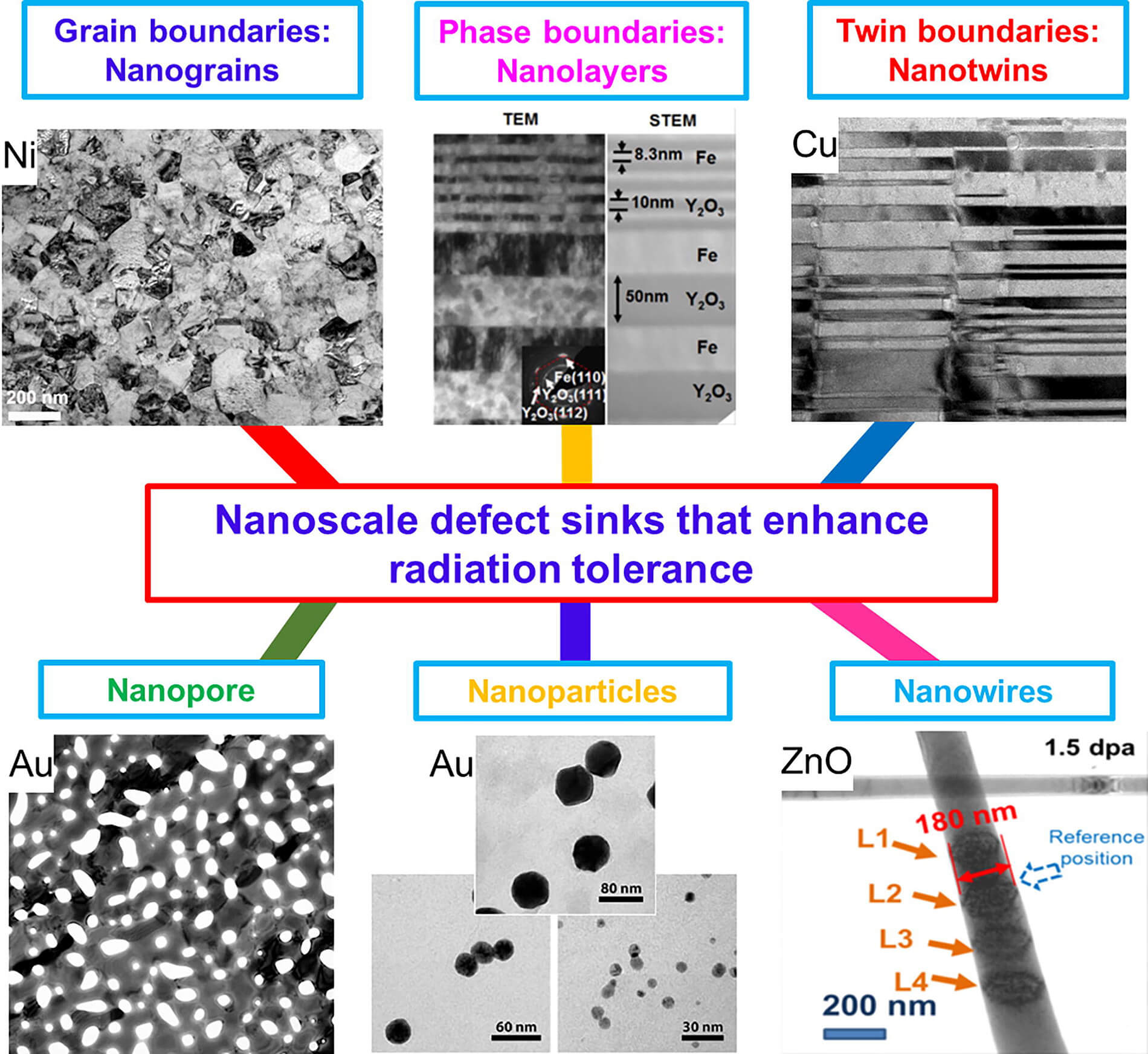
This high-resolution transmission electron micrograph of particles made by the research team shows the particles’ highly uniform size and shape. These are iron oxide particles just 3 nanometers across, coated with a zwitterion layer. Their small size means they can easily be cleared through the kidneys after injection. Courtesy of the researchers

A Feb. 14, 2017 news item on ScienceDaily announces a new MRI (magnetic resonance imaging) contrast agent,

A new, specially coated iron oxide nanoparticle developed by a team at MIT [Massachusetts Institute of Technology] and elsewhere could provide an alternative to conventional gadolinium-based contrast agents used for magnetic resonance imaging (MRI) procedures. In rare cases, the currently used gadolinium agents have been found to produce adverse effects in patients with impaired kidney function.

A Feb. 14, 2017 MIT news release (also on EurekAlert), which originated the news item, provides more technical detail,


The advent of MRI technology, which is used to observe details of specific organs or blood vessels, has been an enormous boon to medical diagnostics over the last few decades. About a third of the 60 million MRI procedures done annually worldwide use contrast-enhancing agents, mostly containing the element gadolinium. While these contrast agents have mostly proven safe over many years of use, some rare but significant side effects have shown up in a very small subset of patients. There may soon be a safer substitute thanks to this new research.

In place of gadolinium-based contrast agents, the researchers have found that they can produce similar MRI contrast with tiny nanoparticles of iron oxide that have been treated with a zwitterion coating. (Zwitterions are molecules that have areas of both positive and negative electrical charges, which cancel out to make them neutral overall.) The findings are being published this week in the Proceedings of the National Academy of Sciences, in a paper by Moungi Bawendi, the Lester Wolfe Professor of Chemistry at MIT; He Wei, an MIT postdoc; Oliver Bruns, an MIT research scientist; Michael Kaul at the University Medical Center Hamburg-Eppendorf in Germany; and 15 others.

Contrast agents, injected into the patient during an MRI procedure and designed to be quickly cleared from the body by the kidneys afterwards, are needed to make fine details of organ structures, blood vessels, and other specific tissues clearly visible in the images. Some agents produce dark areas in the resulting image, while others produce light areas. The primary agents for producing light areas contain gadolinium.

Iron oxide particles have been largely used as negative (dark) contrast agents, but radiologists vastly prefer positive (light) contrast agents such as gadolinium-based agents, as negative contrast can sometimes be difficult to distinguish from certain imaging artifacts and internal bleeding. But while the gadolinium-based agents have become the standard, evidence shows that in some very rare cases they can lead to an untreatable condition called nephrogenic systemic fibrosis, which can be fatal. In addition, evidence now shows that the gadolinium can build up in the brain, and although no effects of this buildup have yet been demonstrated, the FDA is investigating it for potential harm.

“Over the last decade, more and more side effects have come to light” from the gadolinium agents, Bruns says, so that led the research team to search for alternatives. “None of these issues exist for iron oxide,” at least none that have yet been detected, he says.

The key new finding by this team was to combine two existing techniques: making very tiny particles of iron oxide, and attaching certain molecules (called surface ligands) to the outsides of these particles to optimize their characteristics. The iron oxide inorganic core is small enough to produce a pronounced positive contrast in MRI, and the zwitterionic surface ligand, which was recently developed by Wei and coworkers in the Bawendi research group, makes the iron oxide particles water-soluble, compact, and biocompatible.

The combination of a very tiny iron oxide core and an ultrathin ligand shell leads to a total hydrodynamic diameter of 4.7 nanometers, below the 5.5-nanometer renal clearance threshold. This means that the coated iron oxide should quickly clear through the kidneys and not accumulate. This renal clearance property is an important feature where the particles perform comparably to gadolinium-based contrast agents.

Now that initial tests have demonstrated the particles’ effectiveness as contrast agents, Wei and Bruns say the next step will be to do further toxicology testing to show the particles’ safety, and to continue to improve the characteristics of the material. “It’s not perfect. We have more work to do,” Bruns says. But because iron oxide has been used for so long and in so many ways, even as an iron supplement, any negative effects could likely be treated by well-established protocols, the researchers say. If all goes well, the team is considering setting up a startup company to bring the material to production.

For some patients who are currently excluded from getting MRIs because of potential side effects of gadolinium, the new agents “could allow those patients to be eligible again” for the procedure, Bruns says. And, if it does turn out that the accumulation of gadolinium in the brain has negative effects, an overall phase-out of gadolinium for such uses could be needed. “If that turned out to be the case, this could potentially be a complete replacement,” he says.

Ralph Weissleder, a physician at Massachusetts General Hospital who was not involved in this work, says, “The work is of high interest, given the limitations of gadolinium-based contrast agents, which typically have short vascular half-lives and may be contraindicated in renally compromised patients.”

The research team included researchers in MIT’s chemistry, biological engineering, nuclear science and engineering, brain and cognitive sciences, and materials science and engineering departments and its program in Health Sciences and Technology; and at the University Medical Center Hamburg-Eppendorf; Brown University; and the Massachusetts General Hospital. It was supported by the MIT-Harvard NIH Center for Cancer Nanotechnology, the Army Research Office through MIT’s Institute for Soldier Nanotechnologies, the NIH-funded Laser Biomedical Research Center, the MIT Deshpande Center, and the European Union Seventh Framework Program.

Here’s a link to and a citation for the paper,

Exceedingly small iron oxide nanoparticles as positive MRI contrast agents by He Wei, Oliver T. Bruns, Michael G. Kaul, Eric C. Hansen, Mariya Barch, Agata Wiśniowsk, Ou Chen, Yue Chen, Nan Li, Satoshi Okada, Jose M. Cordero, Markus Heine, Christian T. Farrar, Daniel M. Montana, Gerhard Adam, Harald Ittrich, Alan Jasanoff, Peter Nielsen, and Moungi G. Bawendi. PNAS February 13, 2017 doi: 10.1073/pnas.1620145114 Published online before print February 13, 2017

This paper is behind a paywall.

Tweet your nano

Researchers at the University of Wisconsin-Madison have published a study titled, “Tweeting nano: how public discourses about nanotechnology develop in social media environments,”  which analyses, for the first time, nanotechnology discourse on Twitter social media. From the Life Sciences Communication University of Wisconsin-Madison research webpage,

The study, “Tweeting nano: how public discourses about nanotechnology develop in social media environments,” mapped social media traffic about nanotechnology, finding that Twitter traffic expressing opinion about nanotechnology is more likely to originate from states with a federally-funded National Nanotechnology Initiative center or network than states without such centers.

Runge [Kristin K. Runge, doctoral student] and her co-authors used computational linguistic software to analyze a census of all English-language nanotechnology-related tweets expressing opinion posted on Twitter over one calendar year. In addition to mapping tweets by state, the team coded sentiment along two axes: certain vs. uncertain, and optimistic-neutral-pessimistic. They found 55% of nanotechnology-related opinions expressed certainty, 41% expressed pessimistic outlooks and 32% expressed neutral outlooks.

In addition to shedding light on how social media is used in communicating about an emerging technology, this study is believed to be the first published study to use a census of social media messages rather than a sample.

“We likely wouldn’t have captured these results if we had to rely on a sample rather than a complete census,” said Runge. “That would have been unfortunate, because the distinct geographic origins of the tweets and the tendency toward certainty in opinion expression will be useful in helping us understand how key online influencers are shaping the conversation around nanotechnology.”

It’s not obvious from this notice or the title of the study but it is stated clearly in the study that the focus is the world of US nano, not the English language world of nano. After reading the study (very quickly), I can say it’s interesting and, hopefully, will stimulate more work about public opinion that takes social media into account. (I’d love to know how they limited their study to US tweets only and how they determined the region that spawned the tweet. )

The one thing which puzzles me is they don’t mention retweets (RTs) specifically. Did they consider only original tweets? If not, did they take into account the possibility that someone might RT an item that does not reflect their own opinion? I occasionally RT something that doesn’t reflect my opinion when there isn’t sufficient space to include comment indicating otherwise because I want to promote discussion and that doesn’t necessarily take place on Twitter or in Twitter’s public space. This leads to another question, did the researchers include direct messages in their study? Unfortunately, there’s no mention in the two sections  (Discussion and Implications for future research) of the conclusion.

For those who would like to see the research for themselves (Note: The article is behind a paywall),

Tweeting nano: how public discourses about nanotechnology develop in social media environments by Kristin K. Runge, Sara K. Yeo, Michael Cacciatore, Dietram A. Scheufele, Dominique Brossard, Michael Xenos, Ashley Anderson, Doo-hun Choi, Jiyoun Kim, Nan Li, Xuan Liang, Maria Stubbings, and Leona Yi-Fan Su. Journal of Nanoparticle Research; An Interdisciplinary Forum for Nanoscale Science and Technology© Springer 10.1007/s11051-012-1381-8. Published online Jan. 4, 2013

It’s no surprise to see Dietram Scheufele and Dominique Brossard who are both located the University of Wisconsin-Madison and publish steadily on the topic of nanotechnology and public opinion listed as authors.