Tag Archives: magnetic resonance imaging (MRI)

Brain scan variations

The Scientist is a magazine I do not feature here often enough. The latest issue (June 2020) features a May 20, 2020 opinion piece by Ruth Williams on a recent study about interpretating brain scans—70 different teams of neuroimaging experts were involved (Note: Links have been removed),

In a test of scientific reproducibility, multiple teams of neuroimaging experts from across the globe were asked to independently analyze and interpret the same functional magnetic resonance imaging dataset. The results of the test, published in Nature today (May 20), show that each team performed the analysis in a subtly different manner and that their conclusions varied as a result. While highlighting the cause of the irreproducibility—human methodological decisions—the paper also reveals ways to safeguard future studies against it.

Problems with reproducibility plague all areas of science, and have been particularly highlighted in the fields of psychology and cancer through projects run in part by the Center for Open Science. Now, neuroimaging has come under the spotlight thanks to a collaborative project by neuroimaging experts around the world called the Neuroimaging Analysis Replication and Prediction Study (NARPS).

Neuroimaging, specifically functional magnetic resonance imaging (fMRI), which produces pictures of blood flow patterns in the brain that are thought to relate to neuronal activity, has been criticized in the past for problems such as poor study design and statistical methods, and specifying hypotheses after results are known (SHARKing), says neurologist Alain Dagher of McGill University who was not involved in the study. A particularly memorable criticism of the technique was a paper demonstrating that, without needed statistical corrections, it could identify apparent brain activity in a dead fish.

Perhaps because of such criticisms, nowadays fMRI “is a field that is known to have a lot of cautiousness about statistics and . . . about the sample sizes,” says neuroscientist Tom Schonberg of Tel Aviv University, an author of the paper and co-coordinator of NARPS. Also, unlike in many areas of biology, he adds, the image analysis is computational, not manual, so fewer biases might be expected to creep in.

Schonberg was therefore a little surprised to see the NARPS results, admitting, “it wasn’t easy seeing this variability, but it was what it was.”

The study, led by Schonberg together with psychologist Russell Poldrack of Stanford University and neuroimaging statistician Thomas Nichols of the University of Oxford, recruited independent teams of researchers around the globe to analyze and interpret the same raw neuroimaging data—brain scans of 108 healthy adults taken while the subjects were at rest and while they performed a simple decision-making task about whether to gamble a sum of money.

Each of the 70 research teams taking part used one of three different image analysis software packages. But variations in the final results didn’t depend on these software choices, says Nichols. Instead, they came down to numerous steps in the analysis that each require a human’s decision, such as how to correct for motion of the subjects’ heads, how signal-to-noise ratios are enhanced, how much image smoothing to apply—that is, how strictly the anatomical regions of the brain are defined—and which statistical approaches and thresholds to use.

If this topic interests you, I strongly suggest you read Williams’ article in its entirety.

Here are two links to the paper,

Variability in the analysis of a single neuroimaging dataset by many teams. Nature DOI: https://doi.org/10.1038/s41586-020-2314-9 Published online: 20 May 2020 Check for updates

This first one seems to be a free version of the paper.

Variability in the analysis of a single neuroimaging dataset by many teams by R. Botvinik-Nezer, F. Holzmeister, C. F. Camerer, et al. (at least 70 authors in total) Nature 582, 84–88 (2020). DOI: https://doi.org/10.1038/s41586-020-2314-9 Published 20 May 2020 Issue Date 04 June 2020

This version is behind a paywall.

World’s smallest magnetic resonance imaging (MRI) of a single atom

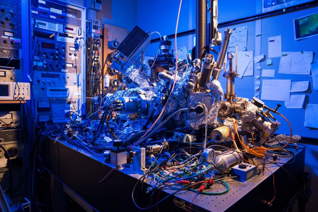
While not science’s sleekest machine, this microscope was able to capture M.R.I. scans of single atoms. Credit: IBM Research

Such a messy looking thing—it makes me feel better about my housekeeping. In any event, it’s fascinating to think this scanning tunneling microscope as seen in the above can actually act as an MRI device and create an image of a single atom.

There’s a wonderful article in the New York Times about the work but I’m starting first with a July 1, 2019 news item on Nanowerk,

Researchers at the Center for Quantum Nanoscience (QNS) within the Institute for Basic Science (IBS) at Ewha Womans University [Seoul, South Korea) have made a major scientific breakthrough by performing the world’s smallest magnetic resonance imaging (MRI). In an international collaboration with colleagues from the US, QNS scientists used their new technique to visualize the magnetic field of single atoms.

A July 2, 2019 IBS news release (also on EurekAlert but published July 1, 2019), which originated the news item, provides some insight into the research,

An MRI is routinely done in hospitals nowadays as a part of imaging for diagnostics. MRI’s detect the density of spins – the fundamental magnets in electrons and protons – in the human body. Traditionally, billions and billions of spins are required for an MRI scan. The new findings, published today [July 1, 2019] in the journal Nature Physics, show that this process is now also possible for an individual atom on a surface. To do this, the team used a Scanning Tunneling Microscope, which consists of an atomically sharp metal tip that allows researchers to image and probe single atoms by scanning the tip across the surface.

The two elements that were investigated in this work, iron and titanium, are both magnetic. Through precise preparation of the sample, the atoms were readily visible in the microscope. The researchers then used the microscope’s tip like an MRI machine to map the three-dimensional magnetic field created by the atoms with unprecedented resolution. In order to do so, they attached another spin cluster to the sharp metal tip of their microscope. Similar to everyday magnets, the two spins would attract or repel each other depending on their relative position. By sweeping the tip spin cluster over the atom on the surface, the researchers were able to map out the magnetic interaction. Lead author, Dr. Philip Willke of QNS says: “It turns out that the magnetic interaction we measured depends on the properties of both spins, the one on the tip and the one on the sample. For example, the signal that we see for iron atoms is vastly different from that for titanium atoms. This allows us to distinguish different kinds of atoms by their magnetic field signature and makes our technique very powerful.”

The researchers plan to use their single-atom MRI to map the spin distribution in more complex structures such as molecules and magnetic materials. “Many magnetic phenomena take place on the nanoscale, including the recent generation of magnetic storage devices.” says Dr. Yujeong Bae also of QNS, a co-author in this study. “We now plan to study a variety of systems using our microscopic MRI.” The ability to analyze the magnetic structure on the nanoscale can help to develop new materials and drugs. Moreover, the research team wants to use this kind of MRI to characterize and control quantum systems. These are of great interest for future computation schemes, also known as quantum computing

“I am very excited about these results. It is certainly a milestone in our field and has very promising implications for future research.” says Prof. Andreas Heinrich, Director of QNS. “The ability to map spins and their magnetic field with previously unimaginable precision, allows us to gain deeper knowledge about the structure of matter and opens new fields of basic research.”

The Center for Quantum Nanoscience, on the campus of Ewha Womans University in Seoul, South Korea, is a world-leading research center merging quantum and nanoscience to engineer the quantum future through basic research. Backed by Korea’s Institute for Basic Science, which was founded in 2011, the Center for Quantum Nanoscience draws on decades of QNS Director Andreas J. Heinrich’s (A Boy and His Atom, IBM, 2013) scientific leadership to lay the foundation for future technology by exploring the use of quantum behavior atom-by-atom on surfaces with highest precision.

You may have noticed that other than a brief mention in the first paragraph (in the Nanowerk news item excerpt), there’s no mention of the US researchers and their contribution to the work.

Interestingly, the July 1, 2019 New York Time article by Knvul Sheikh returns the favour by focusing almost entirely on US researchers while giving the Korean researchers a passing mention (Note: Links have been removed),

Different microscopy techniques allow scientists to see the nucleotide-by-nucleotide genetic sequences in cells down to the resolution of a couple atoms as seen in an atomic force microscopy image. But scientists at the IBM Almaden Research Center in San Jose, Calif., and the Institute for Basic Sciences in Seoul, have taken imaging a step further, developing a new magnetic resonance imaging technique that provides unprecedented detail, right down to the individual atoms of a sample.

When doctors want to detect tumors, measure brain function or visualize the structure of joints, they employ huge M.R.I. machines, which apply a magnetic field across the human body. This temporarily disrupts the protons spinning in the nucleus of every atom in every cell. A subsequent, brief pulse of radio-frequency energy causes the protons to spin perpendicular to the pulse. Afterward, the protons return to their normal state, releasing energy that can be measured by sensors and made into an image.

But to gather enough diagnostic data, traditional hospital M.R.I.s must scan billions and billions of protons in a person’s body, said Christopher Lutz, a physicist at IBM. So he and his colleagues decided to pack the power of an M.R.I. machine into the tip of another specialized instrument known as a scanning tunneling microscope to see if they could image individual atoms.

The tip of a scanning tunneling microscope is just a few atoms wide. And it moves along the surface of a sample, it picks up details about the size and conformation of molecules.

The researchers attached magnetized iron atoms to the tip, effectively combining scanning-tunneling microscope and M.R.I. technologies.

When the magnetized tip swept over a metal wafer of iron and titanium, it applied a magnetic field to the sample, disrupting the electrons (rather than the protons, as a typical M.R.I. would) within each atom. Then the researchers quickly turned a radio-frequency pulse on and off, so that the electrons would emit energy that could be visualized. …

Here’s a link to and a citation for the paper,

Magnetic resonance imaging of single atoms on a surface by Philip Willke, Kai Yang, Yujeong Bae, Andreas J. Heinrich & Christopher P. Lutz. Nature Physics (2019) DOI: https://doi.org/10.1038/s41567-019-0573-x Published 01 July 2019

This paper is behind a paywall.

Robot radiologists (artificially intelligent doctors)

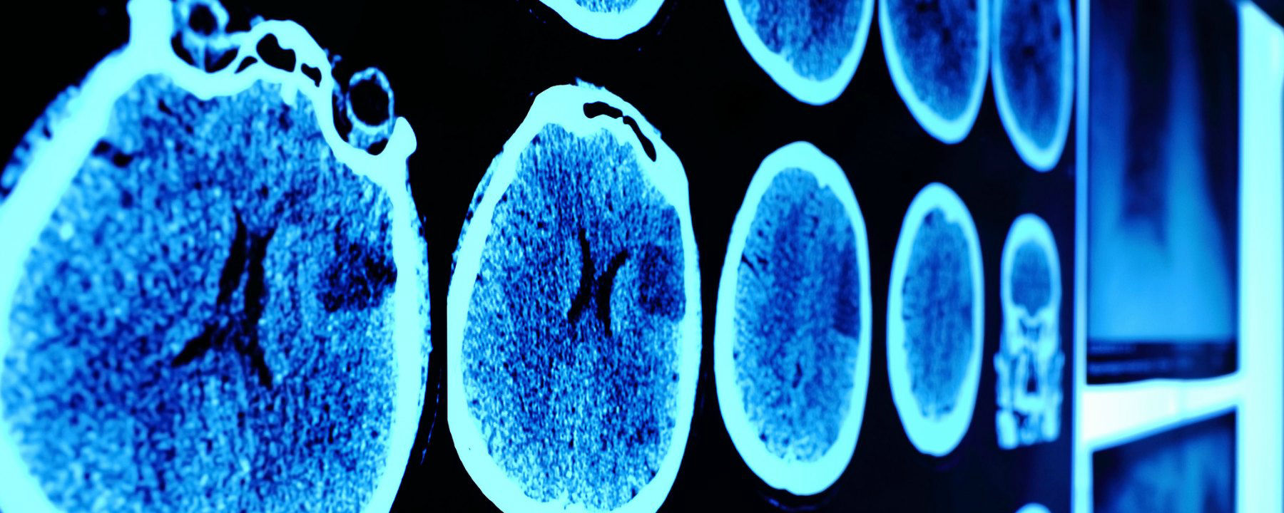
Mutaz Musa, a physician at New York Presbyterian Hospital/Weill Cornell (Department of Emergency Medicine) and software developer in New York City, has penned an eyeopening opinion piece about artificial intelligence (or robots if you prefer) and the field of radiology. From a June 25, 2018 opinion piece for The Scientist (Note: Links have been removed),

Although artificial intelligence has raised fears of job loss for many, we doctors have thus far enjoyed a smug sense of security. There are signs, however, that the first wave of AI-driven redundancies among doctors is fast approaching. And radiologists seem to be first on the chopping block.

Andrew Ng, founder of online learning platform Coursera and former CTO of “China’s Google,” Baidu, recently announced the development of CheXNet, a convolutional neural net capable of recognizing pneumonia and other thoracic pathologies on chest X-rays better than human radiologists. Earlier this year, a Hungarian group developed a similar system for detecting and classifying features of breast cancer in mammograms. In 2017, Adelaide University researchers published details of a bot capable of matching human radiologist performance in detecting hip fractures. And, of course, Google achieved superhuman proficiency in detecting diabetic retinopathy in fundus photographs, a task outside the scope of most radiologists.

Beyond single, two-dimensional radiographs, a team at Oxford University developed a system for detecting spinal disease from MRI data with a performance equivalent to a human radiologist. Meanwhile, researchers at the University of California, Los Angeles, reported detecting pathology on head CT scans with an error rate more than 20 times lower than a human radiologist.

Although these particular projects are still in the research phase and far from perfect—for instance, often pitting their machines against a limited number of radiologists—the pace of progress alone is telling.

Others have already taken their algorithms out of the lab and into the marketplace. Enlitic, founded by Aussie serial entrepreneur and University of San Francisco researcher Jeremy Howard, is a Bay-Area startup that offers automated X-ray and chest CAT scan interpretation services. Enlitic’s systems putatively can judge the malignancy of nodules up to 50 percent more accurately than a panel of radiologists and identify fractures so small they’d typically be missed by the human eye. One of Enlitic’s largest investors, Capitol Health, owns a network of diagnostic imaging centers throughout Australia, anticipating the broad rollout of this technology. Another Bay-Area startup, Arterys, offers cloud-based medical imaging diagnostics. Arterys’s services extend beyond plain films to cardiac MRIs and CAT scans of the chest and abdomen. And there are many others.

Musa has offered a compelling argument with lots of links to supporting evidence.
[downloaded from https://www.the-scientist.com/news-opinion/opinion–rise-of-the-robot-radiologists-64356]

And evidence keeps mounting, I just stumbled across this June 30, 2018 news item on Xinhuanet.com,

An artificial intelligence (AI) system scored 2:0 against elite human physicians Saturday in two rounds of competitions in diagnosing brain tumors and predicting hematoma expansion in Beijing.

The BioMind AI system, developed by the Artificial Intelligence Research Centre for Neurological Disorders at the Beijing Tiantan Hospital and a research team from the Capital Medical University, made correct diagnoses in 87 percent of 225 cases in about 15 minutes, while a team of 15 senior doctors only achieved 66-percent accuracy.

The AI also gave correct predictions in 83 percent of brain hematoma expansion cases, outperforming the 63-percent accuracy among a group of physicians from renowned hospitals across the country.

The outcomes for human physicians were quite normal and even better than the average accuracy in ordinary hospitals, said Gao Peiyi, head of the radiology department at Tiantan Hospital, a leading institution on neurology and neurosurgery.

To train the AI, developers fed it tens of thousands of images of nervous system-related diseases that the Tiantan Hospital has archived over the past 10 years, making it capable of diagnosing common neurological diseases such as meningioma and glioma with an accuracy rate of over 90 percent, comparable to that of a senior doctor.

All the cases were real and contributed by the hospital, but never used as training material for the AI, according to the organizer.

Wang Yongjun, executive vice president of the Tiantan Hospital, said that he personally did not care very much about who won, because the contest was never intended to pit humans against technology but to help doctors learn and improve [emphasis mine] through interactions with technology.

“I hope through this competition, doctors can experience the power of artificial intelligence. This is especially so for some doctors who are skeptical about artificial intelligence. I hope they can further understand AI and eliminate their fears toward it,” said Wang.

Dr. Lin Yi who participated and lost in the second round, said that she welcomes AI, as it is not a threat but a “friend.” [emphasis mine]

AI will not only reduce the workload but also push doctors to keep learning and improve their skills, said Lin.

Bian Xiuwu, an academician with the Chinese Academy of Science and a member of the competition’s jury, said there has never been an absolute standard correct answer in diagnosing developing diseases, and the AI would only serve as an assistant to doctors in giving preliminary results. [emphasis mine]

Dr. Paul Parizel, former president of the European Society of Radiology and another member of the jury, also agreed that AI will not replace doctors, but will instead function similar to how GPS does for drivers. [emphasis mine]

Dr. Gauden Galea, representative of the World Health Organization in China, said AI is an exciting tool for healthcare but still in the primitive stages.

Based on the size of its population and the huge volume of accessible digital medical data, China has a unique advantage in developing medical AI, according to Galea.

China has introduced a series of plans in developing AI applications in recent years.

In 2017, the State Council issued a development plan on the new generation of Artificial Intelligence and the Ministry of Industry and Information Technology also issued the “Three-Year Action Plan for Promoting the Development of a New Generation of Artificial Intelligence (2018-2020).”

The Action Plan proposed developing medical image-assisted diagnostic systems to support medicine in various fields.

I note the reference to cars and global positioning systems (GPS) and their role as ‘helpers’;, it seems no one at the ‘AI and radiology’ competition has heard of driverless cars. Here’s Musa on those reassuring comments abut how the technology won’t replace experts but rather augment their skills,

To be sure, these services frame themselves as “support products” that “make doctors faster,” rather than replacements that make doctors redundant. This language may reflect a reserved view of the technology, though it likely also represents a marketing strategy keen to avoid threatening or antagonizing incumbents. After all, many of the customers themselves, for now, are radiologists.

Radiology isn’t the only area where experts might find themselves displaced.

Eye experts

It seems inroads have been made by artificial intelligence systems (AI) into the diagnosis of eye diseases. It got the ‘Fast Company’ treatment (exciting new tech, learn all about it) as can be seen further down in this posting. First, here’s a more restrained announcement, from an August 14, 2018 news item on phys.org (Note: A link has been removed),

An artificial intelligence (AI) system, which can recommend the correct referral decision for more than 50 eye diseases, as accurately as experts has been developed by Moorfields Eye Hospital NHS Foundation Trust, DeepMind Health and UCL [University College London].

The breakthrough research, published online by Nature Medicine, describes how machine-learning technology has been successfully trained on thousands of historic de-personalised eye scans to identify features of eye disease and recommend how patients should be referred for care.

Researchers hope the technology could one day transform the way professionals carry out eye tests, allowing them to spot conditions earlier and prioritise patients with the most serious eye diseases before irreversible damage sets in.

An August 13, 2018 UCL press release, which originated the news item, describes the research and the reasons behind it in more detail,

More than 285 million people worldwide live with some form of sight loss, including more than two million people in the UK. Eye diseases remain one of the biggest causes of sight loss, and many can be prevented with early detection and treatment.

Dr Pearse Keane, NIHR Clinician Scientist at the UCL Institute of Ophthalmology and consultant ophthalmologist at Moorfields Eye Hospital NHS Foundation Trust said: “The number of eye scans we’re performing is growing at a pace much faster than human experts are able to interpret them. There is a risk that this may cause delays in the diagnosis and treatment of sight-threatening diseases, which can be devastating for patients.”

“The AI technology we’re developing is designed to prioritise patients who need to be seen and treated urgently by a doctor or eye care professional. If we can diagnose and treat eye conditions early, it gives us the best chance of saving people’s sight. With further research it could lead to greater consistency and quality of care for patients with eye problems in the future.”

The study, launched in 2016, brought together leading NHS eye health professionals and scientists from UCL and the National Institute for Health Research (NIHR) with some of the UK’s top technologists at DeepMind to investigate whether AI technology could help improve the care of patients with sight-threatening diseases, such as age-related macular degeneration and diabetic eye disease.

Using two types of neural network – mathematical systems for identifying patterns in images or data – the AI system quickly learnt to identify 10 features of eye disease from highly complex optical coherence tomography (OCT) scans. The system was then able to recommend a referral decision based on the most urgent conditions detected.

To establish whether the AI system was making correct referrals, clinicians also viewed the same OCT scans and made their own referral decisions. The study concluded that AI was able to make the right referral recommendation more than 94% of the time, matching the performance of expert clinicians.

The AI has been developed with two unique features which maximise its potential use in eye care. Firstly, the system can provide information that helps explain to eye care professionals how it arrives at its recommendations. This information includes visuals of the features of eye disease it has identified on the OCT scan and the level of confidence the system has in its recommendations, in the form of a percentage. This functionality is crucial in helping clinicians scrutinise the technology’s recommendations and check its accuracy before deciding the type of care and treatment a patient receives.

Secondly, the AI system can be easily applied to different types of eye scanner, not just the specific model on which it was trained. This could significantly increase the number of people who benefit from this technology and future-proof it, so it can still be used even as OCT scanners are upgraded or replaced over time.

The next step is for the research to go through clinical trials to explore how this technology might improve patient care in practice, and regulatory approval before it can be used in hospitals and other clinical settings.

If clinical trials are successful in demonstrating that the technology can be used safely and effectively, Moorfields will be able to use an eventual, regulatory-approved product for free, across all 30 of their UK hospitals and community clinics, for an initial period of five years.

The work that has gone into this project will also help accelerate wider NHS research for many years to come. For example, DeepMind has invested significant resources to clean, curate and label Moorfields’ de-identified research dataset to create one of the most advanced eye research databases in the world.

Moorfields owns this database as a non-commercial public asset, which is already forming the basis of nine separate medical research studies. In addition, Moorfields can also use DeepMind’s trained AI model for future non-commercial research efforts, which could help advance medical research even further.

Mustafa Suleyman, Co-founder and Head of Applied AI at DeepMind Health, said: “We set up DeepMind Health because we believe artificial intelligence can help solve some of society’s biggest health challenges, like avoidable sight loss, which affects millions of people across the globe. These incredibly exciting results take us one step closer to that goal and could, in time, transform the diagnosis, treatment and management of patients with sight threatening eye conditions, not just at Moorfields, but around the world.”

Professor Sir Peng Tee Khaw, director of the NIHR Biomedical Research Centre at Moorfields Eye Hospital NHS Foundation Trust and UCL Institute of Ophthalmology said: “The results of this pioneering research with DeepMind are very exciting and demonstrate the potential sight-saving impact AI could have for patients. I am in no doubt that AI has a vital role to play in the future of healthcare, particularly when it comes to training and helping medical professionals so that patients benefit from vital treatment earlier than might previously have been possible. This shows the transformative research than can be carried out in the UK combining world leading industry and NIHR/NHS hospital/university partnerships.”

Matt Hancock, Health and Social Care Secretary, said: “This is hugely exciting and exactly the type of technology which will benefit the NHS in the long term and improve patient care – that’s why we fund over a billion pounds a year in health research as part of our long term plan for the NHS.”

Here’s a link to and a citation for the study,

Clinically applicable deep learning for diagnosis and referral in retinal disease by Jeffrey De Fauw, Joseph R. Ledsam, Bernardino Romera-Paredes, Stanislav Nikolov, Nenad Tomasev, Sam Blackwell, Harry Askham, Xavier Glorot, Brendan O’Donoghue, Daniel Visentin, George van den Driessche, Balaji Lakshminarayanan, Clemens Meyer, Faith Mackinder, Simon Bouton, Kareem Ayoub, Reena Chopra, Dominic King, Alan Karthikesalingam, Cían O. Hughes, Rosalind Raine, Julian Hughes, Dawn A. Sim, Catherine Egan, Adnan Tufail, Hugh Montgomery, Demis Hassabis, Geraint Rees, Trevor Back, Peng T. Khaw, Mustafa Suleyman, Julien Cornebise, Pearse A. Keane, & Olaf Ronneberger. Nature Medicine (2018) DOI: https://doi.org/10.1038/s41591-018-0107-6 Published 13 August 2018

This paper is behind a paywall.

And now, Melissa Locker’s August 15, 2018 article for Fast Company (Note: Links have been removed),

In a paper published in Nature Medicine on Monday, Google’s DeepMind subsidiary, UCL, and researchers at Moorfields Eye Hospital showed off their new AI system. The researchers used deep learning to create algorithm-driven software that can identify common patterns in data culled from dozens of common eye diseases from 3D scans. The result is an AI that can identify more than 50 diseases with incredible accuracy and can then refer patients to a specialist. Even more important, though, is that the AI can explain why a diagnosis was made, indicating which part of the scan prompted the outcome. It’s an important step in both medicine and in making AIs slightly more human

The editor or writer has even highlighted the sentence about the system’s accuracy—not just good but incredible!

I will be publishing something soon [my August 21, 2018 posting] which highlights some of the questions one might want to ask about AI and medicine before diving headfirst into this brave new world of medicine.
Democratizing science .. neuroscience that is

What is going on with the neuroscience folks? First it was Montreal Neuro opening up its science  as featured in my January 22, 2016 posting,

The Montreal Neurological Institute (MNI) in Québec, Canada, known informally and widely as Montreal Neuro, has ‘opened’ its science research to the world. David Bruggeman tells the story in a Jan. 21, 2016 posting on his Pasco Phronesis blog (Note: Links have been removed),

The Montreal Neurological Institute (MNI) at McGill University announced that it will be the first academic research institute to become what it calls ‘Open Science.’  As Science is reporting, the MNI will make available all research results and research data at the time of publication.  Additionally it will not seek patents on any of the discoveries made on research at the Institute.

Will this catch on?  I have no idea if this particular combination of open access research data and results with no patents will spread to other university research institutes.  But I do believe that those elements will continue to spread.  More universities and federal agencies are pursuing open access options for research they support.  Elon Musk has opted to not pursue patent litigation for any of Tesla Motors’ patents, and has not pursued patents for SpaceX technology (though it has pursued litigation over patents in rocket technology). …

Whether or not they were inspired by the MNI, the scientists at the University of Washington (UW [state]) have found their own unique way of opening up science. From a March 15, 2018 UW news blog posting (also on EurekAlert) by James Urton, Note: Links have been removed,

Over the past few years, scientists have faced a problem: They often cannot reproduce the results of experiments done by themselves or their peers.

This “replication crisis” plagues fields from medicine to physics, and likely has many causes. But one is undoubtedly the difficulty of sharing the vast amounts of data collected and analyses performed in so-called “big data” studies. The volume and complexity of the information also can make these scientific endeavors unwieldy when it comes time for researchers to share their data and findings with peers and the public.

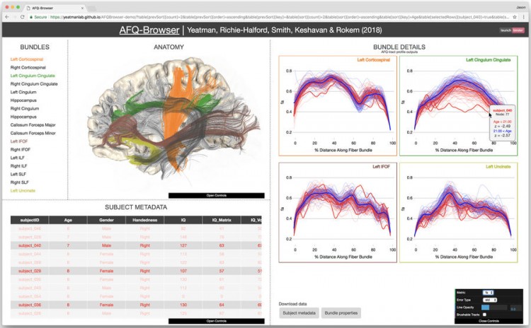
Researchers at the University of Washington have developed a set of tools to make one critical area of big data research — that of our central nervous system — easier to share. In a paper published online March 5 [2018] in Nature Communications, the UW team describes an open-access browser they developed to display, analyze and share neurological data collected through a type of magnetic resonance imaging study known as diffusion-weighted MRI.

“There has been a lot of talk among researchers about the replication crisis,” said lead author Jason Yeatman. “But we wanted a tool — ready, widely available and easy to use — that would actually help fight the replication crisis.”

Yeatman — who is an assistant professor in the UW Department of Speech & Hearing Sciences and the Institute for Learning & Brain Sciences (I-LABS) — is describing AFQ-Browser. This web browser-based tool, freely available online, is a platform for uploading, visualizing, analyzing and sharing diffusion MRI data in a format that is publicly accessible, improving transparency and data-sharing methods for neurological studies. In addition, since it runs in the web browser, AFQ-Browser is portable — requiring no additional software package or equipment beyond a computer and an internet connection.

“One major barrier to data transparency in neuroscience is that so much data collection, storage and analysis occurs on local computers with special software packages,” said senior author Ariel Rokem, a senior data scientist in the UW eScience Institute. “But using AFQ-Browser, we eliminate those requirements and make uploading, sharing and analyzing diffusion-weighted MRI data a simple, straightforward process.”

Diffusion-weighted MRI measures the movement of fluid in the brain and spinal cord, revealing the structure and function of white-matter tracts. These are the connections of the central nervous system, tissue that are made up primarily of axons that transmit long-range signals between neural circuits. Diffusion MRI research on brain connectivity has fundamentally changed the way neuroscientists understand human brain function: The state, organization and layout of white matter tracts are at the core of cognitive functions such as memory, learning and other capabilities. Data collected using diffusion-weighted MRI can be used to diagnose complex neurological conditions such as multiple sclerosis (MS) and amyotrophic lateral sclerosis (ALS). Researchers also use diffusion-weighted MRI data to study the neurological underpinnings of conditions such as dyslexia and learning disabilities.

“This is a widely-used technique in neuroscience research, and it is particularly amenable to the benefits that can be gleaned from big data, so it became a logical starting point for developing browser-based, open-access tools for the field,” said Yeatman.

The AFQ-Browser — the AFQ stands for Automated Fiber-tract Quantification — can receive diffusion-weighted MRI data and perform tract analysis for each individual subject. The analyses occur via a remote server, again eliminating technical and financial barriers for researchers. The AFQ-Browser also contains interactive tools to display data for multiple subjects — allowing a researcher to easily visualize how white matter tracts might be similar or different among subjects, identify trends in the data and generate hypotheses for future experiments. Researchers also can insert additional code to analyze the data, as well as save, upload and share data instantly with fellow researchers.

“We wanted this tool to be as generalizable as possible, regardless of research goals,” said Rokem. “In addition, the format is easy for scientists from a variety of backgrounds to use and understand — so that neuroscientists, statisticians and other researchers can collaborate, view data and share methods toward greater reproducibility.”

The idea for the AFQ-Browser came out of a UW course on data visualization, and the researchers worked with several graduate students to develop and perfect the browser. They tested it on existing diffusion-weighted MRI datasets, including research subjects with ALS and MS. In the future, they hope that the AFQ-Browser can be improved to do automated analyses — and possibly even diagnoses — based on diffusion-weighted MRI data.

“AFQ-Browser is really just the start of what could be a number of tools for sharing neuroscience data and experiments,” said Yeatman. “Our goal here is greater reproducibility and transparency, and a more robust scientific process.”

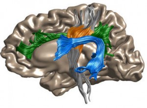
Here are a couple of images the researchers have used to illustrate their work,

AFQ-Browser.Jason Yeatman/Ariel Rokem Courtesy: University of Washington

Depiction of the left hemisphere of the human brain. Colored regions are selected white matter regions that could be measured using diffusion-weighted MRI: Corticospinal tract (orange), arcuate fasciculus (blue) and cingulum (green).Jason Yeatman/Ariel Rokem

You can find an embedded version of the AFQ-Browser here: http://www.washington.edu/news/2018/03/15/democratizing-science-researchers-make-neuroscience-experiments-easier-to-share-reproduce/ (scroll down about 50 – 55% of the way).

As for the paper, here’s a link and a citation,

A browser-based tool for visualization and analysis of diffusion MRI data by Jason D. Yeatman, Adam Richie-Halford, Josh K. Smith, Anisha Keshavan, & Ariel Rokem. Nature Communicationsvolume 9, Article number: 940 (2018) doi:10.1038/s41467-018-03297-7 Published online: 05 March 2018

Fittingly, this paper is open access.

New iron oxide nanoparticle as an MRI (magnetic resonance imaging) contrast agent

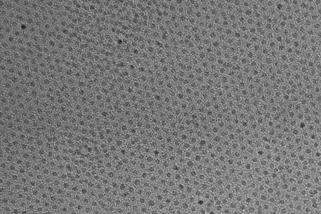
This high-resolution transmission electron micrograph of particles made by the research team shows the particles’ highly uniform size and shape. These are iron oxide particles just 3 nanometers across, coated with a zwitterion layer. Their small size means they can easily be cleared through the kidneys after injection. Courtesy of the researchers

A Feb. 14, 2017 news item on ScienceDaily announces a new MRI (magnetic resonance imaging) contrast agent,

A new, specially coated iron oxide nanoparticle developed by a team at MIT [Massachusetts Institute of Technology] and elsewhere could provide an alternative to conventional gadolinium-based contrast agents used for magnetic resonance imaging (MRI) procedures. In rare cases, the currently used gadolinium agents have been found to produce adverse effects in patients with impaired kidney function.

A Feb. 14, 2017 MIT news release (also on EurekAlert), which originated the news item, provides more technical detail,


The advent of MRI technology, which is used to observe details of specific organs or blood vessels, has been an enormous boon to medical diagnostics over the last few decades. About a third of the 60 million MRI procedures done annually worldwide use contrast-enhancing agents, mostly containing the element gadolinium. While these contrast agents have mostly proven safe over many years of use, some rare but significant side effects have shown up in a very small subset of patients. There may soon be a safer substitute thanks to this new research.

In place of gadolinium-based contrast agents, the researchers have found that they can produce similar MRI contrast with tiny nanoparticles of iron oxide that have been treated with a zwitterion coating. (Zwitterions are molecules that have areas of both positive and negative electrical charges, which cancel out to make them neutral overall.) The findings are being published this week in the Proceedings of the National Academy of Sciences, in a paper by Moungi Bawendi, the Lester Wolfe Professor of Chemistry at MIT; He Wei, an MIT postdoc; Oliver Bruns, an MIT research scientist; Michael Kaul at the University Medical Center Hamburg-Eppendorf in Germany; and 15 others.

Contrast agents, injected into the patient during an MRI procedure and designed to be quickly cleared from the body by the kidneys afterwards, are needed to make fine details of organ structures, blood vessels, and other specific tissues clearly visible in the images. Some agents produce dark areas in the resulting image, while others produce light areas. The primary agents for producing light areas contain gadolinium.

Iron oxide particles have been largely used as negative (dark) contrast agents, but radiologists vastly prefer positive (light) contrast agents such as gadolinium-based agents, as negative contrast can sometimes be difficult to distinguish from certain imaging artifacts and internal bleeding. But while the gadolinium-based agents have become the standard, evidence shows that in some very rare cases they can lead to an untreatable condition called nephrogenic systemic fibrosis, which can be fatal. In addition, evidence now shows that the gadolinium can build up in the brain, and although no effects of this buildup have yet been demonstrated, the FDA is investigating it for potential harm.

“Over the last decade, more and more side effects have come to light” from the gadolinium agents, Bruns says, so that led the research team to search for alternatives. “None of these issues exist for iron oxide,” at least none that have yet been detected, he says.

The key new finding by this team was to combine two existing techniques: making very tiny particles of iron oxide, and attaching certain molecules (called surface ligands) to the outsides of these particles to optimize their characteristics. The iron oxide inorganic core is small enough to produce a pronounced positive contrast in MRI, and the zwitterionic surface ligand, which was recently developed by Wei and coworkers in the Bawendi research group, makes the iron oxide particles water-soluble, compact, and biocompatible.

The combination of a very tiny iron oxide core and an ultrathin ligand shell leads to a total hydrodynamic diameter of 4.7 nanometers, below the 5.5-nanometer renal clearance threshold. This means that the coated iron oxide should quickly clear through the kidneys and not accumulate. This renal clearance property is an important feature where the particles perform comparably to gadolinium-based contrast agents.

Now that initial tests have demonstrated the particles’ effectiveness as contrast agents, Wei and Bruns say the next step will be to do further toxicology testing to show the particles’ safety, and to continue to improve the characteristics of the material. “It’s not perfect. We have more work to do,” Bruns says. But because iron oxide has been used for so long and in so many ways, even as an iron supplement, any negative effects could likely be treated by well-established protocols, the researchers say. If all goes well, the team is considering setting up a startup company to bring the material to production.

For some patients who are currently excluded from getting MRIs because of potential side effects of gadolinium, the new agents “could allow those patients to be eligible again” for the procedure, Bruns says. And, if it does turn out that the accumulation of gadolinium in the brain has negative effects, an overall phase-out of gadolinium for such uses could be needed. “If that turned out to be the case, this could potentially be a complete replacement,” he says.

Ralph Weissleder, a physician at Massachusetts General Hospital who was not involved in this work, says, “The work is of high interest, given the limitations of gadolinium-based contrast agents, which typically have short vascular half-lives and may be contraindicated in renally compromised patients.”

The research team included researchers in MIT’s chemistry, biological engineering, nuclear science and engineering, brain and cognitive sciences, and materials science and engineering departments and its program in Health Sciences and Technology; and at the University Medical Center Hamburg-Eppendorf; Brown University; and the Massachusetts General Hospital. It was supported by the MIT-Harvard NIH Center for Cancer Nanotechnology, the Army Research Office through MIT’s Institute for Soldier Nanotechnologies, the NIH-funded Laser Biomedical Research Center, the MIT Deshpande Center, and the European Union Seventh Framework Program.

Here’s a link to and a citation for the paper,

Exceedingly small iron oxide nanoparticles as positive MRI contrast agents by He Wei, Oliver T. Bruns, Michael G. Kaul, Eric C. Hansen, Mariya Barch, Agata Wiśniowsk, Ou Chen, Yue Chen, Nan Li, Satoshi Okada, Jose M. Cordero, Markus Heine, Christian T. Farrar, Daniel M. Montana, Gerhard Adam, Harald Ittrich, Alan Jasanoff, Peter Nielsen, and Moungi G. Bawendi. PNAS February 13, 2017 doi: 10.1073/pnas.1620145114 Published online before print February 13, 2017

This paper is behind a paywall.

A new graphene-based contrast agent for magnetic resonance imaging (MRI)

After teaching a continuing studies course on bioelectronics for Simon Fraser University (Vancouver, Canada), I’ve developed a mild interest in magnetic resonance imaging and contrast agents which this Nov. 11, 2016 news item on phys.org satisfies,

Graphene, the atomically thin sheets of carbon that materials scientists are hoping to use for everything from nanoelectronics and aircraft de-icers to batteries and bone implants, may also find use as contrast agents for magnetic resonance imaging (MRI), according to new research from Rice University.

“They have a lot of advantages compared with conventionally available contrast agents,” Rice researcher Sruthi Radhakrishnan said of the graphene-based quantum dots she has studied for the past two years. “Virtually all of the widely used contrast agents contain toxic metals, but our material has no metal. It’s just carbon, hydrogen, oxygen and fluorine, and in all of our tests so far it has shown no signs of toxicity.”

The initial findings for Rice’s nanoparticles—disks of graphene that are decorated with fluorine atoms and simply organic molecules that make them magnetic—are described in a new paper in the journal Particle and Particle Systems characterization.

A Nov. 10, 2016 Rice University (Texas, US) news release, which originated the news item, describes the work in more detail,

Pulickel Ajayan, the Rice materials scientist who is directing the work, said the fluorinated graphene oxide quantum dots could be particularly useful as MRI contrast agents because they could be targeted to specific kinds of tissues.

“There are tried-and-true methods for attaching biomarkers to carbon nanoparticles, so one could easily envision using these quantum dots to develop tissue-specific contrast agents,” Ajayan said. “For example, this method could be used to selectively target specific types of cancer or brain lesions caused by Alzheimer’s disease. That kind of specificity isn’t available with today’s contrast agents.”

MRI scanners make images of the body’s internal structures using strong magnetic fields and radio waves. As diagnostic tests, MRIs often provide greater detail than X-rays without the harmful radiation, and as a result, MRI usage has risen sharply over the past decade. More than 30 million MRIs are performed annually in the U.S.

Radhakrishnan said her work began in 2014 after Ajayan’s research team found that adding fluorine to either graphite or graphene caused the materials to show up well on MRI scans.

All materials are influenced by magnetic fields, including animal tissues. In MRI scanners, a powerful magnetic field causes individual atoms throughout the body to become magnetically aligned. A pulse of radio energy is used to disrupt this alignment, and the machine measures how long it takes for the atoms in different parts of the body to become realigned. Based on these measures, the scanner can build up a detailed image of the body’s internal structures.

MRI contrast agents shorten the amount of time it takes for tissues to realign and significantly improve the resolution of MRI scans. Almost all commercially available contrast agents are made from toxic metals like gadolinium, iron or manganese.

“We worked with a team from MD Anderson Cancer Center to assess the cytocompatibility of fluorinated graphene oxide quantum dots,” Radhakrishnan said. “We used a test that measures the metabolic activity of cell cultures and detects toxicity as a drop in metabolic activity. We incubated quantum dots in kidney cell cultures for up to three days and found no significant cell death in the cultures, even at the highest concentrations.”

The fluorinated graphene oxide quantum dots Radhakrishnan studies can be made in less than a day, but she spent two years perfecting the recipe for them. She begins with micron-sized sheets of graphene that have been fluorinated and oxidized. When these are added to a solvent and stirred for several hours, they break into smaller pieces. Making the material smaller is not difficult, but the process for making small particles with the appropriate magnetic properties is exacting. Radhakrishnan said there was no “eureka moment” in which she suddenly achieved the right results by stumbling on the best formula. Rather, the project was marked by incremental improvements through dozens of minor alterations.

“It required a lot of optimization,” she said. “The recipe matters a lot.”

Radhakrishnan said she plans to continue studying the material and hopes to eventually have a hand in proving that it is safe and effective for clinical MRI tests.

“I would like to see it applied commercially in clinical ways because it has a lot of advantages compared with conventionally available agents,” she said.

Here’s a link to and a citation for the paper,

Metal-Free Dual Modal Contrast Agents Based on Fluorographene Quantum Dots by Sruthi Radhakrishnan, Atanu Samanta, Parambath M. Sudeep, Kiersten L. Maldonado, Sendurai A. Mani, Ghanashyam Acharya, Chandra Sekhar Tiwary, Abhishek K. Singh, and Pulickel M. Ajayan. Particle & Particle Systems Characterization DOI: 10.1002/ppsc.201600221 Version of Record online: 21 OCT 2016

This paper is behind a paywall.

Better contrast agents for magnetic resonance imaging with nanoparticles

I wonder what’s going on in the field of magnetic resonance imaging. This is the third news item I’ve stumbled across related to the topic in the last couple of months. (Links to the other two posts follow at the end of this post.) By comparison, that’s the more than in the previous seven years (2008 – 2015) combined.

The latest research concerns a new and better contrast agent. From an Aug. 3, 2016 news item on Nanowerk,

Scientists at the University of Basel [Switzerland] have developed nanoparticles which can serve as efficient contrast agents for magnetic resonance imaging. This new type of nanoparticles [sic] produce around ten times more contrast than the actual contrast agents and are responsive to specific environments.

An Aug. 3, 2016 University of Basel press release (also on EurekAlert), which originated the news item, explains further,

Contrast agents are usually based on the metal Gadolinium, which is injected and serves for an improved imaging of various organs in an MRI. Gadolinium ions should be bound with a carrier compound to avoid the toxicity to the human body of the free ions. Therefore, highly efficient contrast agents requiring lower Gadolinium concentrations represent an important step for advancing diagnosis and improving patient health prognosis.

Smart nanoparticles as contrast agents

The research groups of Prof. Cornelia Palivan and Prof. Wolfgang Meier from the Department of Chemistry at the University of Basel have introduced a new type of nanoparticles [sic], which combine multiple properties required for contrast agents: an increased MRI contrast for lower concentration, a potential for long blood circulation and responsiveness to different biochemical environments. These nanoparticles were obtained by co-assembly of heparin-functionalized polymers with trapped gadolinium ions and stimuli-responsive peptides.

The study shows, that the nanoparticles have the capacity of enhancing the MRI signal tenfold higher than the current agents. In addition, they have an enhanced efficacy in reductive milieu, characteristic for specific regions, such as cancerous tissues. These nanoparticles fulfill numerous key criteria for further development, such as absence of cellular toxicity, no apparent anticoagulation property, and high shelf stability. The concept developed by the researchers at the University of Basel to produce better contrast agents based on nanoparticles highlights a new direction in the design of MRI contrast agents, and supports their implementation for future applications.

Here’s a link to and a citation for the paper,

Nanoparticle-based highly sensitive MRI contrast agents with enhanced relaxivity in reductive milieu by
Severin J. Sigg, Francesco Santini, Adrian Najer, Pascal U. Richard, Wolfgang P. Meier, and Cornelia G. Palivan. Chem. Commun., 2016,52, 9937-9940 DOI: 10.1039/C6CC03396B First published online 13 Jul 2016

This paper is behind a paywall.

The other two MRI items featured here are in a June 10, 2016 posting (pH dependent nanoparticle-based contrast agent for MRIs [magnetic resonance images]) and in an Aug. 1, 2016 posting (Nuclear magnetic resonance microscope breaks records).

pH dependent nanoparticle-based contrast agent for MRIs (magnetic resonance images)

This news about a safer and more effective contrast agent for MRIs (magnetic resonance images) developed by Japanese scientists come from a June 6, 2016 article by Heather Zeiger on phys.org. First some explanations,

Magnetic resonance imaging relies on the excitation and subsequent relaxation of protons. In clinical MRI studies, the signal is determined by the relaxation time of the hydrogen protons in water. To get a stronger signal, scientists can use contrast agents to shorten the relaxation time of the protons.

MRI is non-invasive and does not involve radiation, making it a safe diagnostic tool. However, its weak signal makes tumor detection difficult. The ideal contrast agent would select for malignant tumors, making its location and diagnosis much more obvious.

Nanoparticle contrast agents have been of interested because nanoparticles can be functionalized and, as in this study, can contain various metals. Researchers have attempted to functionalize nanoparticles with ligands that attach to chemical factors on the surface of cancer cells. However, cancer cells tend to be compositionally heterogeneous, leading some researchers to look for nanoparticles that respond to differences in pH or redox potential compared to normal cells.

Now for the research,

Researchers from the University of Tokyo, Tokyo Institute of Technology, Kawasaki Institute of Industry Promotion, and the Japan Agency for Quantum and Radiological Science and Technology have developed a contrast agent from calcium phosphate-based nanoparticles that release a manganese ion an acidic environment. …

Peng Mi, Daisuke Kokuryo, Horacio Cabral, Hailiang Wu, Yasuko Terada, Tsuneo Saga, Ichio Aoki, Nobuhiro Nishiyama, and Kazunori Kataoka developed a contrast agent that is comprised of Mn2+– doped CaP nanoparticles with a PEG shell. They reasoned that using CaP nanoparticles, which are known to be pH sensitive, would allow the targeted release of Mn2+ ions in the tumor microenvironment. The tumor microenvironment tends to have a lower pH than the normal regions to rapid cell metabolism in an oxygen-depleted environment. Manganese ions were tested because they are paramagnetic, which makes for a good contrast agent. They also bind to proteins creating a slowly rotating manganese-protein system that results in sharp contrast enhancement.

These results were promising, so Peng Mi, et al. then tested whether the CaPMnPEG contrast agent worked in solid tumors. Because Mn2+ remains confined within the nanoparticle matrix at physiological pH, CaPMnPEG demonstrate a much lower toxicity [emphasis mine] compared to MnCl2. MRI studies showed a tumor-to-normal contrast of 131% after 30 minute, which is much higher than Gd-DTPA [emphasis mine], a clinically approved contrast agent. After an hour, the tumor-to-normal ratio was 160% and remained around 170% for several hours.

Three-dimensional MRI studies of solid tumors showed that without the addition of CaPMnPEG, only blood vessels were visible. However, upon adding CaPMnPEG, the tumor was easily distinguishable. Additionally, there is evidence that excess Mn2+ leaves the plasma after an hour. The contrast signal remained strong for several hours indicating that protein binding rather than Mn2+ concentration is important for signal enhancement.

Finally, tests with metastatic tumors in the liver (C26 colon cancer cells) showed that CaPMnPEG works well in solid organ analysis and is highly sensitive to detecting millimeter-sized micrometastasis [emphasis mine]. Unlike other contrast agents used in the clinic, CaPMnPEG provided a contrast signal that lasted for several hours after injection. After an hour, the signal was enhanced by 25% and after two hours, the signal was enhanced by 39%.

This is exciting stuff. Bravo to the researchers!

Here’s a link to and citation for the paper,

A pH-activatable nanoparticle with signal-amplification capabilities for non-invasive imaging of tumour malignancy by Peng Mi, Daisuke Kokuryo, Horacio Cabral, Hailiang Wu, Yasuko Terada, Tsuneo Saga, Ichio Aoki, Nobuhiro Nishiyama, & Kazunori Kataoka. Nature Nanotechnology (2016) doi:10.1038/nnano.2016.72 Published online 16 May 2016

This paper is behind a paywall.

Molecular ‘lightbulb’ could mean new form of magnetic resonance imaging (MRI)

A new technique promises to show body chemistry in action according to a March 25, 2016 news item on phys.org,

Duke University researchers have taken a major step towards realizing a new form of MRI that could record biochemical reactions in the body as they happen.

In the March 25 issue of Science Advances, they report the discovery of a new class of molecular tags that enhance MRI signals by 10,000-fold and generate detectable signals that last over an hour. The tags are biocompatible and inexpensive to produce, paving the way for widespread use of magnetic resonance imaging (MRI) to monitor metabolic processes of conditions like cancer and heart disease in real time.

“This represents a completely new class of molecules that doesn’t look anything at all like what people thought could be made into MRI tags,” said Warren S. Warren, James B. Duke Professor and Chair of Physics at Duke, and senior author on the study. “We envision it could provide a whole new way to use MRI to learn about the biochemistry of disease.”

A March 25, 2016 Duke University news release (also on EurekAlert), which originated the news item, offers more information about the new technique,

MRI takes advantage of a property called spin, which makes the nuclei in hydrogen atoms act like tiny magnets. Applying a strong magnetic field, followed by a series of radio waves, induces these hydrogen magnets to broadcast their locations. Since most of the hydrogen atoms in the body are bound up in water, the technique is used in clinical settings to create detailed images of soft tissues like organs, blood vessels and tumors inside the body.

But the technique also has the potential to show body chemistry in action, said Thomas Theis, assistant research professor of chemistry at Duke and co-lead author on the paper. “With magnetic resonance in general, you have this unique sensitivity to chemical transformations. You can see them and track them in real time,” Theis said.

MRI’s ability to track chemical transformations in the body has been limited by the low sensitivity of the technique, which makes small numbers of molecules impossible to detect without using unattainably massive magnetic fields.

For the past decade, researchers have been developing methods to “hyperpolarize” biologically important molecules, converting them into what Warren calls magnetic resonance “lightbulbs.”

With this boosted signal, these “lightbulbs” can be detected even in low numbers. “Hyperpolarization gives them 10,000 times more signal than they would normally have if they had just been magnetized in an ordinary magnetic field,” Warren said.

While promising, Warren says these hyperpolarization techniques face two fundamental problems: incredibly expensive equipment — around 3 million dollars for one machine — and most of these molecular lightbulbs burn out in a matter of seconds.

“It’s hard to take an image with an agent that is only visible for seconds, and there are a lot of biological processes you could never hope to see,” said Warren. “We wanted to try to figure out what molecules could give extremely long-lived signals so that you could look at slower processes.”

Jerry Ortiz Jr., a graduate student at Duke and co-lead author on the paper, synthesized a series of molecules containing diazarines, a chemical structure which is composed of two nitrogen atoms bound together in a ring. Diazirines were a promising target for screening because their geometry traps hyperpolarization in a “hidden state” where it cannot relax quickly.

Using a simple and inexpensive approach to hyperpolarization called SABRE-SHEATH, in which the molecular tags are mixed with a spin-polarized form of hydrogen and a catalyst, the researchers were able to rapidly hyperpolarize one of the diazirine-containing molecules, greatly enhancing its magnetic resonance signals for over an hour.

Qiu Wang, assistant professor of chemistry at Duke and co-author on the paper, said this structure is a particularly exciting target for hyperpolarization because it has already been demonstrated as a tag for other types of biomedical imaging.

“It can be tagged on small molecules, macro molecules, amino acids, without changing the intrinsic properties of the original compound,” said Wang. “We are really interested to see if it would be possible to use it as a general imaging tag.”

The scientists believe their SABRE-SHEATH catalyst could be used to hyperpolarize a wide variety of chemical structures at a fraction of the cost of other methods.

“You could envision, in five or ten years, you’ve got the container with the catalyst, you’ve got the bulb with the hydrogen gas. In a minute, you’ve made the hyperpolarized agent, and on the fly you could actually take an image,” Warren said. “That is something that is simply inconceivable by any other method.”

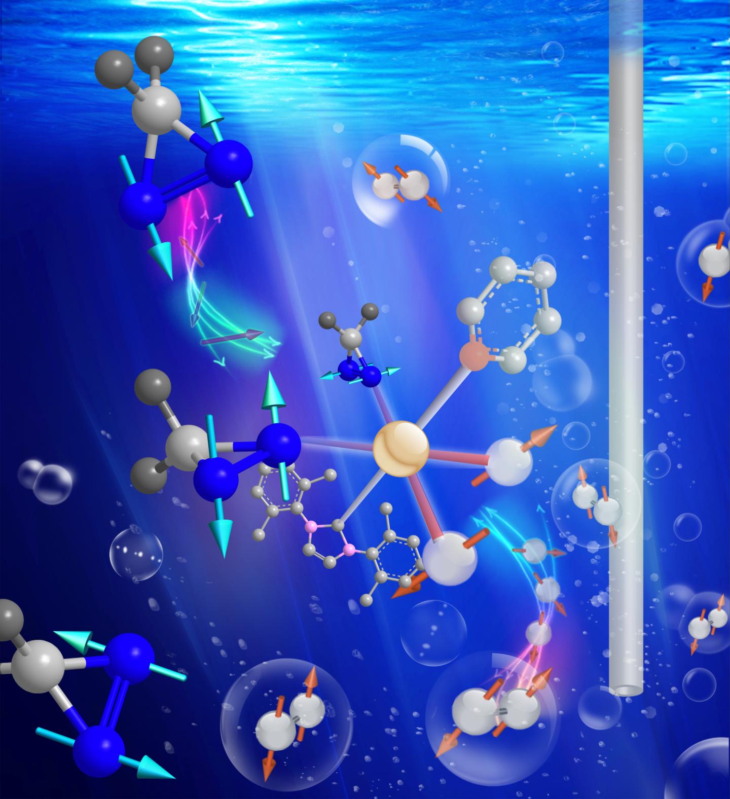
The researchers have provided an artistic representation of the molecular ‘lightbulbs’,

Caption: Duke scientists have discovered a new class of inexpensive and long-lived molecular tags that enhance MRI signals by 10,000-fold. To activate the tags, the researchers mix them with a newly developed catalyst (center) and a special form of hydrogen (gray), converting them into long-lived magnetic resonance 'lightbulbs' that might be used to track disease metabolism in real time. Credit: Thomas Theis, Duke University

Caption: Duke scientists have discovered a new class of inexpensive and long-lived molecular tags that enhance MRI signals by 10,000-fold. To activate the tags, the researchers mix them with a newly developed catalyst (center) and a special form of hydrogen (gray), converting them into long-lived magnetic resonance ‘lightbulbs’ that might be used to track disease metabolism in real time. Credit: Thomas Theis, Duke University

Here’s a link to and a citation for the paper,

Direct and cost-efficient hyperpolarization of long-lived nuclear spin states on universal 15N2-diazirine molecular tags by Thomas Theis, Gerardo X. Ortiz Jr, Angus W. J. Logan, Kevin E. Claytor, Yesu Feng, William P. Huhn, Volker Blum, Steven J. Malcolmson, Eduard Y. Chekmenev, Qiu Wang, and Warren S. Warren. Science Advances  25 Mar 2016: Vol. 2, no. 3, e1501438 DOI: 10.1126/sciadv.1501438

This paper appears to be open access.

Imaging and treating artherosclerosis with a nanoparticle

For anyone concerned about atherosclerosis (build up of plaque in the arteries) and who doesn’t need immediate assistance, this is encouraging news. A March 14, 2016 news item on ScienceDaily announces research into a nanoparticle that could both image and treat the condition,

Atherosclerosis, a disease in which plaque builds up inside arteries, is a prolific and invisible killer, but it may soon lose its ability to hide in the body and wreak havoc. Scientists have now developed a nanoparticle that functionally mimics nature’s own high-density lipoprotein (HDL). The nanoparticle can simultaneously light up and treat atherosclerotic plaques that clog arteries. Therapy with this approach could someday help prevent deadly heart attacks and strokes.

A March 13, 2016 American Chemical Society (ACS) news release (also on EurekAlert), which originated the news item, expands on the theme,

The researchers present their work today [March 13, 2016] at the 251st National Meeting & Exposition of the American Chemical Society (ACS). …

“Other researchers have shown that if you isolate HDL components from donated blood, reconstitute them and inject them into animals, there seems to be a therapeutic effect,” says Shanta Dhar, Ph.D. “However, with donors’ blood, there is the chance of immunological rejection. This technology also suffers scale-up challenges. Our motivation was to avoid immunogenic factors by making a synthetic nanoparticle which can functionally mimic HDL. At the same time, we wanted a way to locate the synthetic particles.”

Current detection strategies often fail to identify dangerous plaques, which can clog arteries over time or break off from arterial walls and block blood flow, causing a heart attack or stroke. Magnetic resonance imaging (MRI) offers a potential approach for plaque visualization, but requires the use of a contrast agent to show the atherosclerotic plaques clearly. But the potential for harmful immune reactions still exists with the use of donor-derived HDL.

Beyond imaging, there is a therapeutic aspect of using HDL. HDL is widely known as “good” cholesterol because of its ability to pull low-density lipoprotein, or “bad” cholesterol, out of plaques. This process shrinks the plaques, making them less likely to clog arteries or break apart.

To simultaneously identify and treat atherosclerosis without triggering an immune response, Dhar and Bhabatosh Banik, Ph.D., a postdoctoral fellow in her lab, created an MRI-active HDL mimic. The researchers, who are at the University of Georgia, Athens, had previously built synthetic HDL particles lacking a contrast agent. These particles lowered levels of total cholesterol and triglycerides in mice.

“The key challenge, then, was designing the contrast agent,” Banik says. “It took time to figure out the optimal lipophilicity and solubility.” The contrast agent, iron oxide, needs to be encapsulated in the synthetic lipoparticle’s hydrophobic core to provide the brightest possible signal. Eventually, the researchers hit on the right chemical combination — iron oxide with a fatty surface coating — for optimal particle encapsulation. They successfully visualized the contrast agent using MRI in cell studies.

The researchers are applying their synthetic nanoparticle to distinguish between unstable plaques and stationary ones. To do this, Dhar targeted the new MRI-active HDL mimics to macrophages, which are white blood cells that, along with lipids and cholesterol, make up atherosclerotic plaques.

The researchers targeted macrophages by decorating the nanoparticles’ surfaces with a molecule that selectively binds to macrophages. The team observed that the nanoparticles were engulfed by these white blood cells. “Then, when the macrophages ruptured, which is a sign of an unstable plaque, the cells spit out the nanoparticles, causing the MRI signal to change in a detectable fashion,” Banik says.

Dhar says her lab is now using MRI to study how well the particles light up and treat plaques in animals, and she hopes to begin clinical trials within two years. [emphasis mine]

Good luck to the researchers!