Tag Archives: protons

World’s smallest magnetic resonance imaging (MRI) of a single atom

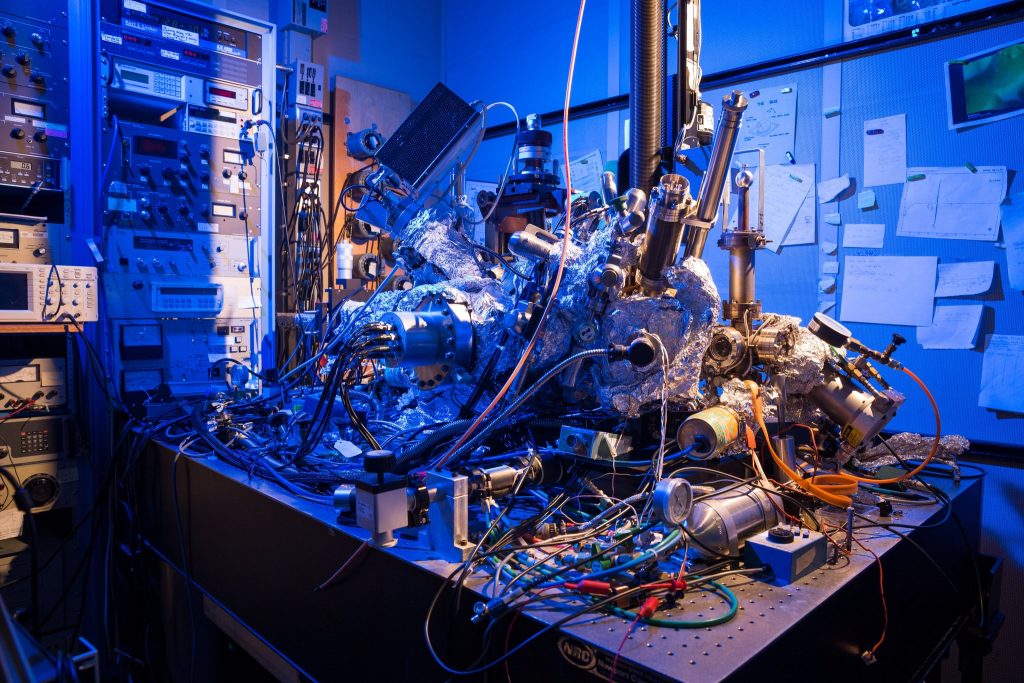
While not science’s sleekest machine, this microscope was able to capture M.R.I. scans of single atoms. Credit: IBM Research

Such a messy looking thing—it makes me feel better about my housekeeping. In any event, it’s fascinating to think this scanning tunneling microscope as seen in the above can actually act as an MRI device and create an image of a single atom.

There’s a wonderful article in the New York Times about the work but I’m starting first with a July 1, 2019 news item on Nanowerk,

Researchers at the Center for Quantum Nanoscience (QNS) within the Institute for Basic Science (IBS) at Ewha Womans University [Seoul, South Korea) have made a major scientific breakthrough by performing the world’s smallest magnetic resonance imaging (MRI). In an international collaboration with colleagues from the US, QNS scientists used their new technique to visualize the magnetic field of single atoms.

A July 2, 2019 IBS news release (also on EurekAlert but published July 1, 2019), which originated the news item, provides some insight into the research,

An MRI is routinely done in hospitals nowadays as a part of imaging for diagnostics. MRI’s detect the density of spins – the fundamental magnets in electrons and protons – in the human body. Traditionally, billions and billions of spins are required for an MRI scan. The new findings, published today [July 1, 2019] in the journal Nature Physics, show that this process is now also possible for an individual atom on a surface. To do this, the team used a Scanning Tunneling Microscope, which consists of an atomically sharp metal tip that allows researchers to image and probe single atoms by scanning the tip across the surface.

The two elements that were investigated in this work, iron and titanium, are both magnetic. Through precise preparation of the sample, the atoms were readily visible in the microscope. The researchers then used the microscope’s tip like an MRI machine to map the three-dimensional magnetic field created by the atoms with unprecedented resolution. In order to do so, they attached another spin cluster to the sharp metal tip of their microscope. Similar to everyday magnets, the two spins would attract or repel each other depending on their relative position. By sweeping the tip spin cluster over the atom on the surface, the researchers were able to map out the magnetic interaction. Lead author, Dr. Philip Willke of QNS says: “It turns out that the magnetic interaction we measured depends on the properties of both spins, the one on the tip and the one on the sample. For example, the signal that we see for iron atoms is vastly different from that for titanium atoms. This allows us to distinguish different kinds of atoms by their magnetic field signature and makes our technique very powerful.”

The researchers plan to use their single-atom MRI to map the spin distribution in more complex structures such as molecules and magnetic materials. “Many magnetic phenomena take place on the nanoscale, including the recent generation of magnetic storage devices.” says Dr. Yujeong Bae also of QNS, a co-author in this study. “We now plan to study a variety of systems using our microscopic MRI.” The ability to analyze the magnetic structure on the nanoscale can help to develop new materials and drugs. Moreover, the research team wants to use this kind of MRI to characterize and control quantum systems. These are of great interest for future computation schemes, also known as quantum computing

“I am very excited about these results. It is certainly a milestone in our field and has very promising implications for future research.” says Prof. Andreas Heinrich, Director of QNS. “The ability to map spins and their magnetic field with previously unimaginable precision, allows us to gain deeper knowledge about the structure of matter and opens new fields of basic research.”

The Center for Quantum Nanoscience, on the campus of Ewha Womans University in Seoul, South Korea, is a world-leading research center merging quantum and nanoscience to engineer the quantum future through basic research. Backed by Korea’s Institute for Basic Science, which was founded in 2011, the Center for Quantum Nanoscience draws on decades of QNS Director Andreas J. Heinrich’s (A Boy and His Atom, IBM, 2013) scientific leadership to lay the foundation for future technology by exploring the use of quantum behavior atom-by-atom on surfaces with highest precision.

You may have noticed that other than a brief mention in the first paragraph (in the Nanowerk news item excerpt), there’s no mention of the US researchers and their contribution to the work.

Interestingly, the July 1, 2019 New York Time article by Knvul Sheikh returns the favour by focusing almost entirely on US researchers while giving the Korean researchers a passing mention (Note: Links have been removed),

Different microscopy techniques allow scientists to see the nucleotide-by-nucleotide genetic sequences in cells down to the resolution of a couple atoms as seen in an atomic force microscopy image. But scientists at the IBM Almaden Research Center in San Jose, Calif., and the Institute for Basic Sciences in Seoul, have taken imaging a step further, developing a new magnetic resonance imaging technique that provides unprecedented detail, right down to the individual atoms of a sample.

When doctors want to detect tumors, measure brain function or visualize the structure of joints, they employ huge M.R.I. machines, which apply a magnetic field across the human body. This temporarily disrupts the protons spinning in the nucleus of every atom in every cell. A subsequent, brief pulse of radio-frequency energy causes the protons to spin perpendicular to the pulse. Afterward, the protons return to their normal state, releasing energy that can be measured by sensors and made into an image.

But to gather enough diagnostic data, traditional hospital M.R.I.s must scan billions and billions of protons in a person’s body, said Christopher Lutz, a physicist at IBM. So he and his colleagues decided to pack the power of an M.R.I. machine into the tip of another specialized instrument known as a scanning tunneling microscope to see if they could image individual atoms.

The tip of a scanning tunneling microscope is just a few atoms wide. And it moves along the surface of a sample, it picks up details about the size and conformation of molecules.

The researchers attached magnetized iron atoms to the tip, effectively combining scanning-tunneling microscope and M.R.I. technologies.

When the magnetized tip swept over a metal wafer of iron and titanium, it applied a magnetic field to the sample, disrupting the electrons (rather than the protons, as a typical M.R.I. would) within each atom. Then the researchers quickly turned a radio-frequency pulse on and off, so that the electrons would emit energy that could be visualized. …

Here’s a link to and a citation for the paper,

Magnetic resonance imaging of single atoms on a surface by Philip Willke, Kai Yang, Yujeong Bae, Andreas J. Heinrich & Christopher P. Lutz. Nature Physics (2019) DOI: https://doi.org/10.1038/s41567-019-0573-x Published 01 July 2019

This paper is behind a paywall.

Gold’s origin in the universe due to cosmic collision

An hypothesis for gold’s origins was first mentioned here in a May 26, 2016 posting,

The link between this research and my side project on gold nanoparticles is a bit tenuous but this work on the origins for gold and other precious metals being found in the stars is so fascinating and I’m determined to find a connection.

An artist's impression of two neutron stars colliding. (Credit: Dana Berry / Skyworks Digital, Inc.) Courtesy: Kavli Foundation

An artist’s impression of two neutron stars colliding. (Credit: Dana Berry / Skyworks Digital, Inc.) Courtesy: Kavli Foundation

From a May 19, 2016 news item on phys.org,

The origin of many of the most precious elements on the periodic table, such as gold, silver and platinum, has perplexed scientists for more than six decades. Now a recent study has an answer, evocatively conveyed in the faint starlight from a distant dwarf galaxy.

In a roundtable discussion, published today [May 19, 2016?], The Kavli Foundation spoke to two of the researchers behind the discovery about why the source of these heavy elements, collectively called “r-process” elements, has been so hard to crack.

From the Spring 2016 Kavli Foundation webpage hosting the  “Galactic ‘Gold Mine’ Explains the Origin of Nature’s Heaviest Elements” Roundtable ,

Astronomers studying a galaxy called Reticulum II have just discovered that its stars contain whopping amounts of these metals—collectively known as “r-process” elements (See “What is the R-Process?”). Of the 10 dwarf galaxies that have been similarly studied so far, only Reticulum II bears such strong chemical signatures. The finding suggests some unusual event took place billions of years ago that created ample amounts of heavy elements and then strew them throughout the galaxy’s reservoir of gas and dust. This r-process-enriched material then went on to form Reticulum II’s standout stars.

Based on the new study, from a team of researchers at the Kavli Institute at the Massachusetts Institute of Technology, the unusual event in Reticulum II was likely the collision of two, ultra-dense objects called neutron stars. Scientists have hypothesized for decades that these collisions could serve as a primary source for r-process elements, yet the idea had lacked solid observational evidence. Now armed with this information, scientists can further hope to retrace the histories of galaxies based on the contents of their stars, in effect conducting “stellar archeology.”

Researchers have confirmed the hypothesis according to an Oct. 16, 2017 news item on phys.org,

Gold’s origin in the Universe has finally been confirmed, after a gravitational wave source was seen and heard for the first time ever by an international collaboration of researchers, with astronomers at the University of Warwick playing a leading role.

Members of Warwick’s Astronomy and Astrophysics Group, Professor Andrew Levan, Dr Joe Lyman, Dr Sam Oates and Dr Danny Steeghs, led observations which captured the light of two colliding neutron stars, shortly after being detected through gravitational waves – perhaps the most eagerly anticipated phenomenon in modern astronomy.

Marina Koren’s Oct. 16, 2017 article for The Atlantic presents a richly evocative view (Note: Links have been removed),

Some 130 million years ago, in another galaxy, two neutron stars spiraled closer and closer together until they smashed into each other in spectacular fashion. The violent collision produced gravitational waves, cosmic ripples powerful enough to stretch and squeeze the fabric of the universe. There was a brief flash of light a million trillion times as bright as the sun, and then a hot cloud of radioactive debris. The afterglow hung for several days, shifting from bright blue to dull red as the ejected material cooled in the emptiness of space.

Astronomers detected the aftermath of the merger on Earth on August 17. For the first time, they could see the source of universe-warping forces Albert Einstein predicted a century ago. Unlike with black-hole collisions, they had visible proof, and it looked like a bright jewel in the night sky.

But the merger of two neutron stars is more than fireworks. It’s a factory.

Using infrared telescopes, astronomers studied the spectra—the chemical composition of cosmic objects—of the collision and found that the plume ejected by the merger contained a host of newly formed heavy chemical elements, including gold, silver, platinum, and others. Scientists estimate the amount of cosmic bling totals about 10,000 Earth-masses of heavy elements.

I’m not sure exactly what this image signifies but it did accompany Koren’s article so presumably it’s a representation of colliding neutron stars,
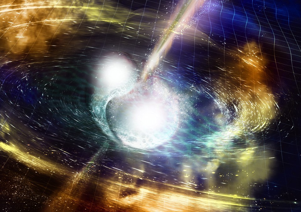
NSF / LIGO / Sonoma State University /A. Simonnet. Downloaded from: https://www.theatlantic.com/science/archive/2017/10/the-making-of-cosmic-bling/543030/

An Oct. 16, 2017 University of Warwick press release (also on EurekAlert), which originated the news item on phys.org, provides more detail,

Huge amounts of gold, platinum, uranium and other heavy elements were created in the collision of these compact stellar remnants, and were pumped out into the universe – unlocking the mystery of how gold on wedding rings and jewellery is originally formed.

The collision produced as much gold as the mass of the Earth. [emphasis mine]

This discovery has also confirmed conclusively that short gamma-ray bursts are directly caused by the merging of two neutron stars.

The neutron stars were very dense – as heavy as our Sun yet only 10 kilometres across – and they collided with each other 130 million years ago, when dinosaurs roamed the Earth, in a relatively old galaxy that was no longer forming many stars.

They drew towards each other over millions of light years, and revolved around each other increasingly quickly as they got closer – eventually spinning around each other five hundred times per second.

Their merging sent ripples through the fabric of space and time – and these ripples are the elusive gravitational waves spotted by the astronomers.

The gravitational waves were detected by the Advanced Laser Interferometer Gravitational-Wave Observatory (Adv-LIGO) on 17 August this year [2017], with a short duration gamma-ray burst detected by the Fermi satellite just two seconds later.

This led to a flurry of observations as night fell in Chile, with a first report of a new source from the Swope 1m telescope.

Longstanding collaborators Professor Levan and Professor Nial Tanvir (from the University of Leicester) used the facilities of the European Southern Observatory to pinpoint the source in infrared light.

Professor Levan’s team was the first one to get observations of this new source with the Hubble Space Telescope. It comes from a galaxy called NGC 4993, 130 million light years away.

Andrew Levan, Professor in the Astronomy & Astrophysics group at the University of Warwick, commented: “Once we saw the data, we realised we had caught a new kind of astrophysical object. This ushers in the era of multi-messenger astronomy, it is like being able to see and hear for the first time.”

Dr Joe Lyman, who was observing at the European Southern Observatory at the time was the first to alert the community that the source was unlike any seen before.

He commented: “The exquisite observations obtained in a few days showed we were observing a kilonova, an object whose light is powered by extreme nuclear reactions. This tells us that the heavy elements, like the gold or platinum in jewellery are the cinders, forged in the billion degree remnants of a merging neutron star.”

Dr Samantha Oates added: “This discovery has answered three questions that astronomers have been puzzling for decades: what happens when neutron stars merge? What causes the short duration gamma-ray bursts? Where are the heavy elements, like gold, made? In the space of about a week all three of these mysteries were solved.”

Dr Danny Steeghs said: “This is a new chapter in astrophysics. We hope that in the next few years we will detect many more events like this. Indeed, in Warwick we have just finished building a telescope designed to do just this job, and we expect it to pinpoint these sources in this new era of multi-messenger astronomy”.

Congratulations to all of the researchers involved in this work!

Many, many research teams were  involved. Here’s a sampling of their news releases which focus on their areas of research,

University of the Witwatersrand (South Africa)

https://www.eurekalert.org/pub_releases/2017-10/uotw-wti101717.php

Weizmann Institute of Science (Israel)

https://www.eurekalert.org/pub_releases/2017-10/wios-cns101717.php

Carnegie Institution for Science (US)

https://www.eurekalert.org/pub_releases/2017-10/cifs-dns101217.php

Northwestern University (US)

https://www.eurekalert.org/pub_releases/2017-10/nu-adc101617.php

National Radio Astronomy Observatory (US)

https://www.eurekalert.org/pub_releases/2017-10/nrao-ru101317.php

Max-Planck-Gesellschaft (Germany)

https://www.eurekalert.org/pub_releases/2017-10/m-gwf101817.php

Penn State (Pennsylvania State University; US)

https://www.eurekalert.org/pub_releases/2017-10/ps-stl101617.php

University of California – Davis

https://www.eurekalert.org/pub_releases/2017-10/uoc–cns101717.php

The American Association for the Advancement of Science’s (AAAS) magazine, Science, has published seven papers on this research. Here’s an Oct. 16, 2017 AAAS news release with an overview of the papers,

https://www.eurekalert.org/pub_releases/2017-10/aaft-btf101617.php

I’m sure there are more news releases out there and that there will be many more papers published in many journals, so if this interests, I encourage you to keep looking.

Two final pieces I’d like to draw your attention to: one answers basic questions and another focuses on how artists knew what to draw when neutron stars collide.

Keith A Spencer’s Oct. 18, 2017 piece on salon.com answers a lot of basic questions for those of us who don’t have a background in astronomy. Here are a couple of examples,

What is a neutron star?

Okay, you know how atoms have protons, neutrons, and electrons in them? And you know how protons are positively charged, and electrons are negatively charged, and neutrons are neutral?

Yeah, I remember that from watching Bill Nye as a kid.

Totally. Anyway, have you ever wondered why the negatively-charged electrons and the positively-charged protons don’t just merge into each other and form a neutral neutron? I mean, they’re sitting there in the atom’s nucleus pretty close to each other. Like, if you had two magnets that close, they’d stick together immediately.

I guess now that you mention it, yeah, it is weird.

Well, it’s because there’s another force deep in the atom that’s preventing them from merging.

It’s really really strong.

The only way to overcome this force is to have a huge amount of matter in a really hot, dense space — basically shove them into each other until they give up and stick together and become a neutron. This happens in very large stars that have been around for a while — the core collapses, and in the aftermath, the electrons in the star are so close to the protons, and under so much pressure, that they suddenly merge. There’s a big explosion and the outer material of the star is sloughed off.

Okay, so you’re saying under a lot of pressure and in certain conditions, some stars collapse and become big balls of neutrons?

Pretty much, yeah.

So why do the neutrons just stick around in a huge ball? Aren’t they neutral? What’s keeping them together? 

Gravity, mostly. But also the strong nuclear force, that aforementioned weird strong force. This isn’t something you’d encounter on a macroscopic scale — the strong force only really works at the type of distances typified by particles in atomic nuclei. And it’s different, fundamentally, than the electromagnetic force, which is what makes magnets attract and repel and what makes your hair stick up when you rub a balloon on it.

So these neutrons in a big ball are bound by gravity, but also sticking together by virtue of the strong nuclear force. 

So basically, the new ball of neutrons is really small, at least, compared to how heavy it is. That’s because the neutrons are all clumped together as if this neutron star is one giant atomic nucleus — which it kinda is. It’s like a giant atom made only of neutrons. If our sun were a neutron star, it would be less than 20 miles wide. It would also not be something you would ever want to get near.

Got it. That means two giant balls of neutrons that weighed like, more than our sun and were only ten-ish miles wide, suddenly smashed into each other, and in the aftermath created a black hole, and we are just now detecting it on Earth?

Exactly. Pretty weird, no?

Spencer does a good job of gradually taking you through increasingly complex explanations.

For those with artistic interests, Neel V. Patel tries to answer a question about how artists knew what draw when neutron stars collided in his Oct. 18, 2017 piece for Slate.com,

All of these things make this discovery easy to marvel at and somewhat impossible to picture. Luckily, artists have taken up the task of imagining it for us, which you’ve likely seen if you’ve already stumbled on coverage of the discovery. Two bright, furious spheres of light and gas spiraling quickly into one another, resulting in a massive swell of lit-up matter along with light and gravitational waves rippling off speedily in all directions, towards parts unknown. These illustrations aren’t just alluring interpretations of a rare phenomenon; they are, to some extent, the translation of raw data and numbers into a tangible visual that gives scientists and nonscientists alike some way of grasping what just happened. But are these visualizations realistic? Is this what it actually looked like? No one has any idea. Which is what makes the scientific illustrators’ work all the more fascinating.

“My goal is to represent what the scientists found,” says Aurore Simmonet, a scientific illustrator based at Sonoma State University in Rohnert Park, California. Even though she said she doesn’t have a rigorous science background (she certainly didn’t know what a kilonova was before being tasked to illustrate one), she also doesn’t believe that type of experience is an absolute necessity. More critical, she says, is for the artist to have an interest in the subject matter and in learning new things, as well as a capacity to speak directly to scientists about their work.

Illustrators like Simmonet usually start off work on an illustration by asking the scientist what’s the biggest takeaway a viewer should grasp when looking at a visual. Unfortunately, this latest discovery yielded a multitude of papers emphasizing different conclusions and highlights. With so many scientific angles, there’s a stark challenge in trying to cram every important thing into a single drawing.

Clearly, however, the illustrations needed to center around the kilonova. Simmonet loves colors, so she began by discussing with the researchers what kind of color scheme would work best. The smash of two neutron stars lends itself well to deep, vibrant hues. Simmonet and Robin Dienel at the Carnegie Institution for Science elected to use a wide array of colors and drew bright cracking to show pressure forming at the merging. Others, like Luis Calcada at the European Southern Observatory, limited the color scheme in favor of emphasizing the bright moment of collision and the signal waves created by the kilonova.

Animators have even more freedom to show the event, since they have much more than a single frame to play with. The Conceptual Image Lab at NASA’s [US National Aeronautics and Space Administration] Goddard Space Flight Center created a short video about the new findings, and lead animator Brian Monroe says the video he and his colleagues designed shows off the evolution of the entire process: the rising action, climax, and resolution of the kilonova event.

The illustrators try to adhere to what the likely physics of the event entailed, soliciting feedback from the scientists to make sure they’re getting it right. The swirling of gas, the direction of ejected matter upon impact, the reflection of light, the proportions of the objects—all of these things are deliberately framed such that they make scientific sense. …

Do take a look at Patel’s piece, if for no other reason than to see all of the images he has embedded there. You may recognize Aurore Simmonet’s name from the credit line in the second image I have embedded here.
Carbon nanotubes transport protons faster than bulk water

An April 4, 2016 news item on Science Daily focuses on carbon nanotubes that measure eight-tenths of a nanometre and transport protons more quickly than bulk water by an order of magnitude,

For the first time, Lawrence Livermore National Laboratory (LLNL) researchers have shown that carbon nanotubes as small as eight-tenths of a nanometer in diameter can transport protons faster than bulk water, by an order of magnitude.

The research validates a 200-year old mechanism of proton transport.

A US Department of Energy Lawrence Livermore National Laboratory (LLNL) news release on EurekAlert, which originated the news item, provides more explanation,

The transport rates in these nanotube pores, which form one-dimensional water wires, also exceed those of biological channels and man-made proton conductors, making carbon nanotubes the fastest known proton conductor. …

Practical applications include proton exchange membranes, proton-based signaling in biological systems and the emerging field of proton bioelectronics (protonics).

“The cool thing about our results is that we found that when you squeeze water into the nanotube, protons move through that water even faster than through normal (bulk) water,” said Aleksandr Noy, an LLNL biophysicist and a lead author of the paper. (Bulk water is similar to what you would find in a cup of water that is much bigger than the size of a single water molecule).

The idea that protons travel fast in solutions by hopping along chains of hydrogen-bonded water molecules dates back 200 years to the work of Theodore von Grotthuss and still remains the foundation of the scientific understanding of proton transport. In the new research, LLNL researchers used carbon nanotube pores to line up water molecules into perfect one-dimensional chains and showed that they allow proton transport rates to approach the ultimate limits for the Grotthuss transport mechanism.

“The possibility to achieve fast proton transport by changing the degree of water confinement is exciting,” Noy said. “So far, the man-made proton conductors, such as polymer Nafion, use a different principle to enhance the proton transport. We have mimicked the way biological systems enhance the proton transport, took it to the extreme, and now our system realizes the ultimate limit of proton conductivity in a nanopore.”

Of all man-made materials, the narrow hydrophobic inner pores of carbon nanotubes (CNT) hold the most promise to deliver the level of confinement and weak interactions with water molecules that facilitate the formation of one-dimensional hydrogen-bonded water chains that enhance proton transport.

Earlier molecular dynamic simulations showed that water in 0.8 nm diameter carbon nanotubes would create such water wires and predicted that these channels would exhibit proton transport rates that would be much faster than those of bulk water. Ramya Tunuguntla, an LLNL postdoctoral researcher and the first author on the paper, said that despite significant efforts in carbon nanotube transport studies, these predictions proved to be hard to validate, mainly because of the difficulties in creating sub-1-nm diameter CNT pores.

However, the Lawrence Livermore team along with colleagues from the Lawrence Berkeley National Lab and UC Berkeley was able to create a simple and versatile experimental system for studying transport in ultra-narrow CNT pores. They used carbon nanotube porins (CNTPs), a technology they developed earlier at LLNL, which uses carbon nanotubes embedded in the lipid membrane to mimic biological ion channel functionality. The key breakthrough was the creation of nanotube porins with a diameter of less than 1 nm, which allowed researchers for the first time to achieve true one-dimensional water confinement.

Here’s a link to and a citation for the paper,

Ultrafast proton transport in sub-1-nm diameter carbon nanotube porins by Ramya H. Tunuguntla, Frances I. Allen, Kyunghoon Kim, Allison Belliveau, & Aleksandr Noy. Nature Nanotechnology (2016) doi:10.1038/nnano.2016.43 Published online 04 April 2016

This paper is behind a paywall.

Gold, acetic acid, and proton shuttles

I think the information has been taken from Russian to English by a machine translator, as well, I’m not a chemist, so please bear with my interpretation. It seems that Russian researchers have determined why gold, inert at the macroscale, is a good catalyst at the nanoscale. From a July 28, 2015 news item on Azonano,

Being found mostly in the native state, gold is one of the oldest elements known to man. The affection to gold was determined by it’s unusual properties – heft, shine and ability to withstand oxidation and corrosion.

The combination of properties determined gold use in the jewelry and as a coinage metal. The ancient alchemists working with gold were struggled by utmost chemical resistance of this element – it did not react with concentrated acids or alkali solutions even at high temperatures. Actually, it is the chemical inertness that makes gold to appear in a native form and not as a part of a mineral.

Later analysis established that gold compounds can not only compete with traditional nickel and palladium-based catalysts in the common reactions, but to surpass them. Besides that, gold compounds often demonstrated principally novel types of reactivity compared to well-established catalysts. This allowed chemists to discover a bunch of new chemical reactions and predetermined a fascinating boom in gold catalysis that we have observed in the recent years.

A July 24, 2015 Institute of Organic Chemistry, Russian Academy of Sciences press release on EurekAlert, which also originated the news item, reveals more about the study,

Professor Ananikov and co-workers introduced gold into well-known catalytic system which led to dramatic change of the reactivity and furnished the formation of novel gold-containing complexes. The complexes appeared to be air stable and were isolated in the individual state. A single crystal X-Ray diffraction study ascertained the existence of unique structural motif in the molecule, which can not be explained within conventional mechanistic framework.

The study was carried out using both theoretical and experimental approaches. Dedicated labeling of the reagents allowed observation of molecular re-organizations. Variation of reaction conditions helped to estimate key factors governing the discovered transformation. In addition, computational study of the reaction provided the models of certain intermediate steps, which were invisible for experimental investigation. The theoretical data obtained was in excellent agreement with experiment, proposing the reaction mechanism, where a molecule of acetic acid serves as a proton shuttle, transferring the hydrogen atom between the reaction centers.

The belief of gold inactivity towards chemical transformations resulted in the fact, that organometallic chemistry of gold was developed significantly later compared to other coinage metals (like silver, nickel or copper). Today, our goal is to “introduce gold catalysis as a valuable practical tool in fine organic chemistry, competitive with other transition metal catalysts”, says Prof. Ananikov.

Here’s a link to and a citation for the paper,

Carboxylic Group-Assisted Proton Transfer in Gold-Mediated Thiolation of Alkynes by Sergey S. Zalesskiy, Victor N. Khrustalev, Alexandr Yu. Kostukovich, and Valentine P. Ananikov. Organometallics, Article ASAP DOI: 10.1021/acs.organomet.5b00210 Publication Date (Web): July 22, 2015

Copyright © 2015 American Chemical Society

This paper is behind a paywall.

Fermionic atoms and the microscopes that can see them

The new fermionic microscope built at the Massachusetts Institute of Technology (MIT) allows you to image 1000 or more fermionic atoms according to a May 13, 2015 news item on ScienceDaily,

Fermions are the building blocks of matter, interacting in a multitude of permutations to give rise to the elements of the periodic table. Without fermions, the physical world would not exist.

Examples of fermions are electrons, protons, neutrons, quarks, and atoms consisting of an odd number of these elementary particles. Because of their fermionic nature, electrons and nuclear matter are difficult to understand theoretically, so researchers are trying to use ultracold gases of fermionic atoms as stand-ins for other fermions.

But atoms are extremely sensitive to light: When a single photon hits an atom, it can knock the particle out of place — an effect that has made imaging individual fermionic atoms devilishly hard.

Now a team of MIT physicists has built a microscope that is able to see up to 1,000 individual fermionic atoms. The researchers devised a laser-based technique to trap and freeze fermions in place, and image the particles simultaneously.

A May 13, 2015 MIT news release, which originated the news item, provides intriguing detail about the microscope and fascinating insight into fermions (for those who are interested but not expert and sufficiently brave to endure certain failure to understand everything in this piece),

The new imaging technique uses two laser beams trained on a cloud of fermionic atoms in an optical lattice. The two beams, each of a different wavelength, cool the cloud, causing individual fermions to drop down an energy level, eventually bringing them to their lowest energy states — cool and stable enough to stay in place. At the same time, each fermion releases light, which is captured by the microscope and used to image the fermion’s exact position in the lattice — to an accuracy better than the wavelength of light.

With the new technique, the researchers are able to cool and image over 95 percent of the fermionic atoms making up a cloud of potassium gas. Martin Zwierlein, a professor of physics at MIT, says an intriguing result from the technique appears to be that it can keep fermions cold even after imaging.

“That means I know where they are, and I can maybe move them around with a little tweezer to any location, and arrange them in any pattern I’d like,” Zwierlein says.

Zwierlein and his colleagues, including first author and graduate student Lawrence Cheuk, have published their results today in the journal Physical Review Letters.

Seeing fermions from bosons

For the past two decades, experimental physicists have studied ultracold atomic gases of the two classes of particles: fermions and bosons — particles such as photons that, unlike fermions, can occupy the same quantum state in limitless numbers. In 2009, physicist Markus Greiner at Harvard University devised a microscope that successfully imaged individual bosons in a tightly spaced optical lattice. This milestone was followed, in 2010, by a second boson microscope, developed by Immanuel Bloch’s group at the Max Planck Institute of Quantum Optics.

These microscopes revealed, in unprecedented detail, the behavior of bosons under strong interactions. However, no one had yet developed a comparable microscope for fermionic atoms.

“We wanted to do what these groups had done for bosons, but for fermions,” Zwierlein says. “And it turned out it was much harder for fermions, because the atoms we use are not so easily cooled. So we had to find a new way to cool them while looking at them.”

Techniques to cool atoms ever closer to absolute zero have been devised in recent decades. Carl Wieman, Eric Cornell, and MIT’s Wolfgang Ketterle were able to achieve Bose-Einstein condensation in 1995, a milestone for which they were awarded the 2001 Nobel Prize in physics. Other techniques include a process using lasers to cool atoms from 300 degrees Celsius to a few ten-thousandths of a degree above absolute zero.

A clever cooling technique

And yet, to see individual fermionic atoms, the particles need to be cooled further still. To do this, Zwierlein’s group created an optical lattice using laser beams, forming a structure resembling an egg carton, each well of which could potentially trap a single fermion. Through various stages of laser cooling, magnetic trapping, and further evaporative cooling of the gas, the atoms were prepared at temperatures just above absolute zero — cold enough for individual fermions to settle onto the underlying optical lattice. The team placed the lattice a mere 7 microns from an imaging lens, through which they hoped to see individual fermions.

However, seeing fermions requires shining light on them, causing a photon to essentially knock a fermionic atom out of its well, and potentially out of the system entirely.

“We needed a clever technique to keep the atoms cool while looking at them,” Zwierlein says.

His team decided to use a two-laser approach to further cool the atoms; the technique manipulates an atom’s particular energy level, or vibrational energy. Each atom occupies a certain energy state — the higher that state, the more active the particle is. The team shone two laser beams of differing frequencies at the lattice. The difference in frequencies corresponded to the energy between a fermion’s energy levels. As a result, when both beams were directed at a fermion, the particle would absorb the smaller frequency, and emit a photon from the larger-frequency beam, in turn dropping one energy level to a cooler, more inert state. The lens above the lattice collects the emitted photon, recording its precise position, and that of the fermion.

Zwierlein says such high-resolution imaging of more than 1,000 fermionic atoms simultaneously would enhance our understanding of the behavior of other fermions in nature — particularly the behavior of electrons. This knowledge may one day advance our understanding of high-temperature superconductors, which enable lossless energy transport, as well as quantum systems such as solid-state systems or nuclear matter.

“The Fermi gas microscope, together with the ability to position atoms at will, might be an important step toward the realization of a quantum computer based on fermions,” Zwierlein says. “One would thus harness the power of the very same intricate quantum rules that so far hamper our understanding of electronic systems.”

Zwierlein says it is a good time for Fermi gas microscopists: Around the same time his group first reported its results, teams from Harvard and the University of Strathclyde in Glasgow also reported imaging individual fermionic atoms in optical lattices, indicating a promising future for such microscopes.

Zoran Hadzibabic, a professor of physics at Trinity College [University of Cambridge, UK], says the group’s microscope is able to detect individual atoms “with almost perfect fidelity.”

“They detect them reliably, and do so without affecting their positions — that’s all you want,” says Hadzibabic, who did not contribute to the research. “So far they demonstrated the technique, but we know from the experience with bosons that that’s the hardest step, and I expect the scientific results to start pouring out.”

Here’s a link to and a citation for the published paper,

Quantum-Gas Microscope for Fermionic Atoms by Lawrence W. Cheuk, Matthew A. Nichols, Melih Okan, Thomas Gersdorf, Vinay V. Ramasesh, Waseem S. Bakr, Thomas Lompe, and Martin W. Zwierlein. Phys. Rev. Lett. 114, 193001 – Published 13 May 2015 (print: Vol. 114, Iss. 19 — 15 May 2015) DOI: http://dx.doi.org/10.1103/PhysRevLett.114.193001

I believe this paper is behind a paywall.

There is an earlier version available on arXiv.org,

A Quantum Gas Microscope for Fermionic Atoms by Lawrence W. Cheuk, Matthew A. Nichols, Melih Okan, Thomas Gersdorf, Vinay V. Ramasesh, Waseem S. Bakr, Thomas Lompe, Martin W. Zwierlein. (Submitted on 9 Mar 2015 (v1), last revised 10 Mar 2015 (this version, v2))

This an open access website.

Graphene not so impermeable after all

I saw the news last week but it took reading Dexter Johnson’s Dec. 2, 2014 post for me to achieve a greater understanding of why graphene’s proton permeability is such a big deal and of the tensions underlying graphene research in the UK.

Let’s start with the news, from a Nov. 26, 2014 news item on Nanowerk (Note: A link has been removed),

Published in the journal Nature (“Proton transport through one-atom-thick crystals”), the discovery could revolutionise fuel cells and other hydrogen-based technologies as they require a barrier that only allow protons – hydrogen atoms stripped off their electrons – to pass through.

In addition, graphene membranes could be used to sieve hydrogen gas out of the atmosphere, where it is present in minute quantities, creating the possibility of electric generators powered by air.

A Nov. 26, 2014 University of Manchester news release, which originated the news item, describes the research in greater detail,

One-atom thick material graphene, first isolated and explored in 2004 by a team at The University of Manchester, is renowned for its barrier properties, which has a number of uses in applications such as corrosion-proof coatings and impermeable packaging.

For example, it would take the lifetime of the universe for hydrogen, the smallest of all atoms, to pierce a graphene monolayer.

Now a group led by Sir Andre Geim tested whether protons are also repelled by graphene. They fully expected that protons would be blocked, as existing theory predicted as little proton permeation as for hydrogen.

Despite the pessimistic prognosis, the researchers found that protons pass through the ultra-thin crystals surprisingly easily, especially at elevated temperatures and if the films were covered with catalytic nanoparticles such as platinum.

The discovery makes monolayers of graphene, and its sister material boron nitride, attractive for possible uses as proton-conducting membranes, which are at the heart of modern fuel cell technology. Fuel cells use oxygen and hydrogen as a fuel and convert the input chemical energy directly into electricity. Without membranes that allow an exclusive flow of protons but prevent other species to pass through, this technology would not exist.

Despite being well-established, fuel-cell technology requires further improvements to make it more widely used. One of the major problems is a fuel crossover through the existing proton membranes, which reduces their efficiency and durability.

The University of Manchester research suggests that the use of graphene or monolayer boron nitride can allow the existing membranes to become thinner and more efficient, with less fuel crossover and poisoning. This can boost competitiveness of fuel cells.

The Manchester group also demonstrated that their one-atom-thick membranes can be used to extract hydrogen from a humid atmosphere. They hypothesise that such harvesting can be combined together with fuel cells to create a mobile electric generator that is fuelled simply by hydrogen present in air.

Marcelo Lozada-Hidalgo, a PhD student and corresponding author of this paper, said: “When you know how it should work, it is a very simple setup. You put a hydrogen-containing gas on one side, apply small electric current and collect pure hydrogen on the other side. This hydrogen can then be burned in a fuel cell.

“We worked with small membranes, and the achieved flow of hydrogen is of course tiny so far. But this is the initial stage of discovery, and the paper is to make experts aware of the existing prospects. To build up and test hydrogen harvesters will require much further effort.”

Dr Sheng Hu, a postdoctoral researcher and the first author in this work, added: “It looks extremely simple and equally promising. Because graphene can be produced these days in square metre sheets, we hope that it will find its way to commercial fuel cells sooner rather than later”.

The work is an international collaboration involving groups from China and the Netherlands who supported theoretical aspects of this research. Marcelo Lozada-Hidalgo is funded by a PhD studentship programme between the National Council of Science and Technology of Mexico and The University of Manchester.

Here’s more about the research and its implications from Dexter Johnson’s Dec. 2, 2014 post on the Nanoclast blog on the IEEE (Institute of Electronics and Electrical Engineers) website (Note: Links have been removed),

This latest development alters the understanding of one of the key properties of graphene: that it is impermeable to all gases and liquids. Even an atom as small as hydrogen would need billions of years for it to pass through the dense electronic cloud of graphene.  In fact, it is this impermeability that has made it attractive for use in gas separation membranes.

But as Geim and his colleagues discovered, in research that was published in the journal Nature, monolayers of graphene and boron nitride are highly permeable to thermal protons under ambient conditions. So hydrogen atoms stripped of their electrons could pass right through the one-atom-thick materials.

The surprising discovery that protons could breach these materials means that that they could be used in proton-conducting membranes (also known as proton exchange membranes), which are central to the functioning of fuel cells. Fuel cells operate through chemical reactions involving hydrogen fuel and oxygen, with the result being electrical energy. The membranes used in the fuel cells are impermeable to oxygen and hydrogen but allow for the passage of protons.

Dexter goes into more detail about hydrogen fuel cells and why this discovery is so exciting. He also provides some insight into the UK’s graphene community (Note: A link has been removed),

While some have been frustrated that Geim has focused his attention on fundamental research rather than becoming more active in the commercialization of graphene, he may have just cracked open graphene’s greatest application possibility to date.

I recommend reading Dexter’s post if you want to learn more about fuel cell technology and the impact this discovery may have.

Richard Van Noorden’s Nov. 27, 2014 article for Nature provides another perspective on this work,

Fuel-cell experts say that the work is proof of principle, but are cautious about its immediate application. Factors such as to how grow a sufficiently clean, large graphene sheet, and its cost and lifetime, would have to be taken into account. “It may or may not be a better membrane for a fuel cell,” says Andrew Herring, a chemical engineer at the Colorado School of Mines in Golden.

Van Noorden also writes about another graphene discovery from last week, which won’t be featured here. Where graphene is concerned I have to draw a line or else this entire blog would be focused on that material alone.

Getting back back to permeability, graphene, and protons, here’s a link to and a citation for the research paper,

Proton transport through one-atom-thick crystals by S. Hu, M. Lozada-Hidalgo, F. C. Wang, A. Mishchenko, F. Schedin, R. R. Nair, E. W. Hill, D. W. Boukhvalov, M. I. Katsnelson, R. A. W. Dryfe, I. V. Grigorieva, H. A. Wu, & A. K. Geim. Nature (2014 doi:10.1038/nature14015 Published online 26 November 2014

This article is behind a paywall.

Step closer to integrating electronics into the body

The Sept. 20, 2011 news item (Proton-based transistor could let machines communicate with living things) on Nanowerk features a rather interesting development,

Human devices, from light bulbs to iPods, send information using electrons. Human bodies and all other living things, on the other hand, send signals and perform work using ions or protons.

Materials scientists at the University of Washington have built a novel transistor that uses protons, creating a key piece for devices that can communicate directly with living things.

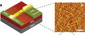
Here’s a diagram from the University of Washington Sept. 20, 2011 article about the proton transistor by Hannah Hickey,


On the left is a colored photo of the UW device overlaid on a graphic of the other components. On the right is a magnified image of the chitosan fibers. The white scale bar is 200 nanometers. (Marco Rolandi, UW)

Here’s a little more about the proton transistor (from the Hickey article),

In the body, protons activate “on” and “off” switches and are key players in biological energy transfer. Ions open and close channels in the cell membrane to pump things in and out of the cell. Animals including humans use ions to flex their muscles and transmit brain signals. A machine that was compatible with a living system in this way could, in the short term, monitor such processes. Someday it could generate proton currents to control certain functions directly.

A first step toward this type of control is a transistor that can send pulses of proton current. The prototype device is a field-effect transistor, a basic type of transistor that includes a gate, a drain and a source terminal for the current. The UW prototype is the first such device to use protons. It measures about 5 microns wide, roughly a twentieth the width of a human hair.

As for the device (from the Hickey article),

The device uses a modified form of the compound chitosan originally extracted from squid pen, a structure that survives from when squids had shells. The material is compatible with living things, is easily manufactured, and can be recycled from crab shells and squid pen discarded by the food industry.

There is a minor Canadian connection,

Computer models of charge transport developed by co-authors M.P. Anantram, a UW professor of electrical engineering, and Anita Fadavi Roudsari at Canada’s University of Waterloo, were a good match for the experimental results.

If I understand this correctly, the computer models were confirmed by the experimental  results, which means the computer models can be used (to augment the use of expensive experiments) with a fair degree of confidence.

I am finding this integration of electronics into the body both fascinating and disturbing as per my paper, Whose electric brain? More about that when I have more time.